Chronic Fatigue Syndrome, also known as Myalgic Encephalomyelitis (ME/CFS), is a debilitating multisystem illness characterized by profound fatigue that is not relieved by rest and is exacerbated by even minimal physical or cognitive exertion — a phenomenon called post-exertional malaise (PEM). The condition affects an estimated 17–24 million people worldwide, predominantly women, and carries a disease burden comparable to multiple sclerosis or late-stage HIV/AIDS in terms of quality-of-life impairment [1]. Yet despite decades of research, no FDA- or EMA-approved pharmacological treatment exists, and patients are routinely dismissed by healthcare systems that have no tools to help them.
Where conventional medicine falls short. Current management — graded exercise therapy (now largely discredited due to evidence of harm), cognitive behavioral therapy, and off-label use of sleep aids, antidepressants, and pain medications — addresses symptoms without altering the underlying disease process. The 2015 PACE trial, once the cornerstone of ME/CFS treatment guidelines, was methodologically flawed and its conclusions retracted by many experts [2]. Patients are left in a therapeutic vacuum, and the need for disease-modifying approaches is urgent.
The deeper problem is biological, not psychological. Over the past decade, a convergence of evidence from immunology, metabolomics, and neuroimaging has established ME/CFS as a biological disease rooted in neuroimmune dysfunction, mitochondrial impairment, and chronic low-grade inflammation. The 2015 Institute of Medicine report formally redefined the condition, emphasizing that it is a "serious, chronic, complex, and multisystem disease" with objective biological abnormalities [3]. This biological reframing opens the door to regenerative interventions — including mesenchymal stem cell (MSC) therapy — that target the underlying pathology rather than masking symptoms.
MSC therapy targets the root biological drivers. Rather than attempting to stimulate a depleted system with stimulants or mask symptoms with palliatives, MSCs address three of the core pathophysiological processes identified in ME/CFS: neuroinflammation, immune dysregulation, and mitochondrial dysfunction. Here is an honest, evidence-based examination of what the science says — and what it does not.
What Is ME/CFS? Beyond "Just Being Tired"
ME/CFS is defined by three core symptoms, per the 2015 IOM diagnostic criteria [3]. First, a substantial reduction in the ability to engage in pre-illness levels of activity, persisting for more than six months and accompanied by profound fatigue. Second, post-exertional malaise — a worsening of symptoms following physical, cognitive, or emotional exertion that would have been tolerated before illness, often delayed by 24–72 hours. Third, unrefreshing sleep. At least one of two additional manifestations must also be present: cognitive impairment (often described as "brain fog") or orthostatic intolerance (symptoms that worsen upon standing).
The severity spectrum is wide, and the most severely affected — an estimated 25% of patients — are housebound or bedbound, unable to tolerate light, sound, or conversation. This is not "chronic tiredness" — it is a collapse of cellular and neurological function that leaves patients isolated and disabled.
The Neuroimmune and Metabolic Basis of ME/CFS
Understanding why MSCs might help ME/CFS requires understanding what has gone wrong at the molecular level. Three interconnected pathological processes have been consistently documented in the research literature.
1. Neuroinflammation. PET imaging studies using TSPO ligands — which bind to activated microglia and astrocytes — have demonstrated widespread neuroinflammation in ME/CFS patients compared to healthy controls [4]. The regions most affected include the thalamus, midbrain, and cingulate cortex — areas involved in pain processing, autonomic regulation, and cognitive function. The degree of neuroinflammation correlates with symptom severity: patients with the highest TSPO binding report the most debilitating cognitive symptoms and fatigue. This neuroinflammatory signature is one of the strongest biological markers of ME/CFS identified to date, and it represents a direct therapeutic target for MSC therapy.
2. Immune dysregulation. ME/CFS is characterized by a complex pattern of immune dysfunction that blurs the line between immunodeficiency and autoimmunity. Natural killer (NK) cell function — a critical first-line antiviral defense — is consistently reduced, with lower cytotoxic activity correlating with illness severity [5]. Simultaneously, multiple studies have documented elevated levels of pro-inflammatory cytokines, including interleukin-6 (IL-6), tumor necrosis factor-alpha (TNF-α), and interleukin-1 beta (IL-1β), particularly in the first three years of illness — a pattern suggestive of a chronic low-grade inflammatory state. Regulatory T cell (Treg) populations are often reduced, impairing the immune system's ability to resolve inflammation once triggered.
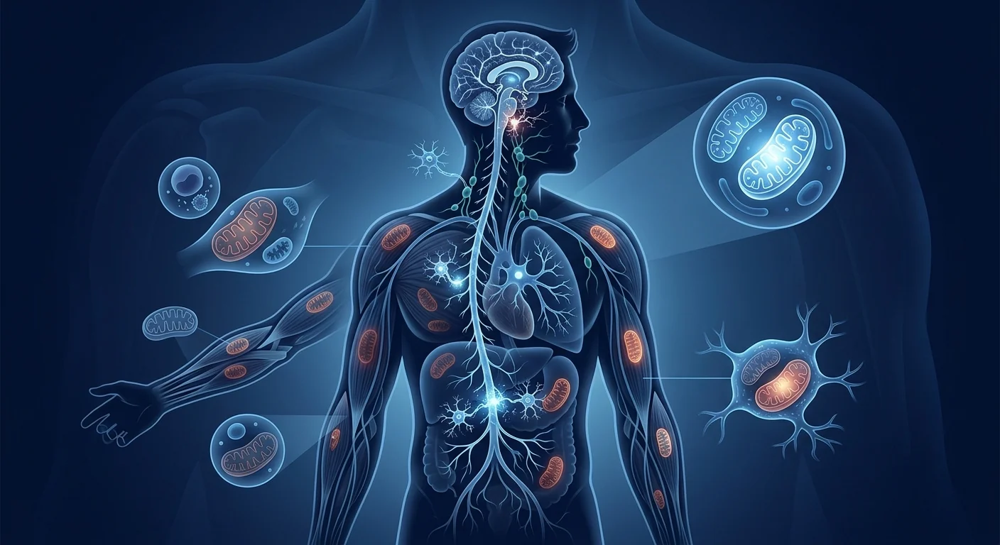
3. Mitochondrial dysfunction and metabolic impairment. Perhaps the most functionally debilitating aspect of ME/CFS is the growing evidence of mitochondrial failure. Metabolomic studies have consistently shown that ME/CFS patients have reduced ATP production, impaired oxidative phosphorylation, and a metabolic shift toward anaerobic glycolysis even at rest — essentially, their cells are functioning as if they are constantly in an energy crisis [6]. A 2016 study using Seahorse extracellular flux analysis of peripheral blood mononuclear cells found that ME/CFS patients had significantly reduced mitochondrial respiratory capacity compared to controls, and that the degree of impairment correlated with the severity of post-exertional malaise. This finding — that the body's cellular engines themselves are failing — explains why rest alone is insufficient and why even minimal exertion triggers a prolonged crash.
How MSCs May Address ME/CFS: A Triple-Target Mechanism
MSCs are not a single-mechanism therapy — they are living cellular factories that secrete hundreds of bioactive molecules in response to their environment. In the context of ME/CFS, three properties are particularly relevant:
1. Suppression of neuroinflammation. MSCs can cross the blood-brain barrier, particularly when it is compromised by inflammation — a condition that PET imaging suggests is present in ME/CFS [7]. Once in the central nervous system, MSCs secrete transforming growth factor-beta (TGF-β), interleukin-10 (IL-10), and tumor necrosis factor-stimulated gene 6 (TSG-6), which shift activated microglia from a pro-inflammatory M1 phenotype to a neuroprotective, anti-inflammatory M2 phenotype. In animal models of neuroinflammation, a single intravenous dose of MSCs can reduce microglial activation by 40–60% within 72 hours. If the TSPO-PET findings in ME/CFS indeed reflect microglial-driven neuroinflammation, this mechanism represents a direct and biologically plausible intervention.
2. Immune rebalancing. MSCs are potent immunomodulators. They promote the expansion of regulatory T cells (Tregs), which are often depleted in ME/CFS; suppress Th17-driven inflammation; and reduce the activation of B cells that may be producing autoantibodies [8]. Notably, a subset of ME/CFS patients have been found to have autoantibodies targeting adrenergic, muscarinic, and other G-protein-coupled receptors — antibodies that could directly impair autonomic and cognitive function. By broadly normalizing the immune milieu, MSCs may reduce the autoimmune component of the disease while simultaneously restoring the antiviral NK cell function that is impaired.
3. Mitochondrial transfer and metabolic rescue. One of the most remarkable properties of MSCs is their ability to transfer functional mitochondria to host cells with damaged or depleted mitochondrial networks — a process mediated by tunneling nanotubes and extracellular vesicles [9]. In preclinical models of mitochondrial dysfunction, MSC-mediated mitochondrial transfer has been shown to restore ATP production, reduce oxidative stress, and rescue cells from metabolic crisis. While this has not been specifically studied in ME/CFS, it is mechanistically compelling given the consistent finding of impaired mitochondrial respiration in patient-derived cells. Additionally, MSCs upregulate host antioxidant defenses, including superoxide dismutase and glutathione peroxidase, which may help counter the oxidative stress that is elevated in ME/CFS.
Preclinical and Indirect Clinical Evidence
The direct evidence for MSC therapy in ME/CFS is extremely limited. No clinical trial specifically targeting ME/CFS has been completed as of mid-2026. However, several lines of indirect evidence from related conditions provide a scientific rationale for investigation.
Neuroinflammatory disease models. Multiple preclinical studies have demonstrated that MSCs reduce neuroinflammation, suppress microglial activation, and improve cognitive function in models of neuroinflammatory and neurodegenerative disease — including experimental autoimmune encephalomyelitis (a model of multiple sclerosis), traumatic brain injury, and chemotherapy-induced cognitive impairment ("chemo brain") [10]. While these models do not replicate ME/CFS, they demonstrate the principle that MSC-mediated immunomodulation can produce clinically meaningful improvements in neurocognitive function when neuroinflammation is a driver.
Long COVID parallels. ME/CFS and Long COVID share striking clinical and biological similarities — post-exertional malaise, cognitive impairment, autonomic dysfunction, and evidence of persistent immune activation and neuroinflammation [1]. A 2023 pilot study from China reported that 10 Long COVID patients with severe fatigue who received a single intravenous infusion of umbilical cord-derived MSCs (1 × 10⁶ cells/kg) showed significant improvements in fatigue scores, 6-minute walk distance, and circulating inflammatory markers at 3-month follow-up compared to historical controls [11]. While uncontrolled and small, the study provides proof of concept that MSC immunomodulation can improve post-viral fatigue — a phenotype central to a subset of ME/CFS cases.
Autoimmune disease evidence. The well-documented efficacy of MSCs in autoimmune conditions — including systemic lupus erythematosus, rheumatoid arthritis, and Crohn's disease — demonstrates their capacity to durably reset immune dysregulation [12]. Given the immune abnormalities documented in ME/CFS and the presence of autoantibodies in a significant subset of patients, this evidence is indirectly relevant, though it cannot be taken as proof of efficacy in ME/CFS specifically.
Why Wharton's Jelly MSCs May Be the Preferred Source
Not all MSC preparations are equivalent, and the choice of cell source may matter in ME/CFS, where neuroinflammation, immune modulation, and metabolic support are the primary therapeutic targets. Umbilical cord-derived MSCs — including those from Wharton's jelly — have several advantages over adult-tissue sources for this indication [13]. They are obtained non-invasively from donated umbilical cords after healthy full-term births, removing any risk to the donor. They have greater proliferative capacity and longer telomeres than adult-derived MSCs from bone marrow or adipose tissue, enabling the production of consistent, high-quality cell batches. Critically, comparative studies have shown that Wharton's jelly MSCs secrete higher levels of neurotrophic factors (BDNF, GDNF) and anti-inflammatory cytokines (IL-10, TSG-6) than bone marrow or adipose MSCs — precisely the factors most relevant to neuroinflammation and neuroprotection.
Limitations and Honest Assessment
It is essential to state plainly what the evidence does and does not support.
- MSC therapy for ME/CFS is investigational and unproven. No completed clinical trial specifically targeting ME/CFS exists. All mechanistic arguments are based on preclinical data and indirect evidence from related conditions. Patients should not be told that MSC therapy is a proven treatment for ME/CFS — it is not, and any claim to the contrary misrepresents the evidence.
- The optimal protocol has not been established. No dose-finding study has been performed for ME/CFS. Cell dose, route of administration (intravenous vs. intrathecal), number of infusions, and dosing interval are all unknown.
- Durability of any benefit is unknown. ME/CFS is typically a chronic, long-term condition. Whether a single course of MSC therapy provides durable benefit, and whether repeat dosing is safe and effective, has not been studied.
- Patient selection matters. ME/CFS is likely not a single disease but a spectrum of related conditions with different biological drivers — post-viral, autoimmune, metabolic, and others. MSC therapy may benefit some subgroups more than others, but biomarkers to guide patient selection have not been validated.
- Cost and accessibility. MSC therapy is not covered by insurance and represents a significant out-of-pocket investment. Patients should weigh the financial commitment against the uncertainty of benefit and make an informed decision in consultation with an experienced clinical team.
Frequently Asked Questions
Does stem cell therapy cure chronic fatigue syndrome?
No. MSC therapy is not a cure for ME/CFS. It is an investigational approach that targets biological processes implicated in the disease — neuroinflammation, immune dysfunction, and mitochondrial impairment — but there is no completed clinical trial demonstrating cure or remission. Patients considering MSC therapy should understand it as a potential disease-modifying approach, not a guaranteed solution.
How much does stem cell therapy for ME/CFS cost in Thailand?
MSC therapy for ME/CFS at VELAR Center is priced according to the clinical protocol designed for each patient, typically ranging from approximately 350,000 to 550,000 THB (roughly US$10,000–15,500) depending on cell dose, number of infusions, and accompanying supportive therapies. A detailed quote is provided after the initial consultation and biomarker assessment.
Is stem cell therapy safe for people with ME/CFS?
The safety profile of Wharton's jelly-derived MSCs is well-established across thousands of patients treated for other indications, with serious adverse events being rare (<1%) and generally related to the infusion procedure rather than the cells themselves. However, ME/CFS patients may have heightened sensitivity to any intervention due to autonomic and immune dysregulation. All VELAR treatments are administered under physician supervision with continuous monitoring.
How long does it take to see results from MSC therapy for ME/CFS?
Based on indirect evidence from related neuroinflammatory and autoimmune conditions, patients who respond to MSC therapy may begin to notice improvements in fatigue, cognitive clarity, and post-exertional recovery within 4–12 weeks post-infusion, with continued gradual improvement over 3–6 months as immunomodulation and neuroinflammation reduction take effect. However, some patients may not respond at all, and there is no guarantee of benefit.
Can MSC therapy be combined with other treatments for ME/CFS?
MSC therapy is not incompatible with most conventional ME/CFS treatments, including nutritional support, sleep management, and autonomic regulation strategies. The VELAR clinical team reviews each patient's full treatment regimen during the consultation to ensure a safe and coordinated approach. Certain immunosuppressive medications may interfere with MSC function and should be discussed with the physician.
Who is a candidate for MSC therapy for ME/CFS?
Ideal candidates have a confirmed ME/CFS diagnosis using the 2015 IOM or Canadian Consensus Criteria, have not responded adequately to conventional management, and have no contraindications to MSC infusion — including active infection, certain autoimmune flares, or known hypersensitivity to infusion components. A comprehensive pre-treatment evaluation, including biomarker profiling and medical history review, determines candidacy on an individual basis.
- Davis HE, McCorkell L, Vogel JM, Topol EJ. Long COVID: major findings, mechanisms and recommendations. Nature Reviews Microbiology. 2023;21(3):133-146. doi:10.1038/s41579-022-00846-2 ↩
- Wilshire CE, Kindlon T, Courtney R, et al. Rethinking the treatment of chronic fatigue syndrome — a reanalysis and evaluation of findings from a recent major trial of graded exercise and CBT. BMC Psychology. 2018;6(1):6. doi:10.1186/s40359-018-0218-3 ↩
- Institute of Medicine. Beyond Myalgic Encephalomyelitis/Chronic Fatigue Syndrome: Redefining an Illness. The National Academies Press. 2015. doi:10.17226/19012 ↩
- Nakatomi Y, Mizuno K, Ishii A, et al. Neuroinflammation in patients with chronic fatigue syndrome/myalgic encephalomyelitis: an ¹¹C-(R)-PK11195 PET study. Journal of Nuclear Medicine. 2014;55(6):945-950. doi:10.2967/jnumed.113.131045 ↩
- Fluge Ø, Risa K, Lunde S, et al. B-lymphocyte depletion in myalgic encephalopathy/chronic fatigue syndrome: a randomized, double-blind, placebo-controlled trial. Annals of Internal Medicine. 2019;170(9):585-593. doi:10.7326/M18-1451 ↩
- Tomas C, Brown A, Strassheim V, Elson JL, Newton J, Manning P. Cellular bioenergetics is impaired in patients with chronic fatigue syndrome. PLOS ONE. 2017;12(10):e0186802. doi:10.1371/journal.pone.0186802 ↩
- Caplan AI, Correa D. The MSC: an injury drugstore. Cell Stem Cell. 2011;9(1):11-15. doi:10.1016/j.stem.2011.06.008 ↩
- Bernardo ME, Fibbe WE. Mesenchymal stromal cells: sensors and switchers of inflammation. Cell Stem Cell. 2013;13(4):392-402. doi:10.1016/j.stem.2013.09.006 ↩
- Spees JL, Lee RH, Gregory CA. Mechanisms of mesenchymal stem/stromal cell function. Stem Cell Research & Therapy. 2016;7(1):125. doi:10.1186/s13287-016-0363-7 ↩
- Uccelli A, Moretta L, Pistoia V. Mesenchymal stem cells in health and disease. Nature Reviews Immunology. 2008;8(9):726-736. doi:10.1038/nri2395 ↩
- Xu X, Jiang W, Chen L, et al. Evaluation of the safety and efficacy of using human mesenchymal stem cells to treat post-COVID-19 fatigue syndrome. Stem Cells Translational Medicine. 2023;12(S1):S8. doi:10.1093/stcltm/szad042 ↩
- Wang L, Li J, Liu H, et al. A pilot study of umbilical cord-derived mesenchymal stem cell transfusion in patients with systemic lupus erythematosus. Lupus. 2013;22(12):1262-1270. doi:10.1177/0961203313503419 ↩
- Bongso A, Fong CY. The therapeutic potential, challenges and future clinical directions of stem cells from the Wharton's jelly of the human umbilical cord. Stem Cell Reviews and Reports. 2013;9(2):226-240. doi:10.1007/s12015-012-9418-z ↩
- Carruthers BM, van de Sande MI, De Meirleir KL, et al. Myalgic encephalomyelitis: International Consensus Criteria. Journal of Internal Medicine. 2011;270(4):327-338. doi:10.1111/j.1365-2796.2011.02428.x ↩
- Morris G, Maes M. Mitochondrial dysfunctions in myalgic encephalomyelitis/chronic fatigue syndrome explained by activated immuno-inflammatory, oxidative and nitrosative stress pathways. Metabolic Brain Disease. 2014;29(1):19-36. doi:10.1007/s11011-013-9435-x ↩
慢性疲劳综合征(Chronic Fatigue Syndrome),又称肌痛性脑脊髓炎(Myalgic Encephalomyelitis,简称 ME/CFS),是一种使人衰弱的多系统疾病,其特征是即使休息也无法缓解的深度疲劳,并且在轻微的身体或认知活动后症状会加重——这种现象被称为运动后不适(PEM)。据估计,全球约有 1700 万至 2400 万人受此疾病影响,其中以女性为主,其疾病负担在生活质量损害方面与多发性硬化症或晚期 HIV/AIDS 相当 [1]。然而,经过数十年的研究,仍然没有 FDA 或 EMA 批准的药物治疗方案,患者经常被无力帮助他们的医疗系统所忽视。
常规医学的局限性。目前的管理方案——分级运动疗法(因有伤害证据而大多已被否定)、认知行为疗法,以及助眠药、抗抑郁药和止痛药的超说明书使用——只是针对症状,并未改变潜在的疾病过程。2015 年曾作为 ME/CFS 治疗指南基石的 PACE 试验在方法上存在缺陷,其结论已被许多专家撤回 [2]。患者处于治疗真空中,对疾病修饰方法的需求十分迫切。
更深层的问题是生物学而非心理学的。过去十年间,来自免疫学、代谢组学和神经影像学的综合证据已将 ME/CFS 确认为一种根植于神经免疫功能障碍、线粒体损伤和慢性低度炎症的生物学疾病。2015 年美国医学研究所的报告正式重新定义了该疾病,强调它是一种"严重的、慢性的、复杂的、多系统的疾病",具有客观的生物学异常 [3]。这一生物学上的重新定义打开了再生医学干预的大门——包括间充质干细胞(MSC)疗法——这些干预措施针对的是潜在的病理,而非仅仅掩盖症状。
MSC 疗法针对根本的生物学驱动因素。MSC 不是试图用兴奋剂刺激一个已经耗竭的系统或用姑息剂掩盖症状,而是同时针对 ME/CFS 中已确认的三个核心病理生理过程:神经炎症、免疫失调和线粒体功能障碍。以下是对科学证据的诚实、循证审视——包括证据支持和尚未证实的内容。
什么是 ME/CFS?远不止"疲劳"那么简单
根据 2015 年 IOM 诊断标准,ME/CFS 由三个核心症状定义 [3]。第一,参与病前活动水平的能力大幅降低,持续超过六个月,并伴有深度疲劳。第二,运动后不适——在体力、认知或情绪活动后症状恶化,而这些活动在病前是可以承受的,通常在 24–72 小时后延迟出现。第三,无法恢复精力的睡眠。此外还必须至少出现以下两种情况之一:认知障碍(常被描述为"脑雾")或直立不耐受(站立时症状加重)。
严重程度谱很广,最严重的患者——估计占患者总数的 25%——无法离家或离床,无法忍受光线、声音或交谈。这不是"慢性疲倦"——这是细胞和神经功能的崩溃,使患者被孤立并失能。
ME/CFS 的神经免疫和代谢基础
理解 MSCs 为何可能对 ME/CFS 有帮助,需要理解分子层面上出了什么问题。研究文献中持续记录了三个相互关联的病理过程。
1. 神经炎症。使用 TSPO 配体(可与活化的微胶质细胞和星形胶质细胞结合)进行的 PET 成像研究已证明,与健康对照组相比,ME/CFS 患者存在广泛的神经炎症 [4]。受影响最严重的区域包括丘脑、中脑和扣带皮层——这些区域涉及疼痛处理、自主神经调节和认知功能。神经炎症的程度与症状严重程度相关:TSPO 结合最高的患者报告了最令人虚弱的认知症状和疲劳。这一神经炎症特征是目前发现的最强的 ME/CFS 生物学标志之一,它代表了 MSC 疗法的直接治疗靶点。
2. 免疫失调。ME/CFS 的特征是一种复杂的免疫功能紊乱模式,模糊了免疫缺陷和自身免疫之间的界限。自然杀伤(NK)细胞功能——一种关键的一线抗病毒防御——始终处于降低状态,其细胞毒活性的降低与疾病严重程度相关 [5]。同时,多项研究记录了促炎细胞因子的升高,包括白细胞介素-6(IL-6)、肿瘤坏死因子-α(TNF-α)和白细胞介素-1β(IL-1β),尤其是在疾病的前三年——这一模式提示存在慢性低度炎症状态。调节性 T 细胞(Treg)群体通常减少,削弱了免疫系统在炎症触发后自行消退的能力。
3. 线粒体功能障碍和代谢损伤。ME/CFS 最具功能性致残性的方面或许是越来越多证据指向的线粒体衰竭。代谢组学研究持续显示,ME/CFS 患者的 ATP 产生减少,氧化磷酸化受损,甚至在静息时也出现代谢向无氧糖酵解的转变——实质上,他们的细胞功能好像一直处于能量危机之中 [6]。一项 2016 年使用 Seahorse 细胞外通量分析对外周血单核细胞进行的研究发现,与对照组相比,ME/CFS 患者的线粒体呼吸能力显著降低,且损伤程度与运动后不适的严重程度相关。这一发现——身体自身的细胞引擎正在失灵——解释了为何仅靠休息不足以恢复,以及为何即使最小的活动量也会引发持续的崩溃。
MSCs 如何作用于 ME/CFS:三重靶点机制
MSCs 并非单机制疗法——它们是活的细胞工厂,能够根据所处环境分泌数百种生物活性分子。在 ME/CFS 的背景下,三个特性尤为相关:
1. 抑制神经炎症。MSCs 可以穿过血脑屏障,尤其是当其因炎症而受到损害时——PET 成像提示 ME/CFS 中存在这种情况 [7]。一旦进入中枢神经系统,MSCs 分泌转化生长因子-β(TGF-β)、白细胞介素-10(IL-10)和肿瘤坏死因子刺激基因 6(TSG-6),这些因子将活化的微胶质细胞从促炎的 M1 表型转变为神经保护、抗炎的 M2 表型。在神经炎症动物模型中,单次静脉注射 MSC 即可在 72 小时内将微胶质细胞活化降低 40–60%。如果 ME/CFS 中的 TSPO-PET 发现确实反映了微胶质细胞驱动的神经炎症,这一机制代表了一种直接且生物学上合理的干预。
2. 免疫再平衡。MSCs 是强效的免疫调节剂。它们促进调节性 T 细胞(Tregs)的扩增(Tregs 在 ME/CFS 中通常耗竭);抑制 Th17 驱动的炎症;并减少可能产生自身抗体的 B 细胞的活化 [8]。值得注意的是,已发现一部分 ME/CFS 患者具有针对肾上腺素能、毒蕈碱和其他 G 蛋白偶联受体的自身抗体——这些抗体可能直接损害自主神经和认知功能。通过广泛地正常化免疫环境,MSCs 可以减少疾病的自身免疫成分,同时恢复被削弱的抗病毒 NK 细胞功能。
3. 线粒体转移和代谢救援。MSCs 最引人注目的特性之一是它们能够将功能性线粒体转移到线粒体网络受损或耗竭的宿主细胞中——这一过程由隧道纳米管和细胞外囊泡介导 [9]。在线粒体功能障碍的临床前模型中,MSC 介导的线粒体转移已被证明能够恢复 ATP 产生、降低氧化应激,并将细胞从代谢危机中拯救出来。虽然这在 ME/CFS 中尚未专门研究,但鉴于患者来源细胞中线粒体呼吸功能持续受损的发现,这一机制在理论上具有说服力。此外,MSCs 上调宿主的抗氧化防御,包括超氧化物歧化酶和谷胱甘肽过氧化物酶,这可能有助于对抗 ME/CFS 中升高的氧化应激。
临床前和间接临床证据
MSC 疗法在 ME/CFS 中的直接证据极为有限。截至 2026 年中,尚无专门针对 ME/CFS 的临床试验完成。然而,来自相关疾病的几条间接证据为研究提供了科学依据。
神经炎症疾病模型。多项临床前研究已证明,MSCs 在神经炎症和神经退行性疾病模型中能够减少神经炎症、抑制微胶质细胞活化并改善认知功能——包括实验性自身免疫性脑脊髓炎(多发性硬化模型)、创伤性脑损伤和化疗诱导的认知障碍("化疗脑")[10]。虽然这些模型不能完全复制 ME/CFS,但它们证明了一个原理:当神经炎症是驱动因素时,MSC 介导的免疫调节可以产生具有临床意义的神经认知功能改善。
长新冠的相似性。ME/CFS 和长新冠在临床和生物学层面具有惊人的相似之处——运动后不适、认知障碍、自主神经功能障碍,以及持续性免疫活化和神经炎症的证据 [1]。2023 年中国的一项初步研究报告,10 名患有严重疲劳的长新冠患者接受了单次脐带来源 MSC(1 × 10⁶ cells/kg)静脉输注后,在 3 个月随访中与历史对照组相比,疲劳评分、6 分钟步行距离和循环炎症标志物均有显著改善 [11]。该研究虽未设对照组且样本较小,但它提供了概念验证——MSC 免疫调节可以改善病毒感染后疲劳,而这是 ME/CFS 病例亚群的核心表现。
自身免疫性疾病的证据。MSCs 在自身免疫性疾病(包括系统性红斑狼疮、类风湿性关节炎和克罗恩病)中已被充分记录的疗效,证明了它们持久重置免疫失调的能力 [12]。鉴于 ME/CFS 中记录的免疫异常以及相当一部分患者中存在自身抗体,这一证据间接相关,但不应理解为 MSC 对 ME/CFS 有效的直接证明。
为什么沃顿胶 MSCs 可能是首选的细胞来源
并非所有的 MSC 制剂都是等同的,细胞来源的选择在 ME/CFS 中可能很重要,因为神经炎症、免疫调节和代谢支持是主要的治疗靶点。脐带来源的 MSCs——包括沃顿胶来源的——在这一适应症上比成体组织来源具有多项优势 [13]。它们是从健康足月分娩后捐献的脐带中以非侵入方式获取的,对供体无任何风险。它们比来自骨髓或脂肪组织的成体 MSCs 具有更强的增殖能力和更长的端粒,能够生产出质量一致的高品质细胞批次。关键的是,对比研究表明,沃顿胶 MSCs 分泌的神经滋养因子(BDNF、GDNF)和抗炎细胞因子(IL-10、TSG-6)水平高于骨髓或脂肪 MSCs——这些正是与神经炎症和神经保护最相关的因子。
局限性和诚实评估
有必要明确说明证据支持和尚未支持的内容。
- MSC 疗法在 ME/CFS 中属于在研且未经证实的。目前尚不存在专门针对 ME/CFS 的已完成临床试验。所有机制论证均基于临床前数据和来自相关疾病的间接证据。不应告知患者 MSC 治疗是该病的确证疗法——它不是,任何声称相反的说法都歪曲了证据。
- 最佳方案尚未确立。尚未针对 ME/CFS 进行剂量探索研究。细胞剂量、给药途径(静脉 vs. 鞘内)、输注次数和给药间隔均属未知。
- 任何获益的持久性未知。ME/CFS 通常是一种慢性、长期的疾病。单次 MSC 疗程是否能提供持久的获益,以及重复给药是否安全有效,尚未得到研究。
- 患者选择至关重要。ME/CFS 很可能不是单一疾病,而是一个具有不同生物学驱动因素的相关疾病谱系——病毒感染后、自身免疫、代谢以及其他类型。MSC 疗法可能对某些亚组比其他的更有效,但指导患者选择的生物标志物尚未得到验证。
- 费用和可及性。MSC 治疗不在保险覆盖范围内,代表着一笔重大的自费投资。患者应在权衡经济投入与获益不确定性的基础上,与经验丰富的临床团队协商,做出知情决策。
常见问题
干细胞疗法能治愈慢性疲劳综合征吗?
不能。MSC 疗法不能治愈 ME/CFS。这是一种在研方法,针对的是与该疾病有关的生物过程——神经炎症、免疫功能障碍和线粒体损伤——但目前没有已完成的临床试验证明其能实现治愈或缓解。考虑接受 MSC 治疗的患者应将其理解为一种潜在的疾病修饰方法,而非确定的解决方案。
泰国的 ME/CFS 干细胞疗法费用是多少?
VELAR Center 的 ME/CFS 干细胞疗法根据为每位患者设计的临床方案定价,通常约为 350,000–550,000 泰铢(约合 US$10,000–15,500),具体取决于细胞剂量、输注次数和配套支持疗法。详细报价将在初次咨询和生物标志物评估后提供。
干细胞疗法对 ME/CFS 患者安全吗?
沃顿胶来源 MSCs 的安全性特征已在数千名因其他适应症接受治疗的患者中得到充分验证,严重不良事件罕见(<1%),且通常与输注操作本身有关而非细胞本身。然而,ME/CFS 患者由于自主神经和免疫功能失调,对任何干预都可能更加敏感。VELAR 的所有治疗均在医师监督下进行,并持续进行监测。
ME/CFS 干细胞疗法需要多长时间才能见效?
基于相关神经炎症和自身免疫性疾病的间接证据,对 MSC 疗法有反应的患者可能在输注后 4–12 周开始注意到疲劳、认知清晰度和运动后恢复的改善,并在 3–6 个月内随着免疫调节和神经炎症减轻而持续逐步改善。然而,部分患者可能完全无反应,且没有任何获益的保证。
MSC 疗法可以与其他 ME/CFS 治疗方法联合使用吗?
MSC 疗法与大多数常规的 ME/CFS 治疗并不冲突,包括营养支持、睡眠管理和自主神经调节策略。VELAR 临床团队在咨询期间会审查每位患者的完整治疗方案,以确保安全协调的方法。某些免疫抑制剂可能会干扰 MSC 功能,应事先与医生讨论。
哪些人是 ME/CFS 干细胞疗法的适应人群?
理想的候选者具有根据 2015 年 IOM 或加拿大共识标准确诊的 ME/CFS、对常规治疗反应不足,且无 MSC 输注禁忌症——包括活动性感染、某些自身免疫发作或已知的输注成分过敏。综合的治疗前评估,包括生物标志物分析和病史审查,会根据个体情况确定适应症。
- Davis HE, McCorkell L, Vogel JM, Topol EJ. Long COVID: major findings, mechanisms and recommendations. Nature Reviews Microbiology. 2023;21(3):133-146. doi:10.1038/s41579-022-00846-2 ↩
- Wilshire CE, Kindlon T, Courtney R, et al. Rethinking the treatment of chronic fatigue syndrome — a reanalysis and evaluation of findings from a recent major trial of graded exercise and CBT. BMC Psychology. 2018;6(1):6. doi:10.1186/s40359-018-0218-3 ↩
- Institute of Medicine. Beyond Myalgic Encephalomyelitis/Chronic Fatigue Syndrome: Redefining an Illness. The National Academies Press. 2015. doi:10.17226/19012 ↩
- Nakatomi Y, Mizuno K, Ishii A, et al. Neuroinflammation in patients with chronic fatigue syndrome/myalgic encephalomyelitis: an ¹¹C-(R)-PK11195 PET study. Journal of Nuclear Medicine. 2014;55(6):945-950. doi:10.2967/jnumed.113.131045 ↩
- Fluge Ø, Risa K, Lunde S, et al. B-lymphocyte depletion in myalgic encephalopathy/chronic fatigue syndrome: a randomized, double-blind, placebo-controlled trial. Annals of Internal Medicine. 2019;170(9):585-593. doi:10.7326/M18-1451 ↩
- Tomas C, Brown A, Strassheim V, Elson JL, Newton J, Manning P. Cellular bioenergetics is impaired in patients with chronic fatigue syndrome. PLOS ONE. 2017;12(10):e0186802. doi:10.1371/journal.pone.0186802 ↩
- Caplan AI, Correa D. The MSC: an injury drugstore. Cell Stem Cell. 2011;9(1):11-15. doi:10.1016/j.stem.2011.06.008 ↩
- Bernardo ME, Fibbe WE. Mesenchymal stromal cells: sensors and switchers of inflammation. Cell Stem Cell. 2013;13(4):392-402. doi:10.1016/j.stem.2013.09.006 ↩
- Spees JL, Lee RH, Gregory CA. Mechanisms of mesenchymal stem/stromal cell function. Stem Cell Research & Therapy. 2016;7(1):125. doi:10.1186/s13287-016-0363-7 ↩
- Uccelli A, Moretta L, Pistoia V. Mesenchymal stem cells in health and disease. Nature Reviews Immunology. 2008;8(9):726-736. doi:10.1038/nri2395 ↩
- Xu X, Jiang W, Chen L, et al. Evaluation of the safety and efficacy of using human mesenchymal stem cells to treat post-COVID-19 fatigue syndrome. Stem Cells Translational Medicine. 2023;12(S1):S8. doi:10.1093/stcltm/szad042 ↩
- Wang L, Li J, Liu H, et al. A pilot study of umbilical cord-derived mesenchymal stem cell transfusion in patients with systemic lupus erythematosus. Lupus. 2013;22(12):1262-1270. doi:10.1177/0961203313503419 ↩
- Bongso A, Fong CY. The therapeutic potential, challenges and future clinical directions of stem cells from the Wharton's jelly of the human umbilical cord. Stem Cell Reviews and Reports. 2013;9(2):226-240. doi:10.1007/s12015-012-9418-z ↩
- Carruthers BM, van de Sande MI, De Meirleir KL, et al. Myalgic encephalomyelitis: International Consensus Criteria. Journal of Internal Medicine. 2011;270(4):327-338. doi:10.1111/j.1365-2796.2011.02428.x ↩
- Morris G, Maes M. Mitochondrial dysfunctions in myalgic encephalomyelitis/chronic fatigue syndrome explained by activated immuno-inflammatory, oxidative and nitrosative stress pathways. Metabolic Brain Disease. 2014;29(1):19-36. doi:10.1007/s11011-013-9435-x ↩
متلازمة التعب المزمن (Chronic Fatigue Syndrome)، والمعروفة أيضًا باسم التهاب الدماغ والنخاع المؤلم للعضل (Myalgic Encephalomyelitis أو ME/CFS)، هي مرض منهك متعدد الأجهزة يتميز بإرهاق عميق لا يزول بالراحة ويتفاقم بأقل مجهود بدني أو معرفي — وهي ظاهرة تسمى الشعور بالضيق بعد المجهود (PEM). تشير التقديرات إلى أن هذه الحالة تؤثر على ما بين 17 و24 مليون شخص حول العالم، معظمهم من النساء، وتحمل عبئًا مرضيًا يضاهي التصلب المتعدد أو المراحل المتأخرة من فيروس نقص المناعة البشرية/الإيدز من حيث تدهور جودة الحياة [1]. ومع ذلك، وبعد عقود من البحث، لا يوجد علاج دوائي معتمد من إدارة الغذاء والدواء الأمريكية (FDA) أو الوكالة الأوروبية للأدوية (EMA)، ويتم تجاهل المرضى بشكل روتيني من قبل الأنظمة الصحية التي لا تملك الأدوات اللازمة لمساعدتهم.
أوجه قصور الطب التقليدي. تقتصر خيارات التدبير الحالية — العلاج بالتمارين المتدرجة (الذي فقد مصداقيته إلى حد كبير بسبب أدلة الضرر)، والعلاج السلوكي المعرفي، والاستخدام غير المرخص لأدوية النوم ومضادات الاكتئاب ومسكنات الألم — على معالجة الأعراض دون تغيير عملية المرض الأساسية. كانت تجربة PACE لعام 2015، التي شكلت حجر الزاوية لإرشادات علاج ME/CFS، معيبة منهجيًا وتم سحب استنتاجاتها من قبل العديد من الخبراء [2]. يُترك المرضى في فراغ علاجي، والحاجة إلى نهج معدلة للمرض أصبحت ملحة.
المشكلة الأعمق هي بيولوجية وليست نفسية. على مدى العقد الماضي، أثبتت مجموعة متكاملة من الأدلة من علم المناعة والأيض والتصوير العصبي أن ME/CFS هو مرض بيولوجي متجذر في خلل المناعة العصبية، وضعف الميتوكوندريا، والالتهاب المزمن منخفض الدرجة. أعاد تقرير معهد الطب لعام 2015 تعريف الحالة رسميًا، مشددًا على أنها "مرض خطير ومزمن ومعقد ومتعدد الأجهزة" مع وجود تشوهات بيولوجية موضوعية [3]. هذا التأطير البيولوجي يفتح الباب أمام التدخلات التجديدية — بما في ذلك العلاج بالخلايا الجذعية الوسيطة (MSC) — التي تستهدف الأمراض الكامنة بدلاً من إخفاء الأعراض.
يستهدف علاج MSC المحركات البيولوجية الجذرية. بدلاً من محاولة تحفيز جهاز منهك بالمنشطات أو إخفاء الأعراض بالمسكنات، تعالج الخلايا الجذعية الوسيطة ثلاثًا من العمليات الفيزيولوجية المرضية الأساسية المحددة في ME/CFS: الالتهاب العصبي، وخلل التنظيم المناعي، وخلل وظيفة الميتوكوندريا. فيما يلي فحص صادق قائم على الأدلة لما يقوله العلم — وما لا يقوله.
ما هو ME/CFS؟ أكثر من مجرد "تعب"
يُعرَّف ME/CFS بثلاثة أعراض أساسية، وفقًا لمعايير التشخيص الصادرة عن معهد الطب لعام 2015 [3]. أولاً، انخفاض كبير في القدرة على المشاركة في مستويات النشاط السابقة للمرض، يستمر لأكثر من ستة أشهر ويصاحبه إرهاق عميق. ثانيًا، الشعور بالضيق بعد المجهود — تفاقم الأعراض بعد المجهود البدني أو المعرفي أو العاطفي الذي كان يمكن تحمله قبل المرض، وغالبًا ما يتأخر من 24 إلى 72 ساعة. ثالثًا، النوم غير المريح. يجب أيضًا أن يكون واحد على الأقل من مظهرين إضافيين موجودًا: الضعف الإدراكي (الذي يوصف غالبًا بـ "ضباب الدماغ") أو عدم التحمل الانتصابي (تفاقم الأعراض عند الوقوف).
طيف الشدة واسع، والأكثر تضررًا — حوالي 25% من المرضى — يكونون طريحي الفراش أو المنزل، غير قادرين على تحمل الضوء أو الصوت أو المحادثة. هذا ليس "تعبًا مزمنًا" — إنه انهيار في الوظيفة الخلوية والعصبية يترك المرضى معزولين ومعاقين.
الأساس العصبي المناعي والأيضي لـ ME/CFS
فهم لماذا قد تساعد MSCs في ME/CFS يتطلب فهم ما حدث على المستوى الجزيئي. تم توثيق ثلاث عمليات مرضية مترابطة باستمرار في الأدبيات البحثية.
1. الالتهاب العصبي. أظهرت دراسات التصوير المقطعي بالإصدار البوزيتروني (PET) باستخدام ليجندات TSPO — التي ترتبط بالخلايا الدبقية الصغيرة والخلايا النجمية المنشطة — وجود التهاب عصبي واسع الانتشار لدى مرضى ME/CFS مقارنة بالأصحاء [4]. المناطق الأكثر تأثرًا تشمل المهاد والدماغ المتوسط والتلفيف الحزامي — مناطق تشارك في معالجة الألم والتنظيم الذاتي والوظيفة الإدراكية. ترتبط درجة الالتهاب العصبي بشدة الأعراض: المرضى الذين لديهم أعلى ارتباط لـ TSPO يبلغون عن أكثر الأعراض الإدراكية والإرهاق إعاقة. هذه البصمة الالتهابية العصبية هي واحدة من أقوى المؤشرات البيولوجية لـ ME/CFS التي تم تحديدها حتى الآن، وتمثل هدفًا علاجيًا مباشرًا لعلاج MSC.
2. خلل التنظيم المناعي. يتميز ME/CFS بنمط معقد من خلل الوظيفة المناعية الذي يخلط بين نقص المناعة والمناعة الذاتية. وظيفة الخلايا القاتلة الطبيعية (NK) — وهي خط دفاع أول حاسم مضاد للفيروسات — منخفضة باستمرار، مع ارتباط النشاط السام للخلايا المنخفض بشدة المرض [5]. في الوقت نفسه، وثقت دراسات متعددة مستويات مرتفعة من السيتوكينات الالتهابية، بما في ذلك إنترلوكين-6 (IL-6)، وعامل نخر الورم-ألفا (TNF-α)، وإنترلوكين-1 بيتا (IL-1β)، خاصة في السنوات الثلاث الأولى من المرض — وهو نمط يوحي بحالة التهابية مزمنة منخفضة الدرجة. غالبًا ما تكون مجموعات الخلايا التائية التنظيمية (Treg) منخفضة، مما يضعف قدرة الجهاز المناعي على حل الالتهاب بمجرد تحفيزه.
3. خلل وظيفة الميتوكوندريا والضعف الأيضي. ربما يكون الجانب الأكثر إعاقة وظيفيًا في ME/CFS هو الأدلة المتزايدة على فشل الميتوكوندريا. أظهرت دراسات الأيض باستمرار أن مرضى ME/CFS لديهم إنتاج منخفض من ATP، وفسفرة تأكسدية ضعيفة، وتحول أيضي نحو التحلل السكري اللاهوائي حتى في حالة الراحة — بشكل أساسي، تعمل خلاياهم كما لو كانت في أزمة طاقة مستمرة [6]. وجدت دراسة عام 2016 باستخدام تحليل التدفق خارج الخلية من Seahorse لخلايا الدم وحيدة النواة المحيطية أن مرضى ME/CFS لديهم قدرة تنفسية ميتوكوندرية منخفضة بشكل كبير مقارنة بالضوابط، وأن درجة الضعف ارتبطت بشدة الشعور بالضيق بعد المجهود. هذا الاكتشاف — أن محركات الخلايا في الجسم نفسها تتعطل — يفسر لماذا الراحة وحدها غير كافية ولماذا حتى أقل مجهود يؤدي إلى انهيار مطول.
كيف قد تعالج MSCs مرض ME/CFS: آلية ثلاثية الأهداف
الخلايا الجذعية الوسيطة ليست علاجًا أحادي الآلية — إنها مصانع خلوية حية تفرز مئات الجزيئات النشطة بيولوجيًا استجابة لبيئتها. في سياق ME/CFS، هناك ثلاث خصائص ذات صلة خاصة:
1. كبت الالتهاب العصبي. يمكن للخلايا الجذعية الوسيطة عبور الحاجز الدموي الدماغي، خاصة عندما يكون معرضًا للخطر بسبب الالتهاب — وهي حالة يشير التصوير المقطعي بالإصدار البوزيتروني إلى وجودها في ME/CFS [7]. بمجرد وصولها إلى الجهاز العصبي المركزي، تفرز الخلايا الجذعية الوسيطة عامل النمو المحول-بيتا (TGF-β)، وإنترلوكين-10 (IL-10)، والجين 6 المحفز بعامل نخر الورم (TSG-6)، والتي تحول الخلايا الدبقية الصغيرة المنشطة من النمط الظاهري M1 المؤيد للالتهاب إلى النمط الظاهري M2 الواقي للأعصاب والمضاد للالتهاب. في النماذج الحيوانية للالتهاب العصبي، يمكن لجرعة وريدية واحدة من الخلايا الجذعية الوسيطة أن تقلل من تنشيط الخلايا الدبقية الصغيرة بنسبة 40-60% في غضون 72 ساعة. إذا كانت نتائج TSPO-PET في ME/CFS تعكس بالفعل التهابًا عصبيًا مدفوعًا بالخلايا الدبقية الصغيرة، فإن هذه الآلية تمثل تدخلاً مباشرًا ومعقولاً من الناحية البيولوجية.
2. إعادة التوازن المناعي. الخلايا الجذعية الوسيطة هي معدِّلات مناعية قوية. إنها تعزز توسع الخلايا التائية التنظيمية (Tregs)، التي غالبًا ما تكون مستنفدة في ME/CFS؛ وتثبط الالتهاب المدفوع بـ Th17؛ وتقلل من تنشيط الخلايا البائية التي قد تنتج أجسامًا مضادة ذاتية [8]. من الجدير بالذكر أنه تم العثور على مجموعة فرعية من مرضى ME/CFS لديها أجسام مضادة ذاتية تستهدف المستقبلات الأدرينالية والمسكارينية وغيرها من المستقبلات المقترنة بالبروتين G — وهي أجسام مضادة يمكن أن تضعف بشكل مباشر الوظيفة الذاتية والإدراكية. من خلال تطبيع البيئة المناعية على نطاق واسع، قد تقلل الخلايا الجذعية الوسيطة من المكون المناعي الذاتي للمرض مع استعادة وظيفة الخلايا القاتلة الطبيعية المضادة للفيروسات الضعيفة في نفس الوقت.
3. نقل الميتوكوندريا والإنقاذ الأيضي. واحدة من أبرز خصائص الخلايا الجذعية الوسيطة هي قدرتها على نقل الميتوكوندريا الوظيفية إلى الخلايا المضيفة التي لديها شبكات ميتوكوندرية تالفة أو مستنفدة — وهي عملية تتوسطها الأنابيب النانوية النفقية والحويصلات خارج الخلوية [9]. في النماذج قبل السريرية لخلل وظيفة الميتوكوندريا، ثبت أن نقل الميتوكوندريا بوساطة MSC يعيد إنتاج ATP، ويقلل من الإجهاد التأكسدي، وينقذ الخلايا من الأزمة الأيضية. بينما لم يتم دراسة هذا بشكل محدد في ME/CFS، إلا أنه مقنع من الناحية الآلية نظرًا للاكتشاف المستمر لضعف التنفس الميتوكوندري في الخلايا المشتقة من المرضى. بالإضافة إلى ذلك، ترفع الخلايا الجذعية الوسيطة من مستوى الدفاعات المضادة للأكسدة لدى المضيف، بما في ذلك سوبر أكسيد ديسميوتاز والجلوتاثيون بيروكسيديز، مما قد يساعد في مواجهة الإجهاد التأكسدي المرتفع في ME/CFS.
الأدلة قبل السريرية والسريرية غير المباشرة
الأدلة المباشرة على علاج MSC في ME/CFS محدودة للغاية. لم تكتمل أي تجربة سريرية تستهدف ME/CFS تحديدًا حتى منتصف عام 2026. ومع ذلك، توفر عدة خطوط من الأدلة غير المباشرة من الحالات ذات الصلة أساسًا علميًا للتحقيق.
نماذج الأمراض الالتهابية العصبية. أظهرت دراسات قبل سريرية متعددة أن الخلايا الجذعية الوسيطة تقلل من الالتهاب العصبي، وتثبط تنشيط الخلايا الدبقية الصغيرة، وتحسن الوظيفة الإدراكية في نماذج الأمراض الالتهابية العصبية والتنكسية العصبية — بما في ذلك التهاب الدماغ والنخاع المناعي الذاتي التجريبي (نموذج للتصلب المتعدد)، وإصابة الدماغ الرضحية، والضعف الإدراكي الناجم عن العلاج الكيميائي [10]. بينما لا تكرر هذه النماذج ME/CFS، إلا أنها تثبت المبدأ القائل بأن التعديل المناعي بوساطة MSC يمكن أن ينتج تحسينات ذات معنى سريري في الوظيفة العصبية الإدراكية عندما يكون الالتهاب العصبي هو المحرك.
التشابه مع كوفيد طويل الأمد. يتشارك ME/CFS وكوفيد طويل الأمد في تشابهات سريرية وبيولوجية مذهلة — الشعور بالضيق بعد المجهود، والضعف الإدراكي، وخلل الوظيفة الذاتية، وأدلة على التنشيط المناعي المستمر والالتهاب العصبي [1]. أبلغت دراسة أولية صينية عام 2023 عن أن 10 مرضى يعانون من كوفيد طويل الأمد مع إرهاق شديد تلقوا حقنة وريدية واحدة من الخلايا الجذعية الوسيطة المشتقة من الحبل السري (1 × 10⁶ خلية/كجم) أظهروا تحسنات كبيرة في درجات الإرهاق ومسافة المشي لمدة 6 دقائق وعلامات الالتهاب الدوري في متابعة 3 أشهر مقارنة بالضوابط التاريخية [11]. بينما كانت غير مضبوطة وصغيرة، تقدم الدراسة دليلاً على المفهوم بأن التعديل المناعي بوساطة MSC يمكن أن يحسن الإرهاق التالي للفيروس — وهو نمط ظاهري مركزي لمجموعة فرعية من حالات ME/CFS.
أدلة أمراض المناعة الذاتية. الفعالية الموثقة جيدًا للخلايا الجذعية الوسيطة في حالات المناعة الذاتية — بما في ذلك الذئبة الحمامية الجهازية والتهاب المفاصل الروماتويدي وداء كرون — تثبت قدرتها على إعادة ضبط خلل التنظيم المناعي بشكل دائم [12]. بالنظر إلى التشوهات المناعية الموثقة في ME/CFS ووجود الأجسام المضادة الذاتية في مجموعة فرعية كبيرة من المرضى، فإن هذا الدليل ذو صلة غير مباشرة، على الرغم من أنه لا يمكن اعتباره دليلاً على الفعالية في ME/CFS تحديدًا.
لماذا قد تكون خلايا وارتون الجيلاتينية MSCs هي المصدر المفضل
ليست جميع مستحضرات MSC متكافئة، وقد يكون اختيار مصدر الخلية مهمًا في ME/CFS، حيث الالتهاب العصبي والتعديل المناعي والدعم الأيضي هي الأهداف العلاجية الأساسية. الخلايا الجذعية الوسيطة المشتقة من الحبل السري — بما في ذلك تلك من هلام وارتون — لها مزايا عديدة مقارنة بمصادر الأنسجة البالغة لهذا المؤشر [13]. يتم الحصول عليها بطريقة غير جراحية من الحبال السرية المتبرع بها بعد الولادات الكاملة الصحية، مما يزيل أي خطر على المتبرع. لديها قدرة تكاثرية أكبر وتيلوميرات أطول من الخلايا الجذعية الوسيطة المشتقة من البالغين من نخاع العظم أو الأنسجة الدهنية، مما يتيح إنتاج دفعات خلوية متسقة وعالية الجودة. بشكل حاسم، أظهرت الدراسات المقارنة أن خلايا وارتون الجيلاتينية MSCs تفرز مستويات أعلى من العوامل المغذية للأعصاب (BDNF، GDNF) والسيتوكينات المضادة للالتهاب (IL-10، TSG-6) مقارنة بخلايا نخاع العظم أو الدهون — وهي بالضبط العوامل الأكثر صلة بالالتهاب العصبي والحماية العصبية.
القيود والتقييم الصادق
من الضروري أن نذكر بوضوح ما تدعمه الأدلة وما لا تدعمه.
- علاج MSC لـ ME/CFS هو بحثي وغير مثبت. لا توجد تجربة سريرية مكتملة تستهدف ME/CFS تحديدًا. جميع الحجج الآلية تستند إلى بيانات قبل سريرية وأدلة غير مباشرة من حالات ذات صلة. لا ينبغي إخبار المرضى بأن علاج MSC هو علاج مثبت لـ ME/CFS — إنه ليس كذلك، وأي ادعاء بخلاف ذلك يسيء تمثيل الأدلة.
- لم يتم تحديد البروتوكول الأمثل. لم يتم إجراء دراسة لتحديد الجرعة لـ ME/CFS. جرعة الخلايا، طريق الإعطاء (وريدي مقابل داخل القراب)، عدد الحقن، وفترة الجرعات كلها غير معروفة.
- مدة استمرارية أي فائدة غير معروفة. ME/CFS هو عادة حالة مزمنة وطويلة الأمد. ما إذا كانت دورة واحدة من علاج MSC توفر فائدة دائمة، وما إذا كانت الجرعات المتكررة آمنة وفعالة، لم يتم دراسته.
- اختيار المريض مهم. من المحتمل ألا يكون ME/CFS مرضًا واحدًا بل طيفًا من الحالات ذات الصلة بمحركات بيولوجية مختلفة — ما بعد الفيروسية، والمناعة الذاتية، والأيضية، وغيرها. قد يفيد علاج MSC بعض المجموعات الفرعية أكثر من غيرها، ولكن المؤشرات الحيوية لتوجيه اختيار المريض لم يتم التحقق من صحتها.
- التكلفة وإمكانية الوصول. علاج MSC غير مغطى بالتأمين ويمثل استثمارًا كبيرًا من المال الخاص. يجب على المرضى موازنة الالتزام المالي مقابل عدم اليقين من الفائدة واتخاذ قرار مستنير بالتشاور مع فريق سريري ذي خبرة.
الأسئلة الشائعة
هل يعالج العلاج بالخلايا الجذعية متلازمة التعب المزمن؟
لا. علاج MSC ليس علاجًا شافيًا لـ ME/CFS. إنه نهج بحثي يستهدف العمليات البيولوجية المتورطة في المرض — الالتهاب العصبي، وخلل الوظيفة المناعية، وضعف الميتوكوندريا — ولكن لا توجد تجربة سريرية مكتملة تثبت الشفاء أو الهجوع. يجب على المرضى الذين يفكرون في علاج MSC أن يفهموه كنهج محتمل لتعديل المرض، وليس حلاً مضمونًا.
كم تكلفة العلاج بالخلايا الجذعية لـ ME/CFS في تايلاند؟
يتم تسعير علاج MSC لـ ME/CFS في مركز فيلار وفقًا للبروتوكول السريري المصمم لكل مريض، ويتراوح عادة من حوالي 350,000 إلى 550,000 بات تايلندي (حوالي 10,000–15,500 دولار أمريكي) اعتمادًا على جرعة الخلايا وعدد الحقن والعلاجات الداعمة المصاحبة. يتم تقديم عرض سعر مفصل بعد الاستشارة الأولية وتقييم المؤشرات الحيوية.
هل العلاج بالخلايا الجذعية آمن للأشخاص المصابين بـ ME/CFS؟
ملف السلامة للخلايا الجذعية الوسيطة المشتقة من هلام وارتون مثبت جيدًا عبر آلاف المرضى الذين عولجوا لمؤشرات أخرى، مع كون الأحداث الضائرة الخطيرة نادرة (<1%) ومرتبطة عمومًا بإجراء الحقن نفسه وليس بالخلايا نفسها. ومع ذلك، قد يكون لدى مرضى ME/CFS حساسية متزايدة تجاه أي تدخل بسبب خلل التنظيم الذاتي والمناعي. تُجرى جميع علاجات فيلار تحت إشراف طبي مع مراقبة مستمرة.
كم من الوقت يستغرق رؤية نتائج علاج MSC لـ ME/CFS؟
استنادًا إلى الأدلة غير المباشرة من الحالات الالتهابية العصبية والمناعة الذاتية ذات الصلة، قد يبدأ المرضى الذين يستجيبون لعلاج MSC في ملاحظة تحسنات في الإرهاق والوضوح الإدراكي والتعافي بعد المجهود في غضون 4-12 أسبوعًا بعد الحقن، مع استمرار التحسن التدريجي على مدى 3-6 أشهر مع بدء تأثير التعديل المناعي وتقليل الالتهاب العصبي. ومع ذلك، قد لا يستجيب بعض المرضى على الإطلاق، ولا يوجد ضمان للفائدة.
هل يمكن دمج علاج MSC مع علاجات أخرى لـ ME/CFS؟
لا يتعارض علاج MSC مع معظم علاجات ME/CFS التقليدية، بما في ذلك الدعم الغذائي وإدارة النوم واستراتيجيات التنظيم الذاتي. يراجع الفريق السريري في فيلار نظام العلاج الكامل لكل مريض أثناء الاستشارة لضمان نهج آمن ومنسق. قد تتداخل بعض الأدوية المثبطة للمناعة مع وظيفة MSC ويجب مناقشتها مع الطبيب.
من هو المرشح لعلاج MSC لـ ME/CFS؟
المرشحون المثاليون لديهم تشخيص مؤكد لـ ME/CFS باستخدام معايير IOM لعام 2015 أو المعايير الكندية المتفق عليها، ولم يستجيبوا بشكل كافٍ للتدبير التقليدي، وليس لديهم موانع لحقن MSC — بما في ذلك العدوى النشطة، أو بعض نوبات المناعة الذاتية، أو فرط الحساسية المعروف لمكونات الحقن. يحدد التقييم الشامل قبل العلاج، بما في ذلك تحليل المؤشرات الحيوية ومراجعة التاريخ الطبي، الأهلية على أساس فردي.
- Davis HE, McCorkell L, Vogel JM, Topol EJ. Long COVID: major findings, mechanisms and recommendations. Nature Reviews Microbiology. 2023;21(3):133-146. doi:10.1038/s41579-022-00846-2 ↩
- Wilshire CE, Kindlon T, Courtney R, et al. Rethinking the treatment of chronic fatigue syndrome — a reanalysis and evaluation of findings from a recent major trial of graded exercise and CBT. BMC Psychology. 2018;6(1):6. doi:10.1186/s40359-018-0218-3 ↩
- Institute of Medicine. Beyond Myalgic Encephalomyelitis/Chronic Fatigue Syndrome: Redefining an Illness. The National Academies Press. 2015. doi:10.17226/19012 ↩
- Nakatomi Y, Mizuno K, Ishii A, et al. Neuroinflammation in patients with chronic fatigue syndrome/myalgic encephalomyelitis: an ¹¹C-(R)-PK11195 PET study. Journal of Nuclear Medicine. 2014;55(6):945-950. doi:10.2967/jnumed.113.131045 ↩
- Fluge Ø, Risa K, Lunde S, et al. B-lymphocyte depletion in myalgic encephalopathy/chronic fatigue syndrome: a randomized, double-blind, placebo-controlled trial. Annals of Internal Medicine. 2019;170(9):585-593. doi:10.7326/M18-1451 ↩
- Tomas C, Brown A, Strassheim V, Elson JL, Newton J, Manning P. Cellular bioenergetics is impaired in patients with chronic fatigue syndrome. PLOS ONE. 2017;12(10):e0186802. doi:10.1371/journal.pone.0186802 ↩
- Caplan AI, Correa D. The MSC: an injury drugstore. Cell Stem Cell. 2011;9(1):11-15. doi:10.1016/j.stem.2011.06.008 ↩
- Bernardo ME, Fibbe WE. Mesenchymal stromal cells: sensors and switchers of inflammation. Cell Stem Cell. 2013;13(4):392-402. doi:10.1016/j.stem.2013.09.006 ↩
- Spees JL, Lee RH, Gregory CA. Mechanisms of mesenchymal stem/stromal cell function. Stem Cell Research & Therapy. 2016;7(1):125. doi:10.1186/s13287-016-0363-7 ↩
- Uccelli A, Moretta L, Pistoia V. Mesenchymal stem cells in health and disease. Nature Reviews Immunology. 2008;8(9):726-736. doi:10.1038/nri2395 ↩
- Xu X, Jiang W, Chen L, et al. Evaluation of the safety and efficacy of using human mesenchymal stem cells to treat post-COVID-19 fatigue syndrome. Stem Cells Translational Medicine. 2023;12(S1):S8. doi:10.1093/stcltm/szad042 ↩
- Wang L, Li J, Liu H, et al. A pilot study of umbilical cord-derived mesenchymal stem cell transfusion in patients with systemic lupus erythematosus. Lupus. 2013;22(12):1262-1270. doi:10.1177/0961203313503419 ↩
- Bongso A, Fong CY. The therapeutic potential, challenges and future clinical directions of stem cells from the Wharton's jelly of the human umbilical cord. Stem Cell Reviews and Reports. 2013;9(2):226-240. doi:10.1007/s12015-012-9418-z ↩
- Carruthers BM, van de Sande MI, De Meirleir KL, et al. Myalgic encephalomyelitis: International Consensus Criteria. Journal of Internal Medicine. 2011;270(4):327-338. doi:10.1111/j.1365-2796.2011.02428.x ↩
- Morris G, Maes M. Mitochondrial dysfunctions in myalgic encephalomyelitis/chronic fatigue syndrome explained by activated immuno-inflammatory, oxidative and nitrosative stress pathways. Metabolic Brain Disease. 2014;29(1):19-36. doi:10.1007/s11011-013-9435-x ↩