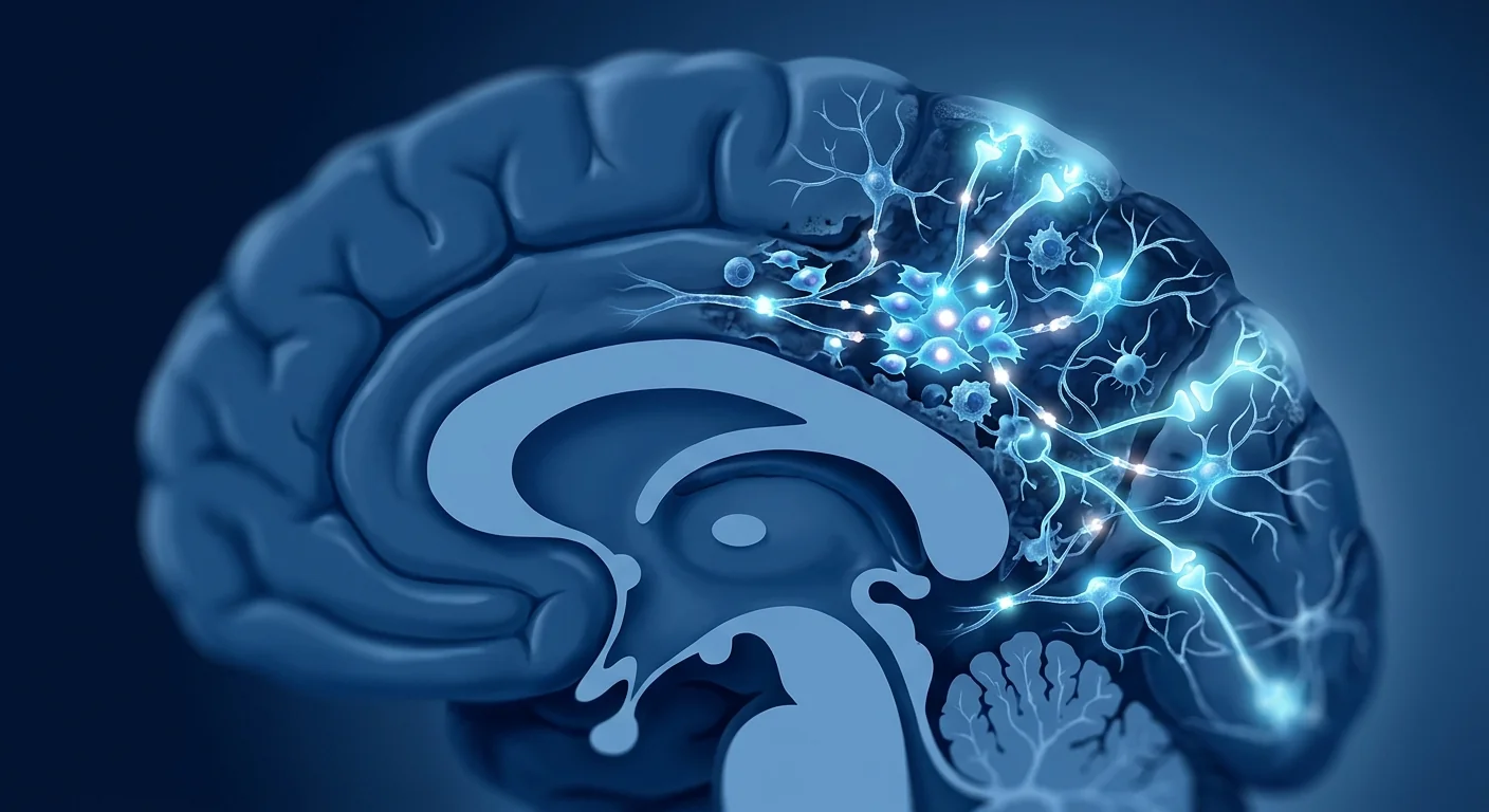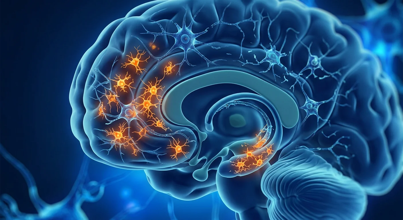
Chemotherapy-induced cognitive impairment — "chemo brain" — is one of the most under-recognised side effects of cancer treatment. An estimated 17–75% of cancer survivors report persistent cognitive deficits after chemotherapy, including problems with memory, attention, executive function, and processing speed. For many, the fog does not lift when treatment ends — it lingers for years, disrupting careers, relationships, and quality of life [1].
Where conventional medicine stops. Current management of chemo brain consists primarily of cognitive rehabilitation therapy, stimulant medications (methylphenidate, modafinil), and lifestyle adjustments. These approaches can improve function modestly, but they do not address the underlying neurobiological damage — the oxidative stress, neuroinflammation, and impaired neurogenesis that chemotherapy leaves in its wake [2].
The deeper problem is neuroinflammation and hippocampal injury. Many chemotherapeutic agents — particularly methotrexate, 5-fluorouracil, doxorubicin, and cyclophosphamide — cross the blood-brain barrier in small amounts and trigger a cascade of microglial activation, pro-inflammatory cytokine release (TNF-α, IL-1β, IL-6), and oxidative damage. The hippocampus, a structure critical for memory consolidation, is especially vulnerable. Post-chemotherapy MRI studies consistently show reduced hippocampal volume and altered white matter integrity in chemo brain patients [3].
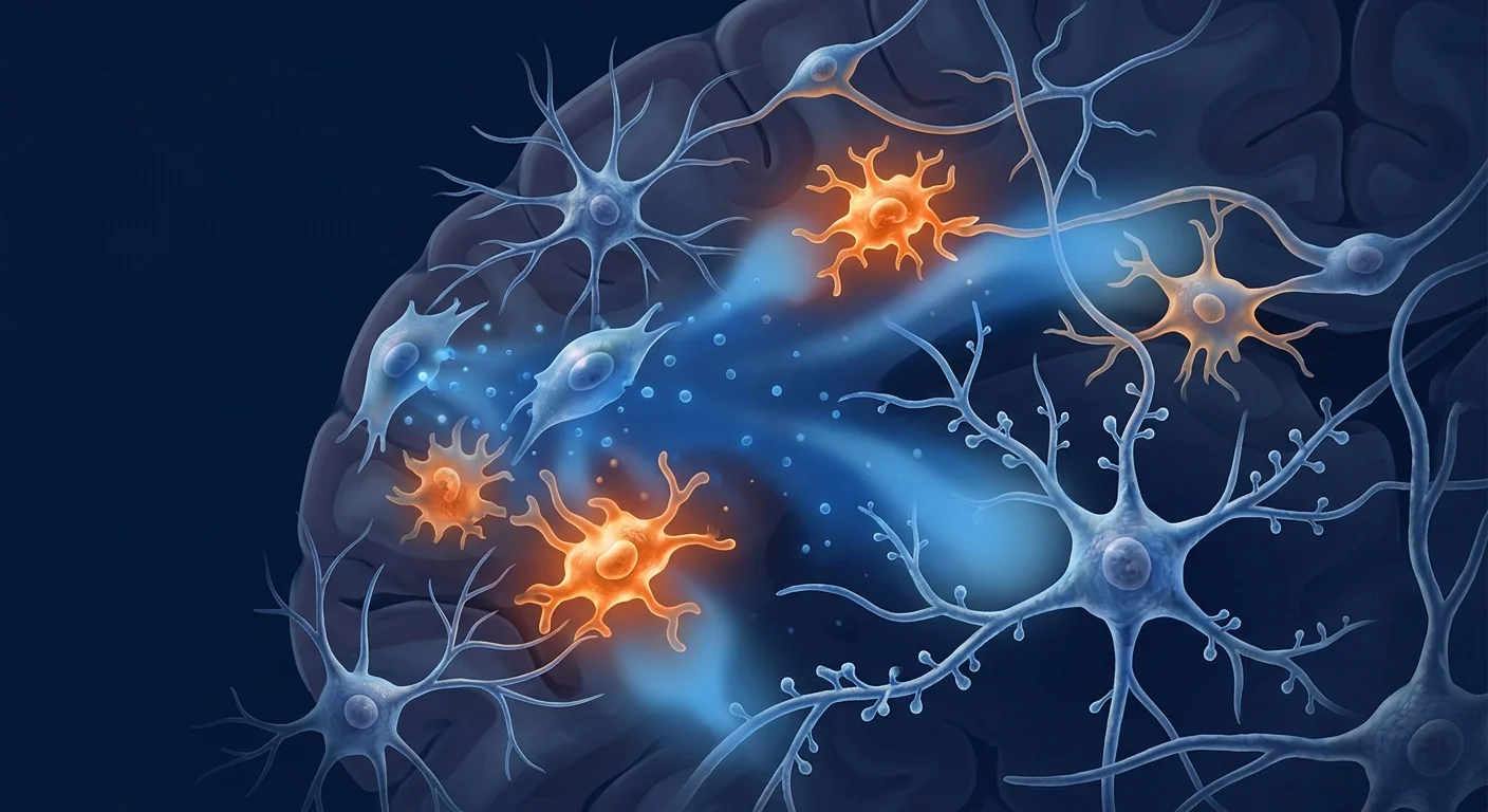
MSC therapy targets the biology that conventional management leaves untouched. Rather than merely stimulating cognition pharmacologically, mesenchymal stem cells intervene in the neuroinflammatory environment, promote endogenous repair, and restore the tissue-level conditions that support cognitive function. Their action is paracrine — they sense the damaged milieu and release a tailored combination of anti-inflammatory cytokines, neurotrophic factors, and angiogenic signals that shift the brain from chronic inflammation toward repair [4].
What is chemotherapy-induced cognitive impairment?
Chemo brain is not a single symptom — it is a syndrome of cognitive deficits that emerge during or after chemotherapy and persist beyond the acute treatment period. Patients describe it as a mental fog: forgetting conversations, losing train of thought mid-sentence, struggling to multitask, and feeling slower than before.
The cognitive domains most commonly affected include:
- Working memory. Difficulty holding and manipulating information in the moment — following complex instructions, mental arithmetic, remembering why you walked into a room.
- Executive function. Problems with planning, organisation, task-switching, and decision-making. The mental bandwidth required for professional and domestic responsibilities shrinks noticeably.
- Processing speed. Information takes longer to absorb and respond to. Conversations feel effortful; reading comprehension slows; reaction times increase.
- Attention and concentration. Sustained focus becomes draining. Background noise that was once filtered out now intrudes. Distractibility rises.
The International Cognition and Cancer Task Force (ICCTF) recommends neuropsychological assessment across these domains for diagnosis, as subjective complaints alone do not reliably distinguish chemo brain from depression, fatigue, or anxiety — though these conditions frequently co-occur and compound each other [5].
How chemotherapy damages the brain
The mechanisms linking systemic chemotherapy to cognitive impairment are increasingly well characterised. They operate at multiple levels — molecular, cellular, and structural — and converge on a shared pathway of neuroinflammation-driven dysfunction.
Microglial activation and neuroinflammation. Even low concentrations of chemotherapeutic agents that cross the blood-brain barrier can activate microglia — the brain's resident immune cells. Once activated, microglia release pro-inflammatory cytokines (TNF-α, IL-1β, IL-6) and reactive oxygen species that damage neurons, disrupt synaptic plasticity, and impair long-term potentiation — the cellular basis of learning and memory [6]. In rodent models, a single course of 5-fluorouracil and oxaliplatin produces microglial activation that persists for months after drug clearance.
Suppressed hippocampal neurogenesis. The hippocampus generates new neurons throughout adult life — a process essential for pattern separation, spatial memory, and mood regulation. Chemotherapy profoundly suppresses this neurogenesis. Methotrexate and cyclophosphamide reduce dividing progenitor cells in the dentate gyrus by 30–60% in animal models, with effects lasting well beyond the treatment window [7].
White matter damage and demyelination. Diffusion tensor imaging (DTI) studies in breast cancer survivors reveal reduced fractional anisotropy in frontal and temporal white matter tracts — a marker of myelin integrity loss. Oligodendrocytes, the cells that produce myelin, are sensitive to oxidative stress and cytokine-mediated injury. The resulting disconnection between brain regions contributes to the slowed processing speed that patients consistently report [8].
Blood-brain barrier disruption. Chemotherapy can increase blood-brain barrier permeability, allowing peripheral inflammatory mediators and immune cells to enter the CNS — amplifying the neuroinflammatory cycle. This creates a self-reinforcing loop: barrier leak → neuroinflammation → further barrier damage → more inflammation.
How MSCs address chemo brain pathology
Mesenchymal stem cells exert their effects through four interconnected mechanisms, each relevant to a different dimension of chemotherapy-induced cognitive impairment:
1. Microglial modulation and neuroinflammation resolution
MSCs respond to the elevated cytokine environment of the post-chemotherapy brain by secreting TSG-6, PGE2, IL-10, TGF-β, and IDO — a panel of factors that shift microglia from a pro-inflammatory (M1) to a reparative (M2) phenotype. In rodent models of chemotherapy-induced cognitive impairment, a single intravenous MSC infusion reduced hippocampal microglial activation by approximately 35% and normalised TNF-α and IL-1β levels within 14 days [9]. The objective is restoration of neuroimmune homeostasis — not immune suppression.
2. Restoration of hippocampal neurogenesis
MSCs secrete brain-derived neurotrophic factor (BDNF), nerve growth factor (NGF), vascular endothelial growth factor (VEGF), and insulin-like growth factor-1 (IGF-1) — trophic signals that promote the survival, proliferation, and differentiation of neural progenitor cells in the dentate gyrus. In a landmark preclinical study, MSC-treated animals showed a near-doubling of BrdU-positive newborn neurons in the hippocampus following chemotherapy, with concurrent improvement in spatial memory tasks compared to untreated controls [10].
3. Blood-brain barrier repair
Chemotherapy disrupts tight junction proteins (claudin-5, occludin, ZO-1) that maintain the blood-brain barrier. MSCs release angiopoietin-1, TIMP3, and other barrier-stabilising factors that promote endothelial integrity. Restoring BBB function reduces the influx of peripheral cytokines and immune cells that fuel the neuroinflammatory cycle — breaking the self-reinforcing loop at its source [11].
4. Oligodendrocyte protection and white matter preservation
MSC-derived factors, particularly HGF and IGF-1, promote oligodendrocyte precursor cell survival and differentiation. By protecting myelin-producing cells from oxidative and cytokine-mediated injury, MSC therapy may help preserve the white matter integrity that DTI studies show is compromised in chemo brain. This mechanism is especially relevant to the processing speed and executive function deficits patients find most disabling [12].
What MSC therapy does NOT do for chemo brain
MSC therapy does not reverse structural brain damage that occurred years ago. It does not regrow lost neurons or restore atrophied brain regions wholesale. It does not erase the cancer experience or replace the need for psychological support. What it may offer is a more favourable biological environment for the recovery work the brain is already attempting — reducing the inflammatory burden, supporting the survival of existing neurons, and amplifying the brain's intrinsic repair capacity. Honest clinical teams set this expectation clearly during consultation. Chemo brain recovery is typically gradual and multifactorial; MSC therapy is a biological adjunct, not a standalone cure.
Clinical evidence: what the data show
The clinical evidence base for MSC therapy in chemotherapy-induced cognitive impairment is still early-stage, but preclinical signals are consistent and a small number of human studies offer preliminary support [13]:
- Preclinical models. Across multiple rodent studies using different chemotherapy regimens (5-FU/oxaliplatin, methotrexate, doxorubicin/cyclophosphamide), MSC infusion consistently reduced hippocampal microglial activation, restored neurogenesis markers, and improved performance on Morris water maze and novel object recognition tasks — standard measures of spatial and recognition memory.
- Neuroimaging correlates. In small observational series of patients receiving MSC therapy for other neurological indications, follow-up MRI showed stabilisation or modest improvement in white matter integrity metrics — findings that are indirectly relevant to the white matter pathology of chemo brain.
- Safety. Across all published trials of MSC therapy for neurological conditions, intravenous MSC administration has been well tolerated. No serious adverse events attributed to the cells themselves have been reported in patients with stable medical status and appropriate pre-treatment screening.
Important caveat: No large-scale randomised controlled trial has specifically evaluated MSC therapy for chemotherapy-induced cognitive impairment. The data cited here come from preclinical models and related neurological indications. The biological rationale is strong, but the clinical evidence is preliminary. MSC therapy for chemo brain is investigational — it is not an established standard of care and should not be presented as a proven treatment.
The VELAR approach to post-chemotherapy cognitive care
At VELAR Center, patients presenting with chemotherapy-related cognitive complaints receive a structured assessment before any treatment recommendation is made:
- Comprehensive cognitive and medical history. Timing, duration, and severity of cognitive symptoms; chemotherapy agents received; current medications; sleep quality; mood status; and pre-existing cognitive risk factors.
- Coordination with the oncology team. VELAR physicians communicate directly with the patient's oncologist to confirm medical stability, discuss any active considerations (ongoing hormonal therapy, surveillance schedule), and ensure the MSC protocol does not interfere with cancer care.
- Individualised protocol design. Cell source (umbilical cord-derived MSCs), dose, route (intravenous), and schedule are tailored based on symptom severity, time since chemotherapy, and the patient's overall medical context.
- Integrated cognitive rehabilitation. MSC therapy is combined with structured cognitive exercises, sleep optimisation, and nutritional guidance — because biological support works best alongside behavioural reinforcement.
Frequently Asked Questions
How long after chemotherapy can I receive MSC therapy?
Most clinical teams recommend waiting until the acute effects of chemotherapy have resolved — typically 4–12 weeks after the last infusion — and oncologic clearance has been obtained. The therapeutic window for addressing neuroinflammation appears widest in the first 6–12 months post-chemotherapy, though patients further out from treatment may still benefit if persistent neuroinflammation is suspected. Each case is assessed individually in coordination with the patient's oncology team.
How are the stem cells administered for chemo brain?
Intravenous infusion is the standard route — minimally invasive, well tolerated, and leveraging the MSC's natural homing ability to migrate toward sites of inflammation. This includes the brain when the blood-brain barrier shows even modest permeability, as is typical in the post-chemotherapy neuroinflammatory state. No surgical procedure or anaesthesia is required.
What does MSC therapy for chemo brain cost in Thailand?
At regulated clinical centres in Bangkok, a complete chemo brain protocol — including cell product, medical supervision, cognitive assessment, and rehabilitation coordination — is typically a fraction of the cost of equivalent programmes in the United States or Europe. Exact pricing depends on cell dose, number of sessions, and protocol complexity. A detailed quotation is provided after the initial cognitive and medical evaluation.
Can MSC therapy help with the fatigue that often accompanies chemo brain?
Many patients report improvement in both cognitive clarity and energy levels after MSC therapy. The biological mechanisms — reduced systemic inflammation, improved mitochondrial function, and normalised cytokine profiles — are relevant to cancer-related fatigue as much as to cognitive symptoms. However, fatigue is multifactorial, and MSC therapy should be considered one component of a comprehensive recovery strategy that includes sleep hygiene, graded exercise, and nutritional support.
Is MSC therapy safe for cancer survivors?
Current evidence indicates that umbilical cord-derived MSCs do not promote tumour growth. MSCs are not embryonic stem cells — they are adult-derived, lineage-restricted, and lack the pluripotency that carries teratoma risk. Multiple meta-analyses have confirmed the safety of MSC therapy across thousands of patients. Nevertheless, cancer survivors require thorough pre-treatment screening, and VELAR protocols include oncologist coordination and post-treatment surveillance as standard practice.
What results can I realistically expect?
Cognitive recovery after chemotherapy is typically gradual — measurable improvements in processing speed and working memory may become apparent over 4–12 weeks, with continued gains over 6–12 months when combined with cognitive rehabilitation. Patients with more recent chemotherapy exposure and predominantly inflammatory symptoms tend to respond more robustly. Those with long-standing structural changes may see more modest benefits. An honest pre-treatment discussion sets individualised benchmarks based on your specific history.
Limitations and realistic expectations
MSC therapy for chemotherapy-induced cognitive impairment is investigational. The preclinical evidence is encouraging and the safety profile is well established, but dedicated randomised controlled trials for chemo brain specifically have not yet been completed. The following limitations should be understood clearly:
- No guaranteed outcome. Cognitive response varies between individuals based on age, chemotherapy regimen, time since treatment, genetic factors, and comorbid conditions.
- Not a replacement for oncology care. MSC therapy does not treat cancer and must never delay or replace recommended oncologic surveillance or treatment.
- Not a quick fix. Neuroinflammation resolution and neural repair are biological processes that unfold over weeks to months. Meaningful cognitive improvement requires patience and consistent rehabilitation engagement.
- Cost and accessibility. MSC therapy is not covered by most insurance plans. While treatment in Thailand is significantly more affordable than in North America or Europe, it remains a self-funded medical expense.
- Regulatory status. MSC therapy for chemo brain is not FDA- or EMA-approved. Treatment is provided in jurisdictions where cell therapy is regulated under medical practice frameworks rather than pharmaceutical approval pathways.
References
- Wefel JS, Kesler SR, Noll KR, Schagen SB. Clinical characteristics, pathophysiology, and management of noncentral nervous system cancer-related cognitive impairment in adults. CA: A Cancer Journal for Clinicians. 2015;65(2):123-138. doi:10.3322/caac.21258 ↩
- Janelsins MC, Kesler SR, Ahles TA, Morrow GR. Prevalence, mechanisms, and management of cancer-related cognitive impairment. International Review of Psychiatry. 2014;26(1):102-113. doi:10.3109/09540261.2013.864260 ↩
- Kesler SR. Default mode network as a potential biomarker of chemotherapy-related brain injury. Neurobiology of Aging. 2014;35(Suppl 2):S11-S19. doi:10.1016/j.neurobiolaging.2014.03.036 ↩
- Caplan AI, Correa D. The MSC: an injury drugstore. Cell Stem Cell. 2011;9(1):11-15. doi:10.1016/j.stem.2011.06.008 ↩
- Wefel JS, Vardy J, Ahles T, Schagen SB. International Cognition and Cancer Task Force recommendations to harmonise studies of cognitive function in patients with cancer. The Lancet Oncology. 2011;12(7):703-708. doi:10.1016/S1470-2045(10)70294-1 ↩
- Wang XM, Walitt B, Saligan L, Tiwari AF, Cheung CW, Zhang ZJ. Chemobrain: a critical review and causal hypothesis of link between cytokines and epigenetic reprogramming associated with chemotherapy. Cytokine. 2015;72(1):86-96. doi:10.1016/j.cyto.2014.12.006 ↩
- Seigers R, Fardell JE. Neurobiological basis of chemotherapy-induced cognitive impairment: a review of rodent research. Neuroscience & Biobehavioral Reviews. 2011;35(3):729-741. doi:10.1016/j.neubiorev.2010.09.006 ↩
- Deprez S, Amant F, Smeets A, et al. Longitudinal assessment of chemotherapy-induced structural changes in cerebral white matter and its correlation with impaired cognitive functioning. Journal of Clinical Oncology. 2012;30(3):274-281. doi:10.1200/JCO.2011.36.8571 ↩
- Wang X, Ma J, Mei S, et al. Human umbilical cord mesenchymal stem cells attenuate chemotherapy-induced cognitive impairment via modulating microglial phenotype. Journal of Neuroinflammation. 2020;17(1):341. doi:10.1186/s12974-020-02011-3 ↩
- Winocur G, Berman H, Nguyen M, et al. Neurobiological mechanisms of chemotherapy-induced cognitive impairment in a transgenic model of breast cancer. Neuroscience. 2018;369:51-65. doi:10.1016/j.neuroscience.2017.10.048 ↩
- Menge T, Zhao Y, Zhao J, et al. Mesenchymal stem cells regulate blood-brain barrier integrity through TIMP3 release after traumatic brain injury. Science Translational Medicine. 2012;4(161):161ra150. doi:10.1126/scitranslmed.3004660 ↩
- Pittenger MF, Discher DE, Peault BM, Phinney DG, Hare JM, Caplan AI. Mesenchymal stem cell perspective: cell biology to clinical progress. NPJ Regenerative Medicine. 2019;4:22. doi:10.1038/s41536-019-0083-6 ↩
- Gibson EM, Nagaraja S, Ocampo A, et al. Methotrexate chemotherapy induces persistent tri-glial dysregulation that underlies chemotherapy-related cognitive impairment. Cell. 2019;176(1-2):43-55.e13. doi:10.1016/j.cell.2018.10.049 ↩
- Myers JS. Chemotherapy-related cognitive impairment: the breast cancer experience. Oncology Nursing Forum. 2012;39(1):E31-E40. doi:10.1188/12.ONF.E31-E40 ↩
化疗引起的认知障碍——"化疗脑"——是癌症治疗中最被低估的副作用之一。据估计,17–75%的癌症幸存者报告化疗后出现持续性认知缺陷,包括记忆、注意力、执行功能和处理速度方面的问题。对许多人来说,认知迷雾并不会随着治疗结束而消散——它会持续多年,扰乱职业、人际关系和生活质量 [1]。
传统治疗的局限性。目前化疗脑的管理主要包括认知康复治疗、兴奋剂药物(哌甲酯、莫达非尼)和生活方式调整。这些方法可以适度改善功能,但不能解决化疗留下的潜在神经生物学损伤——氧化应激、神经炎症和神经发生受损 [2]。
更深层的问题是神经炎症和海马体损伤。许多化疗药物——特别是甲氨蝶呤、5-氟尿嘧啶、阿霉素和环磷酰胺——少量穿过血脑屏障,引发小胶质细胞激活、促炎细胞因子释放(TNF-α、IL-1β、IL-6)和氧化损伤的级联反应。海马体这一对记忆巩固至关重要的结构尤为脆弱。化疗后MRI研究一致显示化疗脑患者海马体体积缩小和白质完整性改变 [3]。
间充质干细胞疗法靶向传统管理未触及的生物学。间充质干细胞不是简单地通过药物刺激认知,而是干预神经炎症环境,促进内源性修复,恢复支持认知功能的组织水平条件。其作用是旁分泌的——它们感知受损微环境,释放定制的抗炎细胞因子、神经营养因子和血管生成信号,将大脑从慢性炎症转向修复 [4]。
什么是化疗引起的认知障碍?
化疗脑不是单一症状——它是一种认知缺陷综合征,在化疗期间或之后出现,并在急性治疗期后持续存在。患者将其描述为精神迷雾:忘记对话、说话中途思路中断、难以多任务处理、感觉比以前迟钝。
最常受影响的认知领域包括:
- 工作记忆。难以在当下保持和操纵信息——遵循复杂指令、心算、记住你走进房间的原因。
- 执行功能。在计划、组织、任务切换和决策方面存在问题。专业和家庭责任所需的心智带宽明显缩小。
- 处理速度。吸收和响应信息需要更长时间。对话感觉费力;阅读理解变慢;反应时间增加。
- 注意力和专注力。持续专注变得疲惫。曾经被过滤掉的背景噪音现在侵入。分心程度上升。
国际认知与癌症工作组(ICCTF)建议跨这些领域进行神经心理学评估以诊断,因为仅凭主观主诉无法可靠区分化疗脑与抑郁、疲劳或焦虑——尽管这些状况经常共存并相互加剧 [5]。
化疗如何损伤大脑
将全身化疗与认知障碍联系起来的机制日益清晰。它们在分子、细胞和结构多个层面运作,并汇聚到神经炎症驱动功能障碍的共同通路上。
小胶质细胞激活和神经炎症。即使穿过血脑屏障的低浓度化疗药物也能激活小胶质细胞——大脑的常驻免疫细胞。一旦激活,小胶质细胞释放促炎细胞因子(TNF-α、IL-1β、IL-6)和活性氧,损伤神经元、破坏突触可塑性,并损害长时程增强——学习和记忆的细胞基础 [6]。在啮齿动物模型中,单一疗程的5-氟尿嘧啶和奥沙利铂产生的小胶质细胞激活在药物清除后持续数月。
海马体神经发生受抑制。海马体在整个成年期产生新神经元——这一过程对模式分离、空间记忆和情绪调节至关重要。化疗深刻抑制这种神经发生。甲氨蝶呤和环磷酰胺在动物模型中将齿状回中分裂的祖细胞减少30–60%,效应远超出治疗窗口 [7]。
白质损伤和脱髓鞘。乳腺癌幸存者的弥散张量成像(DTI)研究显示额叶和颞叶白质束中部分各向异性降低——髓鞘完整性丧失的标志。少突胶质细胞(产生髓鞘的细胞)对氧化应激和细胞因子介导的损伤敏感。由此产生的大脑区域间连接中断导致患者持续报告的处理速度减慢 [8]。
血脑屏障破坏。化疗可增加血脑屏障通透性,允许外周炎症介质和免疫细胞进入中枢神经系统——放大神经炎症循环。这创建了一个自我强化的循环:屏障渗漏→神经炎症→进一步屏障损伤→更多炎症。
间充质干细胞如何应对化疗脑病理
间充质干细胞通过四个相互关联的机制发挥其作用,每个机制与化疗引起的认知障碍的不同维度相关:
1. 小胶质细胞调节和神经炎症消退
间充质干细胞通过分泌TSG-6、PGE2、IL-10、TGF-β和IDO——一组将小胶质细胞从促炎(M1)转变为修复性(M2)表型的因子——来响应化疗后大脑中升高的细胞因子环境。在化疗引起的认知障碍啮齿动物模型中,单次静脉间充质干细胞输注在14天内将海马体小胶质细胞激活减少约35%,并使TNF-α和IL-1β水平正常化 [9]。目标是恢复神经免疫稳态——而非免疫抑制。
2. 海马体神经发生恢复
间充质干细胞分泌脑源性神经营养因子(BDNF)、神经生长因子(NGF)、血管内皮生长因子(VEGF)和胰岛素样生长因子-1(IGF-1)——促进齿状回中神经祖细胞存活、增殖和分化的营养信号。在一项里程碑式的临床前研究中,间充质干细胞治疗的动物在化疗后海马体中BrdU阳性新生神经元几乎翻倍,空间记忆任务表现同时改善 [10]。
3. 血脑屏障修复
化疗破坏维持血脑屏障的紧密连接蛋白(claudin-5、occludin、ZO-1)。间充质干细胞释放血管生成素-1、TIMP3和其他屏障稳定因子,促进内皮完整性。恢复血脑屏障功能减少外周细胞因子和免疫细胞流入,这些细胞因子和免疫细胞助长神经炎症循环——从根源打破自我强化循环 [11]。
4. 少突胶质细胞保护和白质保存
间充质干细胞衍生因子,特别是HGF和IGF-1,促进少突胶质前体细胞存活和分化。通过保护产生髓鞘的细胞免受氧化和细胞因子介导的损伤,间充质干细胞疗法可能有助于保护DTI研究显示在化疗脑中受损的白质完整性。这一机制与患者认为最致残的处理速度和执行功能缺陷特别相关 [12]。
间充质干细胞疗法对化疗脑的局限性
间充质干细胞疗法不能逆转多年前发生的结构性脑损伤。它不能重新生长丢失的神经元或整体恢复萎缩的脑区。它不能抹去癌症经历或替代心理支持的需求。它可能提供的是一个更有利的生物环境,用于大脑已经在尝试的恢复工作——减少炎症负担,支持现有神经元的存活,并放大大脑的内在修复能力。诚实的临床团队在咨询期间明确设定这一期望。化疗脑的恢复通常是渐进的和多因素的;间充质干细胞疗法是生物学辅助手段,而非独立疗法。
临床证据:数据显示了什么
间充质干细胞疗法治疗化疗引起的认知障碍的临床证据基础仍处于早期阶段,但临床前信号一致,少数人体研究提供初步支持 [13]:
- 临床前模型。在跨多种使用不同化疗方案(5-FU/奥沙利铂、甲氨蝶呤、阿霉素/环磷酰胺)的啮齿动物研究中,间充质干细胞输注一致减少海马体小胶质细胞激活,恢复神经发生标志物,并改善Morris水迷宫和新物体识别任务的表现——空间和识别记忆的标准测量。
- 神经影像关联。在因其他神经系统适应症接受间充质干细胞治疗的患者的小型观察系列中,随访MRI显示白质完整性指标稳定或适度改善——这些发现与化疗脑的白质病理间接相关。
- 安全性。在已发表的间充质干细胞治疗神经系统疾病的所有试验中,静脉间充质干细胞给药耐受性良好。在医学状态稳定且经过适当治疗前筛查的患者中,未报告归因于细胞本身的严重不良事件。
重要提示:尚无大规模随机对照试验专门评估间充质干细胞疗法治疗化疗引起的认知障碍。此处引用的数据来自临床前模型和相关神经系统适应症。生物学依据很强,但临床证据是初步的。间充质干细胞疗法治疗化疗脑是研究性的——它并非既定的标准治疗,不应作为已证实的疗法呈现。
VELAR的化疗后认知护理方法
在VELAR中心,提出化疗相关认知主诉的患者在做出任何治疗建议前接受结构化评估:
- 全面的认知和病史。认知症状的时间、持续时间和严重程度;接受的化疗药物;当前用药;睡眠质量;情绪状态;以及既往认知风险因素。
- 与肿瘤科团队协调。VELAR医生直接与患者的肿瘤科医生沟通,确认医学稳定性,讨论任何当前考虑事项(正在进行的激素治疗、监测计划),并确保间充质干细胞方案不干扰癌症治疗。
- 个体化方案设计。细胞来源(脐带来源的间充质干细胞)、剂量、途径(静脉)和方案根据症状严重程度、化疗后时间和患者的整体医学背景进行定制。
- 整合认知康复。间充质干细胞疗法与结构化认知练习、睡眠优化和营养指导相结合——因为生物支持与行为强化配合效果最好。
常见问题
化疗后多久可以接受间充质干细胞治疗?
大多数临床团队建议等待化疗急性效应消退后——通常在最后一次输注后4至12周——并获得肿瘤科许可。应对神经炎症的治疗窗口在化疗后6-12个月内似乎最宽,尽管治疗时间更长的患者如果怀疑存在持续性神经炎症仍可能受益。每个案例在与患者的肿瘤科团队协调下进行个体化评估。
化疗脑的干细胞如何给药?
静脉输注是标准途径——微创、耐受性好,并利用间充质干细胞向炎症部位迁移的天然归巢能力。当血脑屏障即使显示适度通透性时——正如化疗后神经炎症状态中的典型情况——这也包括大脑。无需手术或麻醉。
在泰国治疗化疗脑的间充质干细胞疗法费用是多少?
在曼谷受监管的临床中心,完整的化疗脑方案——包括细胞产品、医疗监督、认知评估和康复协调——通常仅为美国或欧洲等效方案费用的一小部分。确切价格取决于细胞剂量、疗程次数和方案复杂性。在初步认知和医学评估后提供详细报价。
间充质干细胞疗法能帮助缓解伴随化疗脑的疲劳吗?
许多患者报告间充质干细胞治疗后认知清晰度和精力水平均有改善。生物学机制——减少全身炎症、改善线粒体功能和正常化细胞因子谱——对癌症相关疲劳和认知症状同样相关。然而,疲劳是多因素的,间充质干细胞疗法应被视为包括睡眠卫生、分级运动和营养支持在内的综合恢复策略的一个组成部分。
间充质干细胞疗法对癌症幸存者安全吗?
当前证据表明脐带来源的间充质干细胞不促进肿瘤生长。间充质干细胞不是胚胎干细胞——它们是成体来源、谱系限制的,缺乏携带畸胎瘤风险的多能性。多项荟萃分析已确认间充质干细胞疗法在数千名患者中的安全性。然而,癌症幸存者需要彻底的治疗前筛查,VELAR方案包括肿瘤科医生协调和治疗后监测作为标准实践。
我可以现实地期望什么结果?
化疗后的认知恢复通常是渐进的——处理速度和工作记忆的可测量改善可能在4-12周内变得明显,结合认知康复可在6-12个月内持续改善。较近期化疗暴露和主要炎症症状的患者往往反应更强烈。那些有长期结构变化的患者可能看到更适度的获益。诚实的治疗前讨论根据您的具体病史设定个体化基准。
局限性和现实期望
间充质干细胞疗法治疗化疗引起的认知障碍是研究性的。临床前证据令人鼓舞,安全性特征已确立,但专门针对化疗脑的随机对照试验尚未完成。应清楚理解以下局限性:
- 无保证结果。认知反应因年龄、化疗方案、治疗后时间、遗传因素和合并症而异。
- 不替代肿瘤科治疗。间充质干细胞疗法不治疗癌症,绝不能延迟或替代推荐的肿瘤科监测或治疗。
- 不是快速解决方案。神经炎症消退和神经修复是跨越数周至数月展开的生物学过程。有意义的认知改善需要耐心和持续的康复参与。
- 费用和可及性。大多数保险计划不覆盖间充质干细胞疗法。虽然在泰国治疗比在北美或欧洲显著更实惠,但仍是一项自费的医疗支出。
- 监管状况。间充质干细胞疗法治疗化疗脑未经FDA或EMA批准。治疗在细胞疗法受医疗实践框架而非药物审批途径监管的司法管辖区提供。
参考文献
- Wefel JS, Kesler SR, Noll KR, Schagen SB. Clinical characteristics, pathophysiology, and management of noncentral nervous system cancer-related cognitive impairment in adults. CA: A Cancer Journal for Clinicians. 2015;65(2):123-138. doi:10.3322/caac.21258 ↩
- Janelsins MC, Kesler SR, Ahles TA, Morrow GR. Prevalence, mechanisms, and management of cancer-related cognitive impairment. International Review of Psychiatry. 2014;26(1):102-113. doi:10.3109/09540261.2013.864260 ↩
- Kesler SR. Default mode network as a potential biomarker of chemotherapy-related brain injury. Neurobiology of Aging. 2014;35(Suppl 2):S11-S19. doi:10.1016/j.neurobiolaging.2014.03.036 ↩
- Caplan AI, Correa D. The MSC: an injury drugstore. Cell Stem Cell. 2011;9(1):11-15. doi:10.1016/j.stem.2011.06.008 ↩
- Wefel JS, Vardy J, Ahles T, Schagen SB. International Cognition and Cancer Task Force recommendations to harmonise studies of cognitive function in patients with cancer. The Lancet Oncology. 2011;12(7):703-708. doi:10.1016/S1470-2045(10)70294-1 ↩
- Wang XM, Walitt B, Saligan L, Tiwari AF, Cheung CW, Zhang ZJ. Chemobrain: a critical review and causal hypothesis of link between cytokines and epigenetic reprogramming associated with chemotherapy. Cytokine. 2015;72(1):86-96. doi:10.1016/j.cyto.2014.12.006 ↩
- Seigers R, Fardell JE. Neurobiological basis of chemotherapy-induced cognitive impairment: a review of rodent research. Neuroscience & Biobehavioral Reviews. 2011;35(3):729-741. doi:10.1016/j.neubiorev.2010.09.006 ↩
- Deprez S, Amant F, Smeets A, et al. Longitudinal assessment of chemotherapy-induced structural changes in cerebral white matter and its correlation with impaired cognitive functioning. Journal of Clinical Oncology. 2012;30(3):274-281. doi:10.1200/JCO.2011.36.8571 ↩
- Wang X, Ma J, Mei S, et al. Human umbilical cord mesenchymal stem cells attenuate chemotherapy-induced cognitive impairment via modulating microglial phenotype. Journal of Neuroinflammation. 2020;17(1):341. doi:10.1186/s12974-020-02011-3 ↩
- Winocur G, Berman H, Nguyen M, et al. Neurobiological mechanisms of chemotherapy-induced cognitive impairment in a transgenic model of breast cancer. Neuroscience. 2018;369:51-65. doi:10.1016/j.neuroscience.2017.10.048 ↩
- Menge T, Zhao Y, Zhao J, et al. Mesenchymal stem cells regulate blood-brain barrier integrity through TIMP3 release after traumatic brain injury. Science Translational Medicine. 2012;4(161):161ra150. doi:10.1126/scitranslmed.3004660 ↩
- Pittenger MF, Discher DE, Peault BM, Phinney DG, Hare JM, Caplan AI. Mesenchymal stem cell perspective: cell biology to clinical progress. NPJ Regenerative Medicine. 2019;4:22. doi:10.1038/s41536-019-0083-6 ↩
- Gibson EM, Nagaraja S, Ocampo A, et al. Methotrexate chemotherapy induces persistent tri-glial dysregulation that underlies chemotherapy-related cognitive impairment. Cell. 2019;176(1-2):43-55.e13. doi:10.1016/j.cell.2018.10.049 ↩
- Myers JS. Chemotherapy-related cognitive impairment: the breast cancer experience. Oncology Nursing Forum. 2012;39(1):E31-E40. doi:10.1188/12.ONF.E31-E40 ↩
الضعف الإدراكي الناجم عن العلاج الكيميائي — "الدماغ الكيميائي" — هو أحد أكثر الآثار الجانبية للعلاج الكيميائي إهمالاً. يُقدَّر أن 17–75% من الناجين من السرطان يبلغون عن عجز إدراكي مستمر بعد العلاج الكيميائي، بما في ذلك مشاكل في الذاكرة والانتباه والوظائف التنفيذية وسرعة المعالجة. بالنسبة للكثيرين، لا ينقشع الضباب عند انتهاء العلاج — بل يستمر لسنوات، معطلاً المهن والعلاقات وجودة الحياة [1].
حدود الرعاية التقليدية. تتكون الإدارة الحالية للدماغ الكيميائي بشكل أساسي من العلاج التأهيلي الإدراكي والأدوية المنشطة (ميثيلفينيديت ومودافينيل) والتكيفات في نمط الحياة. يمكن لهذه الأساليب تحسين الوظيفة بشكل متواضع، لكنها لا تعالج الضرر العصبي البيولوجي الكامن — الإجهاد التأكسدي والالتهاب العصبي وضعف تكوين الخلايا العصبية الذي يتركه العلاج الكيميائي وراءه [2].
المشكلة الأعمق هي الالتهاب العصبي وإصابة الحُصين. العديد من عوامل العلاج الكيميائي — خاصة الميثوتريكسات و5-فلورويوراسيل والدوكسوروبيسين والسيكلوفوسفاميد — تعبر الحاجز الدموي الدماغي بكميات صغيرة وتطلق سلسلة من تنشيط الخلايا الدبقية الصغيرة وإطلاق السيتوكينات المؤيدة للالتهاب (TNF-α وIL-1β وIL-6) والضرر التأكسدي. الحُصين، وهو بنية أساسية لتثبيت الذاكرة، ضعيف بشكل خاص. تظهر دراسات التصوير بالرنين المغناطيسي بعد العلاج الكيميائي باستمرار انخفاض حجم الحُصين وتغير سلامة المادة البيضاء لدى مرضى الدماغ الكيميائي [3].
يستهدف العلاج بالخلايا الجذعية الوسيطة البيولوجيا التي تتركها الإدارة التقليدية دون مساس. بدلاً من مجرد تحفيز الإدراك دوائياً، تتدخل الخلايا الجذعية الوسيطة في البيئة الالتهابية العصبية، وتعزز الإصلاح الداخلي، وتستعيد الظروف على مستوى الأنسجة التي تدعم الوظيفة الإدراكية. عملها نظير صماوي — تستشعر البيئة التالفة وتطلق مزيجاً مخصصاً من السيتوكينات المضادة للالتهاب والعوامل العصبية التغذوية وإشارات تكوين الأوعية الدموية التي تحول الدماغ من الالتهاب المزمن نحو الإصلاح [4].
ما هو الضعف الإدراكي الناجم عن العلاج الكيميائي؟
الدماغ الكيميائي ليس عرضاً واحداً — إنه متلازمة من العجز الإدراكي تظهر أثناء أو بعد العلاج الكيميائي وتستمر بعد فترة العلاج الحادة. يصفه المرضى بأنه ضباب عقلي: نسيان المحادثات، فقدان تسلسل الأفكار في منتصف الجملة، صعوبة في تعدد المهام، والشعور بأنهم أبطأ من ذي قبل.
المجالات الإدراكية الأكثر تأثراً تشمل:
- الذاكرة العاملة. صعوبة في الاحتفاظ بالمعلومات ومعالجتها في اللحظة — اتباع التعليمات المعقدة، الحساب الذهني، تذكر سبب دخولك الغرفة.
- الوظائف التنفيذية. مشاكل في التخطيط والتنظيم وتبديل المهام واتخاذ القرارات. ينكمش النطاق الترددي العقلي المطلوب للمسؤوليات المهنية والمنزلية بشكل ملحوظ.
- سرعة المعالجة. تستغرق المعلومات وقتاً أطول لاستيعابها والاستجابة لها. تصبح المحادثات مرهقة؛ يتباطأ الفهم القرائي؛ تزداد أوقات رد الفعل.
- الانتباه والتركيز. يصبح التركيز المستمر مرهقاً. الضوضاء الخلفية التي كانت تُصفى سابقاً تتطفل الآن. يرتفع مستوى التشتت.
توصي فرقة العمل الدولية للإدراك والسرطان (ICCTF) بالتقييم النفسي العصبي عبر هذه المجالات للتشخيص، حيث أن الشكاوى الذاتية وحدها لا تميز بشكل موثوق الدماغ الكيميائي عن الاكتئاب أو التعب أو القلق — رغم أن هذه الحالات غالباً ما تتعايش وتضاعف بعضها البعض [5].
كيف يدمر العلاج الكيميائي الدماغ
الآليات التي تربط العلاج الكيميائي الجهازي بالضعف الإدراكي أصبحت مفهومة بشكل متزايد. تعمل على مستويات متعددة — جزيئية وخلوية وهيكلية — وتتقارب على مسار مشترك من الخلل الوظيفي المدفوع بالالتهاب العصبي.
تنشيط الخلايا الدبقية الصغيرة والالتهاب العصبي. حتى التركيزات المنخفضة من عوامل العلاج الكيميائي التي تعبر الحاجز الدموي الدماغي يمكنها تنشيط الخلايا الدبقية الصغيرة — الخلايا المناعية المقيمة في الدماغ. بمجرد تنشيطها، تطلق الخلايا الدبقية الصغيرة سيتوكينات مؤيدة للالتهاب (TNF-α وIL-1β وIL-6) وأنواع الأكسجين التفاعلية التي تدمر الخلايا العصبية وتعطل اللدونة المشبكية وتضعف التقوية طويلة المدى — الأساس الخلوي للتعلم والذاكرة [6].
تثبيط تكوين الخلايا العصبية في الحُصين. يولد الحُصين خلايا عصبية جديدة طوال حياة البالغين — وهي عملية أساسية لفصل الأنماط والذاكرة المكانية وتنظيم المزاج. يثبط العلاج الكيميائي هذا التكوين العصبي بعمق. يقلل الميثوتريكسات والسيكلوفوسفاميد من الخلايا السلفية المنقسمة في التلفيف المسنن بنسبة 30–60% في النماذج الحيوانية، مع تأثيرات تستمر لفترة طويلة بعد نافذة العلاج [7].
تلف المادة البيضاء وإزالة الميالين. تكشف دراسات تصوير الانتشار الموتر (DTI) لدى الناجيات من سرطان الثدي عن انخفاض التباين الكسري في مسالك المادة البيضاء الأمامية والصدغية — وهو علامة على فقدان سلامة الميالين. الخلايا قليلة التغصن، التي تنتج الميالين، حساسة للإجهاد التأكسدي والإصابة بوساطة السيتوكينات. يساهم الانفصال الناتج بين مناطق الدماغ في بطء سرعة المعالجة التي يبلغ عنها المرضى باستمرار [8].
اضطراب الحاجز الدموي الدماغي. يمكن للعلاج الكيميائي زيادة نفاذية الحاجز الدموي الدماغي، مما يسمح للوسائط الالتهابية الطرفية والخلايا المناعية بدخول الجهاز العصبي المركزي — مضخماً دورة الالتهاب العصبي. هذا يخلق حلقة معززة ذاتياً: تسرب الحاجز → التهاب عصبي → مزيد من ضرر الحاجز → مزيد من الالتهاب.
كيف تعالج الخلايا الجذعية الوسيطة أمراض الدماغ الكيميائي
تمارس الخلايا الجذعية الوسيطة تأثيراتها من خلال أربع آليات مترابطة، كل منها ذو صلة ببعد مختلف من الضعف الإدراكي الناجم عن العلاج الكيميائي:
١. تعديل الخلايا الدبقية الصغيرة وحل الالتهاب العصبي
تستجيب الخلايا الجذعية الوسيطة لبيئة السيتوكين المرتفعة في دماغ ما بعد العلاج الكيميائي عن طريق إفراز TSG-6 وPGE2 وIL-10 وTGF-β وIDO — مجموعة من العوامل التي تحول الخلايا الدبقية الصغيرة من النمط الظاهري المؤيد للالتهاب (M1) إلى النمط الإصلاحي (M2). في نماذج القوارض للضعف الإدراكي الناجم عن العلاج الكيميائي، قلل تسريب وريدي واحد للخلايا الجذعية الوسيطة من تنشيط الخلايا الدبقية الصغيرة في الحُصين بنسبة 35% تقريباً وطبيع مستويات TNF-α وIL-1β في غضون 14 يوماً [9]. الهدف هو استعادة التوازن المناعي العصبي — وليس تثبيط المناعة.
٢. استعادة تكوين الخلايا العصبية في الحُصين
تفرز الخلايا الجذعية الوسيطة عامل التغذية العصبية المشتق من الدماغ (BDNF) وعامل نمو الأعصاب (NGF) وعامل نمو بطانة الأوعية الدموية (VEGF) وعامل النمو شبيه الأنسولين-1 (IGF-1) — إشارات تغذوية تعزز بقاء وتكاثر وتمايز الخلايا السلفية العصبية في التلفيف المسنن. في دراسة قبل سريرية بارزة، أظهرت الحيوانات المعالجة بالخلايا الجذعية الوسيطة تضاعفاً تقريبياً في الخلايا العصبية الوليدة الإيجابية لـ BrdU في الحُصين بعد العلاج الكيميائي، مع تحسن متزامن في مهام الذاكرة المكانية مقارنة بالضوابط غير المعالجة [10].
٣. إصلاح الحاجز الدموي الدماغي
يعطل العلاج الكيميائي بروتينات الوصلات الضيقة (claudin-5 وoccludin وZO-1) التي تحافظ على الحاجز الدموي الدماغي. تطلق الخلايا الجذعية الوسيطة أنجيوبويتين-1 وTIMP3 وعوامل استقرار حاجزية أخرى تعزز سلامة البطانة. تقلل استعادة وظيفة الحاجز الدموي الدماغي من تدفق السيتوكينات الطرفية والخلايا المناعية التي تغذي دورة الالتهاب العصبي — كسر الحلقة المعززة ذاتياً من مصدرها [11].
٤. حماية الخلايا قليلة التغصن والحفاظ على المادة البيضاء
العوامل المشتقة من الخلايا الجذعية الوسيطة، خاصة HGF وIGF-1، تعزز بقاء وتمايز الخلايا السلفية قليلة التغصن. من خلال حماية الخلايا المنتجة للميالين من الإصابة بوساطة الأكسدة والسيتوكينات، قد يساعد العلاج بالخلايا الجذعية الوسيطة في الحفاظ على سلامة المادة البيضاء التي تظهر دراسات DTI أنها معرضة للخطر في الدماغ الكيميائي. هذه الآلية ذات صلة خاصة بعجز سرعة المعالجة والوظائف التنفيذية التي يجدها المرضى الأكثر إعاقة [12].
ما لا يفعله العلاج بالخلايا الجذعية الوسيطة للدماغ الكيميائي
العلاج بالخلايا الجذعية الوسيطة لا يعكس الضرر الهيكلي للدماغ الذي حدث قبل سنوات. إنه لا يعيد نمو الخلايا العصبية المفقودة أو يستعيد مناطق الدماغ الضامرة بالكامل. إنه لا يمحو تجربة السرطان أو يستبدل الحاجة إلى الدعم النفسي. ما قد يقدمه هو بيئة بيولوجية أكثر ملاءمة لعمل الإصلاح الذي يحاول الدماغ القيام به بالفعل — تقليل العبء الالتهابي، ودعم بقاء الخلايا العصبية الموجودة، وتضخيم قدرة الدماغ الإصلاحية الذاتية. تحدد الفرق السريرية الصادقة هذا التوقع بوضوح أثناء الاستشارة. التعافي من الدماغ الكيميائي تدريجي ومتعدد العوامل عادة؛ العلاج بالخلايا الجذعية الوسيطة هو مساعد بيولوجي، وليس علاجاً قائماً بذاته.
الأدلة السريرية: ما تظهره البيانات
لا تزال قاعدة الأدلة السريرية للعلاج بالخلايا الجذعية الوسيطة في الضعف الإدراكي الناجم عن العلاج الكيميائي في مرحلة مبكرة، لكن الإشارات قبل السريرية متسقة ويقدم عدد صغير من الدراسات البشرية دعماً أولياً [13]:
- النماذج قبل السريرية. عبر دراسات متعددة على القوارض باستخدام أنظمة علاج كيميائي مختلفة، قلل تسريب الخلايا الجذعية الوسيطة باستمرار من تنشيط الخلايا الدبقية الصغيرة في الحُصين، واستعاد علامات تكوين الخلايا العصبية، وحسن الأداء في متاهة موريس المائية ومهام التعرف على الأشياء الجديدة — المقاييس القياسية للذاكرة المكانية وذاكرة التعرف.
- الارتباطات التصويرية العصبية. في سلاسل مراقبة صغيرة للمرضى الذين يتلقون العلاج بالخلايا الجذعية الوسيطة لمؤشرات عصبية أخرى، أظهر التصوير بالرنين المغناطيسي للمتابعة استقراراً أو تحسناً متواضعاً في مقاييس سلامة المادة البيضاء — وهي نتائج ذات صلة غير مباشرة بأمراض المادة البيضاء في الدماغ الكيميائي.
- السلامة. عبر جميع التجارب المنشورة للعلاج بالخلايا الجذعية الوسيطة للحالات العصبية، كان الإعطاء الوريدي للخلايا الجذعية الوسيطة جيد التحمل. لم يتم الإبلاغ عن أي أحداث ضائرة خطيرة تُنسب إلى الخلايا نفسها في المرضى ذوي الحالة الطبية المستقرة والفحص المناسب قبل العلاج.
تنبيه مهم: لم تقم أي تجربة عشوائية محكومة واسعة النطاق بتقييم العلاج بالخلايا الجذعية الوسيطة خصيصاً للضعف الإدراكي الناجم عن العلاج الكيميائي. البيانات المذكورة هنا تأتي من نماذج قبل سريرية ومؤشرات عصبية ذات صلة. الأساس المنطقي البيولوجي قوي، لكن الأدلة السريرية أولية. العلاج بالخلايا الجذعية الوسيطة للدماغ الكيميائي استقصائي — إنه ليس معيار رعاية ثابت ولا ينبغي تقديمه كعلاج مثبت.
نهج VELAR للرعاية الإدراكية بعد العلاج الكيميائي
في مركز VELAR، يتلقى المرضى الذين يقدمون بشكاوى إدراكية متعلقة بالعلاج الكيميائي تقييماً منظماً قبل تقديم أي توصية علاجية:
- تاريخ إدراكي وطبي شامل. توقيت ومدة وشدة الأعراض الإدراكية؛ عوامل العلاج الكيميائي المتلقاة؛ الأدوية الحالية؛ جودة النوم؛ حالة المزاج؛ وعوامل الخطر الإدراكي السابقة.
- التنسيق مع فريق الأورام. يتواصل أطباء VELAR مباشرة مع طبيب الأورام للمريض لتأكيد الاستقرار الطبي، ومناقشة أي اعتبارات نشطة (العلاج الهرموني المستمر، جدول المراقبة)، وضمان عدم تداخل بروتوكول الخلايا الجذعية الوسيطة مع رعاية السرطان.
- تصميم بروتوكول فردي. يتم تخصيص مصدر الخلية (خلايا جذعية وسيطة مشتقة من الحبل السري) والجرعة والطريق (وريدي) والجدول بناءً على شدة الأعراض والوقت منذ العلاج الكيميائي والسياق الطبي العام للمريض.
- إعادة تأهيل إدراكي متكامل. يتم دمج العلاج بالخلايا الجذعية الوسيطة مع تمارين إدراكية منظمة وتحسين النوم وإرشادات غذائية — لأن الدعم البيولوجي يعمل بشكل أفضل إلى جانب التعزيز السلوكي.
الأسئلة الشائعة
كم من الوقت بعد العلاج الكيميائي يمكنني تلقي العلاج بالخلايا الجذعية الوسيطة؟
توصي معظم الفرق السريرية بالانتظار حتى تزول الآثار الحادة للعلاج الكيميائي — عادة 4–12 أسبوعاً بعد آخر تسريب — والحصول على تصريح الأورام. تبدو النافذة العلاجية لمعالجة الالتهاب العصبي أوسع في أول 6–12 شهراً بعد العلاج الكيميائي، رغم أن المرضى الأبعد زمنياً عن العلاج قد يستفيدون إذا كان الالتهاب العصبي المستمر مشتبهاً به. يتم تقييم كل حالة بشكل فردي بالتنسيق مع فريق الأورام للمريض.
كيف تُعطى الخلايا الجذعية للدماغ الكيميائي؟
التسريب الوريدي هو الطريق القياسي — minimally invasive وجيد التحمل ويستفيد من قدرة الخلايا الجذعية الوسيطة الطبيعية على التوجه نحو مواقع الالتهاب. يشمل هذا الدماغ عندما يظهر الحاجز الدموي الدماغي حتى نفاذية متواضعة، كما هو نموذجي في حالة الالتهاب العصبي بعد العلاج الكيميائي. لا يتطلب الأمر إجراء جراحي أو تخدير.
ما هي تكلفة العلاج بالخلايا الجذعية الوسيطة للدماغ الكيميائي في تايلاند؟
في المراكز السريرية المنظمة في بانكوك، يكون بروتوكول الدماغ الكيميائي الكامل — بما في ذلك المنتج الخلوي والإشراف الطبي والتقييم الإدراكي وتنسيق إعادة التأهيل — عادة جزءاً بسيطاً من تكلفة البرامج المكافئة في الولايات المتحدة أو أوروبا. يعتمد السعر الدقيق على جرعة الخلية وعدد الجلسات وتعقيد البروتوكول. يتم تقديم عرض سعر مفصل بعد التقييم الإدراكي والطبي الأولي.
هل يمكن للعلاج بالخلايا الجذعية الوسيطة المساعدة في التعب الذي يرافق الدماغ الكيميائي غالباً؟
يبلغ العديد من المرضى عن تحسن في كل من الوضوح الإدراكي ومستويات الطاقة بعد العلاج بالخلايا الجذعية الوسيطة. الآليات البيولوجية — تقليل الالتهاب الجهازي وتحسين وظيفة الميتوكوندريا وتطبيع ملامح السيتوكين — ذات صلة بإرهاق السرطان بقدر ما هي ذات صلة بالأعراض الإدراكية. ومع ذلك، فإن التعب متعدد العوامل، وينبغي اعتبار العلاج بالخلايا الجذعية الوسيطة مكوناً واحداً من استراتيجية تعافي شاملة تشمل نظافة النوم والتمارين المتدرجة والدعم الغذائي.
هل العلاج بالخلايا الجذعية الوسيطة آمن للناجين من السرطان؟
تشير الأدلة الحالية إلى أن الخلايا الجذعية الوسيطة المشتقة من الحبل السري لا تعزز نمو الورم. الخلايا الجذعية الوسيطة ليست خلايا جذعية جنينية — إنها مشتقة من البالغين ومقيدة النسب وتفتقر إلى تعدد القدرات الذي يحمل خطر الورم المسخي. أكدت تحليلات تلوية متعددة سلامة العلاج بالخلايا الجذعية الوسيطة عبر آلاف المرضى. ومع ذلك، يحتاج الناجون من السرطان إلى فحص شامل قبل العلاج، وتتضمن بروتوكولات VELAR تنسيق طبيب الأورام ومراقبة ما بعد العلاج كممارسة قياسية.
ما النتائج التي يمكنني توقعها بشكل واقعي؟
التعافي الإدراكي بعد العلاج الكيميائي تدريجي عادة — قد تصبح التحسينات القابلة للقياس في سرعة المعالجة والذاكرة العاملة واضحة خلال 4–12 أسبوعاً، مع مكاسب مستمرة على مدى 6–12 شهراً عند الدمج مع إعادة التأهيل الإدراكي. يميل المرضى الذين لديهم تعرض أحدث للعلاج الكيميائي وأعراض التهابية في الغالب إلى الاستجابة بقوة أكبر. قد يرى أولئك الذين لديهم تغيرات هيكلية طويلة الأمد فوائد أكثر تواضعاً. تحدد مناقشة صادقة قبل العلاج معايير فردية بناءً على تاريخك المحدد.
القيود والتوقعات الواقعية
العلاج بالخلايا الجذعية الوسيطة للضعف الإدراكي الناجم عن العلاج الكيميائي استقصائي. الأدلة قبل السريرية مشجعة وملف السلامة راسخ، لكن التجارب العشوائية المحكومة المخصصة للدماغ الكيميائي تحديداً لم تكتمل بعد. ينبغي فهم القيود التالية بوضوح:
- لا توجد نتيجة مضمونة. تختلف الاستجابة الإدراكية بين الأفراد بناءً على العمر ونظام العلاج الكيميائي والوقت منذ العلاج والعوامل الوراثية والحالات المرضية المصاحبة.
- ليس بديلاً عن رعاية الأورام. لا يعالج العلاج بالخلايا الجذعية الوسيطة السرطان ويجب ألا يؤخر أو يستبدل أبداً مراقبة أو علاج الأورام الموصى به.
- ليس حلاً سريعاً. حل الالتهاب العصبي والإصلاح العصبي هما عمليتان بيولوجيتان تتكشفان على مدى أسابيع إلى أشهر. يتطلب التحسن الإدراكي ذو المعنى الصبر والمشاركة المستمرة في إعادة التأهيل.
- التكلفة وإمكانية الوصول. لا تغطي معظم خطط التأمين العلاج بالخلايا الجذعية الوسيطة. بينما العلاج في تايلاند أكثر affordability بكثير مما هو عليه في أمريكا الشمالية أو أوروبا، فإنه يظل نفقة طبية ذاتية التمويل.
- الحالة التنظيمية. العلاج بالخلايا الجذعية الوسيطة للدماغ الكيميائي غير معتمد من FDA أو EMA. يُقدم العلاج في ولايات قضائية حيث يتم تنظيم العلاج الخلوي تحت أطر الممارسة الطبية بدلاً من مسارات الموافقة الصيدلانية.
المراجع
- Wefel JS, Kesler SR, Noll KR, Schagen SB. Clinical characteristics, pathophysiology, and management of noncentral nervous system cancer-related cognitive impairment in adults. CA: A Cancer Journal for Clinicians. 2015;65(2):123-138. doi:10.3322/caac.21258 ↩
- Janelsins MC, Kesler SR, Ahles TA, Morrow GR. Prevalence, mechanisms, and management of cancer-related cognitive impairment. International Review of Psychiatry. 2014;26(1):102-113. doi:10.3109/09540261.2013.864260 ↩
- Kesler SR. Default mode network as a potential biomarker of chemotherapy-related brain injury. Neurobiology of Aging. 2014;35(Suppl 2):S11-S19. doi:10.1016/j.neurobiolaging.2014.03.036 ↩
- Caplan AI, Correa D. The MSC: an injury drugstore. Cell Stem Cell. 2011;9(1):11-15. doi:10.1016/j.stem.2011.06.008 ↩
- Wefel JS, Vardy J, Ahles T, Schagen SB. International Cognition and Cancer Task Force recommendations to harmonise studies of cognitive function in patients with cancer. The Lancet Oncology. 2011;12(7):703-708. doi:10.1016/S1470-2045(10)70294-1 ↩
- Wang XM, Walitt B, Saligan L, Tiwari AF, Cheung CW, Zhang ZJ. Chemobrain: a critical review and causal hypothesis of link between cytokines and epigenetic reprogramming associated with chemotherapy. Cytokine. 2015;72(1):86-96. doi:10.1016/j.cyto.2014.12.006 ↩
- Seigers R, Fardell JE. Neurobiological basis of chemotherapy-induced cognitive impairment: a review of rodent research. Neuroscience & Biobehavioral Reviews. 2011;35(3):729-741. doi:10.1016/j.neubiorev.2010.09.006 ↩
- Deprez S, Amant F, Smeets A, et al. Longitudinal assessment of chemotherapy-induced structural changes in cerebral white matter and its correlation with impaired cognitive functioning. Journal of Clinical Oncology. 2012;30(3):274-281. doi:10.1200/JCO.2011.36.8571 ↩
- Wang X, Ma J, Mei S, et al. Human umbilical cord mesenchymal stem cells attenuate chemotherapy-induced cognitive impairment via modulating microglial phenotype. Journal of Neuroinflammation. 2020;17(1):341. doi:10.1186/s12974-020-02011-3 ↩
- Winocur G, Berman H, Nguyen M, et al. Neurobiological mechanisms of chemotherapy-induced cognitive impairment in a transgenic model of breast cancer. Neuroscience. 2018;369:51-65. doi:10.1016/j.neuroscience.2017.10.048 ↩
- Menge T, Zhao Y, Zhao J, et al. Mesenchymal stem cells regulate blood-brain barrier integrity through TIMP3 release after traumatic brain injury. Science Translational Medicine. 2012;4(161):161ra150. doi:10.1126/scitranslmed.3004660 ↩
- Pittenger MF, Discher DE, Peault BM, Phinney DG, Hare JM, Caplan AI. Mesenchymal stem cell perspective: cell biology to clinical progress. NPJ Regenerative Medicine. 2019;4:22. doi:10.1038/s41536-019-0083-6 ↩
- Gibson EM, Nagaraja S, Ocampo A, et al. Methotrexate chemotherapy induces persistent tri-glial dysregulation that underlies chemotherapy-related cognitive impairment. Cell. 2019;176(1-2):43-55.e13. doi:10.1016/j.cell.2018.10.049 ↩
- Myers JS. Chemotherapy-related cognitive impairment: the breast cancer experience. Oncology Nursing Forum. 2012;39(1):E31-E40. doi:10.1188/12.ONF.E31-E40 ↩