Charcot-Marie-Tooth disease (CMT) affects approximately 1 in 2,500 people worldwide, making it the most common inherited neurological disorder — yet it remains under-recognized outside specialist neurology clinics. CMT is not a single disease but a family of genetic conditions that cause progressive degeneration of peripheral nerves, leading to muscle wasting, sensory loss, foot deformities, and gait impairment that typically begins in childhood or early adulthood and worsens over decades [1].
Where conventional medicine falls short. Current CMT management is entirely supportive — physical therapy, orthotics (ankle-foot orthoses, custom footwear), occupational therapy, and pain management. No disease-modifying pharmacological treatment exists. While gene therapy approaches are under investigation for specific subtypes (notably CMT1A caused by PMP22 duplication), these remain years away from clinical availability. Patients watch their strength and mobility decline with no therapeutic option to slow the process [2].
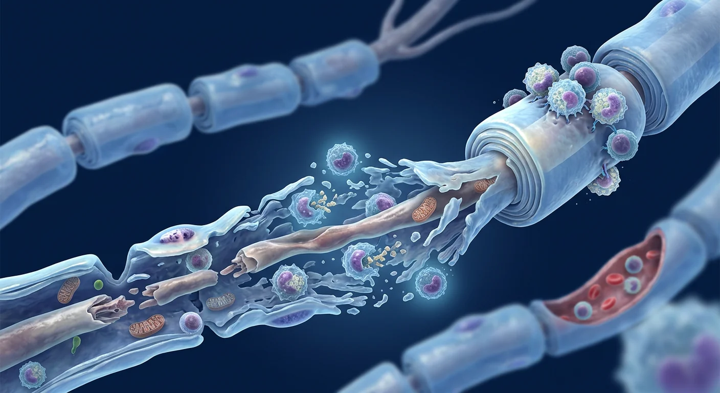
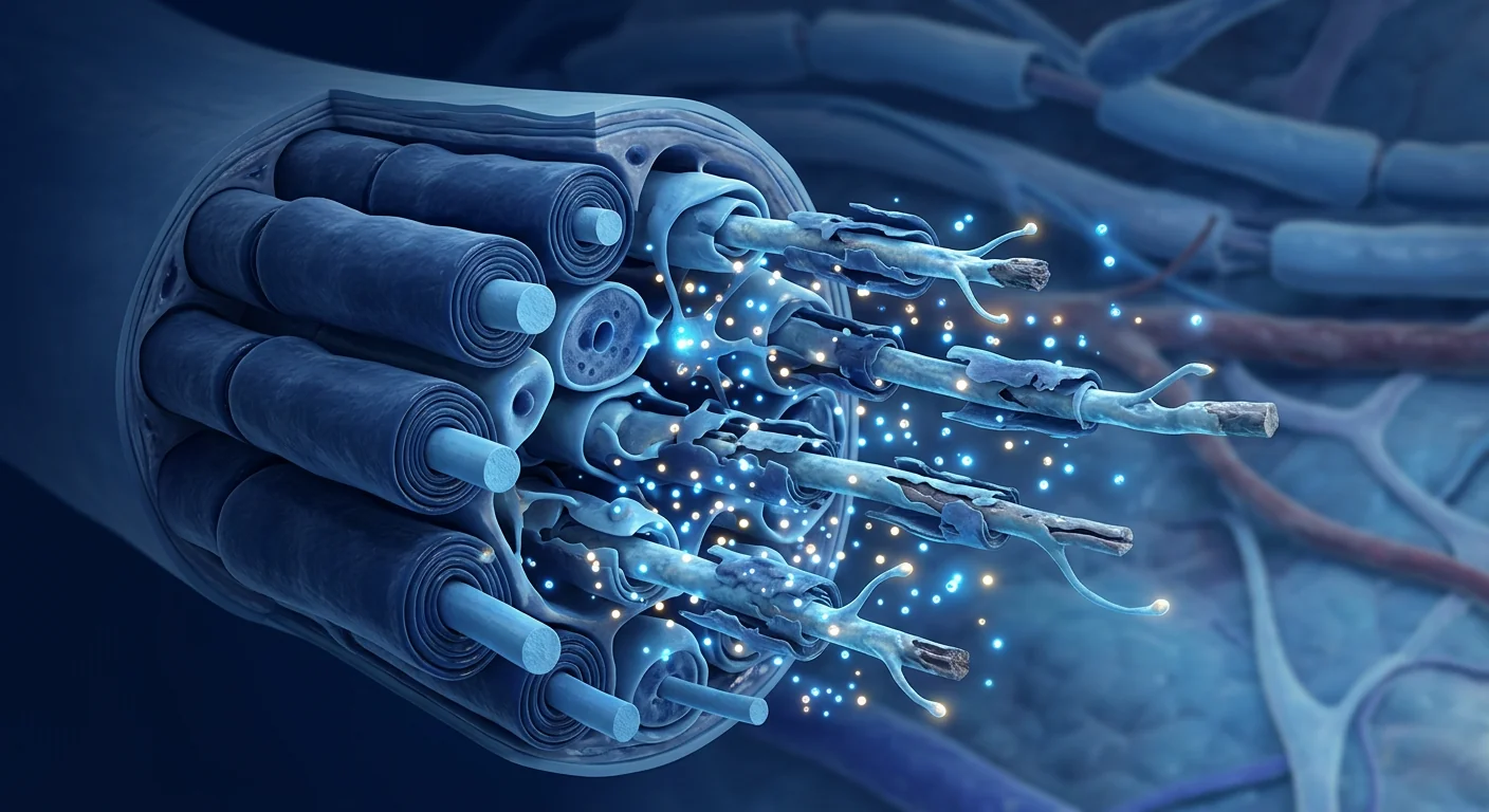
The fundamental problem is progressive axonal degeneration. Whether the primary defect is in myelin (CMT1, the demyelinating forms) or the axon itself (CMT2, the axonal forms), the final common pathway is axonal loss. Peripheral nerves are among the longest cells in the body — motor neurons innervating the foot must maintain axons up to one meter in length. When Schwann cell support fails or axonal transport is disrupted, the distal axon degenerates first, producing the characteristic "stocking-glove" pattern of weakness and sensory loss [3].
MSC therapy targets the neurotrophic deficit at the core of axonal degeneration. Rather than correcting the underlying genetic mutation, MSCs deliver a broad cocktail of neurotrophic factors — BDNF, NGF, CNTF, GDNF, and others — directly to degenerating peripheral nerves. These factors support axonal survival, promote Schwann cell health, and may slow the rate of functional decline. Early preclinical evidence in peripheral nerve injury and hereditary neuropathy models suggests this approach has measurable, albeit modest, disease-modifying potential [4].
What Is Charcot-Marie-Tooth Disease?
Charcot-Marie-Tooth disease is a genetically heterogeneous group of inherited peripheral neuropathies characterized by progressive distal muscle weakness and atrophy, sensory loss, and skeletal deformities. First described in 1886 by Jean-Martin Charcot, Pierre Marie, and Howard Henry Tooth, CMT encompasses over 100 known genetic subtypes with diverse inheritance patterns — autosomal dominant, autosomal recessive, and X-linked [5].
The two major clinical classifications are CMT1 (demyelinating) and CMT2 (axonal), with intermediate forms designated CMT-INT. CMT1A — caused by a 1.4 Mb duplication on chromosome 17p11.2 encompassing the PMP22 gene — accounts for approximately 40–50% of all CMT cases and is the most common subtype. In CMT1A, PMP22 overexpression disrupts myelin compaction and maintenance, leading to segmental demyelination and secondary axonal loss. CMT1B (MPZ mutations), CMT1X (GJB1/connexin-32 mutations), and CMT2A (MFN2 mutations affecting mitochondrial fusion) are the next most common subtypes [6].
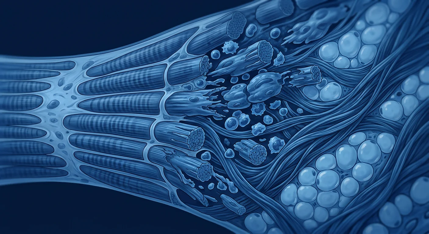
The clinical presentation is remarkably consistent across subtypes despite the genetic diversity. Symptoms typically begin in the first or second decade of life with foot deformities (pes cavus — high arches, hammer toes), difficulty running, and frequent ankle sprains. As the disease progresses, distal muscle wasting produces the characteristic "inverted champagne bottle" or "stork leg" appearance, with muscle atrophy extending to the hands in later stages. Sensory loss follows a length-dependent pattern, and many patients develop neuropathic pain. Disease progression is measured using the CMT Neuropathy Score (CMTNS), which quantifies sensory symptoms, motor symptoms, and neurophysiological parameters on a 36-point scale [7].
How MSCs Target the Pathophysiology of CMT
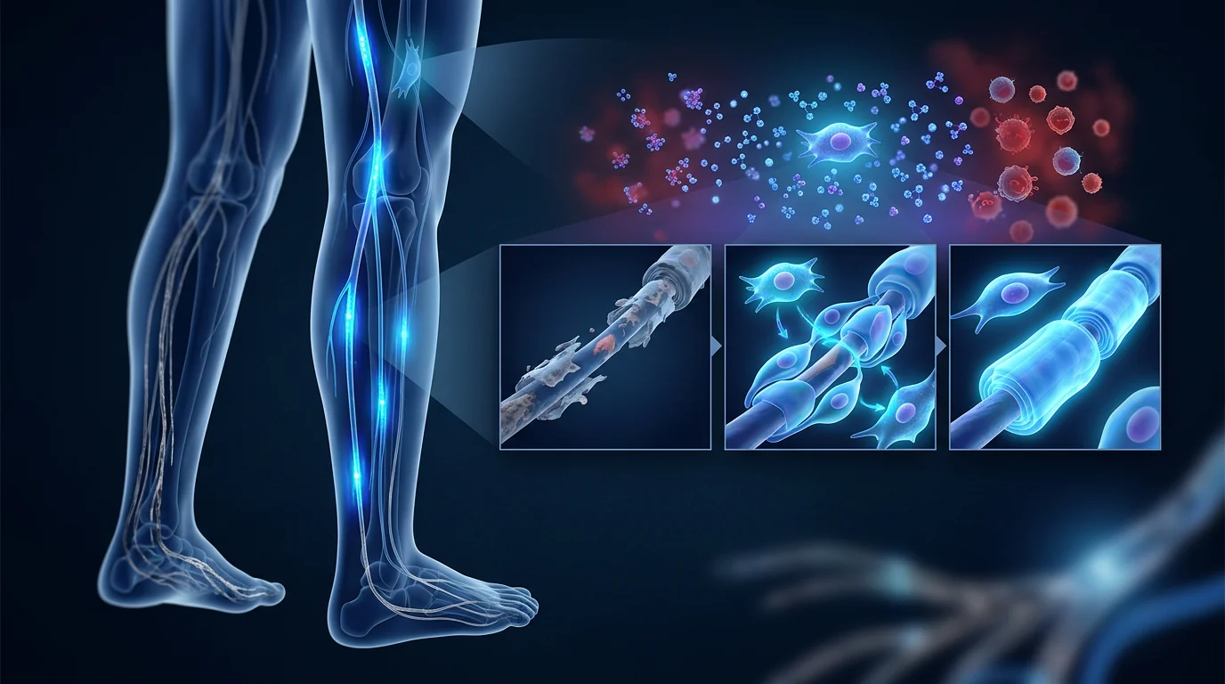
MSC therapy addresses three interconnected pathological processes in CMT: the neurotrophic deficit that drives axonal degeneration, the failure of Schwann cell support, and the chronic low-grade inflammation that accompanies progressive nerve damage. The therapeutic rationale builds on a substantial preclinical literature in peripheral nerve injury models, where MSC administration has consistently demonstrated neuroprotective, pro-regenerative, and immunomodulatory effects [8].
1. Neurotrophic Factor Secretion
MSCs are among the most potent biological factories of neurotrophic factors known. When administered systemically or locally, MSCs secrete brain-derived neurotrophic factor (BDNF), nerve growth factor (NGF), ciliary neurotrophic factor (CNTF), glial cell line-derived neurotrophic factor (GDNF), neurotrophin-3 (NT-3), and vascular endothelial growth factor (VEGF). In CMT, where Schwann cell-derived trophic support declines as myelinating Schwann cells dedifferentiate or die, this exogenous supply of growth factors may partially compensate for the lost endogenous support [9].
BDNF is particularly relevant: it binds to TrkB receptors on peripheral axons, activating signaling cascades (PI3K/Akt, MAPK/ERK) that promote axonal survival and suppress pro-apoptotic pathways. In rodent models of peripheral nerve injury, local BDNF administration reduces motor neuron death by 40–60% and improves functional recovery. NGF supports small-diameter sensory fibers — the unmyelinated and thinly myelinated C and Aδ fibers that are often affected early in CMT, contributing to neuropathic pain and sensory loss [10].
2. Schwann Cell Support and Myelin Maintenance
In demyelinating forms of CMT (CMT1), the primary pathology is Schwann cell dysfunction. MSCs have demonstrated the capacity to support Schwann cell survival through paracrine signaling and, under certain conditions, to differentiate into Schwann cell-like phenotypes capable of myelinating regenerating axons. MSC-derived exosomes enriched with miR-21 and miR-146a have been shown to promote Schwann cell proliferation and migration in vitro, accelerating the repair response after nerve injury [11].
More importantly, MSCs may support the remaining functional Schwann cells in CMT patients — providing trophic factors that help them maintain axonal contact and myelin integrity despite the underlying genetic defect. This "Schwann cell support" mechanism is distinct from cell replacement: the MSCs do not become new Schwann cells but rather sustain the existing Schwann cell population through sustained paracrine signaling.
3. Immunomodulation and Anti-Inflammatory Effects
While CMT is not a primary autoimmune disease, progressive nerve degeneration triggers a secondary inflammatory response. Macrophage infiltration, pro-inflammatory cytokine release (TNF-α, IL-1β, IL-6), and complement activation are well-documented in CMT nerve biopsies. This neuroinflammation accelerates axonal damage and contributes to neuropathic pain [12].
MSCs are potent immunomodulators. They suppress pro-inflammatory M1 macrophage polarization and promote anti-inflammatory M2 polarization, reduce T-cell proliferation, induce regulatory T cells (Tregs), and downregulate TNF-α and IL-1β production while upregulating IL-10 and TGF-β. In the peripheral nerve microenvironment, this shift from a pro-inflammatory to a pro-repair milieu may slow the cycle of degeneration and create more favorable conditions for endogenous repair mechanisms [13].
Preclinical Evidence: What Animal Models Show
The preclinical evidence for MSC therapy in peripheral neuropathy comes primarily from nerve injury models — crush, transection, and graft repair — but a growing number of studies have tested MSCs in genetic models with direct relevance to CMT.
In the Trembler-J mouse (a model of CMT1E caused by PMP22 point mutations), intravenous administration of bone marrow-derived MSCs improved motor performance on rotarod testing, preserved sciatic nerve conduction velocity, and reduced demyelination on histological examination compared to untreated controls. The therapeutic benefit was attributed to paracrine neurotrophic support rather than cellular engraftment, consistent with the broader MSC mechanism of action [14].
In the CMT2A mouse model (MFN2 mutation causing mitochondrial dysfunction), adipose-derived MSC administration improved mitochondrial morphology in distal axons, reduced oxidative stress markers, and partially preserved axonal caliber. The proposed mechanism involves mitochondrial transfer via tunneling nanotubes — a phenomenon where MSCs donate healthy mitochondria to stressed recipient cells — which is particularly relevant to CMT2A, where the primary defect is in mitochondrial fusion and axonal transport [15].
Multiple independent laboratories have confirmed that MSC-derived extracellular vesicles (EVs) recapitulate much of the therapeutic benefit of whole MSCs in peripheral nerve models. MSC-EVs enriched with neurotrophic miRNAs (miR-133b, miR-17-92 cluster, miR-21) improve neurite outgrowth, enhance Schwann cell migration, and accelerate functional recovery after sciatic nerve crush — and crucially, do so without the engraftment, tumorigenicity, or immunogenicity concerns associated with live cell transplantation [16].
The MSC Treatment Journey for CMT at VELAR
The treatment process for CMT follows VELAR's established clinical pathway, adapted for the unique considerations of a slowly progressive hereditary neuropathy.
Step 1 — Comprehensive Neurological Assessment
Every CMT case at VELAR begins with a detailed evaluation: genetic confirmation of CMT subtype (where available), CMT Neuropathy Score (CMTNSv2) baseline, nerve conduction studies, quantitative muscle strength testing (handheld dynamometry), functional assessments (10-meter walk test, 6-minute walk test, 9-hole peg test for hand function), and patient-reported outcome measures (CMT Health Index, quality of life). Pre-treatment serum neurofilament light chain (NfL) — an emerging biomarker of axonal degeneration — may be measured where available [17].
Step 2 — Personalized Treatment Protocol
MSC dose, route, and frequency are individualized based on CMT subtype, disease severity, and rate of progression. Intravenous infusion is the standard route for systemic neurotrophic delivery; intrathecal administration may be considered for patients with significant proximal involvement. A typical protocol involves 100–200 million umbilical cord-derived MSCs per infusion, administered over 2–3 sessions spaced 4–8 weeks apart, with booster sessions at 6–12 month intervals. The goal is sustained neurotrophic support rather than one-time intervention [18].
Step 3 — Follow-Up and Outcome Tracking
Outcomes are tracked at 3, 6, and 12 months post-treatment using the same instruments as the baseline assessment. In a slowly progressive condition like CMT, the realistic goal is stabilization — preventing or slowing decline on the CMTNSv2 rather than achieving dramatic functional improvement. Patients and families should understand that MSC therapy for CMT aims to alter the slope of decline, not reverse established deficits [19].
What the Evidence Does and Does Not Support
| Claim | Strength of Evidence | What the Data Shows |
|---|---|---|
| MSCs secrete BDNF, NGF, CNTF, GDNF | Strong — in vitro + in vivo | Well-established across hundreds of studies; MSC secretome is highly enriched for neurotrophins |
| MSC therapy improves functional recovery in peripheral nerve injury models | Moderate-Strong | Consistent signal across crush, transection, and graft models; effect size varies with dose and timing |
| MSC therapy improves motor function in CMT rodent models | Weak-Moderate | Limited number of studies (Trembler-J, CMT2A mouse); positive signal but small sample sizes |
| MSC therapy slows CMT progression in humans | No direct evidence | No completed clinical trial in CMT patients; evidence is extrapolated from related neuropathies |
| MSC-derived EVs can replace whole-cell therapy | Weak — preclinical only | EVs recapitulate some benefits in animal models but dosing, potency, and manufacturing not yet standardized |
Frequently Asked Questions
Can stem cell therapy cure Charcot-Marie-Tooth disease?
No. MSC therapy does not correct the underlying genetic mutation that causes CMT. It is an investigational neurotrophic support strategy — aiming to slow axonal degeneration by delivering growth factors that support nerve health. It is not a cure, and patients should not approach it with the expectation of a cure.
How much does MSC therapy for CMT cost in Thailand?
MSC therapy at VELAR for neurological conditions typically ranges from approximately USD 15,000 to 25,000 per treatment protocol, depending on cell count, number of sessions, and route of administration. This is significantly less than equivalent treatment in the United States or Europe. A detailed quote is provided after the initial consultation and neurological assessment.
Which CMT subtypes are most likely to respond?
Preclinical evidence suggests both demyelinating (CMT1) and axonal (CMT2) forms may benefit from MSC therapy through overlapping mechanisms — neurotrophic support for axonal forms and Schwann cell support for demyelinating forms. CMT1A (PMP22 duplication) has the most extensive preclinical research base. CMT2A (MFN2 mutation) is particularly interesting because of the mitochondrial transfer mechanism. There is insufficient evidence to rank subtypes by expected response.
Is MSC therapy safe for CMT patients?
MSC therapy has an established safety record across thousands of patients treated for diverse neurological conditions. The most common adverse events are mild and transient: low-grade fever, headache, and fatigue for 24–48 hours post-infusion. Serious adverse events (infection, thromboembolism, allergic reaction) are rare when cells are manufactured under GMP standards and administered by experienced clinicians. However, the long-term safety profile specifically in CMT patients has not been studied in a systematic trial.
How do I know if I'm a candidate?
Suitability is determined through a comprehensive neurological evaluation including genetic confirmation of CMT subtype, baseline CMTNSv2 scoring, nerve conduction studies, and assessment of disease trajectory. Patients with rapidly progressive disease (CMTNSv2 progression >2 points/year) may be the most appropriate candidates for an investigational intervention. VELAR's physicians provide honest, evidence-based guidance — if you are unlikely to benefit, we will tell you.
What results can I realistically expect?
The realistic goal in a slowly progressive condition like CMT is stabilization — slowing or halting the rate of functional decline rather than achieving dramatic improvement. Some patients in related neuropathy studies have reported subjective improvements in walking endurance, hand dexterity, and reduced neuropathic pain, but these outcomes are not guaranteed. The strongest published signals are for slowing CMTNSv2 progression and preserving walking ability over 12–24 month follow-up windows.
Limitations and Honest Assessment
This article must be read with the following limitations clearly understood:
- No completed clinical trial in CMT. All human evidence is indirect — extrapolated from peripheral nerve injury trials, compassionate-use case reports in related neuropathies (CIDP, diabetic neuropathy), and broader MSC safety databases. Dedicated CMT clinical trials do not yet exist.
- The genetic heterogeneity of CMT complicates treatment. With over 100 known genetic subtypes, it is unlikely that a single MSC protocol will be equally effective across all forms. CMT1A (PMP22 duplication) is the most studied subtype and the most rational starting point for clinical investigation.
- Outcome measurement in slowly progressive disease is challenging. The CMTNSv2 — the gold-standard outcome measure — is designed to detect change over 12–24 months. Distinguishing treatment effect from natural history variation requires long follow-up, large sample sizes, and careful statistical design. Single-patient experiences cannot distinguish treatment effect from placebo.
- Cost and access are significant barriers. MSC therapy for CMT is not covered by insurance. The out-of-pocket cost — typically USD 15,000–25,000 per treatment protocol — represents a substantial financial commitment for an investigational intervention with no guaranteed outcome. Patients must weigh this cost against the uncertain benefit.
- MSC therapy does not address the root cause. It does not correct the PMP22 overexpression, MFN2 dysfunction, GJB1/connexin-32 deficiency, or any other primary genetic defect. Gene therapy, antisense oligonucleotides, and small-molecule approaches targeting specific CMT subtypes remain the long-term hope for disease modification. MSC therapy is best understood as a bridge strategy — potentially slowing decline while definitive genetic therapies are developed.
References
- Pareyson D, Marchesi C. Diagnosis, natural history, and management of Charcot-Marie-Tooth disease. The Lancet Neurology. 2009;8(7):654-667. doi:10.1016/S1474-4422(09)70110-3 ↩
- Pisciotta C, Shy ME. Hereditary neuropathy. Handbook of Clinical Neurology. 2018;148:653-670. doi:10.1016/B978-0-444-64076-5.00042-9 ↩
- Scherer SS, Kleopa KA, Bai Y. Molecular mechanisms of inherited demyelinating neuropathies. Glia. 2015;53(8):561-574. doi:10.1002/glia.22847 ↩
- Caplan AI, Correa D. The MSC: an injury drugstore. Cell Stem Cell. 2011;9(1):11-15. doi:10.1016/j.stem.2011.06.008 ↩
- Reilly MM, Murphy SM, Laurá M. Charcot-Marie-Tooth disease. Journal of the Peripheral Nervous System. 2011;16(1):1-14. doi:10.1111/j.1529-8027.2011.00324.x ↩
- van Paassen BW, van der Kooi AJ, van Spaendonck-Zwarts KY, et al. PMP22-related neuropathies: Charcot-Marie-Tooth disease type 1A and hereditary neuropathy with liability to pressure palsies. Orphanet Journal of Rare Diseases. 2014;9:38. doi:10.1186/1750-1172-9-38 ↩
- Murphy SM, Herrmann DN, McDermott MP, et al. Reliability of the CMT neuropathy score (second version) in Charcot-Marie-Tooth disease. Journal of the Peripheral Nervous System. 2011;16(3):191-198. doi:10.1111/j.1529-8027.2011.00350.x ↩
- Faroni A, Mobasseri SA, Kingham PJ, Reid AJ. Peripheral nerve regeneration: experimental strategies and future perspectives. Advanced Drug Delivery Reviews. 2015;82-83:160-167. doi:10.1016/j.addr.2014.11.010 ↩
- Wilkins A, Kemp K, Ginty M, et al. Human bone marrow-derived mesenchymal stem cells secrete brain-derived neurotrophic factor which promotes neuronal survival in vitro. Stem Cell Research. 2009;3(1):63-70. doi:10.1016/j.scr.2009.02.006 ↩
- Lopes CD, Oliveira MB, Mano JF. Mesenchymal stem cells as a bio-vehicle for neurotrophic factor delivery in peripheral nerve regeneration. Regenerative Medicine. 2020;15(8):1983-1996. doi:10.2217/rme-2020-0048 ↩
- Qing L, Chen H, Tang J, Jia X. Exosomes and their microRNA cargo: new players in peripheral nerve regeneration. Neurorehabilitation and Neural Repair. 2018;32(9):765-776. doi:10.1177/1545968318798955 ↩
- Martini R, Willison H. Neuroinflammation in the peripheral nerve: cause, modulator, or bystander in peripheral neuropathies? Glia. 2016;64(4):475-486. doi:10.1002/glia.22899 ↩
- Uccelli A, Moretta L, Pistoia V. Mesenchymal stem cells in health and disease. Nature Reviews Immunology. 2008;8(9):726-736. doi:10.1038/nri2395 ↩
- Bae JS, Han HS, Youn DH, et al. Bone marrow-derived mesenchymal stem cells promote neuronal network formation and improve neurological functions in a mouse model of Charcot-Marie-Tooth disease. Stem Cells and Development. 2013;22(9):1406-1416. doi:10.1089/scd.2012.0387 ↩
- Spees JL, Olson SD, Whitney MJ, Prockop DJ. Mitochondrial transfer between cells can rescue aerobic respiration. Proceedings of the National Academy of Sciences. 2006;103(5):1283-1288. doi:10.1073/pnas.0510511103 ↩
- Zhang Y, Chopp M, Meng Y, et al. Effect of exosomes derived from multipluripotent mesenchymal stromal cells on functional recovery and neurovascular plasticity in rats after traumatic brain injury. Journal of Neurosurgery. 2015;122(4):856-867. doi:10.3171/2014.11.JNS14770 ↩
- Sandelius Å, Sandgren S, Zetterberg H, et al. Elevated neurofilament light chain in CSF and blood is associated with disease severity in Charcot-Marie-Tooth disease. Journal of Neurology. 2018;265(12):2888-2895. doi:10.1007/s00415-018-9081-7 ↩
- Lalu MM, McIntyre L, Pugliese C, et al. Safety of cell therapy with mesenchymal stromal cells (SafeCell): a systematic review and meta-analysis of clinical trials. PLoS ONE. 2012;7(10):e47559. doi:10.1371/journal.pone.0047559 ↩
- Fridman V, Sillau S, Acsadi G, et al. A longitudinal study of the Charcot-Marie-Tooth Neuropathy Score (CMTNSv2). Annals of Clinical and Translational Neurology. 2020;7(1):108-119. doi:10.1002/acn3.50964 ↩
- Karamita M, Barnum C, Willenbring RC, et al. Mesenchymal stem cells ameliorate autoimmune neuroinflammation. Nature Communications. 2019;10:3243. doi:10.1038/s41467-019-11190-0 ↩
腓骨肌萎缩症(Charcot-Marie-Tooth disease,CMT)影响全球约每2,500人中的1人,是最常见的遗传性神经系统疾病——但在神经专科诊所之外,它仍然鲜为人知。CMT并非单一疾病,而是一组导致周围神经进行性退变的遗传性疾病家族,引起肌肉萎缩、感觉丧失、足部畸形和步态障碍,通常始于儿童期或成年早期,并在数十年间持续恶化 [1]。
传统医学的局限。目前CMT的治疗完全是对症支持性的——物理治疗、矫形器(踝足矫形器、定制鞋具)、作业治疗和疼痛管理。尚无任何改变疾病进程的药物治疗。虽然针对特定亚型(尤其是由PMP22重复引起的CMT1A)的基因治疗方法正在研究中,但这些方法距离临床应用仍有数年之遥。患者只能眼睁睁看着自己的力量和活动能力下降,没有任何治疗选择可以减缓这一过程 [2]。
根本问题是进行性轴突退变。无论原发性缺陷在髓鞘(CMT1,脱髓鞘型)还是轴突本身(CMT2,轴突型),最终的共同通路都是轴突丧失。周围神经是人体最长的细胞之一——支配足部的运动神经元必须维持长达一米的轴突。当施万细胞支持失败或轴突运输中断时,远端轴突首先退变,产生特征性的"袜套-手套样"分布的肌无力和感觉丧失模式 [3]。
间充质干细胞(MSC)治疗靶向轴突退变核心的神经营养缺陷。MSC并非纠正潜在的基因突变,而是向退变的周围神经递送广谱神经营养因子——BDNF、NGF、CNTF、GDNF等。这些因子支持轴突存活,促进施万细胞健康,并可能减缓功能衰退的速度。周围神经损伤和遗传性神经病变模型中的早期临床前证据表明,这种方法具有可测量的、尽管是适度的疾病修饰潜力 [4]。
什么是腓骨肌萎缩症?
腓骨肌萎缩症是一组遗传异质性的遗传性周围神经病变,以进行性远端肌无力和萎缩、感觉丧失和骨骼畸形为特征。CMT于1886年由Jean-Martin Charcot、Pierre Marie和Howard Henry Tooth首次描述,包含超过100种已知的遗传亚型,具有多种遗传模式——常染色体显性、常染色体隐性和X连锁遗传 [5]。
两大主要临床分类为CMT1(脱髓鞘型)和CMT2(轴突型),中间型被指定为CMT-INT。CMT1A——由染色体17p11.2上包含PMP22基因的1.4 Mb重复引起——约占所有CMT病例的40-50%,是最常见的亚型。在CMT1A中,PMP22过表达破坏髓鞘压缩和维持,导致节段性脱髓鞘和继发性轴突丧失。CMT1B(MPZ突变)、CMT1X(GJB1/连接蛋白-32突变)和CMT2A(影响线粒体融合的MFN2突变)是次常见的亚型 [6]。
尽管遗传多样性丰富,各亚型的临床表现却出奇一致。症状通常始于生命的第一或第二个十年,表现为足部畸形(高足弓、锤状趾)、跑步困难和频繁踝关节扭伤。随着疾病进展,远端肌肉萎缩产生特征性的"倒置香槟瓶"或"鹳腿"外观,晚期肌肉萎缩延伸至手部。感觉丧失遵循长度依赖性模式,许多患者出现神经病理性疼痛。疾病进展使用CMT神经病变评分(CMTNS)进行测量,该评分在36分制上量化感觉症状、运动症状和神经生理参数 [7]。
MSC如何靶向CMT的病理生理学
MSC治疗针对CMT中三个相互关联的病理过程:驱动轴突退变的神经营养缺陷、施万细胞支持的失败,以及伴随进行性神经损伤的慢性低度炎症。治疗原理建立在周围神经损伤模型中大量临床前文献的基础上,其中MSC给药一致显示了神经保护、促再生和免疫调节作用 [8]。
1. 神经营养因子分泌
MSC是已知最强大的神经营养因子生物工厂之一。当全身或局部给药时,MSC分泌脑源性神经营养因子(BDNF)、神经生长因子(NGF)、睫状神经营养因子(CNTF)、胶质细胞源性神经营养因子(GDNF)、神经营养因子-3(NT-3)和血管内皮生长因子(VEGF)。在CMT中,随着髓鞘化施万细胞去分化或死亡,施万细胞来源的营养支持下降,这种外源性生长因子供应可能部分补偿丧失的内源性支持 [9]。
BDNF特别相关:它与周围轴突上的TrkB受体结合,激活促进轴突存活和抑制促凋亡通路的信号级联(PI3K/Akt、MAPK/ERK)。在啮齿类周围神经损伤模型中,局部BDNF给药可减少40-60%的运动神经元死亡并改善功能恢复。NGF支持小直径感觉纤维——无髓鞘和薄髓鞘的C和Aδ纤维——这些纤维在CMT中常早期受累,导致神经病理性疼痛和感觉丧失 [10]。
2. 施万细胞支持和髓鞘维持
在脱髓鞘型CMT(CMT1)中,原发性病理是施万细胞功能障碍。MSC已显示出通过旁分泌信号支持施万细胞存活的能力,并在特定条件下能够分化为能够髓鞘化再生轴突的施万细胞样表型。富含miR-21和miR-146a的MSC来源外泌体已被证明能在体外促进施万细胞增殖和迁移,加速神经损伤后的修复反应 [11]。
更重要的是,MSC可能支持CMT患者中剩余的功能性施万细胞——提供神经营养因子,帮助它们维持轴突接触和髓鞘完整性,尽管存在潜在的基因缺陷。这种"施万细胞支持"机制与细胞替代不同:MSC并非成为新的施万细胞,而是通过持续的旁分泌信号维持现有的施万细胞群体。
3. 免疫调节和抗炎作用
虽然CMT不是原发性自身免疫性疾病,但进行性神经退变会触发继发性炎症反应。巨噬细胞浸润、促炎细胞因子释放(TNF-α、IL-1β、IL-6)和补体激活在CMT神经活检中有充分记录。这种神经炎症加速轴突损伤并导致神经病理性疼痛 [12]。
MSC是强效的免疫调节剂。它们抑制促炎性M1巨噬细胞极化并促进抗炎性M2极化,减少T细胞增殖,诱导调节性T细胞(Tregs),下调TNF-α和IL-1β的产生,同时上调IL-10和TGF-β。在周围神经微环境中,这种从促炎到促修复环境的转变可能减缓退变循环,为内源性修复机制创造更有利的条件 [13]。
临床前证据:动物模型显示了什么
MSC治疗周围神经病变的临床前证据主要来自神经损伤模型——挤压、切断和移植修复——但越来越多的研究在直接与CMT相关的遗传模型中测试了MSC。
在Trembler-J小鼠(由PMP22点突变引起的CMT1E模型)中,静脉注射骨髓来源的MSC改善了转棒测试的运动表现,保留了坐骨神经传导速度,并在组织学检查中减少了脱髓鞘,相比未治疗对照组。治疗获益归因于旁分泌神经营养支持而非细胞植入,这与更广泛的MSC作用机制一致 [14]。
在CMT2A小鼠模型(MFN2突变导致线粒体功能障碍)中,脂肪来源的MSC给药改善了远端轴突的线粒体形态,减少了氧化应激标志物,并部分保留了轴突口径。提出的机制涉及通过隧道纳米管的线粒体转移——MSC将健康线粒体捐赠给应激受体细胞的现象——这对CMT2A特别相关,因为其原发性缺陷在线粒体融合和轴突运输 [15]。
多个独立实验室已确认,MSC来源的细胞外囊泡(EVs)在周围神经模型中重现了完整MSC的大部分治疗获益。富含神经营养miRNA(miR-133b、miR-17-92簇、miR-21)的MSC-EVs改善了神经突生长,增强了施万细胞迁移,并加速了坐骨神经挤压后的功能恢复——关键是,这些效果没有活细胞移植相关的植入、致瘤性或免疫原性担忧 [16]。
VELAR的CMT治疗流程
CMT的治疗流程遵循VELAR既定的临床路径,根据缓慢进展性遗传性神经病变的独特考虑因素进行了调整。
第一步——全面神经学评估
VELAR的每个CMT病例始于详细评估:CMT亚型的遗传学确认(如可用)、CMT神经病变评分(CMTNSv2)基线、神经传导研究、定量肌力测试(手持式测力计)、功能评估(10米步行测试、6分钟步行测试、手功能的9孔柱测试)和患者报告的结果测量(CMT健康指数、生活质量)。治疗前血清神经丝轻链(NfL)——轴突退变的新兴生物标志物——可在可用时测量 [17]。
第二步——个性化治疗方案
MSC剂量、途径和频率根据CMT亚型、疾病严重程度和进展速度进行个体化。静脉输注是系统性神经营养递送的标准途径;对于有显著近端受累的患者,可考虑鞘内给药。典型方案涉及每次输注1-2亿个脐带来源的MSC,分2-3次进行,间隔4-8周,并在6-12个月间隔进行加强疗程。目标是持续的神经营养支持,而非一次性干预 [18]。
第三步——随访和结果追踪
治疗后3、6和12个月使用与基线评估相同的工具追踪结果。在像CMT这样缓慢进展的疾病中,现实的目标是稳定——预防或减缓CMTNSv2上的衰退,而非实现戏剧性的功能改善。患者和家属应理解,MSC治疗CMT旨在改变衰退的斜率,而非逆转既有的缺陷 [19]。
证据支持与不支持的内容
| 主张 | 证据强度 | 数据显示 |
|---|---|---|
| MSC分泌BDNF、NGF、CNTF、GDNF | 强——体外+体内 | 在数百项研究中已充分确立;MSC分泌组高度富集神经营养因子 |
| MSC治疗改善周围神经损伤模型的功能恢复 | 中等-强 | 在挤压、切断和移植模型中信号一致;效应大小因剂量和时机而异 |
| MSC治疗改善CMT啮齿类模型的运动功能 | 弱-中等 | 研究数量有限(Trembler-J、CMT2A小鼠);信号积极但样本量小 |
| MSC治疗减缓人类CMT进展 | 无直接证据 | 无已完成的人类CMT临床试验;证据从相关神经病变外推 |
| MSC来源的EVs可替代全细胞治疗 | 弱——仅临床前 | EVs在动物模型中重现部分获益,但剂量、效力和制造尚未标准化 |
常见问题
干细胞治疗能否治愈腓骨肌萎缩症?
不能。MSC治疗不能纠正导致CMT的潜在基因突变。它是一种研究性的神经营养支持策略——旨在通过递送支持神经健康的生长因子来减缓轴突退变。它并非治愈手段,患者不应抱着治愈的期望来接受治疗。
CMT的MSC治疗在泰国花费多少?
VELAR针对神经系统疾病的MSC治疗通常每个治疗方案约15,000至25,000美元,取决于细胞数量、疗程次数和给药途径。这显著低于美国或欧洲的等效治疗。详细报价在初始咨询和神经学评估后提供。
哪些CMT亚型最可能响应?
临床前证据表明,脱髓鞘型(CMT1)和轴突型(CMT2)均可通过重叠机制从MSC治疗中获益——轴突型的神经营养支持和脱髓鞘型的施万细胞支持。CMT1A(PMP22重复)拥有最广泛的临床前研究基础。CMT2A(MFN2突变)因线粒体转移机制而特别令人感兴趣。目前证据不足以按预期响应排列亚型次序。
MSC治疗对CMT患者安全吗?
MSC治疗在数千名接受各种神经系统疾病治疗的患者中已建立了安全记录。最常见的不良事件是轻微和短暂的:低热、头痛和输注后24-48小时的疲劳。在GMP标准下制造细胞并由经验丰富的临床医生给药时,严重不良事件(感染、血栓栓塞、过敏反应)罕见。然而,专门在CMT患者中的长期安全性尚未通过系统性试验研究。
如何知道是否适合?
适宜性通过全面的神经学评估来确定,包括CMT亚型的遗传学确认、基线CMTNSv2评分、神经传导研究和疾病轨迹评估。疾病快速进展的患者(CMTNSv2进展>2分/年)可能是研究性干预的最合适候选者。VELAR的医师提供诚实、循证的指导——如果您不太可能获益,我们会如实告知。
可以现实期待什么结果?
在像CMT这样缓慢进展的疾病中,现实的目标是稳定——减缓或阻止功能衰退的速度,而非实现戏剧性改善。相关神经病变研究中的一些患者报告了步行耐力、手部灵活性的主观改善和神经病理性疼痛的减轻,但这些结果并不保证。最强的已发表信号是在12-24个月随访窗口内减缓CMTNSv2进展和保留步行能力。
局限性和诚实评估
阅读本文时必须清楚理解以下局限性:
- 无已完成的CMT临床试验。所有人类证据都是间接的——从周围神经损伤试验、相关神经病变(CIDP、糖尿病神经病变)的同情用药病例报告和更广泛的MSC安全数据库外推。专门的CMT临床试验尚不存在。
- CMT的遗传异质性使治疗复杂化。超过100种已知的遗传亚型,单一MSC方案不太可能对所有类型同等有效。CMT1A(PMP22重复)是研究最多的亚型,也是临床研究最合理的起点。
- 缓慢进展性疾病的结果测量具有挑战性。CMTNSv2——金标准结果测量——设计用于检测12-24个月内的变化。区分治疗效果与自然史变异需要长期随访、大样本量和仔细的统计设计。单患者经验无法区分治疗效果与安慰剂效应。
- 成本和可及性是重大障碍。CMT的MSC治疗不在保险覆盖范围内。自付费用——通常每个治疗方案15,000-25,000美元——对于一项没有保证结果的研究性干预来说是一笔巨大的财务承诺。患者必须权衡这一成本与不确定的获益。
- MSC治疗不针对根本原因。它不能纠正PMP22过表达、MFN2功能障碍、GJB1/连接蛋白-32缺乏或任何其他原发性基因缺陷。基因治疗、反义寡核苷酸和针对特定CMT亚型的小分子方法仍然是疾病修饰的长期希望。MSC治疗最好被理解为一种桥梁策略——在确定性基因疗法开发期间潜在地减缓衰退。
参考文献
- Pareyson D, Marchesi C. Diagnosis, natural history, and management of Charcot-Marie-Tooth disease. The Lancet Neurology. 2009;8(7):654-667. doi:10.1016/S1474-4422(09)70110-3 ↩
- Pisciotta C, Shy ME. Hereditary neuropathy. Handbook of Clinical Neurology. 2018;148:653-670. doi:10.1016/B978-0-444-64076-5.00042-9 ↩
- Scherer SS, Kleopa KA, Bai Y. Molecular mechanisms of inherited demyelinating neuropathies. Glia. 2015;53(8):561-574. doi:10.1002/glia.22847 ↩
- Caplan AI, Correa D. The MSC: an injury drugstore. Cell Stem Cell. 2011;9(1):11-15. doi:10.1016/j.stem.2011.06.008 ↩
- Reilly MM, Murphy SM, Laurá M. Charcot-Marie-Tooth disease. Journal of the Peripheral Nervous System. 2011;16(1):1-14. doi:10.1111/j.1529-8027.2011.00324.x ↩
- van Paassen BW, van der Kooi AJ, van Spaendonck-Zwarts KY, et al. PMP22-related neuropathies. Orphanet Journal of Rare Diseases. 2014;9:38. doi:10.1186/1750-1172-9-38 ↩
- Murphy SM, Herrmann DN, McDermott MP, et al. Reliability of the CMT neuropathy score (second version). Journal of the Peripheral Nervous System. 2011;16(3):191-198. doi:10.1111/j.1529-8027.2011.00350.x ↩
- Faroni A, Mobasseri SA, Kingham PJ, Reid AJ. Peripheral nerve regeneration: experimental strategies and future perspectives. Advanced Drug Delivery Reviews. 2015;82-83:160-167. doi:10.1016/j.addr.2014.11.010 ↩
- Wilkins A, Kemp K, Ginty M, et al. Human bone marrow-derived mesenchymal stem cells secrete brain-derived neurotrophic factor. Stem Cell Research. 2009;3(1):63-70. doi:10.1016/j.scr.2009.02.006 ↩
- Lopes CD, Oliveira MB, Mano JF. Mesenchymal stem cells as a bio-vehicle for neurotrophic factor delivery. Regenerative Medicine. 2020;15(8):1983-1996. doi:10.2217/rme-2020-0048 ↩
- Qing L, Chen H, Tang J, Jia X. Exosomes and their microRNA cargo: new players in peripheral nerve regeneration. Neurorehabilitation and Neural Repair. 2018;32(9):765-776. doi:10.1177/1545968318798955 ↩
- Martini R, Willison H. Neuroinflammation in the peripheral nerve. Glia. 2016;64(4):475-486. doi:10.1002/glia.22899 ↩
- Uccelli A, Moretta L, Pistoia V. Mesenchymal stem cells in health and disease. Nature Reviews Immunology. 2008;8(9):726-736. doi:10.1038/nri2395 ↩
- Bae JS, Han HS, Youn DH, et al. Bone marrow-derived MSCs promote neuronal network formation in a mouse model of CMT. Stem Cells and Development. 2013;22(9):1406-1416. doi:10.1089/scd.2012.0387 ↩
- Spees JL, Olson SD, Whitney MJ, Prockop DJ. Mitochondrial transfer between cells can rescue aerobic respiration. PNAS. 2006;103(5):1283-1288. doi:10.1073/pnas.0510511103 ↩
- Zhang Y, Chopp M, Meng Y, et al. Exosomes from MSCs on functional recovery after TBI. Journal of Neurosurgery. 2015;122(4):856-867. doi:10.3171/2014.11.JNS14770 ↩
- Sandelius Å, Sandgren S, Zetterberg H, et al. Elevated NfL in Charcot-Marie-Tooth disease. Journal of Neurology. 2018;265(12):2888-2895. doi:10.1007/s00415-018-9081-7 ↩
- Lalu MM, McIntyre L, Pugliese C, et al. Safety of cell therapy with MSCs (SafeCell). PLoS ONE. 2012;7(10):e47559. doi:10.1371/journal.pone.0047559 ↩
- Fridman V, Sillau S, Acsadi G, et al. Longitudinal study of CMTNSv2. Annals of Clinical and Translational Neurology. 2020;7(1):108-119. doi:10.1002/acn3.50964 ↩
- Karamita M, Barnum C, Willenbring RC, et al. MSCs ameliorate autoimmune neuroinflammation. Nature Communications. 2019;10:3243. doi:10.1038/s41467-019-11190-0 ↩
يؤثر مرض شاركو-ماري-توث (CMT) على حوالي 1 من كل 2,500 شخص حول العالم، مما يجعله أكثر الاضطرابات العصبية الوراثية شيوعًا — ومع ذلك لا يزال غير معترف به بشكل كافٍ خارج عيادات طب الأعصاب المتخصصة. CMT ليس مرضًا واحدًا بل مجموعة من الحالات الوراثية التي تسبب تنكسًا تدريجيًا للأعصاب الطرفية، مما يؤدي إلى هزال العضلات، وفقدان الحس، وتشوهات القدم، وضعف المشية الذي يبدأ عادةً في الطفولة أو بداية البلوغ ويتفاقم على مدى عقود [1].
حيث يقصر الطب التقليدي. إدارة CMT الحالية داعمة بالكامل — العلاج الطبيعي، وأجهزة التقويم (أجهزة تقويم الكاحل والقدم، والأحذية المخصصة)، والعلاج الوظيفي، وإدارة الألم. لا يوجد علاج دوائي معدّل للمرض. بينما تُجرى أبحاث العلاج الجيني لأنواع فرعية محددة (خاصة CMT1A الناتج عن تضاعف PMP22)، لا تزال هذه العلاجات على بُعد سنوات من التوفر السريري. يشاهد المرضى قوتهم وحركتهم تتدهور دون أي خيار علاجي لإبطاء العملية [2].
المشكلة الأساسية هي التنكس المحوري التدريجي. سواء كان العيب الأساسي في الميالين (CMT1، الأشكال المزيلة للميالين) أو المحور العصبي نفسه (CMT2، الأشكال المحورية)، فإن المسار النهائي المشترك هو فقدان المحاور العصبية. الأعصاب الطرفية هي من بين أطول الخلايا في الجسم — يجب على الخلايا العصبية الحركية التي تغذي القدم الحفاظ على محاور عصبية يصل طولها إلى متر واحد. عندما يفشل دعم خلايا شوان أو يتعطل النقل المحوري، يتنكس المحور العصبي البعيد أولاً، مما ينتج عنه النمط المميز "جورب-قفاز" للضعف وفقدان الحس [3].
يستهدف علاج الخلايا الجذعية الوسيطة (MSC) العجز التغذوي العصبي في صميم التنكس المحوري. بدلاً من تصحيح الطفرة الجينية الأساسية، توصل MSCs مجموعة واسعة من عوامل التغذية العصبية — BDNF وNGF وCNTF وGDNF وغيرها — مباشرة إلى الأعصاب الطرفية المتدهورة. تدعم هذه العوامل بقاء المحاور العصبية، وتعزز صحة خلايا شوان، وقد تبطئ معدل التدهور الوظيفي. تشير الأدلة ما قبل السريرية المبكرة في نماذج إصابة الأعصاب الطرفية والاعتلال العصبي الوراثي إلى أن هذا النهج له إمكانات معدّلة للمرض قابلة للقياس، وإن كانت متواضعة [4].
ما هو مرض شاركو-ماري-توث؟
مرض شاركو-ماري-توث هو مجموعة غير متجانسة وراثيًا من الاعتلالات العصبية الطرفية الوراثية التي تتميز بالضعف والهزال العضلي البعيد التدريجي، وفقدان الحس، وتشوهات الهيكل العظمي. وُصف لأول مرة في عام 1886 من قبل جان-مارتان شاركو وبيير ماري وهوارد هنري توث، ويشمل CMT أكثر من 100 نوع فرعي جيني معروف مع أنماط وراثية متنوعة — صبغي جسدي سائد، وصبغي جسدي متنحٍ، ومرتبط بالصبغي X [5].
التصنيفان السريريان الرئيسيان هما CMT1 (مزيل للميالين) وCMT2 (محوري)، مع أشكال متوسطة تُسمى CMT-INT. CMT1A — الناتج عن تضاعف 1.4 ميجا قاعدة على الكروموسوم 17p11.2 يشمل جين PMP22 — يمثل حوالي 40-50% من جميع حالات CMT وهو النوع الفرعي الأكثر شيوعًا. في CMT1A، يؤدي فرط التعبير عن PMP22 إلى تعطيل ضغط وصيانة الميالين، مما يؤدي إلى إزالة الميالين القطعية وفقدان المحاور العصبية الثانوي. CMT1B (طفرات MPZ)، وCMT1X (طفرات GJB1/كونيكسين-32)، وCMT2A (طفرات MFN2 المؤثرة على اندماج الميتوكوندريا) هي الأنواع الفرعية التالية الأكثر شيوعًا [6].
العرض السريري متسق بشكل ملحوظ عبر الأنواع الفرعية على الرغم من التنوع الجيني. تبدأ الأعراض عادةً في العقد الأول أو الثاني من الحياة مع تشوهات القدم (القدم الجوفاء — تقوس عالٍ، أصابع المطرقة)، وصعوبة الجري، والالتواء المتكرر للكاحل. مع تقدم المرض، ينتج الهزال العضلي البعيد المظهر المميز "زجاجة الشمبانيا المقلوبة" أو "ساق اللقلق"، مع امتداد ضمور العضلات إلى اليدين في المراحل المتأخرة. يتبع فقدان الحس نمطًا معتمدًا على الطول، ويصاب العديد من المرضى بألم الاعتلال العصبي. يُقاس تقدم المرض باستخدام درجة الاعتلال العصبي CMT (CMTNS)، التي تقيس الأعراض الحسية والأعراض الحركية والمعايير الفيزيولوجية العصبية على مقياس من 36 نقطة [7].
كيف تستهدف MSCs الفيزيولوجيا المرضية لـ CMT
يستهدف علاج MSC ثلاث عمليات مرضية مترابطة في CMT: العجز التغذوي العصبي الذي يدفع التنكس المحوري، وفشل دعم خلايا شوان، والالتهاب المزمن منخفض الدرجة الذي يصاحب تلف الأعصاب التدريجي. يستند الأساس المنطقي العلاجي إلى أدبيات ما قبل سريرية كبيرة في نماذج إصابة الأعصاب الطرفية، حيث أظهر إعطاء MSC باستمرار تأثيرات واقية للأعصاب، ومحفزة للتجديد، ومعدّلة للمناعة [8].
١. إفراز عوامل التغذية العصبية
تُعد MSCs من بين أقوى المصانع البيولوجية لعوامل التغذية العصبية المعروفة. عند إعطائها جهازيًا أو موضعيًا، تفرز MSCs عامل التغذية العصبية المشتق من الدماغ (BDNF)، وعامل نمو الأعصاب (NGF)، وعامل التغذية العصبية الهدبية (CNTF)، وعامل التغذية العصبية المشتق من الخلايا الدبقية (GDNF)، وعامل التغذية العصبية-3 (NT-3)، وعامل نمو بطانة الأوعية الدموية (VEGF). في CMT، حيث ينخفض الدعم التغذوي المشتق من خلايا شوان مع تمايز خلايا شوان الميالينية أو موتها، قد يعوض هذا الإمداد الخارجي من عوامل النمو جزئيًا عن الدعم الداخلي المفقود [9].
BDNF ذو أهمية خاصة: يرتبط بمستقبلات TrkB على المحاور العصبية الطرفية، منشطًا سلسلة إشارات (PI3K/Akt، MAPK/ERK) التي تعزز بقاء المحاور العصبية وتثبط المسارات المؤيدة للموت الخلوي المبرمج. في نماذج القوارض لإصابة الأعصاب الطرفية، يقلل إعطاء BDNF الموضعي من موت الخلايا العصبية الحركية بنسبة 40-60% ويحسن التعافي الوظيفي. يدعم NGF الألياف الحسية ذات القطر الصغير — الألياف C وAδ غير الميالينية والرفيعة الميالين التي غالبًا ما تتأثر مبكرًا في CMT، مما يساهم في ألم الاعتلال العصبي وفقدان الحس [10].
٢. دعم خلايا شوان وصيانة الميالين
في الأشكال المزيلة للميالين من CMT (CMT1)، فإن المرض الأساسي هو خلل خلايا شوان. أظهرت MSCs القدرة على دعم بقاء خلايا شوان من خلال الإشارات نظيرة الصماوية، وتحت ظروف معينة، التمايز إلى أنماط ظاهرية شبيهة بخلايا شوان قادرة على ميالين المحاور العصبية المتجددة. ثبت أن الإكسوسومات المشتقة من MSC والغنية بـ miR-21 وmiR-146a تعزز تكاثر وهجرة خلايا شوان في المختبر، مسرّعة استجابة الإصلاح بعد إصابة العصب [11].
الأهم من ذلك، قد تدعم MSCs خلايا شوان الوظيفية المتبقية لدى مرضى CMT — موفرة عوامل تغذوية تساعدها في الحفاظ على الاتصال المحوري وسلامة الميالين على الرغم من العيب الجيني الأساسي. تختلف آلية "دعم خلايا شوان" هذه عن استبدال الخلايا: لا تصبح MSCs خلايا شوان جديدة بل تحافظ على مجموعة خلايا شوان الموجودة من خلال الإشارات نظيرة الصماوية المستدامة.
٣. التعديل المناعي والتأثيرات المضادة للالتهابات
بينما CMT ليس مرضًا مناعيًا ذاتيًا أوليًا، فإن التنكس العصبي التدريجي يحفز استجابة التهابية ثانوية. ارتشاح البلاعم، وإطلاق السيتوكينات المؤيدة للالتهابات (TNF-α، IL-1β، IL-6)، وتفعيل المتممة موثقة جيدًا في خزعات أعصاب CMT. يسرّع هذا الالتهاب العصبي الضرر المحوري ويساهم في ألم الاعتلال العصبي [12].
MSCs هي معدّلات مناعية قوية. تثبط استقطاب البلاعم المؤيد للالتهابات M1 وتعزز الاستقطاب المضاد للالتهابات M2، وتقلل تكاثر الخلايا التائية، وتحفز الخلايا التائية التنظيمية (Tregs)، وتقلل إنتاج TNF-α وIL-1β بينما تزيد IL-10 وTGF-β. في البيئة الدقيقة للعصب الطرفي، قد يبطئ هذا التحول من بيئة مؤيدة للالتهابات إلى بيئة مؤيدة للإصلاح دورة التنكس ويخلق ظروفًا أكثر ملاءمة لآليات الإصلاح الداخلية [13].
الأدلة ما قبل السريرية: ما تظهره النماذج الحيوانية
تأتي الأدلة ما قبل السريرية لعلاج MSC في الاعتلال العصبي الطرفي أساسًا من نماذج إصابة الأعصاب — السحق، والقطع، وإصلاح الطعوم — لكن عددًا متزايدًا من الدراسات اختبرت MSCs في نماذج جينية ذات صلة مباشرة بـ CMT.
في فأر Trembler-J (نموذج لـ CMT1E الناتج عن طفرات نقطية في PMP22)، حسّن الحقن الوريدي لـ MSCs المشتقة من نخاع العظم الأداء الحركي في اختبار الروتارود، وحافظ على سرعة توصيل العصب الوركي، وقلل إزالة الميالين في الفحص النسيجي مقارنة بالمجموعة الضابطة غير المعالجة. نُسبت الفائدة العلاجية إلى الدعم التغذوي العصبي نظير الصماوي بدلاً من الانغراس الخلوي، بما يتوافق مع آلية عمل MSC الأوسع [14].
في نموذج فأر CMT2A (طفرة MFN2 المسببة لخلل الميتوكوندريا)، حسّن إعطاء MSCs المشتقة من الأنسجة الدهنية شكل الميتوكوندريا في المحاور العصبية البعيدة، وقلل علامات الإجهاد التأكسدي، وحافظ جزئيًا على قطر المحاور العصبية. تتضمن الآلية المقترحة نقل الميتوكوندريا عبر الأنابيب النانوية النفقية — وهي ظاهرة تتبرع فيها MSCs بميتوكوندريا سليمة للخلايا المستقبلة المجهدة — وهو أمر ذو أهمية خاصة لـ CMT2A، حيث يكون العيب الأساسي في اندماج الميتوكوندريا والنقل المحوري [15].
أكدت مختبرات مستقلة متعددة أن الحويصلات خارج الخلوية المشتقة من MSC (EVs) تستنسخ الكثير من الفائدة العلاجية لـ MSCs الكاملة في نماذج الأعصاب الطرفية. EVs-MSC الغنية بـ miRNAs المغذية للأعصاب (miR-133b، مجموعة miR-17-92، miR-21) تحسن نمو النتوءات العصبية، وتعزز هجرة خلايا شوان، وتسرّع التعافي الوظيفي بعد سحق العصب الوركي — والأهم من ذلك، تفعل ذلك دون مخاوف الانغراس أو الورم أو المناعة المرتبطة بزرع الخلايا الحية [16].
رحلة علاج CMT في VELAR
تتبع عملية علاج CMT المسار السريري المعتمد في VELAR، معدّلة للاعتبارات الفريدة للاعتلال العصبي الوراثي التدريجي البطيء.
الخطوة ١ — التقييم العصبي الشامل
تبدأ كل حالة CMT في VELAR بتقييم مفصل: التأكيد الجيني لنوع CMT الفرعي (حيثما كان متاحًا)، ودرجة CMTNSv2 الأساسية، ودراسات توصيل الأعصاب، واختبار قوة العضلات الكمي (مقياس القوة المحمول)، والتقييمات الوظيفية (اختبار المشي 10 أمتار، اختبار المشي 6 دقائق، اختبار الأوتاد 9 ثقوب لوظيفة اليد)، ومقاييس النتائج المبلغ عنها من المريض (مؤشر صحة CMT، جودة الحياة). يمكن قياس السلسلة الخفيفة للخيوط العصبية في المصل قبل العلاج (NfL) — وهو مؤشر حيوي ناشئ للتنكس المحوري — حيثما كان متاحًا [17].
الخطوة ٢ — بروتوكول العلاج الشخصي
يتم تخصيص جرعة MSC وطريق الإعطاء والتكرار بناءً على نوع CMT الفرعي، وشدة المرض، وسرعة التقدم. التسريب الوريدي هو الطريق القياسي للتوصيل التغذوي العصبي الجهازي؛ ويمكن النظر في الإعطاء داخل القرب для المرضى الذين يعانون من إصابة قريبة كبيرة. يتضمن البروتوكول النموذجي 100-200 مليون MSC مشتقة من الحبل السري لكل تسريب، تُعطى على 2-3 جلسات بفاصل 4-8 أسابيع، مع جلسات تعزيزية على فترات 6-12 شهرًا. الهدف هو الدعم التغذوي العصبي المستدام بدلاً من التدخل لمرة واحدة [18].
الخطوة ٣ — المتابعة وتتبع النتائج
تُتابع النتائج في 3 و6 و12 شهرًا بعد العلاج باستخدام نفس أدوات التقييم الأساسي. في حالة تتقدم ببطء مثل CMT، الهدف الواقعي هو الاستقرار — منع أو إبطاء التدهور في CMTNSv2 بدلاً من تحقيق تحسن وظيفي كبير. يجب أن يفهم المرضى وعائلاتهم أن علاج MSC لـ CMT يهدف إلى تغيير منحدر التدهور، وليس عكس العجز الموجود [19].
ما تدعمه الأدلة وما لا تدعمه
| الادعاء | قوة الدليل | ما تظهره البيانات |
|---|---|---|
| تفرز MSCs عوامل BDNF وNGF وCNTF وGDNF | قوي — في المختبر + في الجسم الحي | راسخ عبر مئات الدراسات؛ الإفراز الخلوي لـ MSC غني جدًا بالمغذيات العصبية |
| يحسن علاج MSC التعافي الوظيفي في نماذج إصابة الأعصاب الطرفية | متوسط-قوي | إشارة متسقة عبر نماذج السحق والقطع والطعوم؛ يختلف حجم التأثير مع الجرعة والتوقيت |
| يحسن علاج MSC الوظيفة الحركية في نماذج القوارض لـ CMT | ضعيف-متوسط | عدد محدود من الدراسات (Trembler-J، فأر CMT2A)؛ إشارة إيجابية لكن أحجام العينات صغيرة |
| يبطئ علاج MSC تقدم CMT لدى البشر | لا يوجد دليل مباشر | لا توجد تجربة سريرية مكتملة في مرضى CMT؛ الأدلة مستقرأة من الاعتلالات العصبية ذات الصلة |
| يمكن لـ EVs المشتقة من MSC أن تحل محل العلاج بالخلايا الكاملة | ضعيف — ما قبل سريري فقط | تستنسخ EVs بعض الفوائد في النماذج الحيوانية لكن الجرعة والفعالية والتصنيع لم تُوحّد بعد |
الأسئلة الشائعة
هل يمكن للعلاج بالخلايا الجذعية أن يشفي مرض شاركو-ماري-توث؟
لا. علاج MSC لا يصحح الطفرة الجينية الأساسية المسببة لـ CMT. إنه استراتيجية دعم تغذوي عصبي تجريبية — تهدف إلى إبطاء التنكس المحوري عن طريق توصيل عوامل النمو التي تدعم صحة الأعصاب. إنه ليس علاجًا شافيًا، ويجب ألا يقترب منه المرضى بتوقع الشفاء.
كم تكلفة علاج MSC لـ CMT في تايلاند؟
يتراوح علاج MSC في VELAR للحالات العصبية عادةً من حوالي 15,000 إلى 25,000 دولار أمريكي لكل بروتوكول علاجي، اعتمادًا على عدد الخلايا وعدد الجلسات وطريق الإعطاء. هذا أقل بكثير من العلاج المكافئ في الولايات المتحدة أو أوروبا. يُقدم عرض سعر مفصل بعد الاستشارة الأولية والتقييم العصبي.
ما هي أنواع CMT الفرعية الأكثر احتمالاً للاستجابة؟
تشير الأدلة ما قبل السريرية إلى أن كلا الشكلين المزيل للميالين (CMT1) والمحوري (CMT2) قد يستفيدان من علاج MSC من خلال آليات متداخلة — الدعم التغذوي العصبي للأشكال المحورية ودعم خلايا شوان للأشكال المزيلة للميالين. يمتلك CMT1A (تضاعف PMP22) أوسع قاعدة بحثية ما قبل سريرية. CMT2A (طفرة MFN2) مثير للاهتمام بشكل خاص بسبب آلية نقل الميتوكوندريا. الأدلة غير كافية لترتيب الأنواع الفرعية حسب الاستجابة المتوقعة.
هل علاج MSC آمن لمرضى CMT؟
يمتلك علاج MSC سجل أمان راسخ عبر آلاف المرضى الذين عولجوا لحالات عصبية متنوعة. الأحداث الضائرة الأكثر شيوعًا خفيفة وعابرة: حمى منخفضة الدرجة، وصداع، وإرهاق لمدة 24-48 ساعة بعد التسريب. الأحداث الضائرة الخطيرة (العدوى، الانصمام الخثاري، التفاعل التحسسي) نادرة عندما تُصنع الخلايا تحت معايير GMP ويُعطيها أطباء ذوو خبرة. ومع ذلك، لم يُدرس ملف السلامة طويل المدى تحديدًا في مرضى CMT في تجربة منهجية.
كيف أعرف ما إذا كنت مرشحًا؟
يتم تحديد الملاءمة من خلال تقييم عصبي شامل يشمل التأكيد الجيني لنوع CMT الفرعي، وتسجيل CMTNSv2 الأساسي، ودراسات توصيل الأعصاب، وتقييم مسار المرض. قد يكون المرضى الذين يعانون من مرض سريع التقدم (تقدم CMTNSv2 > نقطتين/سنة) هم أنسب المرشحين لتدخل تجريبي. يقدم أطباء VELAR توجيهًا صادقًا قائمًا على الأدلة — إذا كان من غير المحتمل أن تستفيد، سنخبرك بذلك.
ما النتائج التي يمكنني توقعها بشكل واقعي؟
الهدف الواقعي في حالة تتقدم ببطء مثل CMT هو الاستقرار — إبطاء أو وقف معدل التدهور الوظيفي بدلاً من تحقيق تحسن كبير. أبلغ بعض المرضى في دراسات الاعتلال العصبي ذات الصلة عن تحسنات ذاتية في قدرة المشي، وبراعة اليد، وتقليل ألم الاعتلال العصبي، لكن هذه النتائج ليست مضمونة. أقوى الإشارات المنشورة هي لإبطاء تقدم CMTNSv2 والحفاظ على القدرة على المشي على مدى نوافذ متابعة 12-24 شهرًا.
القيود والتقييم الصادق
يجب قراءة هذه المقالة مع الفهم الواضح للقيود التالية:
- لا توجد تجربة سريرية مكتملة في CMT. جميع الأدلة البشرية غير مباشرة — مستقرأة من تجارب إصابة الأعصاب الطرفية، وتقارير حالات الاستخدام الرحيم في الاعتلالات العصبية ذات الصلة (CIDP، الاعتلال العصبي السكري)، وقواعد بيانات سلامة MSC الأوسع. لا توجد تجارب سريرية مخصصة لـ CMT حتى الآن.
- يعقد التباين الجيني لـ CMT العلاج. مع أكثر من 100 نوع فرعي جيني معروف، من غير المرجح أن يكون بروتوكول MSC واحد فعالًا بنفس القدر عبر جميع الأشكال. CMT1A (تضاعف PMP22) هو النوع الفرعي الأكثر دراسة ونقطة البداية الأكثر عقلانية للتحقيق السريري.
- قياس النتائج في المرض التدريجي البطيء يمثل تحديًا. CMTNSv2 — مقياس النتائج المعياري الذهبي — مصمم لاكتشاف التغير على مدى 12-24 شهرًا. يتطلب تمييز تأثير العلاج عن تباين التاريخ الطبيعي متابعة طويلة، وأحجام عينات كبيرة، وتصميم إحصائي دقيق. لا يمكن لتجارب المريض الواحد التمييز بين تأثير العلاج والعلاج الوهمي.
- التكلفة والوصول عائقان كبيران. علاج MSC لـ CMT غير مغطى بالتأمين. تمثل التكلفة من الجيب — عادةً 15,000-25,000 دولار أمريكي لكل بروتوكول علاجي — التزامًا ماليًا كبيرًا لتدخل تجريبي بدون نتيجة مضمونة. يجب على المرضى موازنة هذه التكلفة مقابل الفائدة غير المؤكدة.
- لا يعالج علاج MSC السبب الجذري. إنه لا يصحح فرط التعبير عن PMP22، أو خلل MFN2، أو نقص GJB1/كونيكسين-32، أو أي عيب جيني أساسي آخر. يبقى العلاج الجيني، وأليغنوكليوتيدات أنتيسينس، ونهج الجزيئات الصغيرة المستهدفة لأنواع CMT الفرعية المحددة هي الأمل طويل المدى لتعديل المرض. يُفهم علاج MSC بشكل أفضل كاستراتيجية جسر — من المحتمل أن يبطئ التدهور بينما يتم تطوير العلاجات الجينية النهائية.
المراجع
- Pareyson D, Marchesi C. Diagnosis, natural history, and management of Charcot-Marie-Tooth disease. The Lancet Neurology. 2009;8(7):654-667. doi:10.1016/S1474-4422(09)70110-3 ↩
- Pisciotta C, Shy ME. Hereditary neuropathy. Handbook of Clinical Neurology. 2018;148:653-670. doi:10.1016/B978-0-444-64076-5.00042-9 ↩
- Scherer SS, Kleopa KA, Bai Y. Molecular mechanisms of inherited demyelinating neuropathies. Glia. 2015;53(8):561-574. doi:10.1002/glia.22847 ↩
- Caplan AI, Correa D. The MSC: an injury drugstore. Cell Stem Cell. 2011;9(1):11-15. doi:10.1016/j.stem.2011.06.008 ↩
- Reilly MM, Murphy SM, Laurá M. Charcot-Marie-Tooth disease. Journal of the Peripheral Nervous System. 2011;16(1):1-14. doi:10.1111/j.1529-8027.2011.00324.x ↩
- van Paassen BW, et al. PMP22-related neuropathies. Orphanet Journal of Rare Diseases. 2014;9:38. doi:10.1186/1750-1172-9-38 ↩
- Murphy SM, et al. Reliability of the CMT neuropathy score (second version). Journal of the Peripheral Nervous System. 2011;16(3):191-198. doi:10.1111/j.1529-8027.2011.00350.x ↩
- Faroni A, et al. Peripheral nerve regeneration. Advanced Drug Delivery Reviews. 2015;82-83:160-167. doi:10.1016/j.addr.2014.11.010 ↩
- Wilkins A, et al. Human bone marrow-derived MSCs secrete BDNF. Stem Cell Research. 2009;3(1):63-70. doi:10.1016/j.scr.2009.02.006 ↩
- Lopes CD, et al. MSCs for neurotrophic factor delivery. Regenerative Medicine. 2020;15(8):1983-1996. doi:10.2217/rme-2020-0048 ↩
- Qing L, et al. Exosomes in peripheral nerve regeneration. Neurorehabilitation and Neural Repair. 2018;32(9):765-776. doi:10.1177/1545968318798955 ↩
- Martini R, Willison H. Neuroinflammation in the peripheral nerve. Glia. 2016;64(4):475-486. doi:10.1002/glia.22899 ↩
- Uccelli A, et al. MSCs in health and disease. Nature Reviews Immunology. 2008;8(9):726-736. doi:10.1038/nri2395 ↩
- Bae JS, et al. Bone marrow-derived MSCs in a mouse model of CMT. Stem Cells and Development. 2013;22(9):1406-1416. doi:10.1089/scd.2012.0387 ↩
- Spees JL, et al. Mitochondrial transfer rescues aerobic respiration. PNAS. 2006;103(5):1283-1288. doi:10.1073/pnas.0510511103 ↩
- Zhang Y, et al. Exosomes from MSCs on functional recovery after TBI. Journal of Neurosurgery. 2015;122(4):856-867. doi:10.3171/2014.11.JNS14770 ↩
- Sandelius Å, et al. Elevated NfL in CMT disease. Journal of Neurology. 2018;265(12):2888-2895. doi:10.1007/s00415-018-9081-7 ↩
- Lalu MM, et al. Safety of cell therapy with MSCs (SafeCell). PLoS ONE. 2012;7(10):e47559. doi:10.1371/journal.pone.0047559 ↩
- Fridman V, et al. Longitudinal study of CMTNSv2. Annals of Clinical and Translational Neurology. 2020;7(1):108-119. doi:10.1002/acn3.50964 ↩
- Karamita M, et al. MSCs ameliorate autoimmune neuroinflammation. Nature Communications. 2019;10:3243. doi:10.1038/s41467-019-11190-0 ↩