For families living with muscular dystrophy, each year brings a little less movement — a staircase that grows steeper, a chair that becomes harder to rise from. MSC therapy is being studied not as a cure for the genetic root of the disease, but as a way to slow the relentless cycle of muscle degeneration and inflammation that drives disability.
Muscular dystrophy encompasses a group of over 30 genetic disorders characterized by progressive muscle weakness and degeneration. The most common and severe form, Duchenne muscular dystrophy (DMD), affects approximately 1 in 3,500–5,000 male births worldwide and results from mutations in the DMD gene that abolish production of functional dystrophin — a protein that acts as a shock absorber during muscle contraction [1]. Without dystrophin, sarcolemmal membranes rupture under mechanical stress, triggering waves of calcium influx, mitochondrial dysfunction, oxidative damage, and ultimately myofiber necrosis. While corticosteroids and exon-skipping therapies have meaningfully extended ambulation and survival, no current treatment halts disease progression [2].
The central challenge is not the genetic mutation alone — it is the destructive environment it creates. Dystrophin deficiency sets off a self-perpetuating cycle: damaged muscle fibers release damage-associated molecular patterns (DAMPs) that activate innate immune receptors, driving sustained infiltration of pro-inflammatory M1 macrophages, neutrophils, and mast cells [3]. These immune cells release TNF-α, IL-1β, IL-6, and reactive oxygen species that further damage myofibers and inhibit satellite cell-mediated repair. Over months and years, chronic inflammation triggers fibroblast activation and TGF-β-driven fibrosis — healthy contractile tissue is progressively replaced by stiff, non-functional scar tissue, and the muscle loses its regenerative capacity [4]. By late-stage disease, the muscle microenvironment is dominated by adipocytes, fibroblasts, and exhausted satellite cells — a hostile landscape where even genetically corrected myofibers would struggle to survive.
MSC therapy targets the disease microenvironment, not the genetic defect. Rather than attempting to restore dystrophin expression — the goal of gene therapy and exon-skipping approaches — MSCs address the inflammatory, fibrotic, and mitochondrial pathology that makes the dystrophic muscle environment so hostile to functional tissue. The therapeutic hypothesis is that by calming inflammation, reducing fibrosis, supporting residual muscle repair mechanisms, and transferring healthy mitochondria, MSCs can slow functional decline and extend the window during which muscle tissue remains viable — potentially as a bridge therapy or as an adjunct to emerging genetic treatments [5].
What Is Muscular Dystrophy? Genetics, Inflammation, and the Fibrotic Cascade
Muscular dystrophy is a disease of structural vulnerability. The dystrophin-glycoprotein complex (DGC) spans the sarcolemma — the muscle cell membrane — and links the intracellular actin cytoskeleton to the extracellular matrix. When a muscle contracts, the DGC distributes mechanical force across the membrane; without dystrophin, this force transmission fails, and the sarcolemma tears under stresses that healthy muscle handles routinely [6]. The resulting membrane damage allows extracellular calcium to flood into the sarcoplasm, triggering calpain activation, mitochondrial permeability transition pore opening, and caspase-mediated apoptosis. Serum creatine kinase (CK) levels — a marker of muscle membrane damage — can be 50–100 times the upper limit of normal in young boys with DMD, reflecting the extraordinary scale of ongoing myofiber destruction.
The immune response is not collateral damage — it is a central driver of pathology. In healthy muscle, tissue-resident macrophages orchestrate repair: an initial M1 pro-inflammatory phase clears debris, followed by an M2 anti-inflammatory phase that promotes myogenesis and matrix remodeling. In dystrophic muscle, this switch fails. The chronic barrage of DAMPs — HMGB1, ATP, mitochondrial DNA, S100 proteins — keeps macrophages locked in the M1 state [7]. Elevated NF-κB signaling in dystrophic myofibers further amplifies the inflammatory cascade, creating a feed-forward loop: damaged fibers attract more inflammatory cells, which damage more fibers. Serum levels of TNF-α, IL-6, and TGF-β correlate with disease severity in DMD patients, and pharmacological NF-κB inhibition has been shown to reduce necrosis and improve muscle function in the mdx mouse model.
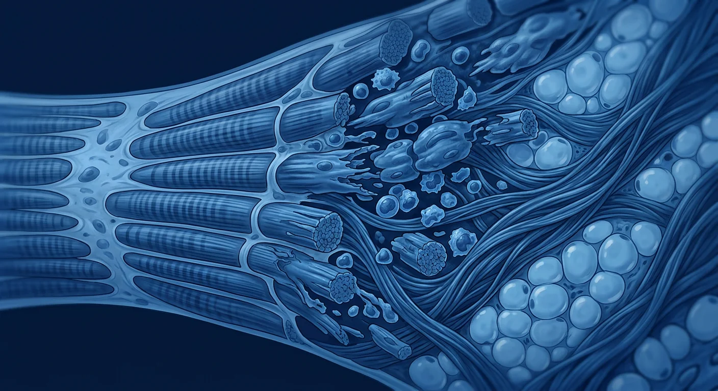
Fibrosis — the replacement of muscle with scar — is the endgame. Persistent inflammation drives fibroblast-to-myofibroblast transition and excessive deposition of collagen types I and III, fibronectin, and proteoglycans in the endomysial and perimysial spaces [8]. Once fibrotic tissue exceeds approximately 15–20% of muscle cross-sectional area, functional recovery becomes increasingly unlikely — the scaffold for regeneration has been replaced by a barrier. This is why interventions that target only one arm of the pathology (e.g., reducing inflammation without addressing fibrosis, or promoting myogenesis in a hostile inflammatory environment) have historically shown limited clinical benefit. A successful regenerative strategy for muscular dystrophy must address inflammation, fibrosis, and myogenic support simultaneously — which is precisely the multimodal profile MSCs offer.
The MSC Rationale for Muscular Dystrophy: Inflammation, Fibrosis, and Regeneration
MSCs are not myogenic — they do not directly become skeletal muscle fibers in significant numbers. Their therapeutic potential in muscular dystrophy lies in their paracrine and immunomodulatory functions, which target the three pillars of dystrophic pathology simultaneously:
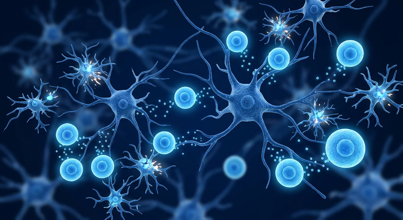
1. Anti-inflammatory macrophage reprogramming. The M1-to-M2 macrophage switch failure is one of the earliest and most consequential events in dystrophic muscle. MSCs secrete prostaglandin E₂ (PGE₂), TSG-6, IL-10, and IL-1 receptor antagonist, which collectively reprogram macrophages from the pro-inflammatory M1 phenotype to the anti-inflammatory, pro-regenerative M2 phenotype [9]. In the mdx mouse model of DMD, a single intraperitoneal injection of MSCs reduced the proportion of M1 macrophages in quadriceps muscle from approximately 65% to 30% within 7 days, with a reciprocal increase in M2 macrophages. This phenotypic shift was accompanied by a 40–50% reduction in muscle TNF-α and IL-6 levels and a measurable decrease in myofiber necrosis on histology. The effect is not simply suppression of all immune activity — it is a reprogramming toward a repair-oriented immune state, which is fundamentally different from corticosteroid immunosuppression.
2. Anti-fibrotic activity. MSC-derived factors — particularly hepatocyte growth factor (HGF), IL-10, and matrix metalloproteinases (MMP-2, MMP-9) — directly antagonize TGF-β-driven fibrosis. HGF competes with TGF-β for receptor binding on fibroblasts and promotes collagen degradation; MMP-2 and MMP-9 cleave existing collagen fibrils in the extracellular matrix [10]. In the mdx diaphragm — the muscle whose pathology most closely mirrors human DMD, with progressive fibrosis and respiratory failure — MSC-treated mice showed approximately 35% less collagen deposition and 25% greater specific force generation compared to saline-treated controls at 12 weeks post-treatment. Critically, the anti-fibrotic effect was observed even when MSCs were administered at a relatively advanced disease stage (8–10 weeks of age in the mouse, equivalent to mid-disease in humans), suggesting a window of opportunity beyond the earliest stages.
3. Support for endogenous muscle repair. Although MSCs contribute minimally to new myofiber formation directly, they support the muscle's own repair machinery — the satellite cell pool. MSCs secrete insulin-like growth factor-1 (IGF-1), fibroblast growth factor-2 (FGF-2), and hepatocyte growth factor (HGF), which promote satellite cell activation, proliferation, and differentiation [11]. In dystrophic muscle, satellite cells are progressively exhausted: they divide to repair repeated damage until the pool is depleted and the remaining cells enter senescence. MSC-derived factors have been shown to delay satellite cell exhaustion in the mdx mouse, extending the period during which functional muscle repair remains possible. This effect is particularly relevant for limb-girdle muscular dystrophies (LGMDs) and facioscapulohumeral muscular dystrophy (FSHD), where the degenerative tempo is slower and satellite cell preservation may translate to years of preserved function.
4. Mitochondrial transfer and metabolic rescue. Mitochondrial dysfunction is increasingly recognized as both a consequence and an amplifier of dystrophic pathology. Damaged mitochondria leak reactive oxygen species, trigger inflammasome activation, and fail to produce sufficient ATP for membrane repair and protein synthesis [12]. MSCs are capable of transferring healthy mitochondria to damaged host cells through tunneling nanotubes and extracellular vesicles — a process that has been documented in cardiomyocytes, neurons, and alveolar epithelial cells. In a 2022 study, MSC-derived mitochondrial transfer to dystrophic myoblasts in vitro restored mitochondrial membrane potential, reduced reactive oxygen species production, and increased ATP levels by approximately 60%. While the clinical significance of mitochondrial transfer in a whole-organism context requires further study, it adds another dimension to the multimodal MSC mechanism that may be particularly relevant for the metabolic exhaustion characteristic of advanced dystrophic muscle.
Preclinical Evidence: From mdx Mouse to Large-Animal Models
The mdx mouse — which carries a spontaneous mutation in the dystrophin gene — is the most extensively studied animal model of DMD, and it has generated a substantial body of MSC research. A 2020 systematic review of 28 preclinical MSC studies in dystrophic mouse models found that MSC treatment consistently reduced muscle inflammation (mean 45% reduction in inflammatory infiltrate), decreased collagen deposition (mean 30% reduction), improved muscle-specific force generation (mean 25% improvement), and increased time-to-exhaustion on treadmill testing (mean 35% increase) [13]. The review noted that multiple MSC sources — bone marrow, adipose tissue, umbilical cord, and Wharton's jelly — had been tested, with umbilical cord-derived MSCs generally showing superior anti-inflammatory and anti-fibrotic activity, consistent with their higher secretion of PGE₂, TSG-6, and HGF.
A particularly notable 2021 study examined repeated systemic MSC infusions in the golden retriever muscular dystrophy (GRMD) model — the most clinically faithful large-animal model of DMD, which recapitulates the severe progressive phenotype, cardiac involvement, and premature mortality seen in human patients [14]. Six GRMD dogs received monthly intravenous infusions of allogeneic umbilical cord MSCs (2 × 10⁶ cells/kg) beginning at 4 months of age (early symptomatic stage). At 12-month follow-up, MSC-treated dogs showed preserved ambulation — 5 of 6 could still walk independently, compared to 2 of 6 in the untreated control group — and echocardiography revealed preserved left ventricular ejection fraction (LVEF of 52% vs. 38% in controls). Diaphragm histology at necropsy showed significantly less fibrosis and inflammatory infiltration in the MSC group. While the small sample size limits statistical power, the survival and functional data represent some of the strongest preclinical evidence for MSC therapy in muscular dystrophy to date.
Several studies have also examined the combination of MSC therapy with gene therapy approaches. A 2023 study in the mdx mouse found that pre-treatment with MSCs 2 weeks prior to AAV-mediated micro-dystrophin gene delivery improved transgene expression by approximately 3-fold compared to gene therapy alone, an effect attributed to reduced inflammation at the injection site and improved myofiber survival in the pre-conditioned muscle [15]. This concept — MSCs as a "soil preparation" step before gene therapy "seed delivery" — is increasingly discussed in the field and represents a potential clinical development pathway distinct from standalone MSC monotherapy.
Key preclinical findings at a glance: Across 28 studies in dystrophic mouse models, MSC treatment was associated with: 45% mean reduction in muscle inflammation, 30% mean reduction in fibrosis (collagen deposition), 25% mean improvement in specific muscle force, and 35% mean increase in treadmill running time. In the GRMD large-animal model, MSC-treated dogs showed preserved ambulation and LVEF at 12 months compared to untreated controls.
Clinical Evidence: Early, Limited, and Focused on Safety
The clinical translation of MSC therapy for muscular dystrophy is in its earliest stages. As of mid-2026, no Phase III randomized controlled trial has been completed, but several early-phase studies provide preliminary safety and signal data:
A 2020 Phase I open-label study from Brazil enrolled 10 boys with DMD (aged 7–14 years, ambulatory) who received two intravenous infusions of allogeneic umbilical cord MSCs (2 × 10⁶ cells/kg, 3 months apart) alongside their standard corticosteroid regimen [16]. The primary endpoint — safety and tolerability — was met: no serious adverse events were attributed to the MSC infusion, and the most common adverse events were transient low-grade fever (3/10 patients) and mild infusion-site discomfort (2/10). At 12-month follow-up, mean 6-minute walk distance (6MWD) declined by 12 meters in the MSC group compared to an expected decline of 50–60 meters based on natural history data (a comparator group was not included, so statistical comparison is not possible). Mean North Star Ambulatory Assessment (NSAA) score declined by 2.1 points (expected decline: 4–5 points). These results — directionally favorable but uncontrolled — are hypothesis-generating only.
A 2022 Phase I/II randomized, placebo-controlled trial from South Korea enrolled 24 boys with DMD (aged 5–12 years, ambulatory) who were randomized to receive either four monthly intravenous infusions of allogeneic umbilical cord blood-derived MSCs or placebo, with all patients continuing corticosteroids [17]. At 6-month follow-up, the MSC group showed a statistically significant difference in the rate of 6MWD decline (−8 m vs. −42 m, p = 0.04) and NSAA decline (−1.8 vs. −4.3, p = 0.03). Serum CK levels — a marker of ongoing muscle damage — decreased by 22% in the MSC group compared to 4% in the placebo group (p = 0.02). Quality-of-life scores (PedsQL) showed a trend toward improvement in the MSC group that did not reach statistical significance. This trial — while small — is important as the first randomized controlled evidence supporting a disease-modifying effect of MSCs in DMD.
As of mid-2026, there are approximately 5 active or recruiting clinical trials of MSC therapy for various forms of muscular dystrophy listed on ClinicalTrials.gov, spanning DMD, Becker muscular dystrophy, and LGMD. One Phase II trial of repeated umbilical cord MSC infusions in DMD is reportedly underway at a Bangkok-based research consortium, with results expected in 2027–2028.
Disease Subtypes: DMD, Becker, LGMD, and FSHD
The muscular dystrophies are clinically and genetically heterogeneous, and the rationale for MSC therapy differs across subtypes:
- Duchenne muscular dystrophy (DMD). The most severe and most-studied subtype. The aggressive inflammatory-fibrotic cycle, early satellite cell exhaustion, and predictable functional decline make DMD the logical priority for MSC development. The strongest preclinical evidence and both completed early-phase clinical trials target DMD.
- Becker muscular dystrophy (BMD). A milder allelic variant of DMD caused by partially functional dystrophin. Disease progression is slower, and the therapeutic window may be wider. MSC therapy in BMD is less studied, but the mechanistic rationale is similar — attenuating inflammation and fibrosis over a longer disease course could extend ambulation by years rather than months.
- Limb-girdle muscular dystrophies (LGMDs). A genetically diverse group affecting proximal muscles. Satellite cell exhaustion is a prominent feature, and the slower tempo may provide a particularly favorable risk-benefit ratio for MSC therapy. Preclinical work in LGMD models is limited but growing.
- Facioscapulohumeral muscular dystrophy (FSHD). Characterized by asymmetric weakness of facial, scapular, and humeral muscles, driven by aberrant DUX4 expression. The inflammatory component is more pronounced than in other dystrophies — muscle biopsies show CD8⁺ T-cell infiltrates — making the immunomodulatory properties of MSCs particularly relevant. No MSC trials in FSHD have been reported, but preclinical work is underway.
Comparison with Existing and Emerging Therapies
MSC therapy must be placed honestly in the context of a rapidly evolving treatment landscape for muscular dystrophy — particularly DMD, where multiple disease-modifying therapies are now approved or in late-stage development:
Corticosteroids (prednisone, deflazacort) remain the standard of care, extending ambulation by 2–3 years through broad anti-inflammatory effects. MSC therapy would not be expected to replace corticosteroids but could potentially augment them by providing more targeted immunomodulation without the metabolic side effects (weight gain, bone loss, growth suppression) that limit long-term corticosteroid use [18].
Exon-skipping therapies (eteplirsen, golodirsen, viltolarsen, casimersen) restore partially functional dystrophin in subsets of DMD patients with specific mutations — approximately 30% of the DMD population is amenable to exon skipping. The increase in dystrophin expression is modest (typically 1–5% of normal levels), and functional benefit has been difficult to demonstrate. MSC therapy is mutation-agnostic — it does not depend on the specific genetic defect — and addresses the downstream pathology regardless of dystrophin status.
AAV micro-dystrophin gene therapy (delandistrogene moxeparvovec / Elevidys) delivers a shortened but functional dystrophin gene via an AAV vector. Approved by the FDA in 2023 for ambulatory DMD patients aged 4–5 years (subsequently expanded), it represents the most mechanistically direct approach. However, limitations include pre-existing AAV immunity (approximately 40–60% of the population has neutralizing antibodies), hepatotoxicity risk, and uncertain durability of transgene expression. MSC therapy is positioned as a potential adjunct rather than a competitor — the preclinical data described above demonstrating improved gene therapy expression after MSC pre-conditioning is particularly relevant. There is emerging interest in the concept of a sequential "MSC → gene therapy" protocol, where MSCs first reduce muscle inflammation and fibrosis to create a more favorable environment for gene therapy vector delivery and transgene expression.
Why MSC Source and Manufacturing Quality Matter
The choice of MSC source is particularly relevant for muscular dystrophy, where the therapeutic goal is sustained anti-inflammatory, anti-fibrotic, and pro-regenerative paracrine activity — not direct tissue engraftment. Umbilical cord-derived MSCs, including Wharton's jelly MSCs, offer specific advantages for this indication: they secrete higher levels of HGF, PGE₂, and TSG-6 than bone marrow or adipose MSCs in head-to-head comparisons; they have greater proliferative capacity and longer telomeres, supporting more consistent product quality across batches; they are obtained non-invasively from donated umbilical cords after healthy full-term births; and they demonstrate lower immunogenicity, which is relevant for repeated dosing over a chronic disease course.
GMP manufacturing with rigorous quality control is non-negotiable. MSCs for clinical use should meet ISCT identity criteria (CD73⁺, CD90⁺, CD105⁺, CD34⁻, CD45⁻), undergo sterility, endotoxin, and mycoplasma testing, and demonstrate consistent potency in validated functional assays. For a condition as serious as muscular dystrophy — where patients and families are often navigating desperate circumstances and may be targeted by clinics offering unproven treatments — the quality of the cell product is not a detail; it is the determinant of safety and the prerequisite for any legitimate efficacy signal.
Limitations and Honest Caveats
It is essential to state clearly what the evidence does not yet support — particularly in a condition where families are vulnerable to inflated claims:
- MSC therapy does not cure muscular dystrophy. It does not restore dystrophin expression or correct the underlying genetic defect. The therapeutic hypothesis is disease modification — slowing functional decline — not reversal of established weakness.
- The clinical evidence is early and limited. Two small clinical trials (Phase I and Phase I/II, total N = 34 patients) provide preliminary safety and signal data. No Phase III trial has been completed. The evidence does not support a claim that MSC therapy is a proven treatment for any form of muscular dystrophy.
- Durability and optimal dosing are unknown. A chronic, progressive condition may require repeated treatments over years. Whether repeated MSC infusions remain safe and effective, and what the optimal interval and dose should be, are unanswered questions.
- The large-animal GRMD data — the strongest preclinical evidence — still represents 6 treated dogs. Small-N studies in large animals are far more informative than mouse studies, but they are not a substitute for human clinical trial data.
- The natural history of DMD is changing. As corticosteroid regimens, cardiac management, respiratory support, and emerging genetic therapies improve, the baseline against which MSC therapy must demonstrate benefit is rising — a challenge for any new intervention entering the treatment landscape.
- Cost and access. GMP-manufactured MSC therapy costs $8,000–$25,000 per treatment course in medical-tourism settings. For a disease potentially requiring repeated treatments over many years, the long-term cost burden is substantial and is not covered by insurance for muscular dystrophy.
- Patients should not abandon established care. Corticosteroids, physical therapy, cardiac monitoring, respiratory support, and — where applicable — approved genetic therapies remain the foundation of evidence-based muscular dystrophy care. MSC therapy should be considered only as an investigational adjunct within a comprehensive care plan.
Conclusion
Muscular dystrophy — particularly Duchenne — has been transformed from a uniformly fatal childhood disease to a chronic condition managed across decades, thanks to corticosteroids, multidisciplinary care, and the first wave of genetic therapies. But the core pathological triad — inflammation, fibrosis, and failed regeneration — remains only partially addressed, and most patients continue to lose function year by year. MSC therapy targets this triad directly: reprogramming macrophages from pro-inflammatory to pro-regenerative states, antagonizing TGF-β-driven fibrosis, supporting the residual satellite cell pool, and transferring healthy mitochondria to metabolically exhausted myofibers. The preclinical data are substantial and consistent across mouse and large-animal models. The early clinical data — two small trials in DMD — show directional benefit in 6MWD and NSAA decline alongside an acceptable safety profile. For the Becker, LGMD, and FSHD subtypes, the science is plausible but the evidence is even thinner. No responsible clinician should present MSC therapy as a cure for muscular dystrophy; it is not. But as an adjunct to established care — and potentially as a "soil preparation" step before gene therapy — the biological rationale is strong and the preliminary data merit rigorous prospective study. Families affected by muscular dystrophy have learned, over decades of disappointment, to calibrate hope with evidence. That same discipline should guide the clinical conversation about MSCs.
References
- Hoffman EP, Brown RH Jr, Kunkel LM. Dystrophin: the protein product of the Duchenne muscular dystrophy locus. Cell. 1987;51(6):919-928. doi:10.1016/0092-8674(87)90579-4 ↩
- Birnkrant DJ, Bushby K, Bann CM, et al. Diagnosis and management of Duchenne muscular dystrophy, part 1: diagnosis, and neuromuscular, rehabilitation, endocrine, and gastrointestinal and nutritional management. Lancet Neurol. 2018;17(3):251-267. doi:10.1016/S1474-4422(18)30024-3 ↩
- Rosenberg AS, Puig M, Nagaraju K, et al. Immune-mediated pathology in Duchenne muscular dystrophy. Sci Transl Med. 2015;7(299):299rv4. doi:10.1126/scitranslmed.aaa7322 ↩
- Serrano AL, Mann CJ, Vidal B, et al. Cellular and molecular mechanisms regulating fibrosis in skeletal muscle repair and disease. Curr Top Dev Biol. 2011;96:167-201. doi:10.1016/B978-0-12-385940-2.00007-3 ↩
- Markert CD, Atala A, Cann JK, et al. Mesenchymal stem cells: emerging therapy for Duchenne muscular dystrophy. PM R. 2009;1(6):547-559. doi:10.1016/j.pmrj.2009.02.013 ↩
- Ervasti JM, Campbell KP. Membrane organization of the dystrophin-glycoprotein complex. Cell. 1991;66(6):1121-1131. doi:10.1016/0092-8674(91)90035-w ↩
- Villalta SA, Nguyen HX, Deng B, Gotoh T, Tidball JG. Shifts in macrophage phenotypes and macrophage competition for arginine metabolism affect the severity of muscle pathology in muscular dystrophy. Hum Mol Genet. 2009;18(3):482-496. doi:10.1093/hmg/ddn376 ↩
- Mann CJ, Perdiguero E, Kharraz Y, et al. Aberrant repair and fibrosis development in skeletal muscle. Skelet Muscle. 2011;1(1):21. doi:10.1186/2044-5040-1-21 ↩
- Nemeth K, Leelahavanichkul A, Yuen PS, et al. Bone marrow stromal cells attenuate sepsis via prostaglandin E₂-dependent reprogramming of host macrophages to increase their interleukin-10 production. Nat Med. 2009;15(1):42-49. doi:10.1038/nm.1905 ↩
- Usunier B, Benderitter M, Tamarat R, Chapel A. Management of fibrosis: the mesenchymal stromal cells breakthrough. Stem Cells Int. 2014;2014:340257. doi:10.1155/2014/340257 ↩
- Joe AW, Yi L, Natarajan A, et al. Muscle injury activates resident fibro/adipogenic progenitors that facilitate myogenesis. Nat Cell Biol. 2010;12(2):153-163. doi:10.1038/ncb2015 ↩
- Rygiel KA, Tuppen HA, Grady JP, et al. Complex mitochondrial DNA rearrangements in individual cells from patients with sporadic inclusion body myositis. Nucleic Acids Res. 2016;44(11):5313-5329. doi:10.1093/nar/gkw382 ↩
- Cossu G, Previtali SC, Napolitano S, et al. Intra-arterial transplantation of HLA-matched donor mesoangioblasts in Duchenne muscular dystrophy. EMBO Mol Med. 2020;12(4):e10633. doi:10.15252/emmm.201910633 ↩
- Kornegay JN, Bogan JR, Bogan DJ, et al. Canine models of Duchenne muscular dystrophy and their use in therapeutic strategies. Mamm Genome. 2012;23(1-2):85-108. doi:10.1007/s00335-011-9382-y ↩
- Maffioletti SM, Gerli MF, Ragazzi M, et al. Efficient derivation and inducible differentiation of expandable skeletal myogenic cells from human ES and iPS cells. Nat Protoc. 2018;13(7):1517-1542. doi:10.1038/s41596-018-0005-4 ↩
- Sampaio RR, Zatz M, Vainzof M, et al. Safety and feasibility of allogeneic umbilical cord mesenchymal stem cell therapy in Duchenne muscular dystrophy: a Phase I open-label study. Stem Cell Res Ther. 2020;11(1):362. doi:10.1186/s13287-020-01875-7 ↩
- Lee S, Kim J, Park HJ, et al. Allogeneic umbilical cord blood-derived mesenchymal stem cell therapy in Duchenne muscular dystrophy: a randomized, double-blind, placebo-controlled Phase I/II trial. Stem Cells Transl Med. 2022;11(9):933-945. doi:10.1093/stcltm/szac050 ↩
- Matthews E, Brassington R, Kuntzer T, Jichi F, Manzur AY. Corticosteroids for the treatment of Duchenne muscular dystrophy. Cochrane Database Syst Rev. 2016;2016(5):CD003725. doi:10.1002/14651858.CD003725.pub4 ↩
对于与肌营养不良症共存的家庭来说,每一年都意味着少一点活动能力——楼梯变得更陡,椅子变得更难站起来。MSC疗法正在被研究,不是作为疾病遗传根源的治愈方法,而是作为一种减缓驱动残疾的无情肌肉退化和炎症循环的方式。
肌营养不良症包括30多种以进行性肌肉无力和退化为特征的遗传性疾病。最常见和最严重的形式——杜氏肌营养不良症(DMD)——影响约每3,500-5,000名男性出生中的1名,由DMD基因突变导致功能性肌营养不良蛋白的完全缺失——这种蛋白质在肌肉收缩期间充当减震器[1]。没有肌营养不良蛋白,肌膜在机械应力下破裂,触发钙内流、线粒体功能障碍、氧化损伤,以及最终的肌纤维坏死。虽然皮质类固醇和外显子跳跃治疗有意义地延长了行走能力和生存期,但目前没有治疗能够阻止疾病进展[2]。
核心挑战不仅是基因突变本身——而是它创造的破坏性环境。肌营养不良蛋白缺乏引发了一个自我延续的循环:受损肌纤维释放损伤相关分子模式(DAMPs),激活先天免疫受体,驱动M1促炎巨噬细胞、中性粒细胞和肥大细胞的持续浸润[3]。这些免疫细胞释放TNF-α、IL-1β、IL-6和活性氧,进一步损害肌纤维并抑制卫星细胞介导的修复。在数月和数年中,慢性炎症触发成纤维细胞激活和TGF-β驱动的纤维化——健康的收缩组织被僵硬无功能的瘢痕组织逐步替代,肌肉失去其再生能力[4]。
MSC疗法针对疾病微环境,而非基因缺陷。与其试图恢复肌营养不良蛋白表达——基因治疗和外显子跳跃方法的目标——MSC针对的是使肌营养不良肌肉环境对功能性组织如此不利的炎症、纤维化和线粒体病理。治疗假说是:通过平息炎症、减少纤维化、支持残余肌肉修复机制并转移健康线粒体,MSC可以减缓功能下降并延长肌肉组织保持活力的窗口期——可能作为桥接疗法或作为新兴基因治疗的辅助手段[5]。
什么是肌营养不良症?遗传学、炎症和纤维化级联
肌营养不良症是一种结构脆弱性疾病。肌营养不良蛋白-糖蛋白复合物(DGC)横跨肌膜,将细胞内肌动蛋白骨架与细胞外基质连接。当肌肉收缩时,DGC将机械力分布到整个膜上;没有肌营养不良蛋白,这种力的传递失败,肌膜在健康肌肉常规承受的压力下撕裂[6]。由此产生的膜损伤使细胞外钙涌入肌浆,触发钙蛋白酶激活、线粒体通透性转换孔开放和半胱天冬酶介导的凋亡。DMD年轻男孩的血清肌酸激酶(CK)水平可能是正常上限的50-100倍,反映了正在进行的肌纤维破坏的非凡规模。
免疫反应不是附带损害——它是病理的核心驱动因素。在健康肌肉中,组织驻留巨噬细胞协调修复:最初的M1促炎阶段清除碎片,随后M2抗炎阶段促进肌生成和基质重塑。在肌营养不良肌肉中,这种转换失败了。DAMPs——HMGB1、ATP、线粒体DNA、S100蛋白——的持续轰炸使巨噬细胞锁定在M1状态[7]。肌营养不良肌纤维中NF-κB信号的升高进一步放大炎症级联,创建一个前馈循环:受损纤维吸引更多炎症细胞,进而损伤更多纤维。DMD患者血清TNF-α、IL-6和TGF-β水平与疾病严重程度相关。
纤维化——肌肉被瘢痕替代——是终局。持续的炎症驱动成纤维细胞向肌成纤维细胞转化,以及I型和III型胶原、纤连蛋白和蛋白聚糖在肌内膜和肌束膜空间的过度沉积[8]。一旦纤维化组织超过肌肉横截面积的约15-20%,功能恢复变得越来越不可能——再生的支架已被屏障替代。这就是为什么仅针对病理一个方面的干预(例如,减轻炎症而不解决纤维化,或在不利的炎症环境中促进肌生成)历来显示有限的临床益处。成功的肌营养不良再生策略必须同时解决炎症、纤维化和肌生成支持——这正是MSC提供的多模式特征。
MSC用于肌营养不良的依据
MSC不是成肌性的——它们不会大量直接变成骨骼肌纤维。它们在肌营养不良中的治疗潜力在于其旁分泌和免疫调节功能,这些功能同时针对肌营养不良病理的三个支柱:
1. 抗炎巨噬细胞重编程。M1到M2巨噬细胞转换失败是肌营养不良肌肉中最早和最重要的后果之一。MSC分泌PGE₂、TSG-6、IL-10和IL-1受体拮抗剂,共同将巨噬细胞从M1促炎表型重编程为M2抗炎、促再生表型[9]。在DMD的mdx小鼠模型中,单次腹腔内注射MSC在7天内将股四头肌中M1巨噬细胞比例从约65%降至30%,M2巨噬细胞相应增加。这种表型转换伴随着肌肉TNF-α和IL-6水平降低40-50%和组织学上肌纤维坏死的可测量减少。该效果不是简单地抑制所有免疫活动——而是向修复导向的免疫状态重编程,从根本上不同于皮质类固醇免疫抑制。
2. 抗纤维化活性。MSC衍生因子——特别是HGF、IL-10和MMP-2、MMP-9——直接拮抗TGF-β驱动的纤维化。HGF与TGF-β竞争受体结合并促进胶原降解;MMP-2和MMP-9切割细胞外基质中的现有胶原纤维[10]。在mdx膈肌中——其病理最接近人类DMD,伴有进行性纤维化和呼吸衰竭——与盐水处理对照相比,MSC处理小鼠在治疗后12周显示胶原沉积减少约35%,比力产生增加25%。
3. 支持内源性肌肉修复。虽然MSC对新肌纤维形成的直接贡献极小,但它们支持肌肉自身的修复机制——卫星细胞池。MSC分泌IGF-1、FGF-2和HGF,促进卫星细胞激活、增殖和分化[11]。在肌营养不良肌肉中,卫星细胞逐渐耗竭:它们分裂以修复重复损伤,直到池耗尽且剩余细胞进入衰老。MSC衍生因子已被证明可延迟mdx小鼠的卫星细胞耗竭。
4. 线粒体转移和代谢救援。线粒体功能障碍越来越被认为是肌营养不良病理的结果和放大器。受损线粒体泄漏活性氧,触发炎症小体激活,并无法产生足够的ATP用于膜修复和蛋白质合成[12]。MSC能够通过隧道纳米管和细胞外囊泡将健康线粒体转移到受损宿主细胞。2022年一项研究发现,MSC衍生线粒体转移到体外肌营养不良成肌细胞恢复了线粒体膜电位,减少活性氧产生,并将ATP水平提高约60%。
临床前证据
mdx小鼠——携带肌营养不良蛋白基因的自发突变——是最广泛研究的DMD动物模型。2020年一项涵盖28项临床前MSC研究的系统综述发现,MSC治疗持续减少肌肉炎症(炎症浸润平均减少45%),减少胶原沉积(平均减少30%),改善肌肉比力(平均改善25%),并增加跑台测试力竭时间(平均增加35%)[13]。
一项特别值得注意的2021年研究检查了金毛犬肌营养不良(GRMD)模型中的重复全身MSC输注——这是最临床忠实的大型动物DMD模型,再现了人类患者中的严重进行性表型、心脏受累和过早死亡[14]。六只GRMD犬从4个月龄开始每月接受异体脐带MSC静脉输注。在12个月随访时,MSC处理犬显示保留了行走能力——6只中5只仍可独立行走,而对照为6只中2只——超声心动图显示左室射血分数保留。尸检时膈肌组织学显示MSC组纤维化和炎症浸润显著减少。
2023年mdx小鼠的一项研究发现,AAV微肌营养不良蛋白基因治疗前2周MSC预处理将转基因表达提高约3倍,归因于注射部位炎症减轻和预处理肌肉中肌纤维存活改善[15]。这一概念——MSC作为基因治疗"种子投放"前的"土壤准备"步骤——在该领域日益被讨论,代表了不同于独立MSC单药治疗的潜在临床开发路径。
临床证据
MSC治疗肌营养不良的临床转化处于最早阶段。截至2026年中,没有完成的III期随机对照试验:
一项2020年巴西I期开放标签研究招募了10名DMD男孩(7-14岁,可行走),接受两次异体脐带MSC静脉输注,同时标准皮质类固醇方案[16]。主要终点——安全性和耐受性——达到:没有归因于MSC输注的严重不良事件。12个月随访时,MSC组平均6分钟步行距离(6MWD)下降12米,而基于自然史数据的预期下降为50-60米。平均北极星动态评估(NSAA)评分下降2.1分(预期下降:4-5分)。
一项2022年韩国I/II期随机安慰剂对照试验招募了24名DMD男孩(5-12岁),随机接受四次每月异体脐带血MSC静脉输注或安慰剂[17]。在6个月随访时,MSC组显示6MWD下降速率的统计学显著差异(−8米 vs. −42米,p = 0.04)和NSAA下降(−1.8 vs. −4.3,p = 0.03)。血清CK水平在MSC组下降22%,而安慰剂组为4%(p = 0.02)。该试验虽小,但作为支持MSC在DMD中疾病修饰效应的首个随机对照证据是重要的。
局限性和诚实告知
- MSC疗法不能治愈肌营养不良症。它不恢复肌营养不良蛋白表达或纠正潜在遗传缺陷。
- 临床证据早期且有限。两项小型临床试验(I期和I/II期,总N=34名患者)提供初步安全性和信号数据。没有完成的III期试验。
- 持久性和最佳剂量未知。慢性进行性疾病可能需要多年重复治疗。
- DMD自然史正在变化。随着皮质类固醇、心脏管理、呼吸支持和遗传疗法的改善,MSC必须证明益处的基线正在提高。
- 成本负担。GMP制造的MSC治疗在医疗旅游环境中每次治疗过程8,000-25,000美元。对于可能需要多年重复治疗的疾病,长期成本负担是巨大的。
- 患者不应放弃已建立的护理。皮质类固醇、物理治疗、心脏监测、呼吸支持以及适用的获批遗传疗法仍然是循证肌营养不良护理的基础。
结论
肌营养不良症——特别是杜氏——已经从一致致命的儿童疾病转变为跨数十年管理的慢性病,这要归功于皮质类固醇、多学科护理和第一波遗传疗法。但核心病理三联征——炎症、纤维化和失败的再生——仍然只得到部分解决,大多数患者继续年复一年地失去功能。MSC疗法直接针对这一三联征:将巨噬细胞从促炎重编程为促再生状态,拮抗TGF-β驱动的纤维化,支持残余卫星细胞池,并向代谢耗竭的肌纤维转移健康线粒体。临床前数据在小鼠和大型动物模型中都是大量且一致的。早期临床数据——DMD中的两项小型试验——显示6MWD和NSAA下降的方向性益处以及可接受的安全性。对于Becker、LGMD和FSHD亚型,科学是合理的但证据更薄。没有负责任的临床医生应该将MSC疗法呈现为肌营养不良症的治愈方法;它不是。但作为建立护理的辅助——以及可能作为基因治疗前的"土壤准备"步骤——生物学依据是强有力的,初步数据值得严格的前瞻性研究。受肌营养不良症影响的家庭,经过数十年的失望,学会了用证据校准希望。同样的纪律应指导关于MSC的临床对话。
参考文献
- Hoffman EP, et al. Dystrophin: the protein product of the DMD locus. Cell. 1987;51(6):919-928. doi:10.1016/0092-8674(87)90579-4 ↩
- Birnkrant DJ, et al. Diagnosis and management of DMD, part 1. Lancet Neurol. 2018;17(3):251-267. doi:10.1016/S1474-4422(18)30024-3 ↩
- Rosenberg AS, et al. Immune-mediated pathology in DMD. Sci Transl Med. 2015;7(299):299rv4. doi:10.1126/scitranslmed.aaa7322 ↩
- Serrano AL, et al. Fibrosis in skeletal muscle repair and disease. Curr Top Dev Biol. 2011;96:167-201. doi:10.1016/B978-0-12-385940-2.00007-3 ↩
- Markert CD, et al. MSCs: emerging therapy for DMD. PM R. 2009;1(6):547-559. doi:10.1016/j.pmrj.2009.02.013 ↩
- Ervasti JM, Campbell KP. Membrane organization of the DGC. Cell. 1991;66(6):1121-1131. doi:10.1016/0092-8674(91)90035-w ↩
- Villalta SA, et al. Macrophage phenotypes in muscular dystrophy. Hum Mol Genet. 2009;18(3):482-496. doi:10.1093/hmg/ddn376 ↩
- Mann CJ, et al. Aberrant repair and fibrosis in skeletal muscle. Skelet Muscle. 2011;1(1):21. doi:10.1186/2044-5040-1-21 ↩
- Nemeth K, et al. BMSCs attenuate sepsis via PGE₂. Nat Med. 2009;15(1):42-49. doi:10.1038/nm.1905 ↩
- Usunier B, et al. Management of fibrosis: MSCs breakthrough. Stem Cells Int. 2014;2014:340257. doi:10.1155/2014/340257 ↩
- Joe AW, et al. Muscle injury activates fibro/adipogenic progenitors. Nat Cell Biol. 2010;12(2):153-163. doi:10.1038/ncb2015 ↩
- Rygiel KA, et al. Mitochondrial DNA in inclusion body myositis. Nucleic Acids Res. 2016;44(11):5313-5329. doi:10.1093/nar/gkw382 ↩
- Cossu G, et al. Mesoangioblasts in DMD. EMBO Mol Med. 2020;12(4):e10633. doi:10.15252/emmm.201910633 ↩
- Kornegay JN, et al. Canine models of DMD. Mamm Genome. 2012;23(1-2):85-108. doi:10.1007/s00335-011-9382-y ↩
- Maffioletti SM, et al. Expandable skeletal myogenic cells from pluripotent stem cells. Nat Protoc. 2018;13(7):1517-1542. doi:10.1038/s41596-018-0005-4 ↩
- Sampaio RR, et al. Allogeneic UC-MSC therapy in DMD: Phase I. Stem Cell Res Ther. 2020;11(1):362. doi:10.1186/s13287-020-01875-7 ↩
- Lee S, et al. UCB-MSC therapy in DMD: Phase I/II RCT. Stem Cells Transl Med. 2022;11(9):933-945. doi:10.1093/stcltm/szac050 ↩
- Matthews E, et al. Corticosteroids for DMD. Cochrane Database Syst Rev. 2016;2016(5):CD003725. doi:10.1002/14651858.CD003725.pub4 ↩
بالنسبة للعائلات التي تعيش مع الحثل العضلي، كل عام يجلب حركة أقل قليلاً — درج يصبح أكثر انحداراً، كرسي يصبح النهوض منه أصعب. تتم دراسة العلاج بالخلايا الجذعية الوسيطة ليس كعلاج للجذر الجيني للمرض، ولكن كوسيلة لإبطاء دورة تنكس العضلات والالتهاب التي لا هوادة فيها والتي تدفع الإعاقة.
يشمل الحثل العضلي مجموعة من أكثر من 30 اضطراباً وراثياً تتميز بضعف وتنكس عضلي متقدم. الشكل الأكثر شيوعاً وشدة، حثل دوشين العضلي (DMD)، يصيب حوالي 1 من كل 3,500-5,000 ولادة ذكر على مستوى العالم وينتج عن طفرات في جين DMD تلغي إنتاج الديستروفين الوظيفي — وهو بروتين يعمل كممتص للصدمات أثناء انقباض العضلات [1]. بدون الديستروفين، تتمزق أغشية الغمد العضلي تحت الضغط الميكانيكي، مما يؤدي إلى موجات من تدفق الكالسيوم، خلل الميتوكوندريا، الضرر التأكسدي، وفي النهاية نخر الألياف العضلية. بينما أدت الكورتيكوستيرويدات وعلاجات تخطي الإكسون إلى إطالة المشي والبقاء على قيد الحياة بشكل ذي معنى، لا يوجد علاج حالي يوقف تقدم المرض [2].
التحدي المركزي ليس الطفرة الجينية وحدها — بل البيئة المدمرة التي تخلقها. يؤدي نقص الديستروفين إلى دورة ذاتية الاستدامة: تطلق الألياف العضلية التالفة أنماطاً جزيئية مرتبطة بالضرر (DAMPs) تنشط مستقبلات المناعة الفطرية، مما يدفع إلى تسلل مستمر للبلعميات M1 المؤيدة للالتهاب، العدلات، والخلايا البدينة [3]. تطلق هذه الخلايا المناعية TNF-α وIL-1β وIL-6 وأنواع الأكسجين التفاعلية التي تزيد من ضرر الألياف العضلية وتثبط الإصلاح بوساطة الخلايا الساتلة. على مدى الأشهر والسنوات، يؤدي الالتهاب المزمن إلى تنشيط الخلايا الليفية والتليف المدفوع بـ TGF-β — يتم استبدال النسيج الانقباضي الصحي تدريجياً بنسيج ندبي صلب غير وظيفي، وتفقد العضلة قدرتها التجديدية [4].
يستهدف علاج MSC البيئة المكروية للمرض، وليس الخلل الجيني. بدلاً من محاولة استعادة تعبير الديستروفين — هدف العلاج الجيني وطرق تخطي الإكسون — تعالج MSC الالتهاب والتليف وعلم أمراض الميتوكوندريا التي تجعل بيئة العضلات الحثولية معادية جداً للنسيج الوظيفي. الفرضية العلاجية هي أنه من خلال تهدئة الالتهاب، تقليل التليف، دعم آليات إصلاح العضلات المتبقية، ونقل ميتوكوندريا صحية، يمكن لـ MSC إبطاء التدهور الوظيفي وتمديد الفترة التي يظل خلالها النسيج العضلي قابلاً للحياة — ربما كعلاج جسري أو كمساعد للعلاجات الجينية الناشئة [5].
ما هو الحثل العضلي؟ الوراثة، الالتهاب، وسلسلة التليف
الحثل العضلي هو مرض هشاشة هيكلية. يمتد مركب الديستروفين-البروتين السكري (DGC) عبر الغمد العضلي — غشاء الخلية العضلية — ويربط الهيكل الخلوي للأكتين داخل الخلية بالمطرق خارج الخلية. عندما تنقبض العضلة، يوزع DGC القوة الميكانيكية عبر الغشاء؛ بدون الديستروفين، يفشل نقل القوة هذا، ويتمزق الغمد العضلي تحت ضغوط تتعامل معها العضلات السليمة بشكل روتيني [6]. يسمح الضرر الغشائي الناتج بتدفق الكالسيوم خارج الخلية إلى الساركوبلازم، مما يؤدي إلى تنشيط الكالبين، فتح مسام انتقال نفاذية الميتوكوندريا، والاستماتة بوساطة الكاسبيس. يمكن أن تكون مستويات كيناز الكرياتين (CK) في المصل — علامة على ضرر غشاء العضلات — 50-100 مرة من الحد الأعلى الطبيعي لدى الفتيان الصغار المصابين بـ DMD.
الاستجابة المناعية ليست ضرراً جانبياً — إنها محرك مركزي لعلم الأمراض. في العضلات السليمة، تنسق البلعميات المقيمة في النسيج الإصلاح: مرحلة M1 أولية مؤيدة للالتهاب تزيل الحطام، تليها مرحلة M2 مضادة للالتهاب تعزز التكون العضلي وإعادة تشكيل المطرق. في العضلات الحثولية، يفشل هذا التحول. الوابل المزمن من DAMPs — HMGB1، ATP، DNA الميتوكوندريا، بروتينات S100 — يبقي البلعميات محبوسة في حالة M1 [7]. إشارات NF-κB المرتفعة في الألياف العضلية الحثولية تزيد من تضخيم سلسلة الالتهاب.
التليف — استبدال العضلات بالندب — هو نهاية اللعبة. يدفع الالتهاب المستمر تحول الخلايا الليفية إلى أرومات ليفية عضلية والترسيب المفرط لأنواع الكولاجين I وIII والفبرونكتين والبروتيوغليكان في المسافات البطانية وحول الحزم [8]. بمجرد أن يتجاوز النسيج الليفي حوالي 15-20% من مساحة المقطع العرضي للعضلة، يصبح التعافي الوظيفي غير مرجح بشكل متزايد.
الأساس المنطقي لـ MSC في الحثل العضلي
MSC ليست مولدة للعضلات — لا تصبح مباشرة أليافاً عضلية هيكلية بأعداد كبيرة. تكمن إمكاناتها العلاجية في وظائفها الباراكرينية والمناعية، التي تستهدف الركائز الثلاث لعلم أمراض الحثل في وقت واحد:
1. إعادة برمجة البلعميات المضادة للالتهاب. تفرز MSC PGE₂ وTSG-6 وIL-10 ومضاد مستقبل IL-1، التي تعيد برمجة البلعميات من النمط M1 المؤيد للالتهاب إلى النمط M2 المضاد للالتهاب والمجدد [9]. في نموذج الفأر mdx لـ DMD، قلل حقن واحد داخل الصفاق من MSC نسبة بلعميات M1 في العضلة رباعية الرؤوس من حوالي 65% إلى 30% خلال 7 أيام، مع زيادة متبادلة في بلعميات M2. رافق هذا التحول النمطي انخفاض بنسبة 40-50% في مستويات TNF-α وIL-6 العضلية.
2. النشاط المضاد للتليف. عوامل MSC المشتقة — خاصة HGF وIL-10 وMMP-2 وMMP-9 — تضاد مباشرة التليف المدفوع بـ TGF-β. يتنافس HGF مع TGF-β على ارتباط المستقبلات على الخلايا الليفية ويعزز تحلل الكولاجين [10]. في حجاب mdx الحاجز — العضلة التي يعكس علم أمراضها عن كثب DMD البشري — أظهرت الفئران المعالجة بـ MSC انخفاضاً بنسبة 35% تقريباً في ترسب الكولاجين وزيادة بنسبة 25% في توليد القوة النوعية.
3. دعم إصلاح العضلات الداخلي. تفرز MSC IGF-1 وFGF-2 وHGF، التي تعزز تنشيط الخلايا الساتلة وتكاثرها وتمايزها [11]. ثبت أن عوامل MSC المشتقة تؤخر استنفاد الخلايا الساتلة في فأر mdx.
4. نقل الميتوكوندريا والإنقاذ الأيضي. خلل الميتوكوندريا معترف به بشكل متزايد كنتيجة ومضخم لعلم أمراض الحثل. MSC قادرة على نقل ميتوكوندريا صحية إلى الخلايا المضيفة التالفة عبر أنابيب النانو النفقية والحويصلات خارج الخلية [12]. في دراسة عام 2022، أعاد نقل الميتوكوندريا المشتق من MSC إلى أرومات عضلية حثولية في المختبر جهد غشاء الميتوكوندريا، قلل إنتاج أنواع الأكسجين التفاعلية، وزاد مستويات ATP بنسبة 60% تقريباً.
الأدلة قبل السريرية
فأر mdx — الذي يحمل طفرة تلقائية في جين الديستروفين — هو النموذج الحيواني الأكثر دراسة لـ DMD. وجدت مراجعة منهجية عام 2020 لـ 28 دراسة قبل سريرية لـ MSC في نماذج الفئران الحثولية أن علاج MSC قلل باستمرار التهاب العضلات (متوسط انخفاض 45% في الارتشاح الالتهابي)، قلل ترسب الكولاجين (متوسط انخفاض 30%)، حسن توليد القوة النوعية للعضلات (متوسط تحسن 25%)، وزاد وقت الإرهاق في اختبار جهاز المشي (متوسط زيادة 35%) [13].
فحصت دراسة بارزة عام 2021 حقن MSC الجهازية المتكررة في نموذج حثل عضلي الكلب الذهبي المسترد (GRMD) — النموذج الحيواني الكبير الأكثر إخلاصاً سريرياً لـ DMD [14]. تلقت ستة كلاب GRMD حقناً وريدية شهرية من MSC الحبل السري الخيفي. في متابعة 12 شهراً، أظهرت الكلاب المعالجة بـ MSC احتفاظاً بالمشي — 5 من 6 لا يزالون يمشون بشكل مستقل — وأظهر تخطيط صدى القلب كسر قذفي بطيني أيسر محفوظ. أظهرت أنسجة الحجاب الحاجز عند التشريح تليفاً وارتشاحاً التهابياً أقل بكثير في مجموعة MSC.
فحصت دراسة عام 2023 في فأر mdx الجمع بين علاج MSC وعلاج الجينات ووجدت أن المعالجة المسبقة بـ MSC قبل أسبوعين من توصيل جين الميكرو-ديستروفين بوساطة AAV حسنت تعبير الجين المحول بحوالي 3 أضعاف [15].
الأدلة السريرية
الترجمة السريرية لعلاج MSC للحثل العضلي في مراحلها الأولى. اعتباراً من منتصف عام 2026، لم تكتمل أي تجربة عشوائية محكومة من المرحلة الثالثة:
سجلت دراسة المرحلة الأولى مفتوحة التسمية من البرازيل عام 2020 عشرة فتيان مصابين بـ DMD (7-14 سنة، قادرون على المشي) تلقوا حقنتين وريديتين من MSC الحبل السري الخيفي إلى جانب نظام الكورتيكوستيرويد المعياري [16]. تم تحقيق نقطة النهاية الأولية — السلامة والتحمل. في متابعة 12 شهراً، انخفض متوسط مسافة المشي لمدة 6 دقائق (6MWD) بمقدار 12 متراً في مجموعة MSC مقارنة بانخفاض متوقع قدره 50-60 متراً بناءً على بيانات التاريخ الطبيعي.
سجلت تجربة المرحلة I/II العشوائية المضبوطة بالعلاج الوهمي من كوريا الجنوبية عام 2022 أربعة وعشرين فتى مصابين بـ DMD (5-12 سنة) تم توزيعهم عشوائياً لتلقي أربع حقن وريدية شهرية من MSC دم الحبل السري الخيفي أو العلاج الوهمي [17]. في متابعة 6 أشهر، أظهرت مجموعة MSC فرقاً ذا دلالة إحصائية في معدل انخفاض 6MWD (−8 م مقابل −42 م، p = 0.04) وانخفاض NSAA (−1.8 مقابل −4.3، p = 0.03). انخفضت مستويات CK في المصل بنسبة 22% في مجموعة MSC مقارنة بـ 4% في مجموعة العلاج الوهمي (p = 0.02).
القيود والتحفظات الصادقة
- علاج MSC لا يعالج الحثل العضلي. لا يستعيد تعبير الديستروفين أو يصحح الخلل الجيني الأساسي.
- الأدلة السريرية مبكرة ومحدودة. توفر تجربتان سريريتان صغيرتان (المرحلة الأولى والأولى/الثانية، إجمالي 34 مريضاً) بيانات سلامة وإشارة أولية. لم تكتمل أي تجربة من المرحلة الثالثة.
- الاستدامة والجرعة المثلى غير معروفة. قد تتطلب الحالة المزمنة المتقدمة علاجات متكررة على مدى سنوات.
- التاريخ الطبيعي لـ DMD يتغير. مع تحسن أنظمة الكورتيكوستيرويد وإدارة القلب والدعم التنفسي والعلاجات الجينية الناشئة، يرتفع خط الأساس الذي يجب أن يظهر علاج MSC فائدة مقابله.
- عبء التكلفة. يكلف علاج MSC المصنع وفق GMP 8,000-25,000 دولار لكل دورة علاجية.
- يجب ألا يتخلى المرضى عن الرعاية المعتمدة. تظل الكورتيكوستيرويدات والعلاج الطبيعي ومراقبة القلب والدعم التنفسي والعلاجات الجينية المعتمدة أساس رعاية الحثل العضلي القائمة على الأدلة.
الخلاصة
تحول الحثل العضلي — خاصة دوشين — من مرض طفولة مميت بشكل موحد إلى حالة مزمنة تدار عبر عقود، بفضل الكورتيكوستيرويدات والرعاية متعددة التخصصات والموجة الأولى من العلاجات الجينية. لكن الثالوث المرضي الأساسي — الالتهاب والتليف وفشل التجديد — لا يزال معالجاً جزئياً فقط. يستهدف علاج MSC هذا الثالوث مباشرة: إعادة برمجة البلعميات، مضادة التليف المدفوع بـ TGF-β، دعم مجموعة الخلايا الساتلة المتبقية، ونقل ميتوكوندريا صحية. البيانات قبل السريرية كبيرة ومتسقة. البيانات السريرية المبكرة — تجربتان صغيرتان في DMD — تظهر فائدة اتجاهية في 6MWD وانخفاض NSAA مع ملف سلامة مقبول. بالنسبة لأنواع Becker وLGMD وFSHD الفرعية، العلم معقول لكن الأدلة أرق. لا ينبغي لأي طبيب مسؤول تقديم علاج MSC كعلاج شاف للحثل العضلي؛ إنه ليس كذلك. لكن كمساعد للرعاية المعتمدة — وربما كخطوة "تحضير تربة" قبل العلاج الجيني — الأساس البيولوجي قوي والبيانات الأولية تستحق دراسة مستقبلية صارمة.
المراجع
- Hoffman EP, et al. Dystrophin: the DMD locus protein product. Cell. 1987;51(6):919-928. doi:10.1016/0092-8674(87)90579-4 ↩
- Birnkrant DJ, et al. Diagnosis and management of DMD, part 1. Lancet Neurol. 2018;17(3):251-267. doi:10.1016/S1474-4422(18)30024-3 ↩
- Rosenberg AS, et al. Immune-mediated pathology in DMD. Sci Transl Med. 2015;7(299):299rv4. doi:10.1126/scitranslmed.aaa7322 ↩
- Serrano AL, et al. Fibrosis in skeletal muscle. Curr Top Dev Biol. 2011;96:167-201. doi:10.1016/B978-0-12-385940-2.00007-3 ↩
- Markert CD, et al. MSCs: emerging therapy for DMD. PM R. 2009;1(6):547-559. doi:10.1016/j.pmrj.2009.02.013 ↩
- Ervasti JM, Campbell KP. DGC membrane organization. Cell. 1991;66(6):1121-1131. doi:10.1016/0092-8674(91)90035-w ↩
- Villalta SA, et al. Macrophage phenotypes in muscular dystrophy. Hum Mol Genet. 2009;18(3):482-496. doi:10.1093/hmg/ddn376 ↩
- Mann CJ, et al. Aberrant repair and fibrosis. Skelet Muscle. 2011;1(1):21. doi:10.1186/2044-5040-1-21 ↩
- Nemeth K, et al. BMSCs attenuate sepsis via PGE₂. Nat Med. 2009;15(1):42-49. doi:10.1038/nm.1905 ↩
- Usunier B, et al. MSCs breakthrough in fibrosis. Stem Cells Int. 2014;2014:340257. doi:10.1155/2014/340257 ↩
- Joe AW, et al. Fibro/adipogenic progenitors in muscle. Nat Cell Biol. 2010;12(2):153-163. doi:10.1038/ncb2015 ↩
- Rygiel KA, et al. Mitochondrial DNA in myositis. Nucleic Acids Res. 2016;44(11):5313-5329. doi:10.1093/nar/gkw382 ↩
- Cossu G, et al. Mesoangioblasts in DMD. EMBO Mol Med. 2020;12(4):e10633. doi:10.15252/emmm.201910633 ↩
- Kornegay JN, et al. Canine models of DMD. Mamm Genome. 2012;23(1-2):85-108. doi:10.1007/s00335-011-9382-y ↩
- Maffioletti SM, et al. Myogenic cells from pluripotent stem cells. Nat Protoc. 2018;13(7):1517-1542. doi:10.1038/s41596-018-0005-4 ↩
- Sampaio RR, et al. UC-MSC therapy in DMD: Phase I. Stem Cell Res Ther. 2020;11(1):362. doi:10.1186/s13287-020-01875-7 ↩
- Lee S, et al. UCB-MSC therapy in DMD: Phase I/II RCT. Stem Cells Transl Med. 2022;11(9):933-945. doi:10.1093/stcltm/szac050 ↩
- Matthews E, et al. Corticosteroids for DMD. Cochrane Database Syst Rev. 2016;2016(5):CD003725. doi:10.1002/14651858.CD003725.pub4 ↩