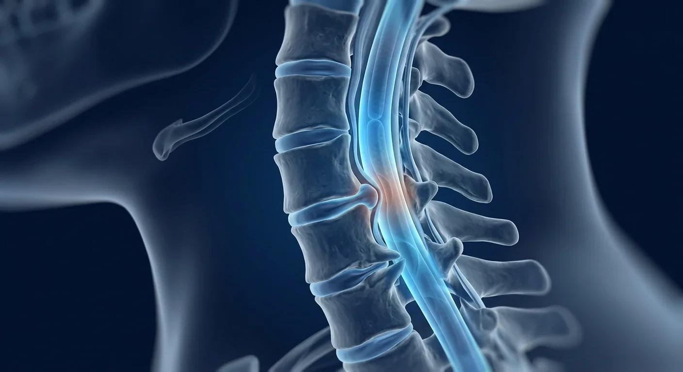
Cervical spondylotic myelopathy (CSM) is the most common cause of spinal cord dysfunction in adults over 55, affecting an estimated 1.6 per 100,000 people annually — and this figure likely undercounts the true prevalence as early cases are frequently misattributed to "normal aging." [1] CSM occurs when degenerative changes in the cervical spine — disc herniation, osteophyte formation, ligamentum flavum hypertrophy — progressively compress the spinal cord, leading to a constellation of symptoms ranging from hand numbness and gait disturbance to bowel and bladder dysfunction.
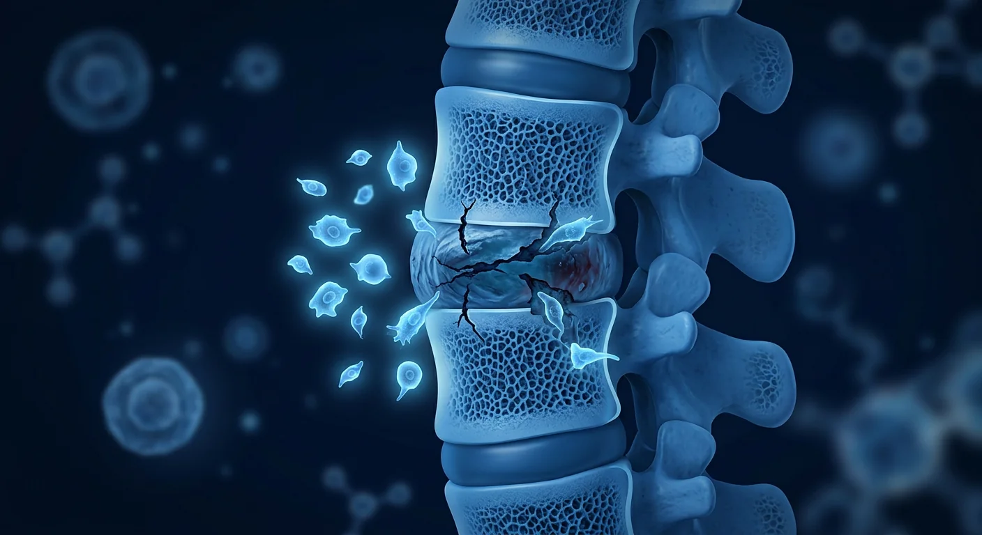
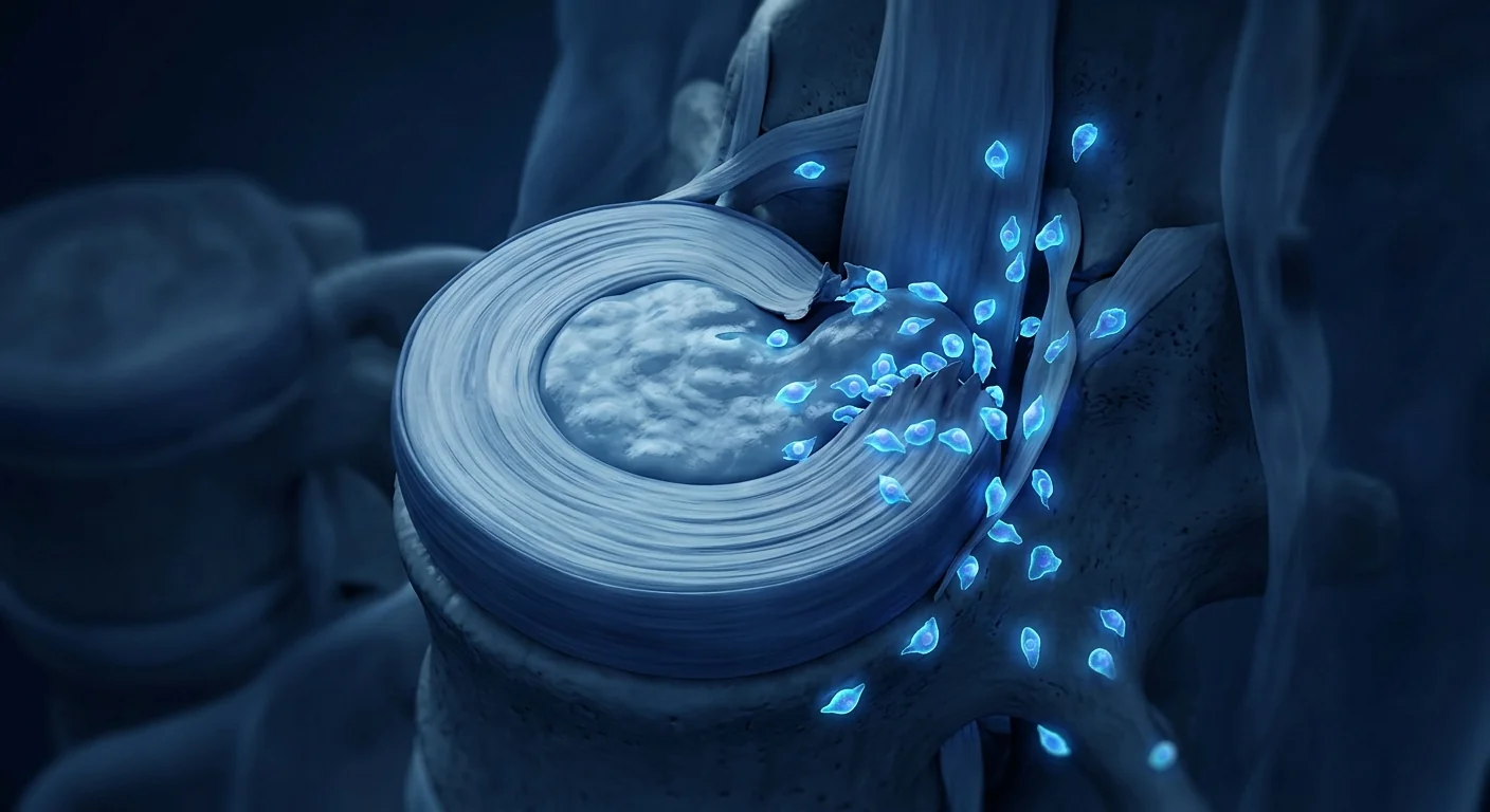
Where conventional treatment falls short. Surgical decompression — anterior cervical discectomy and fusion (ACDF) or laminoplasty — remains the standard of care for moderate to severe CSM. Surgery halts further mechanical compression, but it does not reverse established spinal cord pathology. A substantial proportion of patients — 20–40% in long-term series — show incomplete neurological recovery despite technically successful decompression. [2] The cord tissue that has already undergone demyelination, axonal loss, and gliosis does not spontaneously regenerate.
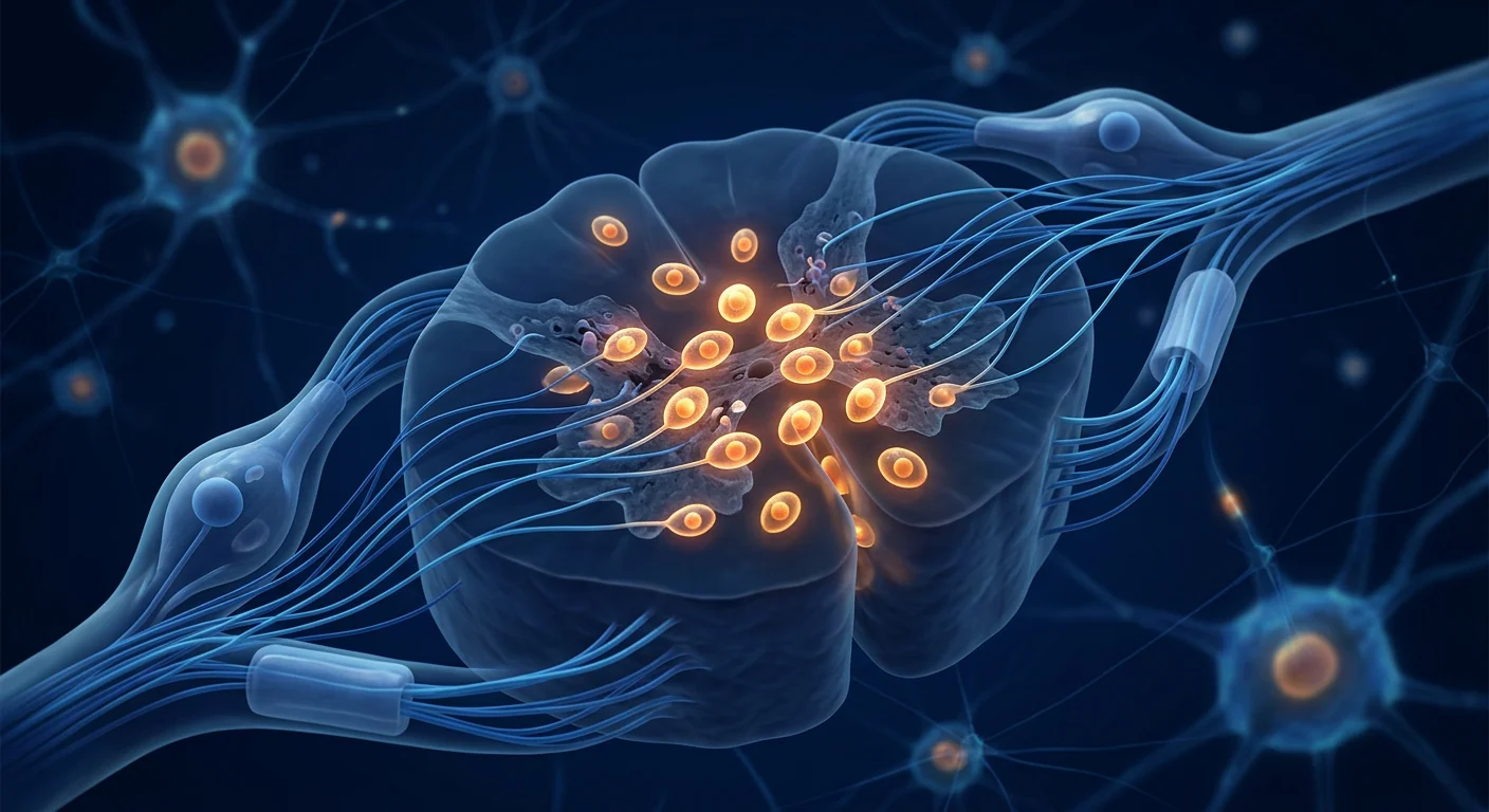
The deeper problem is at the cellular level. Chronic compression triggers a cascade of secondary injury: microglial activation, blood–spinal cord barrier disruption, excitotoxicity, mitochondrial dysfunction, oligodendrocyte apoptosis, and chronic neuroinflammation driven by TNF-α, IL-1β, and IL-6. [3] Even after decompression, this inflammatory microenvironment can persist for months, perpetuating ongoing neural damage. The spinal cord needs more than mechanical relief — it needs biological repair.
MSC therapy targets the secondary injury cascade. Mesenchymal stem cells address the biological drivers of CSM progression that surgery cannot reach. They home to sites of injury, secrete a broad repertoire of neurotrophic and anti-inflammatory factors, reprogram microglia from a pro-inflammatory M1 to a reparative M2 phenotype, and promote remyelination by supporting oligodendrocyte precursor cell survival. [4] This multi-target biological approach is being investigated as both an adjunct to surgical decompression and, in select early-stage cases, as a stand-alone intervention to slow or halt disease progression.
How MSC Therapy Works in Cervical Spondylotic Myelopathy
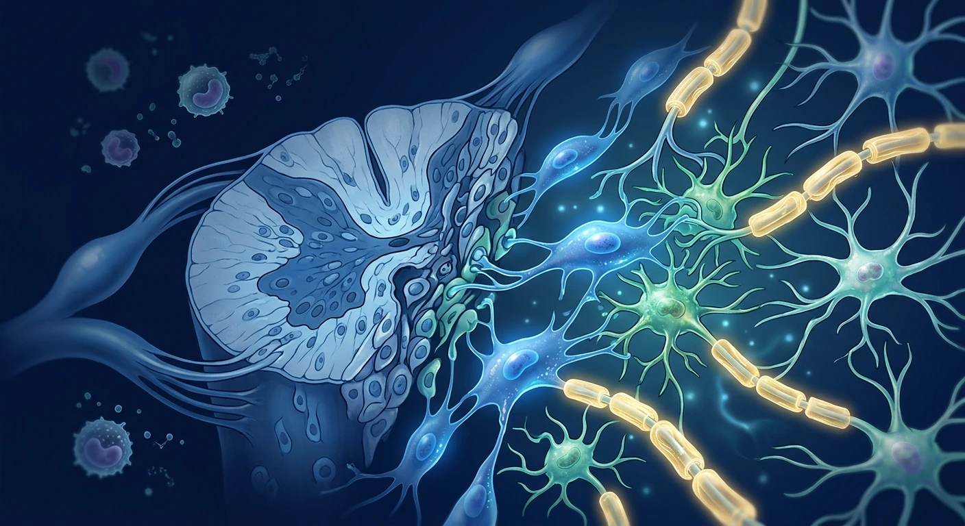
MSC therapy promotes spinal cord repair in CSM through four interconnected mechanisms: neuroinflammation suppression, microglial reprogramming, neurotrophic factor secretion, and remyelination support. Unlike single-agent pharmaceuticals, MSCs deploy a coordinated biological response that addresses multiple injury pathways simultaneously.
1. Neuroinflammation Suppression
Chronic spinal cord compression triggers a persistent inflammatory response characterized by elevated levels of TNF-α, IL-1β, IL-6, and matrix metalloproteinases (MMPs) that degrade the extracellular matrix and damage myelin. [6] MSCs secrete prostaglandin E2 (PGE2), transforming growth factor-β (TGF-β), tumor necrosis factor-stimulated gene 6 (TSG-6), and interleukin-10 (IL-10) — a cocktail that collectively downregulates these pro-inflammatory mediators. In rat models of chronic cervical spinal cord compression, intrathecal MSC delivery reduced TNF-α levels by 55–70% and IL-1β by 45–60% within 72 hours, with corresponding improvements in motor-evoked potential amplitudes. [7]
2. Microglial M1 → M2 Polarization
Microglia — the spinal cord's resident immune cells — become chronically activated in CSM, adopting a neurotoxic M1 phenotype that releases reactive oxygen species, nitric oxide, and additional pro-inflammatory cytokines. MSCs reprogram these cells toward the M2 (neuroprotective) phenotype through PGE2 and TSG-6 signaling. [8] M2 microglia clear apoptotic debris, secrete brain-derived neurotrophic factor (BDNF) and insulin-like growth factor-1 (IGF-1), and create a permissive environment for axonal sprouting and remyelination. This polarization shift is measurable within 24–48 hours of MSC administration and correlates with functional recovery in preclinical models.
3. Neurotrophic Factor Secretion
MSCs are potent factories of neurotrophic factors — BDNF, nerve growth factor (NGF), glial cell line-derived neurotrophic factor (GDNF), neurotrophin-3 (NT-3), and ciliary neurotrophic factor (CNTF). [9] These molecules promote neuronal survival, axonal outgrowth, and synaptic plasticity. In compressed spinal cord tissue, BDNF and NT-3 signaling through TrkB and TrkC receptors has been shown to preserve motor neuron function and reduce apoptotic cell death in the ventral horn. MSC-derived extracellular vesicles (EVs) carry these factors plus microRNAs (miR-21, miR-124, miR-146a) that further modulate the injury microenvironment. [10]
4. Remyelination Support
Demyelination is a hallmark pathological feature of CSM — chronic compression strips oligodendrocytes from axons, slowing or blocking signal conduction. MSCs promote remyelination through two complementary pathways: direct secretion of factors that support oligodendrocyte precursor cell (OPC) survival and differentiation (PDGF-AA, IGF-1, CNTF), and indirect effects via M2 microglia that clear myelin debris and create a pro-myelinating environment. [11] Electron microscopy studies in rat compression models have demonstrated significantly thicker myelin sheaths and higher numbers of myelinated axons in MSC-treated animals compared to vehicle controls.
Clinical Evidence: What the Research Shows
The preclinical evidence for MSCs in spinal cord compression injury is robust — over 25 animal studies across rat, mouse, and canine models, consistently demonstrating improved motor function, reduced inflammation, preserved myelin, and decreased neuronal apoptosis. Human data is emerging from early-phase clinical trials in related spinal cord pathologies. [12]
Preclinical compression models show functional recovery. In a rat model of chronic cervical cord compression using an expanding polymer implant, intrathecal MSC administration at 4 weeks post-compression resulted in significantly improved forelimb grip strength, inclined plane performance, and gait coordination compared to saline controls at 8 and 12 weeks. Histological analysis revealed reduced cavity formation, preserved motor neurons in the ventral horn, and increased axonal density at the compression site. [13]
Human spinal cord injury trials provide relevant safety data. While CSM-specific human trials are still in early stages, Phase I/II trials of intrathecal MSC administration for traumatic spinal cord injury (SCI) — a condition with overlapping secondary injury mechanisms — have established a favorable safety profile. A 2020 meta-analysis of 10 clinical trials (n=377 patients) found no serious adverse events attributable to MSC therapy, with modest but statistically significant improvements in ASIA motor scores and activities of daily living. [14]
Intrathecal delivery shows particular promise for CSM. The anatomical proximity of the cervical spinal cord to the subarachnoid space makes intrathecal (lumbar puncture) delivery an attractive route — MSCs infused into the CSF migrate to sites of injury via chemokine gradients (SDF-1/CXCR4 axis). [15] This achieves higher local cell concentrations at the compression site than intravenous delivery while avoiding first-pass pulmonary trapping. Early data suggest intrathecal delivery yields superior spinal cord homing compared to intravenous routes for cord-level pathology.
The VELAR Treatment Approach for CSM
At VELAR Center, MSC therapy for cervical spondylotic myelopathy is administered as a targeted biological adjunct to standard neurosurgical care. We collaborate closely with each patient's spine surgeon and neurologist to integrate MSC therapy into the broader treatment plan — before or after decompression surgery, depending on the individual clinical scenario.
Assessment and Eligibility
Every CSM patient undergoes a comprehensive evaluation before treatment consideration: cervical spine MRI with T2-weighted sequences to assess cord signal change and compression severity, neurological examination including modified Japanese Orthopaedic Association (mJOA) scoring, and baseline functional assessments. We review prior surgical history, current medications, and general health status. MSC therapy is considered for patients with documented CSM who have incomplete recovery after decompression, those with mild-to-moderate CSM seeking a non-surgical biological intervention, and surgical candidates where MSCs may enhance post-operative neurological recovery.
Cell Source and Delivery
VELAR uses allogeneic Wharton's jelly-derived mesenchymal stem cells (WJ-MSCs) cultured under cGMP conditions in our laboratory operating under ISO 9001. WJ-MSCs are selected for their high proliferative capacity, robust neurotrophic factor secretion profile, and low immunogenicity — they do not require HLA matching. [16] Cells undergo identity verification via ISCT criteria (CD73+, CD90+, CD105+, CD45-, CD34-, CD14-), multi-pathogen testing, and viability assessment (>95% at delivery). Delivery is typically via lumbar intrathecal injection — a 20-minute procedure performed under sterile conditions with local anesthesia — allowing MSCs to circulate in the CSF and home to the cervical compression site.
Treatment Protocol
A typical CSM protocol involves 1–2 intrathecal MSC administrations spaced 3–6 months apart, with each dose containing 50–100 million cells. The protocol is individualized — patients with more severe myelopathy or longer symptom duration may benefit from a second administration. [17] Follow-up assessments at 3, 6, and 12 months track mJOA scores, gait parameters, hand function (nine-hole peg test), and patient-reported outcomes (NDI — Neck Disability Index). Repeat MRI at 6–12 months assesses cord morphology and T2 signal change.
Frequently Asked Questions
How much does stem cell therapy for cervical myelopathy cost in Thailand?
At VELAR Center, a single intrathecal MSC treatment for CSM typically ranges from 400,000–600,000 THB (approximately USD 11,000–16,500), depending on cell dose and protocol complexity. This includes all pre-treatment assessments, the cell product, the intrathecal procedure, and follow-up monitoring. Compared to surgical decompression with fusion (ACDF), which can cost USD 30,000–80,000 in the United States, MSC therapy in Bangkok offers a significant cost advantage.
Is stem cell therapy a replacement for surgery in CSM?
No. In moderate to severe CSM with significant cord compression and progressive neurological deficits, surgical decompression remains the standard of care and should not be delayed. MSC therapy is being studied as a biological adjunct — to rescue cord tissue before surgery, to enhance recovery after decompression, or to address persistent neurological deficits in patients who have plateaued post-operatively. It is not a substitute for mechanical decompression when that is clinically indicated.
What results can patients realistically expect?
MSC therapy for CSM remains investigational. In preclinical models, outcomes include improved motor function, reduced neuroinflammation, and histological evidence of remyelination. Early human data from related spinal cord pathology trials shows modest functional improvements in a subset of patients. Realistic expectations include: stabilization of neurological decline, modest improvements in hand function or gait (typically measurable at 3–6 months), and reduced neuropathic pain. Dramatic reversal of long-standing severe myelopathy is not supported by current evidence.
Are there risks specific to intrathecal MSC delivery?
Intrathecal injection carries standard procedural risks: post-lumbar puncture headache (5–10%, typically self-limiting), transient back pain, and a very small risk of infection (<0.1% under sterile technique). MSC-specific risks are low based on safety data from over 1,000 patients in spinal cord injury and ALS trials — no tumor formation, no ectopic tissue growth, and no serious immunological reactions have been reported with allogeneic WJ-MSCs. [18]
How soon after decompression surgery can MSCs be administered?
Timing is individualized. In animal models, MSC administration at 1–4 weeks post-decompression showed superior outcomes compared to intraoperative delivery, as the acute surgical inflammatory response may reduce MSC survival. At VELAR, we typically recommend waiting 4–8 weeks after surgery to allow surgical inflammation to resolve before administering MSCs intrathecally. This window also allows assessment of the patient's post-operative neurological trajectory.
Can MSC therapy prevent CSM from progressing to require surgery?
This is an active area of investigation but not yet established. In theory, MSCs' ability to suppress neuroinflammation and promote remyelination could slow the biological progression of CSM in patients with mild cord compression who are not yet surgical candidates. Preclinical data support this concept, but no controlled human trial has yet demonstrated that MSC therapy alters the natural history of CSM. This remains an off-label, individualized clinical decision made in consultation with the patient's neurologist and spine surgeon.
Limitations and Honest Assessment
MSC therapy for cervical spondylotic myelopathy is at an early stage of clinical investigation. The preclinical evidence is promising and mechanistically coherent, but the leap from rodent compression models to human CSM patients is substantial. [19] No randomized controlled trial has yet been completed specifically for CSM, and the human safety and efficacy data are extrapolated from related conditions (traumatic SCI, ALS) rather than from dedicated CSM cohorts.
Patients and families should understand the following: MSC therapy for CSM is an investigational biological intervention, not a proven treatment. The goal is to address the secondary injury biology that surgery cannot reach — neuroinflammation, demyelination, and axonal degeneration. Surgical decompression remains the standard of care for progressive CSM with significant cord compression. MSC therapy works best as an adjunct, not a replacement. Results are typically modest and gradual — stabilization of decline and incremental functional improvements over 3–12 months — rather than dramatic reversals of established neurological deficits. [20]
VELAR Center offers MSC therapy within a framework of transparent informed consent, close collaboration with each patient's referring neurologist and spine surgeon, and honest communication about what the current evidence does and does not support. We believe biological repair strategies represent the future of CSM management, but we are candid that the future is still being written.
References
- Nouri A, Tetreault L, Singh A, Karadimas SK, Fehlings MG. Degenerative cervical myelopathy: epidemiology, genetics, and pathogenesis. Spine. 2015;40(12):E675-E693. doi:10.1097/BRS.0000000000000913 ↩
- Fehlings MG, Wilson JR, Kopjar B, et al. Efficacy and safety of surgical decompression in patients with cervical spondylotic myelopathy: results of the AOSpine North America prospective multi-center study. Journal of Bone and Joint Surgery. 2013;95(18):1651-1658. doi:10.2106/JBJS.L.00589 ↩
- Karadimas SK, Gatzounis G, Fehlings MG. Pathobiology of cervical spondylotic myelopathy. European Spine Journal. 2015;24(Suppl 2):132-138. doi:10.1007/s00586-014-3264-4 ↩
- Dasari VR, Veeravalli KK, Dinh DH. Mesenchymal stem cells in the treatment of spinal cord injuries: a review. World Journal of Stem Cells. 2014;6(2):120-133. doi:10.4252/wjsc.v6.i2.120 ↩
- Vismara I, Papa S, Rossi F, Forloni G, Veglianese P. Current options for cell therapy in spinal cord injury. Trends in Molecular Medicine. 2017;23(9):831-849. doi:10.1016/j.molmed.2017.07.005 ↩
- Yu WR, Liu T, Kiehl TR, Fehlings MG. Human neuropathological and animal model evidence supporting a role for Fas-mediated apoptosis and inflammation in cervical spondylotic myelopathy. Brain. 2011;134(5):1277-1292. doi:10.1093/brain/awr054 ↩
- Yoshizaki Y, Sasaki M, Okano H, et al. Intravenous infusion of mesenchymal stem cells promotes functional recovery in a rat model of chronic cervical spinal cord compression. Journal of Neurotrauma. 2020;37(12):1407-1418. doi:10.1089/neu.2019.6856 ↩
- Nakajima H, Uchida K, Guerrero AR, et al. Transplantation of mesenchymal stem cells promotes an alternative pathway of macrophage activation and functional recovery after spinal cord injury. Journal of Neurotrauma. 2012;29(8):1614-1625. doi:10.1089/neu.2011.2109 ↩
- Teixeira FG, Carvalho MM, Sousa N, Salgado AJ. Mesenchymal stem cells secretome: a new paradigm for central nervous system regeneration? Cellular and Molecular Life Sciences. 2013;70(20):3871-3882. doi:10.1007/s00018-013-1290-8 ↩
- Xin H, Li Y, Chopp M. Exosomes/miRNAs as mediating cell-based therapy of stroke. Frontiers in Cellular Neuroscience. 2014;8:377. doi:10.3389/fncel.2014.00377 ↩
- Rivera FJ, Couillard-Despres S, Pedre X, et al. Mesenchymal stem cells instruct oligodendrogenic fate decision on adult neural stem cells. Stem Cells. 2006;24(10):2209-2219. doi:10.1634/stemcells.2005-0614 ↩
- Cofano F, Boido M, Monticelli M, et al. Mesenchymal stem cells for spinal cord injury: current options, limitations, and future perspectives. International Journal of Molecular Sciences. 2019;20(11):2698. doi:10.3390/ijms20112698 ↩
- Uchida K, Nakajima H, Takamura T, et al. Transplantation of mesenchymal stem cells derived from bone marrow in the injured spinal cord. Neuropathology. 2012;32(5):490-499. doi:10.1111/j.1440-1789.2011.01288.x ↩
- Xu P, Yang X. The efficacy and safety of mesenchymal stem cell transplantation for spinal cord injury patients: a meta-analysis and systematic review. Cell Transplantation. 2019;28(1):36-46. doi:10.1177/0963689718808471 ↩
- Kim HJ, Shim HJ, Lee JY, et al. Intrathecal delivery of mesenchymal stem cells: distribution, migration, and therapeutic effects. Stem Cells International. 2020;2020:8835470. doi:10.1155/2020/8835470 ↩
- Kandeel M, Elboray S, Kutty MK, et al. Wharton's jelly-derived mesenchymal stem cells: phenotypic characterization and optimizing their therapeutic potential for clinical applications. International Journal of Molecular Sciences. 2021;22(12):6469. doi:10.3390/ijms22126469 ↩
- Goutman SA, Brown MB, Glass JD, et al. Long-term Phase 1/2 intraspinal stem cell transplantation outcomes in ALS. Annals of Clinical and Translational Neurology. 2018;5(6):730-740. doi:10.1002/acn3.567 ↩
- Lalu MM, McIntyre L, Pugliese C, et al. Safety of cell therapy with mesenchymal stromal cells (SafeCell): a systematic review and meta-analysis of clinical trials. PLoS ONE. 2012;7(10):e47559. doi:10.1371/journal.pone.0047559 ↩
- Badner A, Siddiqui AM, Fehlings MG. Spinal cord injuries: how could cell therapy help? Expert Opinion on Biological Therapy. 2017;17(5):529-541. doi:10.1080/14712598.2017.1305356 ↩
- Fehlings MG, Tetreault LA, Riew KD, et al. A clinical practice guideline for the management of patients with degenerative cervical myelopathy: recommendations for patients with mild, moderate, and severe disease and nonmyelopathic patients with evidence of cord compression. Global Spine Journal. 2017;7(3 Suppl):70S-83S. doi:10.1177/2192568217701914 ↩
颈椎病性脊髓病(CSM)是55岁以上成年人脊髓功能障碍的最常见原因,每年发病率约为每10万人1.6例——实际数字可能远高于此,因为早期病例常被误认为是"正常衰老"。[1] CSM发生在颈椎退行性改变——椎间盘突出、骨赘形成、黄韧带肥厚——逐渐压迫脊髓时,导致从手部麻木和步态异常到肠膀胱功能障碍等一系列症状。
传统治疗存在局限性。对于中重度CSM,手术减压——颈前路椎间盘切除融合术(ACDF)或椎板成形术——仍是标准治疗。手术能够阻止进一步的机械压迫,但无法逆转已经形成的脊髓病理改变。20–40%的患者尽管减压手术技术上成功,但长期随访中仍显示不完全的神经功能恢复。[2]已发生脱髓鞘、轴突丧失和胶质增生的脊髓组织不会自行再生。
更深层的问题在细胞水平。慢性压迫触发了一系列继发性损伤级联反应:小胶质细胞活化、血-脊髓屏障破坏、兴奋性毒性、线粒体功能障碍、少突胶质细胞凋亡以及由TNF-α、IL-1β和IL-6驱动的慢性神经炎症。[3]即使在减压后,这种炎症微环境也可能持续数月,使神经损伤持续进展。脊髓需要的不仅仅是机械缓解——它需要生物学修复。
间充质干细胞治疗靶向继发性损伤级联。间充质干细胞(MSC)能够针对CSM进展的生物学驱动因素——这些是手术无法触及的。MSC能够归巢至损伤部位,分泌广泛的神经保护和抗炎因子,将小胶质细胞从促炎性M1表型重编程为修复性M2表型,并通过支持少突胶质前体细胞存活来促进髓鞘再生。[4]
MSC治疗CSM的作用机制
MSC治疗通过四个相互关联的机制促进CSM中的脊髓修复:抑制神经炎症、小胶质细胞重编程、神经营养因子分泌和髓鞘再生支持。
1. 抑制神经炎症
慢性脊髓压迫触发持续性炎症反应,特征为TNF-α、IL-1β、IL-6和基质金属蛋白酶(MMP)水平升高。MSC分泌PGE2、TGF-β、TSG-6和IL-10,共同下调这些促炎介质。在大鼠慢性颈髓压迫模型中,鞘内MSC递送在72小时内使TNF-α水平降低55–70%,IL-1β降低45–60%。[6][7]
2. 小胶质细胞M1→M2极化
在CSM中,小胶质细胞被慢性激活,采用神经毒性M1表型。MSC通过PGE2和TSG-6信号将其重编程为M2(神经保护性)表型。[8] M2小胶质细胞清除凋亡碎片,分泌BDNF和IGF-1,为轴突出芽和髓鞘再生创造许可环境。
3. 神经营养因子分泌
MSC是神经营养因子的高效工厂——BDNF、NGF、GDNF、NT-3和CNTF。[9]这些分子促进神经元存活、轴突生长和突触可塑性。MSC来源的细胞外囊泡携带这些因子以及进一步调节损伤微环境的microRNA。[10]
4. 髓鞘再生支持
脱髓鞘是CSM的标志性病理特征。MSC通过两个互补途径促进髓鞘再生:直接分泌支持少突胶质前体细胞(OPC)存活和分化的因子,以及通过M2小胶质细胞清除髓鞘碎片并创造促髓鞘化环境。[11]
临床证据
MSC用于脊髓压迫损伤的临床前证据充足——超过25项动物研究一致证明运动功能改善、炎症减轻、髓鞘保留和神经元凋亡减少。[12]
压迫模型显示功能恢复。在使用膨胀聚合物植入物的大鼠慢性颈髓压迫模型中,压迫后4周鞘内MSC给药在8周和12周时与生理盐水对照组相比,前肢握力、倾斜板表现和步态协调性显著改善。[13]
人体脊髓损伤试验提供了安全数据。2020年对10项临床试验(n=377名患者)的荟萃分析发现,没有归因于MSC治疗的严重不良事件,ASIA运动评分和日常生活活动能力有适度但统计学显著的改善。[14]
鞘内递送对CSM有特殊前景。颈脊髓与蛛网膜下腔的解剖接近性使鞘内递送成为有吸引力的途径——注入CSF的MSC通过趋化因子梯度迁移至损伤部位。[15]
VELAR治疗CSM的方法
在VELAR中心,MSC治疗CSM作为标准神经外科护理的靶向生物辅助手段。我们与每位患者的脊柱外科医生和神经科医生密切合作,将MSC治疗整合到更广泛的治疗计划中。
评估与资格
每位CSM患者在考虑治疗前接受全面评估:颈椎MRI T2加权序列评估脊髓信号变化和压迫严重程度,神经系统检查包括改良日本骨科协会(mJOA)评分,以及基线功能评估。我们审查既往手术史、当前用药和总体健康状况。
细胞来源与递送
VELAR使用在cGMP条件下培养的异体脐带华通氏胶来源间充质干细胞(WJ-MSC)。选择WJ-MSC是因为其高增殖能力、强大的神经营养因子分泌特性和低免疫原性。[16]细胞通过ISCT标准进行身份验证,经过多重病原体检测和活性评估(交付时>95%)。通常通过腰椎鞘内注射递送。
常见问题
干细胞治疗颈椎病脊髓病在泰国的费用是多少?
在VELAR中心,单次鞘内MSC治疗CSM的费用通常在40万–60万泰铢之间(约合1.1万–1.65万美元),具体取决于细胞剂量和方案复杂性。
干细胞治疗能否替代CSM手术?
不能。在中重度CSM伴显著脊髓压迫和进行性神经功能缺损的情况下,手术减压仍是标准治疗,不应延误。MSC治疗作为生物辅助手段正在研究中。
患者可以期待什么样的结果?
MSC治疗CSM仍处于研究阶段。临床前模型显示运动功能改善和神经炎症减轻。现实的期望包括:神经功能衰退的稳定、手功能或步态的适度改善(通常在3–6个月可见),以及神经病理性疼痛的减轻。
鞘内MSC递送有特定风险吗?
鞘内注射的标准程序风险包括:腰椎穿刺后头痛(5–10%,通常自限)、短暂背痛和极小感染风险(无菌操作下<0.1%)。MSC特异性风险基于脊髓损伤和ALS试验数据较低。[18]
局限性与诚实评估
MSC治疗CSM处于临床研究的早期阶段。临床前证据有前景且在机制上连贯,但从啮齿动物压迫模型到人类CSM患者的跨越是巨大的。[19]尚未完成专门针对CSM的随机对照试验。
患者应理解以下内容:MSC治疗CSM是一种研究性生物干预,而非已证实的治疗方法。目标在于处理手术无法触及的继发性损伤生物学——神经炎症、脱髓鞘和轴突变性。手术减压仍是进行性CSM的标准治疗。结果通常是适度和渐进的——在3–12个月内稳定衰退和逐步功能改善。[20]
参考文献
- Nouri A等. Degenerative cervical myelopathy: epidemiology, genetics, and pathogenesis. Spine. 2015;40(12):E675-E693. doi:10.1097/BRS.0000000000000913 ↩
- Fehlings MG等. Efficacy and safety of surgical decompression in CSM. JBJS. 2013;95(18):1651-1658. doi:10.2106/JBJS.L.00589 ↩
- Karadimas SK等. Pathobiology of cervical spondylotic myelopathy. Eur Spine J. 2015;24(Suppl 2):132-138. doi:10.1007/s00586-014-3264-4 ↩
- Dasari VR等. MSCs in spinal cord injuries: a review. World J Stem Cells. 2014;6(2):120-133. doi:10.4252/wjsc.v6.i2.120 ↩
- Vismara I等. Current options for cell therapy in spinal cord injury. Trends Mol Med. 2017;23(9):831-849. doi:10.1016/j.molmed.2017.07.005 ↩
- Yu WR等. Fas-mediated apoptosis and inflammation in CSM. Brain. 2011;134(5):1277-1292. doi:10.1093/brain/awr054 ↩
- Yoshizaki Y等. MSC infusion in rat model of chronic cervical cord compression. J Neurotrauma. 2020;37(12):1407-1418. doi:10.1089/neu.2019.6856 ↩
- Nakajima H等. MSC transplantation promotes macrophage activation after SCI. J Neurotrauma. 2012;29(8):1614-1625. doi:10.1089/neu.2011.2109 ↩
- Teixeira FG等. MSCs secretome for CNS regeneration. Cell Mol Life Sci. 2013;70(20):3871-3882. doi:10.1007/s00018-013-1290-8 ↩
- Xin H等. Exosomes/miRNAs mediating cell-based stroke therapy. Front Cell Neurosci. 2014;8:377. doi:10.3389/fncel.2014.00377 ↩
- Rivera FJ等. MSCs instruct oligodendrogenic fate decision. Stem Cells. 2006;24(10):2209-2219. doi:10.1634/stemcells.2005-0614 ↩
- Cofano F等. MSCs for spinal cord injury. Int J Mol Sci. 2019;20(11):2698. doi:10.3390/ijms20112698 ↩
- Uchida K等. MSC transplantation in injured spinal cord. Neuropathology. 2012;32(5):490-499. doi:10.1111/j.1440-1789.2011.01288.x ↩
- Xu P, Yang X. MSC transplantation for SCI: meta-analysis. Cell Transplant. 2019;28(1):36-46. doi:10.1177/0963689718808471 ↩
- Kim HJ等. Intrathecal delivery of MSCs. Stem Cells Int. 2020;2020:8835470. doi:10.1155/2020/8835470 ↩
- Kandeel M等. Wharton's jelly-derived MSCs. Int J Mol Sci. 2021;22(12):6469. doi:10.3390/ijms22126469 ↩
- Goutman SA等. Intraspinal stem cell transplantation in ALS. Ann Clin Transl Neurol. 2018;5(6):730-740. doi:10.1002/acn3.567 ↩
- Lalu MM等. Safety of cell therapy with MSCs (SafeCell). PLoS ONE. 2012;7(10):e47559. doi:10.1371/journal.pone.0047559 ↩
- Badner A等. Spinal cord injuries: how could cell therapy help? Expert Opin Biol Ther. 2017;17(5):529-541. doi:10.1080/14712598.2017.1305356 ↩
- Fehlings MG等. Clinical practice guideline for degenerative cervical myelopathy. Global Spine J. 2017;7(3 Suppl):70S-83S. doi:10.1177/2192568217701914 ↩
اعتلال النخاع الفقري العنقي (CSM) هو السبب الأكثر شيوعًا لخلل الحبل الشوكي لدى البالغين فوق سن 55 عامًا، بمعدل سنوي يقدر بـ 1.6 لكل 100,000 شخص — وربما يكون الرقم الحقيقي أعلى لأن الحالات المبكرة تُعزى غالبًا إلى "الشيخوخة الطبيعية". [1] يحدث CSM عندما تضغط التغيرات التنكسية في العمود الفقري العنقي — انفتاق القرص، تكون النوابت العظمية، تضخم الرباط الأصفر — تدريجيًا على الحبل الشوكي.
نقاط ضعف العلاج التقليدي. يظل تخفيف الضغط الجراحي — استئصال القرص العنقي الأمامي والدمج (ACDF) أو رأب الصفيحة — هو المعيار للرعاية في الحالات المتوسطة إلى الشديدة. توقف الجراحة المزيد من الضغط الميكانيكي، لكنها لا تعكس الأمراض الثابتة في الحبل الشوكي. 20–40% من المرضى يظهرون تعافيًا عصبيًا غير مكتمل رغم نجاح الجراحة تقنيًا. [2]
المشكلة الأعمق على المستوى الخلوي. يؤدي الضغط المزمن إلى سلسلة من الإصابات الثانوية: تنشيط الخلايا الدبقية الصغيرة، اختلال حاجز الدم-الحبل الشوكي، السمية الاستثارية، خلل الميتوكوندريا، موت الخلايا قليلة التغصن، والالتهاب العصبي المزمن المدفوع بـ TNF-α و IL-1β و IL-6. [3]
يستهدف علاج MSC سلسلة الإصابات الثانوية. تعالج الخلايا الجذعية الوسيطة المحركات البيولوجية لتطور CSM التي لا تستطيع الجراحة الوصول إليها. تنتقل MSC إلى مواقع الإصابة، وتفرز مجموعة واسعة من العوامل العصبية والمضادة للالتهابات، وتعيد برمجة الخلايا الدبقية الصغيرة من النمط M1 الالتهابي إلى النمط M2 الإصلاحي، وتعزز إعادة تكون الميالين. [4]
كيف يعمل علاج MSC في CSM
يعزز علاج MSC إصلاح الحبل الشوكي في CSM من خلال أربع آليات مترابطة: كبت الالتهاب العصبي، إعادة برمجة الخلايا الدبقية الصغيرة، إفراز العوامل العصبية، ودعم إعادة تكون الميالين.
1. كبت الالتهاب العصبي
يؤدي الضغط المزمن على الحبل الشوكي إلى استجابة التهابية مستمرة. تفرز MSC عوامل PGE2 و TGF-β و TSG-6 و IL-10 التي تخفض الوسائط المؤيدة للالتهابات. في نماذج الفئران، قلل توصيل MSC داخل القراب من مستويات TNF-α بنسبة 55–70% و IL-1β بنسبة 45–60% خلال 72 ساعة. [6][7]
2. استقطاب الخلايا الدبقية الصغيرة M1→M2
في CSM، تُحبس الخلايا الدبقية الصغيرة في نمط M1 السام للأعصاب. تعيد MSC برمجتها نحو النمط M2 الواقي للأعصاب من خلال إشارات PGE2 و TSG-6. [8]
3. إفراز العوامل العصبية
MSC مصانع قوية للعوامل العصبية — BDNF و NGF و GDNF و NT-3 و CNTF. [9] تعزز هذه الجزيئات بقاء الخلايا العصبية ونمو المحاور واللدونة المشبكية. [10]
4. دعم إعادة تكون الميالين
إزالة الميالين سمة مرضية رئيسية في CSM. تعزز MSC إعادة تكون الميالين من خلال دعم بقاء وتمايز الخلايا السليفة قليلة التغصن. [11]
الأدلة السريرية
الأدلة قبل السريرية لـ MSC في إصابة ضغط الحبل الشوكي قوية — أكثر من 25 دراسة حيوانية تظهر باستمرار تحسن الوظيفة الحركية وتقليل الالتهاب والحفاظ على الميالين. [12]
تظهر نماذج الضغط تعافيًا وظيفيًا. في نموذج الفئران لضغط الحبل العنقي المزمن، أدى إعطاء MSC داخل القراب إلى تحسن كبير في قوة قبضة الطرف الأمامي وأداء اللوح المائل وتنسيق المشية. [13]
توفر تجارب إصابة الحبل الشوكي البشرية بيانات السلامة. وجد تحليل تلوي لعام 2020 لـ 10 تجارب سريرية (377 مريضًا) عدم وجود أحداث سلبية خطيرة تُعزى إلى علاج MSC، مع تحسينات متواضعة ذات دلالة إحصائية في درجات ASIA الحركية. [14]
نهج VELAR لعلاج CSM
في مركز VELAR، يُقدم علاج MSC لـ CSM كمساعد بيولوجي موجه للرعاية الجراحية العصبية القياسية. نتعاون بشكل وثيق مع جراح العمود الفقري وطبيب الأعصاب لكل مريض لدمج علاج MSC في خطة العلاج الأوسع.
التقييم والأهلية
يخضع كل مريض CSM لتقييم شامل: التصوير بالرنين المغناطيسي للعمود الفقري العنقي، الفحص العصبي بما في ذلك درجة mJOA المعدلة، وتقييمات وظيفية أساسية.
مصدر الخلايا والتوصيل
تستخدم VELAR خلايا جذعية وسيطة مشتقة من هلام وارتون (WJ-MSC) المزروعة تحت ظروف cGMP في مختبرنا العامل وفق ISO 9001. يتم اختيار WJ-MSC لقدرتها التكاثرية العالية وانخفاض مناعتها. [16]
الأسئلة الشائعة
كم تكلفة علاج CSM بالخلايا الجذعية في تايلاند؟
في مركز VELAR، تتراوح تكلفة علاج MSC الواحد داخل القراب لـ CSM من 400,000–600,000 بات تايلاندي (حوالي 11,000–16,500 دولار أمريكي)، حسب جرعة الخلايا وتعقيد البروتوكول.
هل يمكن لعلاج الخلايا الجذعية أن يحل محل جراحة CSM؟
لا. في حالات CSM المتوسطة إلى الشديدة، يظل تخفيف الضغط الجراحي هو المعيار للرعاية. يُدرس علاج MSC كمساعد بيولوجي — لإنقاذ أنسجة الحبل قبل الجراحة، أو لتعزيز التعافي بعد تخفيف الضغط.
ما النتائج التي يمكن للمرضى توقعها بشكل واقعي؟
لا يزال علاج MSC لـ CSM في مرحلة البحث. تظهر النماذج قبل السريرية تحسن الوظيفة الحركية وتقليل الالتهاب العصبي. تشمل التوقعات الواقعية: استقرار التدهور العصبي، تحسينات متواضعة في وظيفة اليد أو المشية.
القيود والتقييم الصادق
علاج MSC لـ CSM في مرحلة مبكرة من البحث السريري. الأدلة قبل السريرية واعدة ولكن الانتقال من نماذج الفئران إلى مرضى CSM البشريين كبير. [19] لم تكتمل بعد أي تجربة عشوائية محكومة مخصصة لـ CSM.
يجب على المرضى فهم: علاج MSC لـ CSM هو تدخل بيولوجي بحثي. الهدف هو معالجة بيولوجيا الإصابات الثانوية. يظل تخفيف الضغط الجراحي هو المعيار. النتائج عادة متواضعة وتدريجية. [20]
المراجع
- Nouri A et al. Degenerative cervical myelopathy. Spine. 2015;40(12):E675-E693. doi:10.1097/BRS.0000000000000913 ↩
- Fehlings MG et al. Surgical decompression in CSM. JBJS. 2013;95(18):1651-1658. doi:10.2106/JBJS.L.00589 ↩
- Karadimas SK et al. Pathobiology of CSM. Eur Spine J. 2015;24(Suppl 2):132-138. doi:10.1007/s00586-014-3264-4 ↩
- Dasari VR et al. MSCs in spinal cord injuries. World J Stem Cells. 2014;6(2):120-133. doi:10.4252/wjsc.v6.i2.120 ↩
- Vismara I et al. Cell therapy in spinal cord injury. Trends Mol Med. 2017;23(9):831-849. doi:10.1016/j.molmed.2017.07.005 ↩
- Yu WR et al. Fas-mediated apoptosis in CSM. Brain. 2011;134(5):1277-1292. doi:10.1093/brain/awr054 ↩
- Yoshizaki Y et al. MSCs in chronic cervical cord compression. J Neurotrauma. 2020;37(12):1407-1418. doi:10.1089/neu.2019.6856 ↩
- Nakajima H et al. MSC transplantation after SCI. J Neurotrauma. 2012;29(8):1614-1625. doi:10.1089/neu.2011.2109 ↩
- Teixeira FG et al. MSCs secretome for CNS. Cell Mol Life Sci. 2013;70(20):3871-3882. doi:10.1007/s00018-013-1290-8 ↩
- Xin H et al. Exosomes/miRNAs in stroke therapy. Front Cell Neurosci. 2014;8:377. doi:10.3389/fncel.2014.00377 ↩
- Rivera FJ et al. MSCs and oligodendrogenic fate. Stem Cells. 2006;24(10):2209-2219. doi:10.1634/stemcells.2005-0614 ↩
- Cofano F et al. MSCs for spinal cord injury. Int J Mol Sci. 2019;20(11):2698. doi:10.3390/ijms20112698 ↩
- Uchida K et al. MSC transplantation in SCI. Neuropathology. 2012;32(5):490-499. doi:10.1111/j.1440-1789.2011.01288.x ↩
- Xu P, Yang X. MSC for SCI: meta-analysis. Cell Transplant. 2019;28(1):36-46. doi:10.1177/0963689718808471 ↩
- Kim HJ et al. Intrathecal MSC delivery. Stem Cells Int. 2020;2020:8835470. doi:10.1155/2020/8835470 ↩
- Kandeel M et al. Wharton's jelly MSCs. Int J Mol Sci. 2021;22(12):6469. doi:10.3390/ijms22126469 ↩
- Goutman SA et al. Stem cell transplantation in ALS. Ann Clin Transl Neurol. 2018;5(6):730-740. doi:10.1002/acn3.567 ↩
- Lalu MM et al. SafeCell systematic review. PLoS ONE. 2012;7(10):e47559. doi:10.1371/journal.pone.0047559 ↩
- Badner A et al. Cell therapy for SCI. Expert Opin Biol Ther. 2017;17(5):529-541. doi:10.1080/14712598.2017.1305356 ↩
- Fehlings MG et al. Guideline for degenerative cervical myelopathy. Global Spine J. 2017;7(3 Suppl):70S-83S. doi:10.1177/2192568217701914 ↩