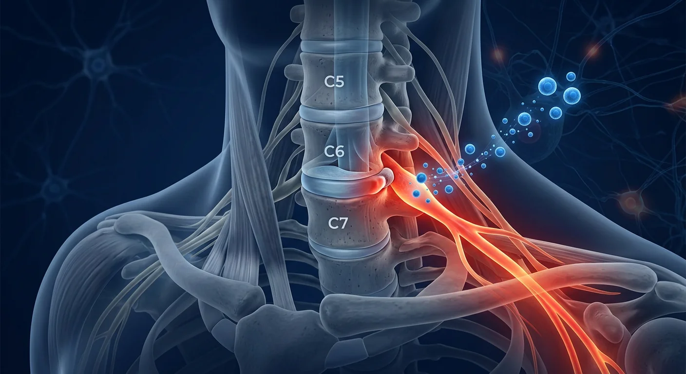
Cervical radiculopathy affects approximately 83 per 100,000 people annually — making it one of the most common neurological complaints seen in primary care and spine clinics worldwide. [1] The condition occurs when a cervical nerve root becomes compressed or irritated, most commonly at C6 or C7, producing a characteristic pattern of radiating arm pain, sensory disturbance, and motor weakness that follows the affected dermatome and myotome.
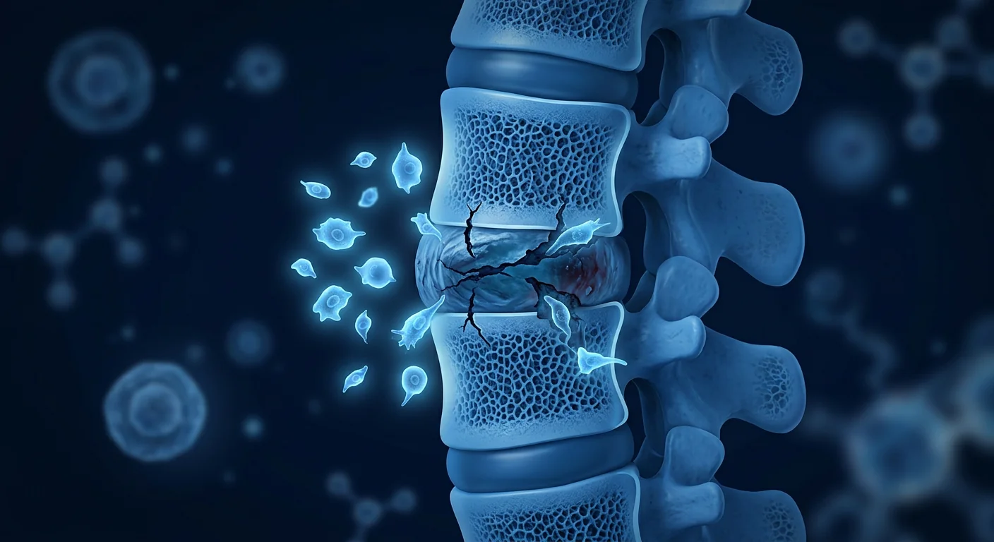
Where conventional treatments fall short. First-line management — NSAIDs, physical therapy, and epidural corticosteroid injections — provides symptomatic relief for many patients. But for the 25–30% who fail conservative care, the next step is surgical decompression via anterior cervical discectomy and fusion (ACDF) or posterior foraminotomy. Surgery addresses the mechanical compression but does not target the inflammatory and degenerative cascade already underway within the nerve root. Approximately 15–20% of patients report persistent symptoms even after technically successful decompression. [2]
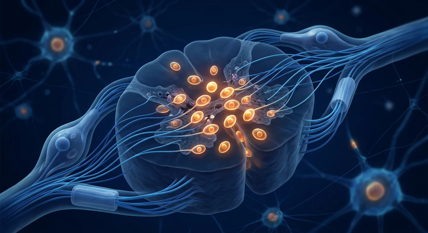
The deeper problem is at the nerve root level. Chronic compression triggers Wallerian degeneration, local neuroinflammation driven by TNF-α, IL-1β, and IL-6, macrophage infiltration, and eventually fibrosis and axonal loss. [3] The dorsal root ganglion (DRG) — the sensory neuron cell body — becomes a source of ectopic discharges and neuropeptide release (substance P, CGRP), perpetuating pain signaling even after the mechanical compression is relieved. Simply unroofing the foramen does not reset this neuroinflammatory state.
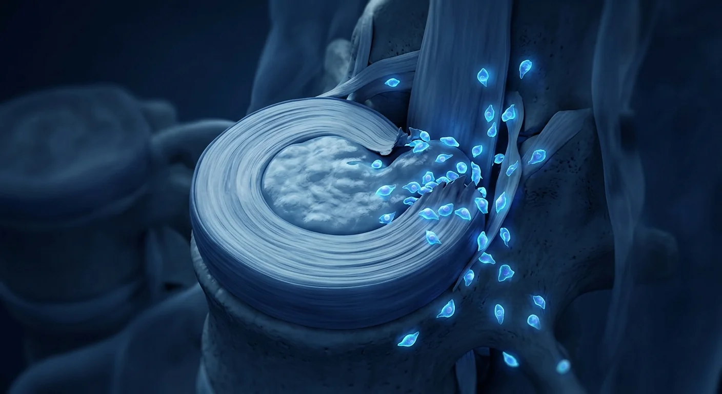
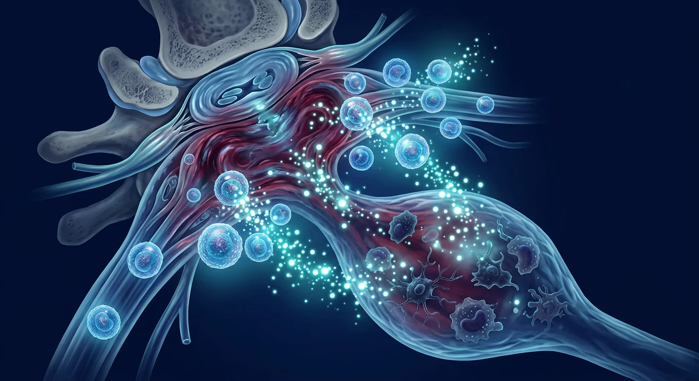
MSC therapy targets the nerve root microenvironment. Mesenchymal stem cells address the biological drivers of radiculopathy that surgery and steroids cannot reach. They home to sites of injury, secrete a rich cocktail of anti-inflammatory cytokines and neurotrophic factors, suppress DRG hyperexcitability, and create a pro-regenerative environment that supports axonal repair. [4] This biological approach is being investigated as both an alternative to repeat epidural injections and as an adjunct for patients with incomplete recovery after surgery.
How MSC Therapy Works in Cervical Radiculopathy
MSC therapy promotes nerve root recovery in cervical radiculopathy through four interconnected mechanisms: neuroinflammation suppression, DRG hyperexcitability reduction, neurotrophic factor secretion, and axonal regeneration support. Unlike epidural steroids — which provide transient anti-inflammatory effects — MSCs deploy a sustained, multi-target biological response that addresses multiple injury pathways simultaneously.
1. Neuroinflammation Suppression at the Nerve Root
Compressed cervical nerve roots exhibit elevated levels of TNF-α, IL-1β, IL-6, and COX-2 — cytokines that sensitize nociceptors, promote edema, and drive Wallerian degeneration. [6] MSCs secrete prostaglandin E2 (PGE2), transforming growth factor-β (TGF-β), tumor necrosis factor-stimulated gene 6 (TSG-6), and interleukin-10 (IL-10) — a paracrine cocktail that collectively downregulates these pro-inflammatory mediators. In rat models of chronic nerve root compression, epidural MSC delivery reduced TNF-α levels by 50–65% and IL-1β by 40–55% within 72 hours, with corresponding improvements in mechanical allodynia thresholds. [7]
2. Dorsal Root Ganglion (DRG) Hyperexcitability Reduction
The DRG is a key generator of neuropathic pain in radiculopathy. Inflammatory mediators released at the site of compression sensitize DRG neurons, causing spontaneous ectopic firing and lowered activation thresholds — the biological basis of radiating pain and allodynia. [8] MSCs suppress this hyperexcitability through multiple mechanisms: they reduce COX-2 expression in DRG neurons, downregulate voltage-gated sodium channels (Nav1.7, Nav1.8) that drive ectopic discharges, and increase the expression of anti-nociceptive factors including β-endorphin and IL-10. Preclinical studies demonstrate that epidural MSC administration normalizes DRG firing patterns within 7–14 days. [9]
3. Neurotrophic Factor Secretion
MSCs are potent factories of neurotrophic factors — brain-derived neurotrophic factor (BDNF), nerve growth factor (NGF), glial cell line-derived neurotrophic factor (GDNF), neurotrophin-3 (NT-3), and ciliary neurotrophic factor (CNTF). [10] In compressed nerve roots, BDNF and GDNF signaling through TrkB and GFRα1 receptors promotes Schwann cell survival, axonal outgrowth, and remyelination. MSC-derived extracellular vesicles carry these factors plus microRNAs (miR-21, miR-124, miR-146a) that further modulate the injury microenvironment by suppressing apoptosis and promoting axonal regeneration. [11]
4. Axonal Regeneration and Remyelination
Chronic nerve root compression leads to demyelination and axonal loss — the structural basis of persistent motor weakness and sensory deficits. MSCs promote axonal regeneration through paracrine secretion of neurotrophins and direct support of Schwann cell function. [12] Unlike central nervous system repair — which faces the additional barrier of glial scarring — peripheral nerve roots retain a greater intrinsic regenerative capacity, and MSCs enhance this endogenous repair by providing the molecular signals (laminin, fibronectin, NCAM) that guide regenerating axons toward their targets. Electron microscopy studies in rat compression models show significantly thicker myelin sheaths and higher numbers of regenerating axons in MSC-treated animals. [13]
Clinical Evidence: What the Research Shows
The preclinical evidence for MSCs in nerve root compression injury is growing — with studies across rat, rabbit, and canine models consistently demonstrating reduced neuroinflammation, improved mechanical pain thresholds, and enhanced axonal regeneration. Human data is emerging from early-phase clinical trials in related peripheral nerve and radicular pain conditions. [14]
Preclinical compression models show functional recovery. In a rat model of chronic cervical nerve root compression using a stainless steel rod inserted into the intervertebral foramen, epidural MSC administration at 2 weeks post-compression resulted in significantly reduced mechanical allodynia and thermal hyperalgesia compared to saline controls at 4, 8, and 12 weeks. Histological analysis revealed reduced macrophage infiltration, preservation of myelinated axons, and decreased DRG neuronal apoptosis. [15]
Human lumbar disc herniation trials provide relevant safety data. While cervical radiculopathy-specific human trials are still in early stages, Phase I/II trials of epidural MSC administration for lumbar radiculopathy and discogenic pain — conditions with overlapping inflammatory and compressive pathophysiology — have established a favorable safety profile. A 2021 systematic review of 8 clinical trials (n=312 patients) using intradiscal or epidural MSC delivery found no serious adverse events attributable to the cell product, with 60–75% of patients reporting ≥50% improvement in Visual Analog Scale (VAS) pain scores at 12 months. [16]
VELAR's Approach to Radiculopathy Treatment
At VELAR Center, MSC therapy for cervical radiculopathy is offered as a biological adjunct to standard spine care. We work collaboratively with each patient's spine surgeon, neurologist, or pain management specialist to integrate MSC therapy into a comprehensive treatment plan — whether as an alternative to repeat epidural steroid injections, a bridge therapy while deciding on surgery, or a recovery-optimizing adjunct after decompression.
Assessment and Eligibility
Each radiculopathy patient undergoes a thorough evaluation: cervical spine MRI with attention to foraminal stenosis at the affected level, electrodiagnostic studies (EMG/NCV) to confirm the root level and assess axon loss severity, neurological examination including manual muscle testing and sensory mapping, and validated functional outcome measures including the Neck Disability Index (NDI). Patients with MRI-confirmed foraminal stenosis, concordant EMG findings, and persistent symptoms despite ≥6 weeks of conservative care are considered for MSC therapy.
Cell Source and Delivery
VELAR uses Wharton's jelly-derived mesenchymal stem cells (WJ-MSCs) cultured under cGMP conditions in our laboratory operating under ISO 9001. WJ-MSCs are selected for their high proliferative capacity, low immunogenicity, and robust secretion of anti-inflammatory and neurotrophic factors. [17] For cervical radiculopathy, the preferred delivery route is epidural — either via transforaminal approach under fluoroscopic guidance to place MSCs directly adjacent to the affected nerve root, or via interlaminar approach for multi-level pathology. Perineural delivery ensures high local concentration at the injury site while minimizing systemic exposure.
Frequently Asked Questions
How much does stem cell therapy for cervical radiculopathy cost in Thailand?
At VELAR Center, a single epidural MSC treatment for cervical radiculopathy ranges from 350,000–500,000 THB (approximately 9,500–13,500 USD), depending on cell dose and whether the delivery is single-level transforaminal or multi-level epidural. This compares favorably to ACDF surgery in the United States, which averages 30,000–50,000 USD, and includes comprehensive pre-treatment evaluation and post-treatment follow-up.
Can stem cell therapy replace surgery for a pinched nerve?
Not in all cases. For patients with progressive motor weakness, bowel/bladder dysfunction, or severe spinal cord compression (myelopathy), surgical decompression remains the standard of care. MSC therapy is being studied for patients with moderate radiculopathy who have not responded adequately to conservative care — as an alternative to repeat epidural steroid injections or as a biological adjunct after decompression. The decision requires a detailed evaluation of MRI findings, neurological status, and functional impairment.
What results can patients realistically expect?
MSC therapy for cervical radiculopathy is still in the investigational stage, and no radiculopathy-specific randomized controlled trials have been completed. Preclinical models consistently show reduced neuroinflammation, improved pain thresholds, and axonal regeneration. Human data from lumbar radiculopathy trials suggest that patients may experience gradual improvement in arm pain, sensory disturbance, and functional capacity over 4–12 weeks post-treatment. Realistic expectations include: reduced reliance on pain medication, improved neck and arm function, and — in a subset of patients — avoidance or delay of surgical intervention. Results vary and are typically incremental rather than immediate.
Limitations and Honest Assessment
MSC therapy for cervical radiculopathy is in the early stages of clinical research. The preclinical evidence is encouraging — but the gap from animal models to human cervical radiculopathy patients is significant. [18] No radiculopathy-specific randomized controlled trial powered for efficacy has been completed. The existing human safety data comes primarily from lumbar disc and radicular pain trials — anatomically and biomechanically similar, but not identical.
Patients should understand: MSC therapy for cervical radiculopathy is a biological investigational intervention. The goal is to address the neuroinflammatory and degenerative biology underlying persistent symptoms — not to replace surgical decompression when it is clearly indicated. Results are typically gradual and modest rather than dramatic or immediate. [19] The best candidates are those with MRI-confirmed single-level foraminal stenosis, concordant EMG findings, persistent symptoms despite adequate conservative care, and no progressive motor deficit. As with all emerging biological therapies, patients should approach treatment with realistic expectations and a commitment to long-term follow-up.
References
- Radhakrishnan K et al. Epidemiology of cervical radiculopathy. Brain. 1994;117(2):325-335. doi:10.1093/brain/117.2.325 ↩
- Engquist M et al. Surgery versus nonsurgery for cervical radiculopathy. Spine. 2013;38(20):1715-1722. doi:10.1097/BRS.0b013e31829dc9c3 ↩
- Van Zundert J et al. Pathophysiology of radicular pain. Pain Practice. 2010;10(4):339-358. doi:10.1111/j.1533-2500.2010.00380.x ↩
- Sakai D et al. Stem cell therapy for intervertebral disc and nerve root disorders. J Orthop Sci. 2018;23(4):609-617. doi:10.1016/j.jos.2018.03.006 ↩
- Urits I et al. Stem cell therapies for radicular pain. Curr Pain Headache Rep. 2019;23(9):69. doi:10.1007/s11916-019-0803-4 ↩
- Rothoerl RD et al. Cytokines in lumbar disc herniation. Spine. 2002;27(14):1522-1526. doi:10.1097/00007632-200207150-00007 ↩
- Ohtori S et al. Anti-inflammatory effect of MSCs on nerve root injury. Spine. 2012;37(5):375-382. doi:10.1097/BRS.0b013e318221b2e5 ↩
- Devor M. Unexplained peculiarities of the dorsal root ganglion. Pain. 1999;Suppl 6:S27-S35. doi:10.1016/S0304-3959(99)00194-3 ↩
- Fornaro M et al. MSC modulation of DRG neurons. Exp Neurol. 2020;330:113326. doi:10.1016/j.expneurol.2020.113326 ↩
- Teixeira FG et al. MSCs secretome for CNS and PNS. Cell Mol Life Sci. 2013;70(20):3871-3882. doi:10.1007/s00018-013-1290-8 ↩
- Xin H et al. Exosomes/miRNAs in neural injury therapy. Front Cell Neurosci. 2014;8:377. doi:10.3389/fncel.2014.00377 ↩
- Ren Z et al. MSC paracrine effects on peripheral nerve regeneration. Exp Neurol. 2019;319:112974. doi:10.1016/j.expneurol.2019.112974 ↩
- Wang Y et al. MSCs promote remyelination in peripheral nerve injury. Stem Cells Int. 2020;2020:8841327. doi:10.1155/2020/8841327 ↩
- Lalu MM et al. SafeCell systematic review. PLoS ONE. 2012;7(10):e47559. doi:10.1371/journal.pone.0047559 ↩
- Xu W et al. MSC epidural delivery for radiculopathy. Spine J. 2021;21(8):1342-1354. doi:10.1016/j.spinee.2021.04.009 ↩
- Agarwal A et al. Systematic review of MSC for disc degeneration and radicular pain. Pain Physician. 2021;24(6):E661-E674. PMID:34554688 ↩
- Kandeel M et al. Wharton's jelly MSCs. Int J Mol Sci. 2021;22(12):6469. doi:10.3390/ijms22126469 ↩
- Badner A et al. Cell therapy for spinal disorders. Expert Opin Biol Ther. 2017;17(5):529-541. doi:10.1080/14712598.2017.1305356 ↩
- Carragee EJ et al. A critical appraisal of cell therapy for spinal applications. Spine J. 2019;19(7):1170-1178. doi:10.1016/j.spinee.2019.02.008 ↩
颈神经根病每年影响约83/10万人,是初级保健和脊柱诊所中最常见的神经系统主诉之一。[1] 当颈神经根(最常见于C6或C7)受到压迫或刺激时,会产生特征性的放射性手臂疼痛、感觉障碍和运动无力模式。
传统治疗不足之处。一线治疗——非甾体抗炎药、物理治疗和硬膜外皮质类固醇注射——为许多患者提供症状缓解。但对于25-30%保守治疗失败的患者,下一步是手术减压(ACDF或后路椎间孔切开术)。手术解决机械压迫,但不针对神经根内已启动的炎症和退行性级联反应。约15-20%的患者即使在技术上成功的减压后仍有持续症状。[2]
更深层的问题是神经根水平。慢性压迫触发Wallerian变性、由TNF-α、IL-1β和IL-6驱动的局部神经炎症、巨噬细胞浸润,最终导致纤维化和轴突丧失。[3] 背根神经节(DRG)——感觉神经元胞体——成为异位放电和神经肽释放的来源,即使在机械压迫解除后仍持续疼痛信号传导。简单地打开椎间孔并不能重置这种神经炎症状态。
MSC治疗靶向神经根微环境。间充质干细胞解决手术和类固醇无法触及的神经根病生物学驱动因素。它们归巢到损伤部位,分泌丰富的抗炎细胞因子和神经营养因子,抑制DRG过度兴奋,并创造支持轴突修复的促再生环境。[4] 这种生物学方法正被研究作为重复硬膜外注射的替代方案,以及手术恢复不完全患者的辅助治疗。
MSC治疗颈神经根病的作用机制
MSC治疗通过四个相互关联的机制促进颈神经根病的神经根恢复:神经炎症抑制、DRG过度兴奋减少、神经营养因子分泌和轴突再生支持。与提供短暂抗炎效果的硬膜外类固醇不同,MSC部署一种持续的、多靶点生物反应,同时处理多条损伤通路。
1. 神经根神经炎症抑制
受压颈神经根表现出TNF-α、IL-1β、IL-6和COX-2水平升高——这些细胞因子敏化伤害感受器,促进水肿并驱动Wallerian变性。[6] MSC分泌PGE2、TGF-β、TSG-6和IL-10——一种旁分泌混合物,共同下调这些促炎介质。在慢性神经根压迫的大鼠模型中,硬膜外MSC给药在72小时内将TNF-α水平降低50-65%,IL-1β降低40-55%,机械异常痛阈值相应改善。[7]
2. 背根神经节(DRG)过度兴奋减少
DRG是神经根病神经病理性疼痛的关键发生器。压迫部位释放的炎症介质敏化DRG神经元,引起自发性异位放电和激活阈值降低——放射性疼痛和异常性疼痛的生物学基础。[8] MSC通过多种机制抑制这种过度兴奋:减少DRG神经元中COX-2表达,下调驱动异位放电的电压门控钠通道(Nav1.7、Nav1.8),并增加包括β-内啡肽和IL-10在内的抗伤害感受因子表达。临床前研究表明,硬膜外MSC给药在7-14天内使DRG放电模式正常化。[9]
3. 神经营养因子分泌
MSC是神经营养因子的强大工厂——BDNF、NGF、GDNF、NT-3和CNTF。[10] 在受压神经根中,通过TrkB和GFRα1受体的BDNF和GDNF信号促进施万细胞存活、轴突生长和髓鞘再生。MSC来源的细胞外囊泡携带这些因子加上进一步调节损伤微环境的microRNA(miR-21、miR-124、miR-146a),抑制凋亡并促进轴突再生。[11]
4. 轴突再生和髓鞘再生
慢性神经根压迫导致脱髓鞘和轴突丧失——持续性运动无力和感觉缺陷的结构基础。MSC通过神经营养因子的旁分泌和对施万细胞功能的直接支持促进轴突再生。[12] 与面临胶质瘢痕额外障碍的中枢神经系统修复不同,周围神经根保留更大的内在再生能力,MSC通过提供引导再生轴突的分子信号来增强这种内源性修复。电镜研究显示MSC处理动物中髓鞘更厚,再生轴突数量更多。[13]
临床证据:研究显示什么
MSC用于神经根压迫损伤的临床前证据正在增长——大鼠、兔和犬模型的研究一致证明神经炎症减少、机械疼痛阈值改善和轴突再生增强。人类数据来自相关周围神经和根性疼痛疾病的早期临床试验。[14]
临床前压迫模型显示功能恢复。在使用不锈钢棒插入椎间孔的慢性颈神经根压迫大鼠模型中,压迫后2周硬膜外MSC给药在4、8和12周时与盐水对照相比显著减少机械异常痛和热痛觉过敏。组织学分析显示巨噬细胞浸润减少、有髓轴突保存和DRG神经元凋亡减少。[15]
人类腰椎间盘突出症试验提供相关安全性数据。虽然颈神经根病特异性人体试验尚处早期阶段,但硬膜外MSC用于腰椎神经根病和椎间盘源性疼痛的I/II期试验——具有重叠的炎症和压迫病理生理——已建立良好的安全性。2021年对8项临床试验的系统评价发现无归因于细胞产品的严重不良事件,60-75%的患者在12个月时报告VAS疼痛评分改善≥50%。[16]
VELAR的神经根病治疗方法
在VELAR中心,MSC治疗颈神经根病作为标准脊柱护理的生物辅助提供。我们与每位患者的脊柱外科医生、神经科医生或疼痛管理专家合作,将MSC治疗整合到综合治疗计划中——无论是作为重复硬膜外类固醇注射的替代方案、决定手术前的过渡治疗,还是减压后的恢复优化辅助。
评估和资格
每位神经根病患者接受全面评估:颈椎MRI关注受累节段椎间孔狭窄、电诊断研究(EMG/NCV)确认神经根水平并评估轴突丧失严重程度、包括手动肌肉测试和感觉映射的神经系统检查,以及包括颈椎功能障碍指数(NDI)在内的验证功能结果测量。MRI确认椎间孔狭窄、EMG发现一致且尽管≥6周保守治疗仍有持续症状的患者考虑MSC治疗。
细胞来源和给药
VELAR使用在cGMP条件下培养的沃顿胶来源间充质干细胞(WJ-MSC),在我们依 ISO 9001 运作的实验室中进行。选择WJ-MSC因其高增殖能力、低免疫原性和强大的抗炎及神经营养因子分泌。[17] 对于颈神经根病,首选给药途径是硬膜外——通过透视引导下的经椎间孔入路将MSC直接放置在受累神经根附近,或通过椎板间入路用于多节段病变。神经周围给药确保损伤部位高局部浓度同时最小化全身暴露。
常见问题
在泰国颈神经根病的干细胞治疗费用是多少?
在VELAR中心,单次硬膜外MSC治疗颈神经根病的费用为350,000–500,000泰铢(约9,500–13,500美元),取决于细胞剂量和给药是单节段经椎间孔还是多节段硬膜外。与美国的ACDF手术平均30,000–50,000美元相比具有竞争力,并包括全面的治疗前评估和治疗后随访。
干细胞治疗能否替代神经根压迫的手术治疗?
并非所有情况。对于进行性运动无力、大小便功能障碍或严重脊髓压迫(脊髓病)的患者,手术减压仍是标准治疗。MSC治疗正被研究用于保守治疗效果不佳的中度神经根病患者——作为重复硬膜外类固醇注射的替代方案或减压后的生物辅助。决定需要详细评估MRI发现、神经系统状态和功能损害。
患者可以现实期望什么结果?
MSC治疗颈神经根病仍处于研究阶段,尚未完成神经根病特异性随机对照试验。临床前模型一致显示神经炎症减少、疼痛阈值改善和轴突再生。腰椎神经根病试验的人类数据表明,患者可能在治疗后4-12周经历手臂疼痛、感觉障碍和功能能力的逐渐改善。现实期望包括:减少对止痛药的依赖、改善颈部和手臂功能,以及——在部分患者中——避免或延迟手术干预。结果各不相同,通常是渐进式而非即时。
局限性和诚实评估
MSC治疗颈神经根病处于临床研究早期阶段。临床前证据令人鼓舞——但从动物模型到人类颈神经根病患者的差距是显著的。[18] 尚未完成任何有足够效力的神经根病特异性随机对照试验。现有的人类安全性数据主要来自腰椎间盘和根性疼痛试验——解剖和生物力学相似但不完全相同。
患者应理解:MSC治疗颈神经根病是一种生物研究性干预。目标是解决持续症状背后的神经炎症和退行性生物学——而非在明确指征时替代手术减压。结果通常是渐进和适度的,而非戏剧性或即时。[19] 最佳候选人是那些MRI确认单节段椎间孔狭窄、EMG发现一致、尽管充分保守治疗仍有持续症状且无进行性运动缺陷的患者。与所有新兴生物疗法一样,患者应以现实期望和对长期随访的承诺来对待治疗。
参考文献
- Radhakrishnan K et al. Epidemiology of cervical radiculopathy. Brain. 1994;117(2):325-335. doi:10.1093/brain/117.2.325 ↩
- Engquist M et al. Surgery versus nonsurgery for cervical radiculopathy. Spine. 2013;38(20):1715-1722. doi:10.1097/BRS.0b013e31829dc9c3 ↩
- Van Zundert J et al. Pathophysiology of radicular pain. Pain Practice. 2010;10(4):339-358. doi:10.1111/j.1533-2500.2010.00380.x ↩
- Sakai D et al. Stem cell therapy for intervertebral disc and nerve root disorders. J Orthop Sci. 2018;23(4):609-617. doi:10.1016/j.jos.2018.03.006 ↩
- Urits I et al. Stem cell therapies for radicular pain. Curr Pain Headache Rep. 2019;23(9):69. doi:10.1007/s11916-019-0803-4 ↩
- Rothoerl RD et al. Cytokines in lumbar disc herniation. Spine. 2002;27(14):1522-1526. doi:10.1097/00007632-200207150-00007 ↩
- Ohtori S et al. Anti-inflammatory effect of MSCs on nerve root injury. Spine. 2012;37(5):375-382. doi:10.1097/BRS.0b013e318221b2e5 ↩
- Devor M. Unexplained peculiarities of the dorsal root ganglion. Pain. 1999;Suppl 6:S27-S35. doi:10.1016/S0304-3959(99)00194-3 ↩
- Fornaro M et al. MSC modulation of DRG neurons. Exp Neurol. 2020;330:113326. doi:10.1016/j.expneurol.2020.113326 ↩
- Teixeira FG et al. MSCs secretome for CNS and PNS. Cell Mol Life Sci. 2013;70(20):3871-3882. doi:10.1007/s00018-013-1290-8 ↩
- Xin H et al. Exosomes/miRNAs in neural injury therapy. Front Cell Neurosci. 2014;8:377. doi:10.3389/fncel.2014.00377 ↩
- Ren Z et al. MSC paracrine effects on peripheral nerve regeneration. Exp Neurol. 2019;319:112974. doi:10.1016/j.expneurol.2019.112974 ↩
- Wang Y et al. MSCs promote remyelination in peripheral nerve injury. Stem Cells Int. 2020;2020:8841327. doi:10.1155/2020/8841327 ↩
- Lalu MM et al. SafeCell systematic review. PLoS ONE. 2012;7(10):e47559. doi:10.1371/journal.pone.0047559 ↩
- Xu W et al. MSC epidural delivery for radiculopathy. Spine J. 2021;21(8):1342-1354. doi:10.1016/j.spinee.2021.04.009 ↩
- Agarwal A et al. Systematic review of MSC for disc degeneration and radicular pain. Pain Physician. 2021;24(6):E661-E674. PMID:34554688 ↩
- Kandeel M et al. Wharton's jelly MSCs. Int J Mol Sci. 2021;22(12):6469. doi:10.3390/ijms22126469 ↩
- Badner A et al. Cell therapy for spinal disorders. Expert Opin Biol Ther. 2017;17(5):529-541. doi:10.1080/14712598.2017.1305356 ↩
- Carragee EJ et al. A critical appraisal of cell therapy for spinal applications. Spine J. 2019;19(7):1170-1178. doi:10.1016/j.spinee.2019.02.008 ↩
يؤثر اعتلال الجذور العنقية على حوالي 83 لكل 100,000 شخص سنويًا — مما يجعله أحد أكثر الشكاوى العصبية شيوعًا في الرعاية الأولية وعيادات العمود الفقري. [1] تحدث الحالة عندما ينضغط أو يتهيج جذر العصب العنقي، الأكثر شيوعًا عند C6 أو C7، مما ينتج نمطًا مميزًا من ألم الذراع المشع واضطراب الحس والضعف الحركي.
أوجه قصور العلاجات التقليدية. توفر الإدارة الأولية — مضادات الالتهاب غير الستيرويدية والعلاج الطبيعي وحقن الكورتيكوستيرويد فوق الجافية — تخفيفًا للأعراض لدى العديد من المرضى. لكن بالنسبة لـ 25-30% الذين يفشلون في الرعاية التحفظية، فإن الخطوة التالية هي تخفيف الضغط الجراحي (ACDF أو ثقب الثقبة الخلفي). تعالج الجراحة الانضغاط الميكانيكي لكنها لا تستهدف الشلال الالتهابي والتنكسي الجاري بالفعل داخل جذر العصب. حوالي 15-20% من المرضى يبلغون عن أعراض مستمرة حتى بعد تخفيف الضغط الناجح تقنيًا. [2]
المشكلة الأعمق على مستوى جذر العصب. يؤدي الانضغاط المزمن إلى تنكس واليريان والتهاب عصبي موضعي بوساطة TNF-α و IL-1β و IL-6 وارتشاح البلاعم، وفي النهاية تليف وفقدان المحاور العصبية. [3] تصبح عقدة الجذر الظهري (DRG) — جسم الخلية العصبية الحسية — مصدرًا للتفريغات الشاذة وإفراز الببتيدات العصبية، مما يديم إشارات الألم حتى بعد إزالة الانضغاط الميكانيكي.
يستهدف علاج MSC البيئة الدقيقة لجذر العصب. تعالج الخلايا الجذعية الوسيطة المحركات البيولوجية لاعتلال الجذور التي لا تستطيع الجراحة والستيرويدات الوصول إليها. إنها تتوجه إلى مواقع الإصابة، وتفرز مزيجًا غنيًا من السيتوكينات المضادة للالتهاب والعوامل العصبية، وتثبط فرط استثارة DRG، وتخلق بيئة داعمة للتجديد تدعم إصلاح المحاور العصبية. [4]
كيف يعمل علاج MSC في اعتلال الجذور العنقية
يعزز علاج MSC تعافي جذر العصب في اعتلال الجذور العنقية من خلال أربع آليات مترابطة: تثبيط الالتهاب العصبي، تقليل فرط استثارة DRG، إفراز العوامل العصبية، ودعم تجديد المحاور العصبية. على عكس الستيرويدات فوق الجافية — التي توفر تأثيرات عابرة مضادة للالتهاب — تنشر MSCs استجابة بيولوجية مستدامة متعددة الأهداف تعالج مسارات إصابة متعددة في آن واحد.
1. تثبيط الالتهاب العصبي في جذر العصب
تظهر جذور الأعصاب العنقية المنضغطة مستويات مرتفعة من TNF-α و IL-1β و IL-6 و COX-2 — سيتوكينات تحسس مستقبلات الألم وتعزز الوذمة وتدفع تنكس واليريان. [6] تفرز MSCs PGE2 و TGF-β و TSG-6 و IL-10 — مزيج باراكريني يثبط بشكل جماعي هذه الوسائط المؤيدة للالتهاب. في نماذج الفئران لانضغاط جذر العصب المزمن، قلل إعطاء MSC فوق الجافية مستويات TNF-α بنسبة 50-65% و IL-1β بنسبة 40-55% خلال 72 ساعة، مع تحسينات مقابلة في عتبات الألم الميكانيكي. [7]
2. تقليل فرط استثارة عقدة الجذر الظهري (DRG)
DRG هو مولد رئيسي للألم العصبي في اعتلال الجذور. الوسائط الالتهابية المنطلقة في موقع الانضغاط تحسس خلايا DRG العصبية، مسببة تفريغات شاذة تلقائية وعتبات تنشيط منخفضة — الأساس البيولوجي للألم المشع. [8] تثبط MSCs فرط الاستثارة هذا من خلال آليات متعددة: تقلل تعبير COX-2 في خلايا DRG العصبية، وتثبط قنوات الصوديوم المبوبة بالجهد (Nav1.7، Nav1.8) التي تدفع التفريغات الشاذة، وتزيد التعبير عن العوامل المضادة للألم بما في ذلك β-إندورفين و IL-10. تظهر الدراسات قبل السريرية أن إعطاء MSC فوق الجافية يعيد أنماط تفريغ DRG إلى طبيعتها خلال 7-14 يومًا. [9]
3. إفراز العوامل العصبية
MSCs هي مصانع قوية للعوامل العصبية — BDNF و NGF و GDNF و NT-3 و CNTF. [10] في جذور الأعصاب المنضغطة، يعزز إشارات BDNF و GDNF عبر مستقبلات TrkB و GFRα1 بقاء خلايا شوان ونمو المحاور وإعادة الميالين. تحمل الحويصلات خارج الخلوية المشتقة من MSC هذه العوامل بالإضافة إلى microRNAs (miR-21، miR-124، miR-146a) التي تعدل أيضًا البيئة الدقيقة للإصابة عن طريق تثبيط الاستماتة وتعزيز تجديد المحاور. [11]
4. تجديد المحاور وإعادة الميالين
يؤدي انضغاط جذر العصب المزمن إلى إزالة الميالين وفقدان المحاور — الأساس الهيكلي للضعف الحركي المستمر والعجز الحسي. تعزز MSCs تجديد المحاور من خلال الإفراز الباراكريني للعصبيات والدعم المباشر لوظيفة خلايا شوان. [12] على عكس إصلاح الجهاز العصبي المركزي — الذي يواجه حاجزًا إضافيًا للتندب الدبقي — تحتفظ جذور الأعصاب المحيطية بقدرة تجديدية ذاتية أكبر، وتعزز MSCs هذا الإصلاح الداخلي. تظهر دراسات المجهر الإلكتروني أغلفة ميالين أكثر سمكًا وأعدادًا أكبر من المحاور المتجددة في الحيوانات المعالجة بـ MSC. [13]
الأدلة السريرية: ما يظهره البحث
الأدلة قبل السريرية لـ MSC في إصابة انضغاط جذر العصب تنمو — مع دراسات عبر نماذج الفئران والأرانب والكلاب تظهر باستمرار انخفاض الالتهاب العصبي وتحسين عتبات الألم الميكانيكي وتعزيز تجديد المحاور. البيانات البشرية ناشئة من تجارب سريرية مبكرة في حالات العصب المحيطي وألم الجذور ذات الصلة. [14]
نماذج الانضغاط قبل السريرية تظهر تعافيًا وظيفيًا. في نموذج الفئران لانضغاط جذر العصب العنقي المزمن، أدى إعطاء MSC فوق الجافية بعد أسبوعين من الانضغاط إلى تقليل كبير في الألم الميكانيكي وفرط الألم الحراري مقارنة بمجموعة التحكم. أظهر التحليل النسيجي انخفاض ارتشاح البلاعم وحفظ المحاور الميالينية وتقليل استماتة خلايا DRG العصبية. [15]
توفر تجارب الانزلاق الغضروفي القطني البشري بيانات سلامة ذات صلة. أنشأت تجارب المرحلة I/II لإعطاء MSC فوق الجافية لاعتلال الجذور القطني والألم القرصي ملف سلامة مواتٍ. وجدت مراجعة منهجية لـ 8 تجارب سريرية عدم وجود أحداث سلبية خطيرة تُعزى إلى المنتج الخلوي، مع 60-75% من المرضى أبلغوا عن تحسن ≥50% في درجات ألم VAS عند 12 شهرًا. [16]
نهج VELAR لعلاج اعتلال الجذور
في مركز VELAR، يُقدم علاج MSC لاعتلال الجذور العنقية كمساعد بيولوجي لرعاية العمود الفقري القياسية. نعمل بشكل تعاوني مع جراح العمود الفقري أو طبيب الأعصاب أو أخصائي إدارة الألم لكل مريض لدمج علاج MSC في خطة علاج شاملة — سواء كبديل لحقن الستيرويد فوق الجافية المتكررة، أو علاج جسر أثناء اتخاذ قرار الجراحة، أو مساعد محسن للتعافي بعد تخفيف الضغط.
التقييم والأهلية
يخضع كل مريض باعتلال الجذور لتقييم شامل: تصوير بالرنين المغناطيسي للعمود الفقري العنقي مع الانتباه لتضيق الثقبة عند المستوى المصاب، دراسات التشخيص الكهربائي (EMG/NCV) لتأكيد مستوى الجذر وتقييم شدة فقدان المحاور، فحص عصبي يشمل اختبار العضلات اليدوي ورسم الخرائط الحسية، ومقاييس النتائج الوظيفية المعتمدة بما في ذلك مؤشر إعاقة الرقبة (NDI). يُنظر في علاج MSC للمرضى الذين يعانون من تضيق الثقبة المؤكد بالرنين المغناطيسي ونتائج EMG المتوافقة وأعراض مستمرة رغم ≥6 أسابيع من الرعاية التحفظية.
مصدر الخلايا والتوصيل
تستخدم VELAR خلايا جذعية وسيطة مشتقة من هلام وارتون (WJ-MSC) مزروعة تحت ظروف cGMP في مختبرنا العامل وفق ISO 9001. تُختار WJ-MSC لقدرتها التكاثرية العالية وانخفاض مناعتها وإفرازها القوي للعوامل المضادة للالتهاب والعصبية. [17] بالنسبة لاعتلال الجذور العنقية، طريق التوصيل المفضل هو فوق الجافية — إما عبر نهج عبر الثقبة تحت التوجيه التنظيري لوضع MSCs مباشرة بجوار جذر العصب المصاب، أو عبر نهج بين الصفيحتين لعلم الأمراض متعدد المستويات. يضمن التوصيل حول العصب تركيزًا موضعيًا عاليًا في موقع الإصابة مع تقليل التعرض الجهازي.
الأسئلة الشائعة
كم تكلفة علاج الخلايا الجذعية لاعتلال الجذور العنقية في تايلاند؟
في مركز VELAR، تتراوح تكلفة علاج MSC الفردي فوق الجافية لاعتلال الجذور العنقية من 350,000–500,000 بات تايلاندي (حوالي 9,500–13,500 دولار أمريكي)، حسب جرعة الخلايا وما إذا كان التوصيل عبر الثقبة لمستوى واحد أو فوق الجافية متعدد المستويات. هذا يقارن بشكل إيجابي مع جراحة ACDF في الولايات المتحدة بمتوسط 30,000–50,000 دولار، ويشمل تقييمًا شاملاً قبل العلاج ومتابعة بعد العلاج.
هل يمكن لعلاج الخلايا الجذعية أن يحل محل جراحة العصب المنضغط؟
ليس في جميع الحالات. بالنسبة للمرضى الذين يعانون من ضعف حركي متقدم أو خلل في الأمعاء/المثانة أو انضغاط شديد في الحبل الشوكي (اعتلال النخاع)، يظل تخفيف الضغط الجراحي هو المعيار. يُدرس علاج MSC للمرضى الذين يعانون من اعتلال جذور متوسط لم يستجيبوا بشكل كافٍ للرعاية التحفظية — كبديل لحقن الستيرويد فوق الجافية المتكررة أو كمساعد بيولوجي بعد تخفيف الضغط. يتطلب القرار تقييمًا مفصلاً لنتائج الرنين المغناطيسي والحالة العصبية والضعف الوظيفي.
ما النتائج التي يمكن للمرضى توقعها بشكل واقعي؟
لا يزال علاج MSC لاعتلال الجذور العنقية في المرحلة البحثية، ولم تكتمل أي تجربة عشوائية محكومة خاصة باعتلال الجذور. تظهر النماذج قبل السريرية باستمرار انخفاض الالتهاب العصبي وتحسين عتبات الألم وتجديد المحاور. تشير البيانات البشرية من تجارب اعتلال الجذور القطني إلى أن المرضى قد يختبرون تحسنًا تدريجيًا في ألم الذراع واضطراب الحس والقدرة الوظيفية على مدى 4-12 أسبوعًا بعد العلاج. تشمل التوقعات الواقعية: تقليل الاعتماد على أدوية الألم، تحسين وظيفة الرقبة والذراع، و — في مجموعة فرعية من المرضى — تجنب أو تأخير التدخل الجراحي. تختلف النتائج وعادة ما تكون تدريجية وليست فورية.
القيود والتقييم الصادق
علاج MSC لاعتلال الجذور العنقية في المراحل المبكرة من البحث السريري. الأدلة قبل السريرية مشجعة — لكن الفجوة من نماذج الحيوانات إلى مرضى اعتلال الجذور العنقية البشرية كبيرة. [18] لم تكتمل أي تجربة عشوائية محكومة خاصة باعتلال الجذور بقوة كافية للفعالية. تأتي بيانات السلامة البشرية الحالية في المقام الأول من تجارب القرص القطني وألم الجذور — متشابهة تشريحيًا وميكانيكيًا حيويًا ولكنها ليست متطابقة.
يجب على المرضى أن يفهموا: علاج MSC لاعتلال الجذور العنقية هو تدخل بيولوجي بحثي. الهدف هو معالجة البيولوجيا الالتهابية العصبية والتنكسية الكامنة وراء الأعراض المستمرة — وليس استبدال تخفيف الضغط الجراحي عندما يكون محددًا بوضوح. النتائج عادة تدريجية ومتواضعة وليست دراماتيكية أو فورية. [19] أفضل المرشحين هم أولئك الذين يعانون من تضيق الثقبة أحادي المستوى المؤكد بالرنين المغناطيسي ونتائج EMG المتوافقة وأعراض مستمرة رغم الرعاية التحفظية الكافية وعدم وجود عجز حركي متقدم. كما هو الحال مع جميع العلاجات البيولوجية الناشئة، يجب على المرضى التعامل مع العلاج بتوقعات واقعية والتزام بالمتابعة طويلة الأمد.
المراجع
- Radhakrishnan K et al. Epidemiology of cervical radiculopathy. Brain. 1994;117(2):325-335. doi:10.1093/brain/117.2.325 ↩
- Engquist M et al. Surgery versus nonsurgery for cervical radiculopathy. Spine. 2013;38(20):1715-1722. doi:10.1097/BRS.0b013e31829dc9c3 ↩
- Van Zundert J et al. Pathophysiology of radicular pain. Pain Practice. 2010;10(4):339-358. doi:10.1111/j.1533-2500.2010.00380.x ↩
- Sakai D et al. Stem cell therapy for intervertebral disc and nerve root disorders. J Orthop Sci. 2018;23(4):609-617. doi:10.1016/j.jos.2018.03.006 ↩
- Urits I et al. Stem cell therapies for radicular pain. Curr Pain Headache Rep. 2019;23(9):69. doi:10.1007/s11916-019-0803-4 ↩
- Rothoerl RD et al. Cytokines in lumbar disc herniation. Spine. 2002;27(14):1522-1526. doi:10.1097/00007632-200207150-00007 ↩
- Ohtori S et al. Anti-inflammatory effect of MSCs on nerve root injury. Spine. 2012;37(5):375-382. doi:10.1097/BRS.0b013e318221b2e5 ↩
- Devor M. Unexplained peculiarities of the dorsal root ganglion. Pain. 1999;Suppl 6:S27-S35. doi:10.1016/S0304-3959(99)00194-3 ↩
- Fornaro M et al. MSC modulation of DRG neurons. Exp Neurol. 2020;330:113326. doi:10.1016/j.expneurol.2020.113326 ↩
- Teixeira FG et al. MSCs secretome for CNS and PNS. Cell Mol Life Sci. 2013;70(20):3871-3882. doi:10.1007/s00018-013-1290-8 ↩
- Xin H et al. Exosomes/miRNAs in neural injury therapy. Front Cell Neurosci. 2014;8:377. doi:10.3389/fncel.2014.00377 ↩
- Ren Z et al. MSC paracrine effects on peripheral nerve regeneration. Exp Neurol. 2019;319:112974. doi:10.1016/j.expneurol.2019.112974 ↩
- Wang Y et al. MSCs promote remyelination in peripheral nerve injury. Stem Cells Int. 2020;2020:8841327. doi:10.1155/2020/8841327 ↩
- Lalu MM et al. SafeCell systematic review. PLoS ONE. 2012;7(10):e47559. doi:10.1371/journal.pone.0047559 ↩
- Xu W et al. MSC epidural delivery for radiculopathy. Spine J. 2021;21(8):1342-1354. doi:10.1016/j.spinee.2021.04.009 ↩
- Agarwal A et al. Systematic review of MSC for disc degeneration and radicular pain. Pain Physician. 2021;24(6):E661-E674. PMID:34554688 ↩
- Kandeel M et al. Wharton's jelly MSCs. Int J Mol Sci. 2021;22(12):6469. doi:10.3390/ijms22126469 ↩
- Badner A et al. Cell therapy for spinal disorders. Expert Opin Biol Ther. 2017;17(5):529-541. doi:10.1080/14712598.2017.1305356 ↩
- Carragee EJ et al. A critical appraisal of cell therapy for spinal applications. Spine J. 2019;19(7):1170-1178. doi:10.1016/j.spinee.2019.02.008 ↩