Celiac disease is a chronic autoimmune enteropathy triggered by dietary gluten in genetically susceptible individuals, affecting approximately 1% of the global population — yet an estimated 80% of cases remain undiagnosed. [1] The only treatment available today is a strict lifelong gluten-free diet, which, despite diligent adherence, leaves 30–50% of patients with persistent symptoms, ongoing intestinal inflammation, and impaired quality of life.
Where conventional management falls short. The gluten-free diet eliminates the trigger but does not restore immune tolerance to gluten. Cross-contamination is nearly unavoidable — a crumb of bread containing 10 mg of gluten is enough to provoke villous damage in sensitive individuals. [2] More importantly, the diet does nothing to address the underlying immunological defect: a breakdown of oral tolerance that allows gliadin-reactive CD4+ T cells to orchestrate the destruction of intestinal epithelium. Patients remain immunologically vulnerable for life.
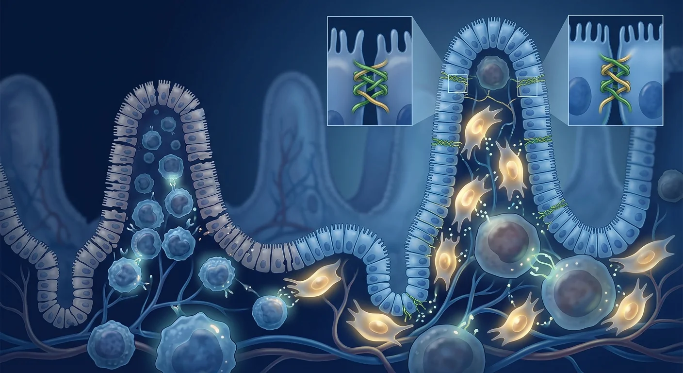
The deeper problem is barrier failure and immune dysregulation. In celiac disease, deamidated gluten peptides presented by HLA-DQ2 or HLA-DQ8 molecules trigger a Th1/Th17-dominant inflammatory cascade within the lamina propria. [3] Pro-inflammatory cytokines — particularly interferon-gamma (IFN-γ), interleukin-15 (IL-15), and tumor necrosis factor-alpha (TNF-α) — drive enterocyte apoptosis, villous atrophy, crypt hyperplasia, and increased intestinal permeability. The resulting "leaky gut" allows luminal antigens to cross the epithelial barrier, perpetuating systemic immune activation far beyond the gut itself.
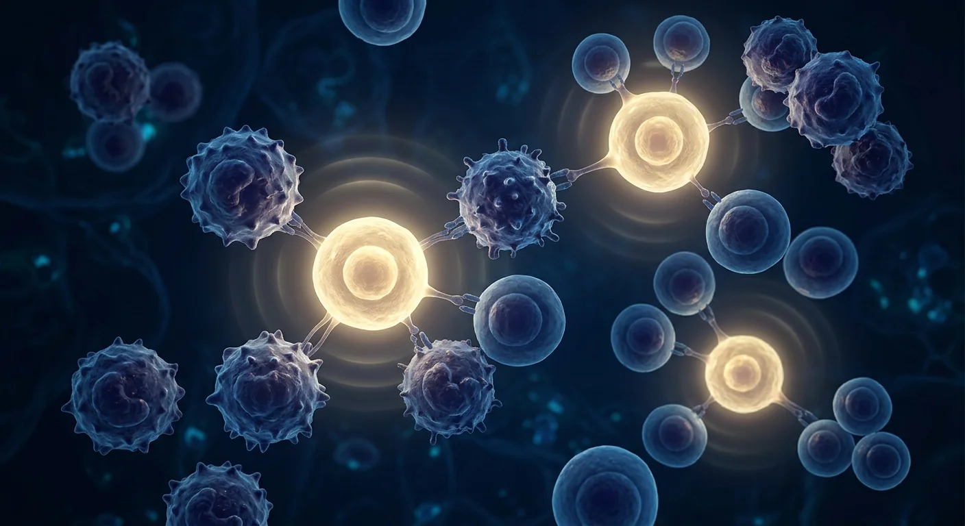
MSC therapy targets the immunological root cause. Mesenchymal stem cells are being investigated for their unique ability to restore immune tolerance through multiple complementary mechanisms: suppressing gliadin-reactive effector T cells, expanding regulatory T cell (Treg) populations, repairing the intestinal epithelial barrier via paracrine trophic factors, and resetting the local cytokine milieu from pro-inflammatory to regulatory. Rather than simply avoiding the trigger, MSC therapy aims to re-establish the immunological peace that the gluten-free diet cannot restore. [4]
Understanding Celiac Disease: Autoimmunity at the Gut Interface
Celiac disease is unique among autoimmune conditions in that the environmental trigger — gluten — is known, the genetic predisposition — HLA-DQ2/DQ8 — is well-defined, and the target organ — the small intestinal mucosa — is accessible. This triad makes celiac disease an ideal model for studying antigen-specific immune tolerance, and a logical target for tolerance-restoring therapies like MSCs.
The pathological hallmark of active celiac disease is villous atrophy: the finger-like projections of the small intestine that provide the enormous surface area for nutrient absorption are flattened by immune-mediated destruction. [5] Intraepithelial lymphocytes (IELs) infiltrate the epithelium in dramatically increased numbers — a finding so characteristic that an elevated IEL count with normal villous architecture (Marsh grade 1) is considered the earliest detectable lesion. As the disease progresses through Marsh grades 2–3, crypt hyperplasia and partial-to-total villous atrophy develop, leading to malabsorption of iron, calcium, vitamin D, folate, and fat-soluble vitamins.
Systemic consequences beyond the gut. Although celiac disease originates in the small intestine, its effects are systemic. Dermatitis herpetiformis — a blistering skin rash — is the classic extra-intestinal manifestation, but the list also includes iron-deficiency anemia, osteoporosis, peripheral neuropathy, ataxia, infertility, and an increased risk of enteropathy-associated T-cell lymphoma. [6] The common thread is chronic immune activation and systemic inflammation driven by a breached gut barrier.
Key point: Celiac disease is not simply "gluten intolerance." It is a systemic autoimmune disease in which gliadin-reactive T cells, intraepithelial lymphocytes, and pro-inflammatory cytokines collaborate to destroy the intestinal lining — with consequences that extend to every organ system. A therapy that restores immune tolerance to gluten would address the disease at its source, not just its dietary trigger.
How MSC Therapy Works in Celiac Disease
Mesenchymal stem cells bring a uniquely suited therapeutic toolkit to celiac disease — one that addresses immunopathology, epithelial barrier integrity, and tissue repair simultaneously. The gastrointestinal tract is already a natural target for intravenous MSCs, as a substantial fraction of infused cells home to the intestinal microvasculature and mesenteric lymph nodes within hours of administration.
Suppression of gliadin-reactive effector T cells. MSCs potently inhibit the proliferation and cytokine production of CD4+ Th1 and Th17 cells — the very lymphocyte subsets that orchestrate villous destruction in active celiac disease. Through secretion of indoleamine 2,3-dioxygenase (IDO), prostaglandin E2 (PGE2), and transforming growth factor-beta (TGF-β), MSCs shift the intestinal immune environment from IFN-γ/IL-17-dominant to IL-10/TGF-β-dominant. [7] In experimental models of intestinal inflammation, MSC infusion reduced IFN-γ levels in mesenteric lymph nodes by over 50% and decreased the number of activated CD4+ T cells infiltrating the lamina propria.
Expansion of regulatory T cells (Tregs). The most therapeutically compelling property of MSCs in celiac disease is their capacity to induce and expand CD4+CD25+FoxP3+ regulatory T cells — the immune system's natural guardians of oral tolerance. [8] In a healthy gut, Tregs actively suppress responses to food antigens and commensal bacteria, maintaining the immunological peace that allows nutrient absorption to proceed without inflammation. Celiac disease represents a failure of precisely this tolerance mechanism. By expanding the Treg compartment, MSCs may help re-establish the intestinal immune homeostasis that the gluten-free diet alone cannot.
Repair of the intestinal epithelial barrier. Beyond immunomodulation, MSCs secrete a rich cocktail of trophic factors — including hepatocyte growth factor (HGF), vascular endothelial growth factor (VEGF), epidermal growth factor (EGF), and keratinocyte growth factor (KGF) — that directly promote epithelial cell proliferation, tight junction protein expression, and barrier integrity. [9] In preclinical models of intestinal injury, MSC-conditioned medium alone accelerated restoration of transepithelial electrical resistance (TEER) — a direct measure of barrier function — by approximately 40% within 48 hours of epithelial wounding. This dual action — immunomodulation plus barrier repair — is precisely what celiac disease therapy needs.
Modulation of B-cell and antibody responses. Celiac disease is characterized by autoantibodies against tissue transglutaminase (anti-tTG), endomysium (EMA), and deamidated gliadin peptides (DGP). While these antibodies serve primarily as diagnostic markers rather than direct mediators of tissue injury, their production reflects ongoing B-cell activation within the intestinal mucosa. MSCs inhibit B-cell proliferation, plasma cell differentiation, and antibody secretion through both soluble mediators and direct cell–cell contact, potentially reducing the immunological memory that sustains the disease. [10]
Preclinical and Clinical Evidence
The clinical evidence base for MSC therapy in celiac disease specifically is nascent — no completed randomized controlled trials exist. However, the mechanistic case is strongly supported by research in closely related intestinal inflammatory conditions and by what is known about MSC biology in the gut.
Preclinical intestinal inflammation models. In murine models of T-cell-mediated enteropathy — including TNBS-induced colitis and gliadin-sensitized HLA-DQ8 transgenic mice — intravenous MSC administration consistently reduced intestinal inflammation scores, lowered pro-inflammatory cytokine levels (IFN-γ, TNF-α, IL-17) in intestinal tissue homogenates, and preserved villous architecture compared to untreated controls. [11] Histological analysis showed reduced crypt hyperplasia, fewer intraepithelial lymphocytes, and restoration of normal villus-to-crypt ratios in MSC-treated animals.
Evidence from human inflammatory bowel disease trials. The strongest clinical evidence for MSC efficacy in intestinal autoimmunity comes from Crohn's disease, where allogeneic MSC infusion has demonstrated fistula closure rates of 50–75% in patients refractory to anti-TNF therapy, and significant reductions in Crohn's Disease Activity Index (CDAI) scores in phase II/III trials. [12] While Crohn's disease and celiac disease differ in etiology — one is a transmural granulomatous inflammation, the other an intraepithelial T-cell attack on villi — both involve Th1/Th17-driven mucosal immunopathology and epithelial barrier disruption. The mechanistic overlap is substantial enough that celiac disease researchers have called for exploration of MSC-based tolerance-restoring strategies.
Refractory celiac disease type II — a high-stakes niche. Refractory celiac disease type II (RCDII) — characterized by clonal expansion of aberrant intraepithelial lymphocytes and a 50% risk of progression to enteropathy-associated T-cell lymphoma — represents an area of particular unmet need. [13] No approved therapy exists for RCDII, and the aberrant IEL clone is typically resistant to immunosuppressants. Given the capacity of MSCs to suppress lymphocyte proliferation and induce apoptosis in aberrant T-cell clones through FAS/FASL and TRAIL pathways, MSC therapy is being explored as a potential intervention to reduce the malignant risk in this high-risk population.
Candid assessment: The preclinical rationale for MSC therapy in celiac disease is mechanistically strong — stronger, arguably, than for many conditions where clinical trials are already underway. However, direct human evidence in celiac disease remains limited to case reports and preclinical inference. Patients considering this approach should understand it is investigational and not a substitute for a gluten-free diet.
The VELAR Treatment Approach for Celiac Disease
At VELAR Center in Bangkok, the clinical team designs individualized protocols for patients with celiac disease based on Marsh grade, serological profile (anti-tTG, EMA, DGP levels), symptom burden, and coexisting autoimmune conditions. The approach is designed to complement — not replace — dietary management.
What to Realistically Expect
MSC therapy for celiac disease is best understood as an investigational disease-modifying strategy designed to restore immunological tolerance — not as a cure, and not as a license to resume gluten consumption. Realistic expectations are essential.
Symptom improvement. The largest potential benefit may be in symptom domains that persist despite a strict gluten-free diet: fatigue, brain fog, joint pain, and residual gastrointestinal symptoms. These persistent complaints — reported by 30–50% of treated celiac patients — likely reflect ongoing low-grade systemic inflammation rather than active villous atrophy. [14] By reducing systemic inflammatory burden and improving gut barrier integrity, MSC therapy may improve quality-of-life metrics even when villous architecture remains stable on biopsy.
Reduced sensitivity to cross-contamination. Perhaps the most meaningful real-world outcome would be an increased threshold for gluten-triggered immune activation — reducing the clinical consequences of the inadvertent gluten exposure that is essentially unavoidable in daily life. While MSCs cannot eliminate the immunological memory of gluten, a more robust Treg compartment and strengthened epithelial barrier may dampen the inflammatory response to trace gluten exposure.
Stabilization of coexisting autoimmunity. Celiac disease frequently coexists with other autoimmune conditions — type 1 diabetes, autoimmune thyroiditis, Sjögren's syndrome, and autoimmune hepatitis among them. By addressing the shared immunological defect (failed peripheral tolerance), MSC therapy may offer benefit across multiple autoimmune targets simultaneously.
Who Is a Candidate?
MSC therapy for celiac disease is most likely to benefit patients in the following categories:
- Persistent symptoms despite strict gluten-free diet — patients with ongoing fatigue, brain fog, joint pain, or gastrointestinal symptoms despite confirmed dietary compliance and normalized serology
- Refractory celiac disease type I — patients with persistent villous atrophy and symptoms despite >12 months of strict gluten-free diet, without clonal IEL expansion
- Multiple coexisting autoimmune conditions — patients with celiac disease plus one or more additional autoimmune diseases where MSC immunomodulation may address multiple targets
- Severe extra-intestinal manifestations — patients with dermatitis herpetiformis, gluten ataxia, or peripheral neuropathy that persist despite dietary gluten elimination
- High cross-contamination sensitivity — patients who experience debilitating symptoms from trace gluten exposure despite rigorous dietary precautions
Patients with uncomplicated celiac disease that is well-controlled on a gluten-free diet, with normal serology and no persistent symptoms, are unlikely to derive sufficient benefit to justify an investigational therapy. The gluten-free diet remains the foundation of celiac disease management.
Important consideration: Celiac disease is associated with an elevated risk of other autoimmune conditions — up to 30% of patients develop a second autoimmune disorder. During your initial consultation at VELAR, the clinical team conducts a comprehensive autoimmune screen to identify concurrent conditions that may influence treatment strategy. Treating the shared immunological root with a single MSC protocol may offer greater benefit than addressing each autoimmune disease in isolation.
Frequently Asked Questions
Can stem cell therapy allow me to eat gluten again?
This is not a realistic expectation. MSC therapy aims to restore immune tolerance and reduce the inflammatory response to trace gluten exposure — it does not erase the immunological memory of gluten that is hardwired into the adaptive immune system. A patient who has developed gliadin-reactive memory T cells will retain those cells. The goal is to strengthen the regulatory mechanisms that normally keep those cells in check, potentially reducing sensitivity to cross-contamination. A gluten-free diet remains essential.
How is MSC therapy for celiac disease different from treatments for Crohn's or colitis?
While both conditions involve intestinal inflammation, the immunological targets differ. Crohn's disease is a transmural granulomatous inflammation often requiring fistula closure, while celiac disease is an intraepithelial T-cell attack on villi with a known antigenic trigger. MSCs are being investigated for both conditions, but the therapeutic rationale in celiac disease focuses on restoring oral tolerance and barrier integrity rather than suppressing transmural inflammation. The MSC protocols share similarities — intravenous delivery of Wharton's jelly-derived cells — but the monitoring framework is tailored to celiac-specific endpoints (serology, nutritional status, symptom inventory).
Does MSC therapy help with dermatitis herpetiformis (the celiac skin rash)?
Dermatitis herpetiformis (DH) is the cutaneous manifestation of celiac disease, characterized by IgA deposition in the dermal papillae and intensely pruritic blistering. The skin lesions are driven by the same immunological process as the intestinal disease. Since MSCs modulate systemic immune activity — not just gut-localized inflammation — there is a plausible mechanistic basis for improvement in DH. However, no studies have specifically examined MSC effects on DH, and any skin benefit would likely parallel the systemic immunomodulatory response rather than being a direct effect on dermal IgA deposits.
How much does stem cell therapy for celiac disease cost in Thailand?
MSC therapy at VELAR Center in Bangkok typically ranges from $8,000–$15,000 USD depending on cell dose, protocol complexity, and whether multiple infusions are recommended. This is substantially less than equivalent protocols in the United States or Europe, where costs commonly exceed $25,000–$40,000. The cost includes comprehensive pre-treatment evaluation, the MSC infusion(s), post-treatment monitoring, and a structured follow-up plan. A detailed quote is provided after your initial consultation and laboratory assessment.
Will I need repeat infusions, or is one treatment enough?
Autoimmune diseases are chronic conditions driven by persistent immunological memory, and a single MSC infusion is unlikely to permanently reset immune tolerance. Many clinicians recommend an initial course of 2–3 infusions spaced 4–8 weeks apart, with maintenance infusions at 6–12 month intervals depending on clinical response. The VELAR team designs an individualized protocol based on your serological profile, symptom burden, Marsh grade, and response to initial treatment.
Limitations and Candid Assessment
It is essential to be direct about what MSC therapy cannot do for celiac disease. MSCs cannot eliminate the immunological memory of gluten — patients will retain gliadin-reactive memory T cells and must continue a gluten-free diet. MSCs cannot reverse long-standing villous atrophy if the intestinal architecture has been structurally remodeled — although some degree of mucosal healing is plausible, complete restoration of normal villous height cannot be guaranteed. And MSCs have not been evaluated in randomized controlled trials for celiac disease specifically — the evidence base is preclinical and inferential.
The evidence supporting MSC therapy for celiac disease comes primarily from mechanistic studies of MSC immunobiology, preclinical models of T-cell-mediated enteropathy, and clinical trial data from related conditions (Crohn's disease, graft-versus-host disease of the gut). Direct human evidence in celiac disease is limited to case reports and small observational series. [4]
Patients considering MSC therapy should view it as an investigational adjunct to strict dietary management — not an alternative to it. Continue your gluten-free diet. Continue working with your gastroenterologist. Continue your nutritional monitoring. MSC therapy is a complementary strategy aimed at restoring the immunological tolerance that the diet alone cannot achieve, while the gluten-free diet remains the indispensable foundation of celiac disease care.
References
- Lebwohl B, Sanders DS, Green PHR. Coeliac disease. The Lancet. 2018;391(10115):70-81. doi:10.1016/S0140-6736(17)31796-8 ↩
- Catassi C, Fabiani E, Iacono G, et al. A prospective, double-blind, placebo-controlled trial to establish a safe gluten threshold for patients with celiac disease. American Journal of Clinical Nutrition. 2007;85(1):160-166. doi:10.1093/ajcn/85.1.160 ↩
- Jabri B, Sollid LM. T cells in celiac disease. Journal of Immunology. 2017;198(8):3005-3014. doi:10.4049/jimmunol.1601693 ↩
- Uccelli A, Moretta L, Pistoia V. Mesenchymal stem cells in health and disease. Nature Reviews Immunology. 2008;8(9):726-736. doi:10.1038/nri2395 ↩
- Marsh MN. Gluten, major histocompatibility complex, and the small intestine. A molecular and immunobiologic approach to the spectrum of gluten sensitivity ('celiac sprue'). Gastroenterology. 1992;102(1):330-354. doi:10.1016/0016-5085(92)91819-p ↩
- Leffler DA, Green PHR, Fasano A. Extraintestinal manifestations of coeliac disease. Nature Reviews Gastroenterology & Hepatology. 2015;12(10):561-571. doi:10.1038/nrgastro.2015.131 ↩
- Di Nicola M, Carlo-Stella C, Magni M, et al. Human bone marrow stromal cells suppress T-lymphocyte proliferation induced by cellular or nonspecific mitogenic stimuli. Blood. 2002;99(10):3838-3843. doi:10.1182/blood.V99.10.3838 ↩
- Selmani Z, Naji A, Zidi I, et al. Human leukocyte antigen-G5 secretion by human mesenchymal stem cells is required to suppress T lymphocyte and natural killer function and to induce CD4+CD25highFOXP3+ regulatory T cells. Stem Cells. 2008;26(1):212-222. doi:10.1634/stemcells.2007-0554 ↩
- Yabana T, Arimura Y, Tanaka H, et al. Enhancing epithelial engraftment of rat mesenchymal stem cells restores epithelial barrier integrity. Journal of Pathology. 2009;218(3):350-359. doi:10.1002/path.2545 ↩
- Corcione A, Benvenuto F, Ferretti E, et al. Human mesenchymal stem cells modulate B-cell functions. Blood. 2006;107(1):367-372. doi:10.1182/blood-2005-07-2657 ↩
- Gonzalez-Rey E, Anderson P, Gonzalez MA, et al. Human adult stem cells derived from adipose tissue protect against experimental colitis and sepsis. Gut. 2009;58(7):929-939. doi:10.1136/gut.2008.168534 ↩
- Panés J, García-Olmo D, Van Assche G, et al. Expanded allogeneic adipose-derived mesenchymal stem cells (Cx601) for complex perianal fistulas in Crohn's disease: a phase 3 randomised, double-blind controlled trial. The Lancet. 2016;388(10051):1281-1290. doi:10.1016/S0140-6736(16)31203-X ↩
- Malamut G, Cellier C. Refractory celiac disease. Gastroenterology Clinics of North America. 2019;48(1):137-144. doi:10.1016/j.gtc.2018.09.010 ↩
- Murray JA, Watson T, Clearman B, Mitros F. Effect of a gluten-free diet on gastrointestinal symptoms in celiac disease. American Journal of Clinical Nutrition. 2004;79(4):669-673. doi:10.1093/ajcn/79.4.669 ↩
- Lalu MM, McIntyre L, Pugliese C, et al. Safety of cell therapy with mesenchymal stromal cells (SafeCell): a systematic review and meta-analysis of clinical trials. PLoS ONE. 2012;7(10):e47559. doi:10.1371/journal.pone.0047559 ↩
乳糜泻是一种慢性自身免疫性肠病,由膳食麸质在遗传易感个体中触发,影响全球约1%的人口——然而估计80%的病例仍未确诊。[1]目前唯一可用的治疗方法是严格的终身无麸质饮食,尽管患者严格遵守,仍有30–50%的患者持续出现症状、持续的肠道炎症和生活质量受损。
常规管理的不足之处。无麸质饮食消除了触发因素,但并未恢复对麸质的免疫耐受。交叉污染几乎不可避免——一片含有10毫克麸质的面包屑足以在敏感个体中引发绒毛损伤。[2]更重要的是,饮食并未解决潜在的免疫缺陷:口腔耐受性的破坏使醇溶蛋白反应性CD4+ T细胞协调破坏肠上皮。患者终生保持免疫学脆弱性。
更深层的问题是屏障失效和免疫失调。在乳糜泻中,由HLA-DQ2或HLA-DQ8分子呈递的去酰胺化麸质肽在固有层内触发Th1/Th17主导的炎症级联反应。[3]促炎细胞因子——特别是干扰素-γ(IFN-γ)、白介素-15(IL-15)和肿瘤坏死因子-α(TNF-α)——驱动肠上皮细胞凋亡、绒毛萎缩、隐窝增生和肠道通透性增加。由此产生的"肠漏"使管腔抗原穿过上皮屏障,使全身免疫激活远超肠道本身。
MSC疗法靶向免疫学根源。间充质干细胞正在被研究其独特的能力,通过多种互补机制恢复免疫耐受:抑制醇溶蛋白反应性效应T细胞、扩增调节性T细胞(Treg)群体、通过旁分泌营养因子修复肠上皮屏障,以及将局部细胞因子微环境从促炎性重置为调节性。MSC疗法不是简单地避免触发因素,而是旨在重新建立无麸质饮食无法恢复的免疫学和平。[4]
理解乳糜泻:肠道界面的自身免疫
乳糜泻在自身免疫性疾病中是独特的,因为环境触发因素——麸质——是已知的,遗传易感性——HLA-DQ2/DQ8——已被明确,靶器官——小肠黏膜——是可及的。这三要素使乳糜泻成为研究抗原特异性免疫耐受的理想模型,也是MSCs等耐受恢复疗法的合理靶点。
活动性乳糜泻的病理标志是绒毛萎缩:为营养吸收提供巨大表面积的小肠指状突起被免疫介导的破坏所夷平。[5]上皮内淋巴细胞(IELs)以显著增加的数量浸润上皮——这一发现如此具有特征性,以至于正常绒毛结构下IEL计数升高(Marsh 1级)被认为是最早可检测的病变。随着疾病进展至Marsh 2–3级,隐窝增生和部分至完全绒毛萎缩发展,导致铁、钙、维生素D、叶酸和脂溶性维生素的吸收不良。
肠道以外的系统性后果。尽管乳糜泻起源于小肠,其影响是系统性的。疱疹样皮炎——一种水疱性皮疹——是典型的肠外表现,但列表中还包括缺铁性贫血、骨质疏松、周围神经病变、共济失调、不孕症以及肠病相关T细胞淋巴瘤的风险增加。[6]共同线索是由受损肠道屏障驱动的慢性免疫激活和系统性炎症。
关键点:乳糜泻不仅仅是"麸质不耐受"。它是一种系统性自身免疫性疾病,醇溶蛋白反应性T细胞、上皮内淋巴细胞和促炎细胞因子协同破坏肠道内壁——其后果延伸到每个器官系统。恢复对麸质免疫耐受的疗法将从源头上解决疾病,而非仅针对其饮食触发因素。
MSC疗法如何在乳糜泻中发挥作用
间充质干细胞为乳糜泻带来了独特适合的治疗工具箱——同时针对免疫病理学、上皮屏障完整性和组织修复。胃肠道已经是静脉MSCs的天然靶点,因为相当大比例的输注细胞在给药后数小时内归巢至肠道微血管和肠系膜淋巴结。
抑制醇溶蛋白反应性效应T细胞。MSCs有效抑制CD4+ Th1和Th17细胞的增殖和细胞因子产生——这正是协调活动性乳糜泻中绒毛破坏的淋巴细胞亚群。通过分泌吲哚胺2,3-双加氧酶(IDO)、前列腺素E2(PGE2)和转化生长因子-β(TGF-β),MSCs将肠道免疫环境从IFN-γ/IL-17主导转变为IL-10/TGF-β主导。[7]在肠道炎症实验模型中,MSC输注使肠系膜淋巴结中IFN-γ水平降低超过50%,并减少浸润固有层的活化CD4+ T细胞数量。
扩增调节性T细胞(Tregs)。MSCs在乳糜泻中最具治疗吸引力的特性是其诱导和扩增CD4+CD25+FoxP3+调节性T细胞的能力——这是免疫系统口腔耐受的自然守护者。[8]在健康肠道中,Tregs主动抑制对食物抗原和共生细菌的应答,维持允许营养吸收在无炎症状态下进行的免疫学和平。乳糜泻恰恰代表了这种耐受机制的失败。通过扩增Treg群体,MSCs可能有助于重建仅靠无麸质饮食无法实现的肠道免疫稳态。
修复肠道上皮屏障。除免疫调节外,MSCs分泌丰富的营养因子——包括肝细胞生长因子(HGF)、血管内皮生长因子(VEGF)、表皮生长因子(EGF)和角质形成细胞生长因子(KGF)——直接促进上皮细胞增殖、紧密连接蛋白表达和屏障完整性。[9]在肠道损伤的临床前模型中,仅MSC条件培养基在上皮损伤后48小时内加速了跨上皮电阻(TEER)的恢复——这是屏障功能的直接测量——约40%。这种双重作用——免疫调节加屏障修复——正是乳糜泻治疗所需要的。
调节B细胞和抗体应答。乳糜泻的特征是针对组织转谷氨酰胺酶(anti-tTG)、肌内膜(EMA)和去酰胺化醇溶蛋白肽(DGP)的自身抗体。尽管这些抗体主要作为诊断标志物而非组织损伤的直接介质,它们的产生反映了肠道黏膜内持续的B细胞活化。MSCs通过可溶性介质和直接细胞间接触抑制B细胞增殖、浆细胞分化和抗体分泌,可能减少维持疾病的免疫记忆。[10]
临床前和临床证据
专门针对乳糜泻的MSC疗法临床证据基础尚处于新生阶段——尚无已完成的随机对照试验。然而,机制学依据得到了密切相关肠道炎症状况研究以及对MSC在肠道中生物学特性了解的强有力支持。
临床前肠道炎症模型。在T细胞介导肠病的小鼠模型中——包括TNBS诱导的结肠炎和醇溶蛋白致敏的HLA-DQ8转基因小鼠——静脉MSC给药一致减少肠道炎症评分,降低肠道组织匀浆中的促炎细胞因子水平(IFN-γ、TNF-α、IL-17),并与未治疗对照组相比保留绒毛结构。[11]组织学分析显示MSC治疗动物中隐窝增生减少、上皮内淋巴细胞减少以及正常绒毛-隐窝比例恢复。
来自人类炎症性肠病试验的证据。MSC在肠道自身免疫中疗效的最强临床证据来自克罗恩病,异体MSC输注在抗TNF治疗无效的患者中显示出50–75%的瘘管闭合率,并在II/III期试验中显著降低克罗恩病活动指数(CDAI)评分。[12]尽管克罗恩病和乳糜泻在病因学上不同——一种是透壁肉芽肿性炎症,另一种是对绒毛的上皮内T细胞攻击——两者都涉及Th1/Th17驱动的黏膜免疫病理学和上皮屏障破坏。机制学上的重叠是显著的,以至于乳糜泻研究人员已呼吁探索基于MSC的耐受恢复策略。
难治性乳糜泻II型——高风险利基。难治性乳糜泻II型(RCDII)——特征为异常上皮内淋巴细胞的克隆扩增和50%进展为肠病相关T细胞淋巴瘤的风险——代表了特别未满足需求的领域。[13]RCDII没有获批的治疗方法,异常IEL克隆通常对免疫抑制剂耐药。鉴于MSCs通过FAS/FASL和TRAIL途径抑制淋巴细胞增殖和诱导异常T细胞克隆凋亡的能力,MSC疗法正在被探索作为减少这一高风险人群恶性风险的潜在干预手段。
坦诚评估:MSC疗法治疗乳糜泻的临床前理论基础在机制上是强有力的——可以说比许多已经进行临床试验的疾病更强。然而,在乳糜泻中的直接人类证据仍局限于病例报告和临床前推断。考虑此方法的患者应理解这属于研究性质,不能替代无麸质饮食。
VELAR针对乳糜泻的治疗方案
在曼谷VELAR中心,临床团队根据Marsh分级、血清学特征(anti-tTG、EMA、DGP水平)、症状负担和共存的自身免疫性疾病,为乳糜泻患者设计个性化方案。该方案旨在补充——而非替代——饮食管理。
现实预期
MSC疗法治疗乳糜泻最好理解为旨在恢复免疫耐受的研究性疾病修饰策略——而非治愈,也非恢复麸质摄入的许可。现实预期至关重要。
症状改善。最大的潜在益处可能在于尽管严格无麸质饮食仍持续存在的症状领域:疲劳、脑雾、关节痛和残余胃肠道症状。这些持续性主诉——在30–50%的已治疗乳糜泻患者中报告——可能反映持续的低度全身性炎症而非活动性绒毛萎缩。[14]通过减轻系统性炎症负担和改善肠道屏障完整性,MSC疗法可能即使在活检绒毛结构保持稳定时也能改善生存质量指标。
降低对交叉污染的敏感性。最有意义的现实世界结果或许是提高麸质触发免疫激活的阈值——减少日常生活中基本上不可避免的无意麸质暴露的临床后果。虽然MSCs不能消除麸质的免疫记忆,但更强大的Treg群体和增强的上皮屏障可能会抑制对微量麸质暴露的炎症反应。
稳定共存的自身免疫。乳糜泻经常与其他自身免疫性疾病共存——包括1型糖尿病、自身免疫性甲状腺炎、干燥综合征和自身免疫性肝炎。通过解决共享的免疫学缺陷(外周耐受失败),MSC疗法可能同时为多个自身免疫靶点提供益处。
谁是适合的候选人?
以下类别的患者最可能从MSC疗法治疗乳糜泻中获益:
- 尽管严格无麸质饮食仍持续有症状——尽管确认饮食依从且血清学正常化,仍持续出现疲劳、脑雾、关节痛或胃肠道症状的患者
- 难治性乳糜泻I型——尽管>12个月严格无麸质饮食仍持续有绒毛萎缩和症状,但无克隆性IEL扩增的患者
- 多种共存的自身免疫性疾病——患有乳糜泻加上一种或多种其他自身免疫性疾病的患者,MSC免疫调节可能解决多个靶点
- 严重的肠外表现——患有疱疹样皮炎、麸质共济失调或周围神经病变且尽管饮食消除麸质仍持续存在的患者
- 高度交叉污染敏感性——尽管严格的饮食预防措施,仍因微量麸质暴露而经历虚弱症状的患者
无并发症的乳糜泻且在无麸质饮食上控制良好、血清学正常且无持续症状的患者,不太可能获得足够的受益来证明研究性治疗的合理性。无麸质饮食仍然是乳糜泻管理的基础。
重要考虑:乳糜泻与其他自身免疫性疾病的风险升高相关——多达30%的患者发展为第二种自身免疫性疾病。在VELAR的初次咨询期间,临床团队进行全面的自身免疫筛查以识别可能影响治疗策略的并发疾病。用单一的MSC方案治疗共享的免疫学根源可能比单独处理每种自身免疫性疾病提供更大的益处。
常见问题
干细胞疗法能让我再次食用麸质吗?
这不是现实的期望。MSC疗法旨在恢复免疫耐受并减少对微量麸质暴露的炎症反应——它不会消除硬连接到适应性免疫系统中的麸质免疫记忆。已经发展出醇溶蛋白反应性记忆T细胞的患者将保留这些细胞。目标是加强通常控制这些细胞的调节机制,可能降低对交叉污染的敏感性。无麸质饮食仍然至关重要。
MSC疗法治疗乳糜泻与治疗克罗恩病或结肠炎有何不同?
虽然两种疾病都涉及肠道炎症,但免疫学靶点不同。克罗恩病是一种通常需要瘘管闭合的透壁肉芽肿性炎症,而乳糜泻是对绒毛的上皮内T细胞攻击,具有已知的抗原触发因素。MSCs正在被研究用于两种疾病,但乳糜泻中的治疗原理侧重于恢复口腔耐受和屏障完整性,而非抑制透壁炎症。MSC方案有相似之处——静脉给予华通胶来源的细胞——但监测框架针对乳糜泻特异性终点(血清学、营养状况、症状清单)。
MSC疗法对疱疹样皮炎(乳糜泻皮疹)有帮助吗?
疱疹样皮炎(DH)是乳糜泻的皮肤表现,特征为真皮乳头中IgA沉积和剧烈瘙痒的水疱。皮肤病变由与肠道疾病相同的免疫过程驱动。由于MSCs调节系统性免疫活动——而不仅仅是肠道局部炎症——DH的改善存在合理的机制基础。然而,没有研究专门检查MSC对DH的影响,任何皮肤益处可能平行于全身免疫调节反应,而非对真皮IgA沉积物的直接作用。
在泰国治疗乳糜泻的干细胞疗法费用是多少?
曼谷VELAR中心的MSC疗法费用通常在8,000–15,000美元之间,具体取决于细胞剂量、方案复杂程度以及是否推荐多次输注。这显著低于美国或欧洲的等效方案(通常超过25,000–40,000美元)。费用包括综合治疗前评估、MSC输注、治疗后监测和结构化随访计划。详细报价在您的初次咨询和实验室评估后提供。
我需要重复输注,还是单次治疗就足够了?
自身免疫性疾病是由持续性免疫记忆驱动的慢性疾病,单次MSC输注不太可能永久重置免疫耐受。许多临床医生推荐初始疗程为2–3次输注,间隔4–8周,并根据临床反应每6–12个月进行维持输注。VELAR团队根据您的血清学特征、症状负担、Marsh分级和对初始治疗的反应设计个性化方案。
局限性与坦诚评估
必须直接说明MSC疗法对乳糜泻不能做什么。MSCs不能消除麸质的免疫记忆——患者将保留醇溶蛋白反应性记忆T细胞,必须继续无麸质饮食。如果肠道结构已被结构重塑,MSCs不能逆转长期存在的绒毛萎缩——尽管一定程度的黏膜愈合是可能的,但不能保证完全恢复正常的绒毛高度。MSCs尚未在专门针对乳糜泻的随机对照试验中进行评估——证据基础是临床前和推断性的。
支持MSC疗法治疗乳糜泻的证据主要来自MSC免疫生物学的机制研究、T细胞介导肠病的临床前模型以及相关疾病(克罗恩病、肠道移植物抗宿主病)的临床试验数据。在乳糜泻中的直接人类证据仅限于病例报告和小型观察系列。[4]
考虑MSC疗法的患者应将其视为严格饮食管理的研究性辅助手段——而非替代方案。继续您的无麸质饮食。继续与您的胃肠科医生合作。继续您的营养监测。MSC疗法是针对饮食单独无法实现的免疫耐受恢复的补充策略,而无麸质饮食仍然是乳糜泻护理的不可或缺的基础。
参考文献
- Lebwohl B, Sanders DS, Green PHR. Coeliac disease. The Lancet. 2018;391(10115):70-81. doi:10.1016/S0140-6736(17)31796-8 ↩
- Catassi C, et al. A prospective, double-blind, placebo-controlled trial to establish a safe gluten threshold for patients with celiac disease. Am J Clin Nutr. 2007;85(1):160-166. doi:10.1093/ajcn/85.1.160 ↩
- Jabri B, Sollid LM. T cells in celiac disease. J Immunol. 2017;198(8):3005-3014. doi:10.4049/jimmunol.1601693 ↩
- Uccelli A, Moretta L, Pistoia V. Mesenchymal stem cells in health and disease. Nat Rev Immunol. 2008;8(9):726-736. doi:10.1038/nri2395 ↩
- Marsh MN. Gluten, major histocompatibility complex, and the small intestine. Gastroenterology. 1992;102(1):330-354. doi:10.1016/0016-5085(92)91819-p ↩
- Leffler DA, Green PHR, Fasano A. Extraintestinal manifestations of coeliac disease. Nat Rev Gastroenterol Hepatol. 2015;12(10):561-571. doi:10.1038/nrgastro.2015.131 ↩
- Di Nicola M, et al. Human bone marrow stromal cells suppress T-lymphocyte proliferation. Blood. 2002;99(10):3838-3843. doi:10.1182/blood.V99.10.3838 ↩
- Selmani Z, et al. HLA-G5 secretion by hMSCs required to suppress T lymphocyte and NK function. Stem Cells. 2008;26(1):212-222. doi:10.1634/stemcells.2007-0554 ↩
- Yabana T, et al. Enhancing epithelial engraftment of rat MSCs restores epithelial barrier integrity. J Pathol. 2009;218(3):350-359. doi:10.1002/path.2545 ↩
- Corcione A, et al. Human mesenchymal stem cells modulate B-cell functions. Blood. 2006;107(1):367-372. doi:10.1182/blood-2005-07-2657 ↩
- Gonzalez-Rey E, et al. Human adult stem cells derived from adipose tissue protect against experimental colitis and sepsis. Gut. 2009;58(7):929-939. doi:10.1136/gut.2008.168534 ↩
- Panés J, et al. Expanded allogeneic adipose-derived MSCs (Cx601) for complex perianal fistulas in Crohn's disease. The Lancet. 2016;388(10051):1281-1290. doi:10.1016/S0140-6736(16)31203-X ↩
- Malamut G, Cellier C. Refractory celiac disease. Gastroenterol Clin North Am. 2019;48(1):137-144. doi:10.1016/j.gtc.2018.09.010 ↩
- Murray JA, et al. Effect of a gluten-free diet on gastrointestinal symptoms in celiac disease. Am J Clin Nutr. 2004;79(4):669-673. doi:10.1093/ajcn/79.4.669 ↩
- Lalu MM, et al. Safety of cell therapy with MSCs (SafeCell). PLoS ONE. 2012;7(10):e47559. doi:10.1371/journal.pone.0047559 ↩
الداء البطني هو اعتلال معوي مناعي ذاتي مزمن يُحفَّز بالغلوتين الغذائي لدى الأفراد ذوي القابلية الوراثية، ويصيب حوالي 1% من سكان العالم — ومع ذلك يُقدَّر أن 80% من الحالات لا تزال غير مشخَّصة. [1] العلاج الوحيد المتاح اليوم هو نظام غذائي صارم خالٍ من الغلوتين مدى الحياة، والذي رغم الالتزام الدؤوب به، يترك 30–50% من المرضى يعانون من أعراض مستمرة والتهاب معوي مستمر وجودة حياة متضررة.
حيث يفشل التدبير التقليدي. يزيل النظام الغذائي الخالي من الغلوتين المحفز لكنه لا يستعيد التحمل المناعي للغلوتين. التلوث المتبادل يكاد يكون لا مفر منه — فتاتة خبز تحتوي على 10 ملغ من الغلوتين كافية لإثارة ضرر زغابي لدى الأفراد الحساسين. [2] الأهم من ذلك، أن النظام الغذائي لا يفعل شيئًا لمعالجة الخلل المناعي الأساسي: انهيار التحمل الفموي الذي يسمح للخلايا التائية CD4+ المتفاعلة مع الغليادين بتنظيم تدمير الظهارة المعوية. يبقى المرضى عرضة مناعيًا مدى الحياة.
المشكلة الأعمق هي فشل الحاجز وخلل التنظيم المناعي. في الداء البطني، تُحفِّز ببتيدات الغلوتين المنزوعة الأميد المقدَّمة بواسطة جزيئات HLA-DQ2 أو HLA-DQ8 سلسلة التهابية يهيمن عليها Th1/Th17 داخل الصفيحة المخصوصة. [3] السيتوكينات الالتهابية — خاصة الإنترفيرون-غاما (IFN-γ) والإنترلوكين-15 (IL-15) وعامل نخر الورم-ألفا (TNF-α) — تدفع استماتة الخلايا المعوية وضمور الزغابات وتضخم الخبايا وزيادة النفاذية المعوية. يسمح "الأمعاء المتسربة" الناتجة للمستضدات اللمعية بعبور الحاجز الظهاري، مما يديم التنشيط المناعي الجهازي بعيدًا عن الأمعاء نفسها.
يستهدف علاج MSC الجذر المناعي. يتم دراسة الخلايا الجذعية الوسيطة لقدرتها الفريدة على استعادة التحمل المناعي من خلال آليات تكاملية متعددة: تثبيط الخلايا التائية المستجيبة المتفاعلة مع الغليادين، توسيع مجموعات الخلايا التائية التنظيمية (Treg)، إصلاح الحاجز الظهاري المعوي عبر عوامل التغذية نظيرة الصماوية، وإعادة ضبط بيئة السيتوكينات المحلية من الالتهابية إلى التنظيمية. بدلاً من مجرد تجنب المحفز، يهدف علاج MSC إلى إعادة تأسيس السلام المناعي الذي لا يستطيع النظام الغذائي الخالي من الغلوتين استعادته. [4]
فهم الداء البطني: المناعة الذاتية عند واجهة الأمعاء
الداء البطني فريد بين أمراض المناعة الذاتية من حيث أن المحفز البيئي — الغلوتين — معروف، والاستعداد الوراثي — HLA-DQ2/DQ8 — محدد جيدًا، والعضو المستهدف — الغشاء المخاطي للأمعاء الدقيقة — قابل للوصول. هذا الثالوث يجعل الداء البطني نموذجًا مثاليًا لدراسة التحمل المناعي الخاص بالمستضد، وهدفًا منطقيًا لعلاجات استعادة التحمل مثل MSCs.
العلامة المرضية للداء البطني النشط هي ضمور الزغابات: النتوءات الإصبعية للأمعاء الدقيقة التي توفر المساحة السطحية الهائلة لامتصاص المغذيات يتم تسويتها بواسطة التدمير بوساطة المناعة. [5] تتسلل الخلايا اللمفاوية داخل الظهارة (IELs) إلى الظهارة بأعداد متزايدة بشكل كبير — وهذه النتيجة مميزة جدًا لدرجة أن ارتفاع تعداد IEL مع بنية زغابية طبيعية (درجة مارش 1) يُعتبر أول آفة قابلة للكشف. مع تقدم المرض عبر درجات مارش 2–3، يتطور تضخم الخبايا وضمور الزغابات الجزئي إلى الكلي، مما يؤدي إلى سوء امتصاص الحديد والكالسيوم وفيتامين د والفولات والفيتامينات الذائبة في الدهون.
عواقب جهازية خارج الأمعاء. رغم أن الداء البطني ينشأ في الأمعاء الدقيقة، فإن آثاره جهازية. التهاب الجلد الحلئي الشكل — طفح جلدي متقرح — هو المظهر الخارجي المعوي الكلاسيكي، لكن القائمة تشمل أيضًا فقر الدم بعوز الحديد وهشاشة العظام والاعتلال العصبي المحيطي والرنح والعقم وزيادة خطر اللمفوما التائية المرتبطة بالاعتلال المعوي. [6] الخيط المشترك هو التنشيط المناعي المزمن والالتهاب الجهازي المدفوع بحاجز معوي مخترق.
نقطة رئيسية: الداء البطني ليس مجرد "عدم تحمل الغلوتين". إنه مرض مناعي ذاتي جهازي تتعاون فيه الخلايا التائية المتفاعلة مع الغليادين والخلايا اللمفاوية داخل الظهارة والسيتوكينات الالتهابية لتدمير بطانة الأمعاء — مع عواقب تمتد إلى كل جهاز عضوي. العلاج الذي يستعيد التحمل المناعي للغلوتين سيعالج المرض من مصدره، وليس فقط محفزه الغذائي.
كيف يعمل علاج MSC في الداء البطني
تجلب الخلايا الجذعية الوسيطة مجموعة أدوات علاجية مناسبة بشكل فريد للداء البطني — تعالج الأمراض المناعية وسلامة الحاجز الظهاري وإصلاح الأنسجة في وقت واحد. الجهاز الهضمي هو بالفعل هدف طبيعي لـ MSCs الوريدية، حيث تتوطن نسبة كبيرة من الخلايا المحقونة إلى الأوعية الدقيقة المعوية والعقد اللمفاوية المساريقية في غضون ساعات من الإعطاء.
تثبيط الخلايا التائية المستجيبة المتفاعلة مع الغليادين. تثبط MSCs بقوة تكاثر وإنتاج السيتوكينات للخلايا التائية CD4+ Th1 و Th17 — وهي بالضبط المجموعات اللمفاوية التي تنظم تدمير الزغابات في الداء البطني النشط. من خلال إفراز إندوليامين 2,3-ديوكسيجيناز (IDO) وبروستاغلاندين E2 (PGE2) وعامل النمو المحول-بيتا (TGF-β)، تحول MSCs البيئة المناعية المعوية من هيمنة IFN-γ/IL-17 إلى هيمنة IL-10/TGF-β. [7] في النماذج التجريبية لالتهاب الأمعاء، قلل تسريب MSC من مستويات IFN-γ في العقد اللمفاوية المساريقية بأكثر من 50% وقلل عدد الخلايا التائية CD4+ المنشطة المتسللة إلى الصفيحة المخصوصة.
توسيع الخلايا التائية التنظيمية (Tregs). الخاصية الأكثر جاذبية علاجيًا لـ MSCs في الداء البطني هي قدرتها على تحفيز وتوسيع الخلايا التائية التنظيمية CD4+CD25+FoxP3+ — الحراس الطبيعيون للتحمل الفموي في الجهاز المناعي. [8] في الأمعاء السليمة، تثبط Tregs بنشاط الاستجابات لمستضدات الطعام والبكتيريا المتعايشة، محافظة على السلام المناعي الذي يسمح بامتصاص المغذيات دون التهاب. يمثل الداء البطني فشلًا في آلية التحمل هذه تحديدًا. بتوسيع مجموعة Treg، قد تساعد MSCs في إعادة تأسيس الاستتباب المناعي المعوي الذي لا يستطيع النظام الغذائي الخالي من الغلوتين وحده تحقيقه.
إصلاح الحاجز الظهاري المعوي. بالإضافة إلى تعديل المناعة، تفرز MSCs مزيجًا غنيًا من عوامل التغذية — بما في ذلك عامل نمو الخلايا الكبدية (HGF) وعامل نمو بطانة الأوعية الدموية (VEGF) وعامل نمو البشرة (EGF) وعامل نمو الخلايا الكيراتينية (KGF) — التي تعزز مباشرة تكاثر الخلايا الظهارية وتعبير بروتينات الوصلات الضيقة وسلامة الحاجز. [9] في النماذج قبل السريرية لإصابة الأمعاء، سرَّع وسط MSCs المكيف وحده استعادة المقاومة الكهربائية عبر الظهارة (TEER) — مقياس مباشر لوظيفة الحاجز — بحوالي 40% في غضون 48 ساعة من تجريح الظهارة. هذا العمل المزدوج — تعديل المناعة بالإضافة إلى إصلاح الحاجز — هو بالضبط ما يحتاجه علاج الداء البطني.
تعديل استجابات الخلايا البائية والأجسام المضادة. يتميز الداء البطني بالأجسام المضادة الذاتية ضد ناقلة الغلوتامين النسيجية (anti-tTG) والإندوميسيوم (EMA) وببتيدات الغليادين منزوعة الأميد (DGP). بينما تعمل هذه الأجسام المضادة في المقام الأول كعلامات تشخيصية وليس كوسطاء مباشرين لإصابة الأنسجة، يعكس إنتاجها تنشيط الخلايا البائية المستمر داخل الغشاء المخاطي المعوي. تثبط MSCs تكاثر الخلايا البائية وتمايز الخلايا البلازمية وإفراز الأجسام المضادة من خلال الوسطاء القابلين للذوبان والتلامس المباشر بين الخلايا، مما قد يقلل الذاكرة المناعية التي تديم المرض. [10]
الأدلة قبل السريرية والسريرية
قاعدة الأدلة السريرية لعلاج MSC في الداء البطني تحديدًا لا تزال ناشئة — لا توجد تجارب عشوائية مضبوطة مكتملة. ومع ذلك، فإن الحالة الآلية مدعومة بقوة بالأبحاث في حالات الالتهاب المعوي وثيقة الصلة وبما هو معروف عن بيولوجيا MSC في الأمعاء.
نماذج الالتهاب المعوي قبل السريرية. في النماذج الفأرية للاعتلال المعوي بوساطة الخلايا التائية — بما في ذلك التهاب القولون المحفَّز بـ TNBS والفئران المعدلة وراثيًا HLA-DQ8 المحسَّسة بالغليادين — قلل إعطاء MSC الوريدي باستمرار درجات الالتهاب المعوي، وخفض مستويات السيتوكينات الالتهابية (IFN-γ، TNF-α، IL-17) في متجانسات الأنسجة المعوية، وحافظ على البنية الزغابية مقارنة بالضوابط غير المعالجة. [11] أظهر التحليل النسيجي انخفاض تضخم الخبايا وعدد أقل من الخلايا اللمفاوية داخل الظهارة واستعادة النسب الطبيعية للزغابات إلى الخبايا في الحيوانات المعالجة بـ MSC.
أدلة من تجارب أمراض الأمعاء الالتهابية البشرية. أقوى الأدلة السريرية على فعالية MSC في المناعة الذاتية المعوية تأتي من داء كرون، حيث أظهر تسريب MSC الخيفي معدلات إغلاق ناسور 50–75% في المرضى المقاومين للعلاج بمضادات TNF، وانخفاضات كبيرة في درجات مؤشر نشاط داء كرون (CDAI) في تجارب المرحلة II/III. [12] بينما يختلف داء كرون والداء البطني في المسببات — أحدهما التهاب حبيبي عبر جداري، والآخر هجوم خلايا تائية داخل الظهارة على الزغابات — كلاهما يشمل أمراض مناعية مخاطية مدفوعة بـ Th1/Th17 واختلال الحاجز الظهاري. التداخل الآلي كبير بما يكفي لدرجة أن باحثي الداء البطني دعوا إلى استكشاف استراتيجيات استعادة التحمل القائمة على MSC.
الداء البطني المقاوم من النوع الثاني — مجال عالي المخاطر. الداء البطني المقاوم من النوع الثاني (RCDII) — الذي يتميز بالتوسع النسيلي للخلايا اللمفاوية داخل الظهارة الشاذة وخطر 50% للتطور إلى لمفوما الخلايا التائية المرتبطة بالاعتلال المعوي — يمثل مجالًا ذا احتياج خاص غير ملبى. [13] لا يوجد علاج معتمد لـ RCDII، والنسيلة الشاذة لـ IEL مقاومة عادة لمثبطات المناعة. نظرًا لقدرة MSCs على تثبيط تكاثر الخلايا اللمفاوية وتحفيز الاستماتة في نسائل الخلايا التائية الشاذة من خلال مسارات FAS/FASL و TRAIL، يتم استكشاف علاج MSC كتدخل محتمل لتقليل الخطر الخبيث في هذه الفئة عالية الخطورة.
تقييم صريح: الأساس قبل السريري لعلاج MSC في الداء البطني قوي آليًا — أقوى، يمكن القول، من العديد من الحالات التي تجري فيها تجارب سريرية بالفعل. ومع ذلك، تبقى الأدلة البشرية المباشرة في الداء البطني محدودة بتقارير الحالات والاستدلال قبل السريري. يجب على المرضى الذين يفكرون في هذا النهج أن يفهموا أنه استقصائي وليس بديلاً عن النظام الغذائي الخالي من الغلوتين.
نهج VELAR العلاجي للداء البطني
في مركز VELAR في بانكوك، يصمم الفريق السريري بروتوكولات فردية للمرضى المصابين بالداء البطني بناءً على درجة مارش والملف المصلي (مستويات anti-tTG و EMA و DGP) وعبء الأعراض وأمراض المناعة الذاتية المتزامنة. تم تصميم النهج لتكميل — وليس استبدال — التدبير الغذائي.
ما يمكن توقعه بواقعية
يُفهم علاج MSC للداء البطني بشكل أفضل كاستراتيجية استقصائية معدلة للمرض مصممة لاستعادة التحمل المناعي — وليس كعلاج شافٍ، وليس كرخصة لاستئناف استهلاك الغلوتين. التوقعات الواقعية ضرورية.
تحسن الأعراض. قد تكون أكبر فائدة محتملة في مجالات الأعراض التي تستمر رغم النظام الغذائي الخالي من الغلوتين الصارم: التعب، ضباب الدماغ، آلام المفاصل، والأعراض المعدية المعوية المتبقية. هذه الشكاوى المستمرة — المبلغ عنها من قبل 30–50% من مرضى الداء البطني المعالجين — من المرجح أنها تعكس التهابًا جهازيًا منخفض الدرجة مستمرًا وليس ضمور زغابات نشط. [14] بتقليل عبء الالتهاب الجهازي وتحسين سلامة الحاجز المعوي، قد يحسن علاج MSC مقاييس جودة الحياة حتى عندما تبقى البنية الزغابية مستقرة على الخزعة.
انخفاض الحساسية للتلوث المتبادل. ربما تكون النتيجة الأكثر معنى في العالم الواقعي هي زيادة عتبة التنشيط المناعي المحفَّز بالغلوتين — تقليل العواقب السريرية للتعرض غير المقصود للغلوتين الذي لا يمكن تجنبه أساسًا في الحياة اليومية. بينما لا تستطيع MSCs القضاء على الذاكرة المناعية للغلوتين، فإن مجموعة Treg الأكثر قوة والحاجز الظهاري المعزز قد يخففان الاستجابة الالتهابية للتعرض للغلوتين بكميات ضئيلة.
استقرار المناعة الذاتية المتزامنة. يتعايش الداء البطني بشكل متكرر مع أمراض مناعة ذاتية أخرى — السكري من النوع الأول والتهاب الغدة الدرقية المناعي الذاتي ومتلازمة شوغرن والتهاب الكبد المناعي الذاتي من بينها. بمعالجة الخلل المناعي المشترك (فشل التحمل المحيطي)، قد يقدم علاج MSC فائدة عبر أهداف مناعية ذاتية متعددة في وقت واحد.
من هو المرشح المناسب؟
من المرجح أن يستفيد علاج MSC للداء البطني المرضى في الفئات التالية:
- أعراض مستمرة رغم نظام غذائي خالٍ من الغلوتين صارم — المرضى الذين يعانون من تعب مستمر أو ضباب دماغي أو آلام مفاصل أو أعراض معدية معوية رغم الامتثال الغذائي المؤكد وعودة المصل إلى طبيعته
- الداء البطني المقاوم من النوع الأول — المرضى الذين يعانون من ضمور زغابات وأعراض مستمرة رغم >12 شهرًا من نظام غذائي خالٍ من الغلوتين صارم، دون توسع نسيلي لـ IEL
- أمراض مناعة ذاتية متزامنة متعددة — المرضى المصابون بالداء البطني بالإضافة إلى واحد أو أكثر من أمراض المناعة الذاتية الأخرى حيث قد يعالج تعديل المناعة بـ MSC أهدافًا متعددة
- مظاهر خارج معوية شديدة — المرضى المصابون بالتهاب الجلد الحلئي الشكل أو رنح الغلوتين أو الاعتلال العصبي المحيطي الذي يستمر رغم الإزالة الغذائية للغلوتين
- حساسية عالية للتلوث المتبادل — المرضى الذين يعانون من أعراض منهكة من التعرض للغلوتين بكميات ضئيلة رغم الاحتياطات الغذائية الصارمة
المرضى المصابون بداء بطني غير معقد ومتحكم به جيدًا على نظام غذائي خالٍ من الغلوتين، مع مصل طبيعي ولا أعراض مستمرة، من غير المرجح أن يستمدوا فائدة كافية لتبرير علاج استقصائي. يبقى النظام الغذائي الخالي من الغلوتين أساس تدبير الداء البطني.
أسئلة شائعة
هل يمكن للعلاج بالخلايا الجذعية أن يسمح لي بتناول الغلوتين مرة أخرى؟
هذا ليس توقعًا واقعيًا. يهدف علاج MSC إلى استعادة التحمل المناعي وتقليل الاستجابة الالتهابية للتعرض للغلوتين بكميات ضئيلة — وهو لا يمحو الذاكرة المناعية للغلوتين المبرمجة في الجهاز المناعي التكيفي. المريض الذي طور خلايا تائية ذاكرة متفاعلة مع الغليادين سيحتفظ بتلك الخلايا. الهدف هو تقوية الآليات التنظيمية التي تبقي تلك الخلايا عادة تحت السيطرة، مما قد يقلل الحساسية للتلوث المتبادل. يبقى النظام الغذائي الخالي من الغلوتين ضروريًا.
كيف يختلف علاج MSC للداء البطني عن علاجات داء كرون أو التهاب القولون؟
بينما تشمل كلتا الحالتين التهابًا معويًا، تختلف الأهداف المناعية. داء كرون هو التهاب حبيبي عبر جداري يتطلب غالبًا إغلاق الناسور، بينما الداء البطني هو هجوم خلايا تائية داخل الظهارة على الزغابات مع محفز مستضدي معروف. يتم دراسة MSCs لكلتا الحالتين، لكن الأساس المنطقي العلاجي في الداء البطني يركز على استعادة التحمل الفموي وسلامة الحاجز بدلاً من تثبيط الالتهاب عبر الجداري. تشترك بروتوكولات MSC في أوجه التشابه — التسريب الوريدي لخلايا مشتقة من هلام وارتون — لكن إطار المراقبة مصمم لنقاط نهاية خاصة بالداء البطني (المصل، الحالة الغذائية، جرد الأعراض).
هل يساعد علاج MSC في التهاب الجلد الحلئي الشكل (طفح الداء البطني الجلدي)؟
التهاب الجلد الحلئي الشكل (DH) هو المظهر الجلدي للداء البطني، يتميز بترسب IgA في الحليمات الجلدية وبثور شديدة الحكة. الآفات الجلدية مدفوعة بنفس العملية المناعية مثل المرض المعوي. نظرًا لأن MSCs تعدل النشاط المناعي الجهازي — وليس فقط الالتهاب الموضعي في الأمعاء — هناك أساس آلي معقول لتحسن DH. ومع ذلك، لم تفحص أي دراسات تأثيرات MSC على DH تحديدًا، وأي فائدة جلدية من المرجح أن توازي الاستجابة المناعية الجهازية بدلاً من أن تكون تأثيرًا مباشرًا على ترسبات IgA الجلدية.
ما هي تكلفة العلاج بالخلايا الجذعية للداء البطني في تايلاند؟
يتراوح علاج MSC في مركز VELAR في بانكوك عادة من 8,000–15,000 دولار أمريكي اعتمادًا على جرعة الخلايا وتعقيد البروتوكول وما إذا كان يوصى بتسريبات متعددة. هذا أقل بكثير من البروتوكولات المكافئة في الولايات المتحدة أو أوروبا، حيث تتجاوز التكاليف عادة 25,000–40,000 دولار. تشمل التكلفة التقييم الشامل قبل العلاج وتسريب(ات) MSC والمراقبة بعد العلاج وخطة متابعة منظمة. يتم تقديم عرض سعر مفصل بعد استشارتك الأولية وتقييم المختبر.
هل سأحتاج إلى تسريبات متكررة، أم أن علاجًا واحدًا يكفي؟
أمراض المناعة الذاتية هي حالات مزمنة مدفوعة بذاكرة مناعية مستمرة، ومن غير المرجح أن يعيد تسريب MSC واحد ضبط التحمل المناعي بشكل دائم. يوصي العديد من الأطباء بدورة أولية من 2–3 تسريبات بفاصل 4–8 أسابيع، مع تسريبات صيانة على فترات 6–12 شهرًا اعتمادًا على الاستجابة السريرية. يصمم فريق VELAR بروتوكولًا فرديًا بناءً على ملفك المصلي وعبء الأعراض ودرجة مارش والاستجابة للعلاج الأولي.
القيود والتقييم الصريح
من الضروري أن نكون مباشرين بشأن ما لا يمكن لعلاج MSC فعله للداء البطني. لا يمكن لـ MSCs القضاء على الذاكرة المناعية للغلوتين — سيحتفظ المرضى بالخلايا التائية الذاكرة المتفاعلة مع الغليادين ويجب أن يستمروا في النظام الغذائي الخالي من الغلوتين. لا يمكن لـ MSCs عكس ضمور الزغابات طويل الأمد إذا تمت إعادة تشكيل البنية المعوية هيكليًا — رغم أن درجة ما من شفاء الغشاء المخاطي معقولة، لا يمكن ضمان الاستعادة الكاملة لارتفاع الزغابات الطبيعي. ولم يتم تقييم MSCs في تجارب عشوائية مضبوطة للداء البطني تحديدًا — قاعدة الأدلة قبل سريرية واستدلالية.
تأتي الأدلة التي تدعم علاج MSC للداء البطني في المقام الأول من الدراسات الآلية لبيولوجيا MSC المناعية والنماذج قبل السريرية للاعتلال المعوي بوساطة الخلايا التائية وبيانات التجارب السريرية من الحالات ذات الصلة (داء كرون، داء الطعم ضد الثوي المعوي). الأدلة البشرية المباشرة في الداء البطني محدودة بتقارير الحالات وسلاسل المراقبة الصغيرة. [4]
يجب على المرضى الذين يفكرون في علاج MSC أن ينظروا إليه كعامل مساعد استقصائي للتدبير الغذائي الصارم — وليس بديلاً عنه. استمر في نظامك الغذائي الخالي من الغلوتين. استمر في العمل مع طبيب الجهاز الهضمي الخاص بك. استمر في مراقبتك الغذائية. علاج MSC هو استراتيجية تكميلية تهدف إلى استعادة التحمل المناعي الذي لا يستطيع النظام الغذائي وحده تحقيقه، بينما يبقى النظام الغذائي الخالي من الغلوتين الأساس الذي لا غنى عنه لرعاية الداء البطني.
المراجع
- Lebwohl B, Sanders DS, Green PHR. Coeliac disease. The Lancet. 2018;391(10115):70-81. doi:10.1016/S0140-6736(17)31796-8 ↩
- Catassi C, et al. A prospective, double-blind, placebo-controlled trial to establish a safe gluten threshold. Am J Clin Nutr. 2007;85(1):160-166. doi:10.1093/ajcn/85.1.160 ↩
- Jabri B, Sollid LM. T cells in celiac disease. J Immunol. 2017;198(8):3005-3014. doi:10.4049/jimmunol.1601693 ↩
- Uccelli A, Moretta L, Pistoia V. Mesenchymal stem cells in health and disease. Nat Rev Immunol. 2008;8(9):726-736. doi:10.1038/nri2395 ↩
- Marsh MN. Gluten, major histocompatibility complex, and the small intestine. Gastroenterology. 1992;102(1):330-354. doi:10.1016/0016-5085(92)91819-p ↩
- Leffler DA, Green PHR, Fasano A. Extraintestinal manifestations of coeliac disease. Nat Rev Gastroenterol Hepatol. 2015;12(10):561-571. doi:10.1038/nrgastro.2015.131 ↩
- Di Nicola M, et al. Human bone marrow stromal cells suppress T-lymphocyte proliferation. Blood. 2002;99(10):3838-3843. doi:10.1182/blood.V99.10.3838 ↩
- Selmani Z, et al. HLA-G5 secretion by hMSCs required to suppress T lymphocyte and NK function. Stem Cells. 2008;26(1):212-222. doi:10.1634/stemcells.2007-0554 ↩
- Yabana T, et al. Enhancing epithelial engraftment of rat MSCs restores epithelial barrier integrity. J Pathol. 2009;218(3):350-359. doi:10.1002/path.2545 ↩
- Corcione A, et al. Human mesenchymal stem cells modulate B-cell functions. Blood. 2006;107(1):367-372. doi:10.1182/blood-2005-07-2657 ↩
- Gonzalez-Rey E, et al. Human adult stem cells derived from adipose tissue protect against experimental colitis and sepsis. Gut. 2009;58(7):929-939. doi:10.1136/gut.2008.168534 ↩
- Panés J, et al. Expanded allogeneic adipose-derived MSCs (Cx601) for complex perianal fistulas in Crohn's disease. The Lancet. 2016;388(10051):1281-1290. doi:10.1016/S0140-6736(16)31203-X ↩
- Malamut G, Cellier C. Refractory celiac disease. Gastroenterol Clin North Am. 2019;48(1):137-144. doi:10.1016/j.gtc.2018.09.010 ↩
- Murray JA, et al. Effect of a gluten-free diet on gastrointestinal symptoms in celiac disease. Am J Clin Nutr. 2004;79(4):669-673. doi:10.1093/ajcn/79.4.669 ↩
- Lalu MM, et al. Safety of cell therapy with MSCs (SafeCell). PLoS ONE. 2012;7(10):e47559. doi:10.1371/journal.pone.0047559 ↩