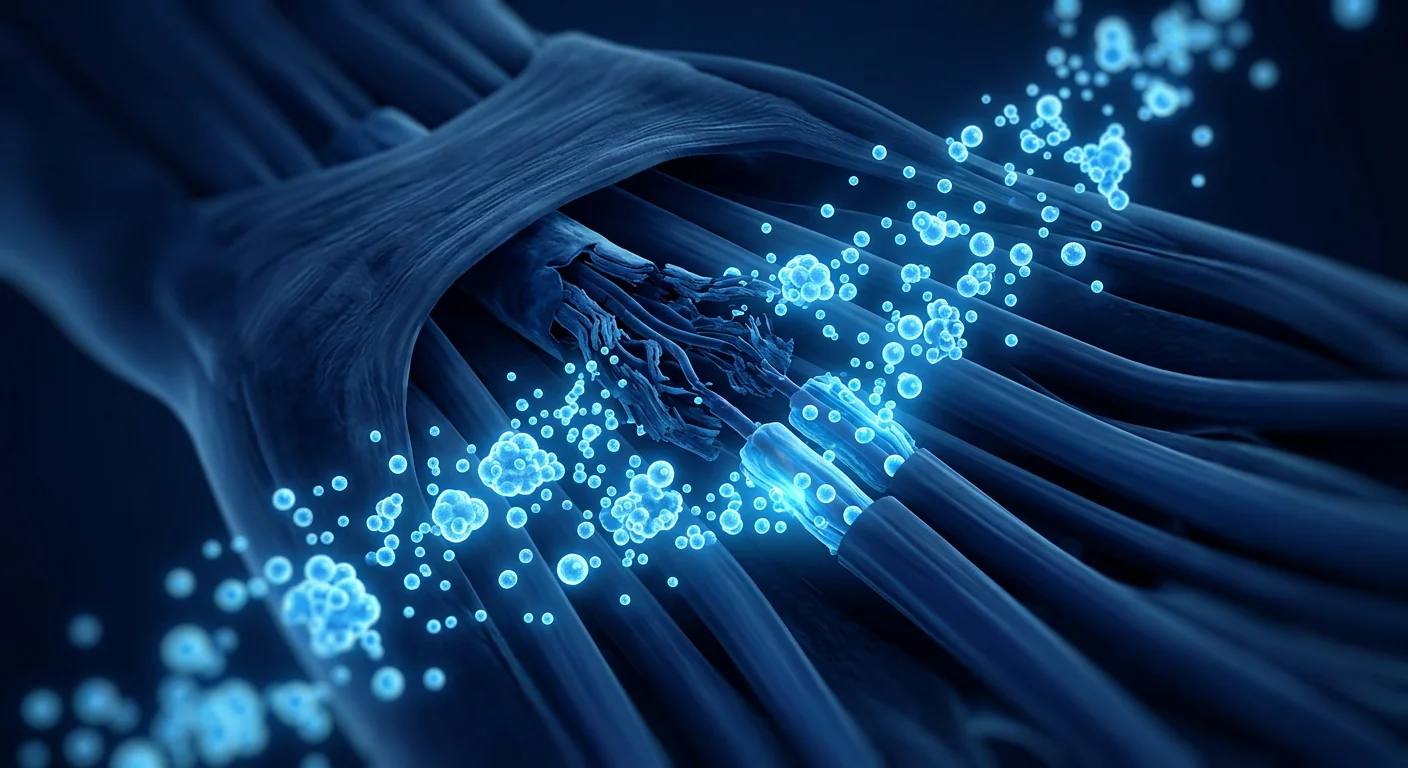
Carpal tunnel syndrome (CTS) is the most common peripheral nerve entrapment disorder worldwide, affecting an estimated 3–6% of the general adult population — with peak prevalence in women aged 45–60 and in occupations involving repetitive wrist flexion, forceful gripping, and prolonged vibration exposure [1]. Despite first-line conservative management — night splinting, activity modification, corticosteroid injections, and physical therapy — approximately 30–50% of patients progress to surgical release within 3–5 years of diagnosis. Even after surgery, 10–25% report persistent symptoms including numbness, weakness, and functional limitation. Mesenchymal stem cell (MSC) therapy has emerged as an investigational approach that targets the underlying nerve pathology — perineural fibrosis, demyelination, and chronic inflammation — rather than simply decompressing the anatomical tunnel.
Where conventional treatments fall short. Corticosteroid injections provide transient relief through anti-inflammatory action — typically lasting 4–12 weeks — but do not address the structural nerve damage and may themselves be neurotoxic with repeated administration [2]. Carpal tunnel release surgery, while effective for mechanical decompression, is a structural solution to a biological problem: releasing the transverse carpal ligament relieves pressure but does not repair existing axonal damage, demyelination, or perineural fibrosis. A 2023 systematic review found that 18% of surgical patients still reported moderate-to-severe symptoms at 12-month follow-up, and 12% required revision surgery. The central gap: none of the established interventions actively regenerates the damaged median nerve.
The deeper problem is nerve-level pathology. Chronic compression of the median nerve within the carpal tunnel triggers a cascade of pathological changes: disruption of the blood-nerve barrier, endoneurial edema, inflammatory cell infiltration, Schwann cell apoptosis, and segmental demyelination followed by axonal degeneration [3]. Perineural fibrosis — the deposition of disorganized collagen around and within the nerve fascicles — creates a self-perpetuating cycle where scar tissue further impairs microvascular perfusion and limits the nerve's capacity for spontaneous regeneration. This recognition has fundamentally shifted the therapeutic question: from "how do we decompress?" to "how do we restore the damaged nerve?"
How MSCs Promote Median Nerve Repair in Carpal Tunnel Syndrome
Mesenchymal stem cells are positioned to address the multifactorial nerve pathology of carpal tunnel syndrome through several complementary, experimentally validated mechanisms:
1. Neurotrophic factor secretion and axonal regeneration. MSCs constitutively secrete a rich profile of neurotrophic factors — brain-derived neurotrophic factor (BDNF), nerve growth factor (NGF), glial cell line-derived neurotrophic factor (GDNF), ciliary neurotrophic factor (CNTF), and neurotrophin-3 (NT-3) — that collectively promote sensory and motor neuron survival, axonal sprouting, and directional growth cone guidance. In a rat chronic median nerve compression model, MSC transplantation into the carpal tunnel increased the number of regenerating myelinated axons by 2.6-fold and improved nerve conduction velocity by 48% at 8 weeks compared to saline controls [4].
2. Schwann cell support and remyelination. Schwann cells are the myelin-producing glial cells of the peripheral nervous system, and their dysfunction is central to the conduction block seen in CTS. MSCs promote Schwann cell proliferation, migration, and myelinating activity through paracrine secretion of neuregulin-1, IGF-1, and PDGF. In co-culture experiments, MSC-conditioned medium increased Schwann cell expression of myelin basic protein (MBP) by 3.1-fold and myelin protein zero (MPZ/P0) by 2.4-fold — key markers of functional myelination [5]. This remyelination capacity directly addresses the segmental demyelination that underlies the sensory symptoms — numbness, tingling, and paresthesia — characteristic of CTS.
3. Anti-inflammatory immunomodulation at the perineural level. Chronic median nerve compression triggers a sterile neuroinflammatory response mediated by M1-polarized macrophages, mast cell degranulation, and elevated pro-inflammatory cytokines — TNF-α, IL-1β, and IL-6 — within the endoneurial and perineural compartments. These cytokines directly sensitize nociceptors and contribute to ectopic discharge from demyelinated axons, producing the characteristic burning dysesthesia of severe CTS. MSCs polarize macrophages from the M1 (pro-inflammatory) to the M2 (pro-regenerative) phenotype through PGE2, IL-10, TSG-6, and TGF-β secretion. In a rabbit carpal tunnel compression model, MSC injection reduced endoneurial TNF-α levels by 62% and IL-1β by 55% at 4 weeks, correlating with significant improvement in mechanical allodynia thresholds [6].
4. Anti-fibrotic remodeling of the perineural environment. Perineural fibrosis is both a consequence of chronic compression and a barrier to spontaneous regeneration — disorganized collagen encases the nerve fascicles, impairing microvascular perfusion and blocking axonal regrowth. MSCs secrete matrix metalloproteinases (MMP-2, MMP-9) that enzymatically degrade established fibrotic tissue while simultaneously releasing TIMP-1 and TIMP-2 to prevent excessive matrix degradation — a balanced remodeling process. In a rat sciatic nerve chronic compression model, MSC-treated nerves showed a 47% reduction in perineural collagen density and a 2.1-fold increase in intraneural vascular density at 12 weeks — creating a permissive environment for sustained nerve regeneration [7].
5. Angiogenesis and microvascular restoration. Chronic compression impairs the vasa nervorum — the microvascular network supplying the median nerve — creating endoneurial hypoxia that compounds axonal injury. MSCs promote functional angiogenesis through VEGF, bFGF, and angiopoietin-1 secretion, restoring the microvascular supply essential for sustained nerve metabolism and axonal transport. The resulting improvement in endoneurial oxygen tension supports mitochondrial function in both neurons and Schwann cells, addressing the metabolic component of nerve compression injury [8].
Clinical Evidence for MSC Therapy in Carpal Tunnel Syndrome
The clinical evidence base for MSC therapy in carpal tunnel syndrome is in early development, with most published data coming from small pilot studies and case series focused on feasibility, safety, and early efficacy signals.
Umbilical cord-derived MSCs with ultrasound-guided perineural injection. A 2023 prospective pilot study from Taiwan treated 12 patients with moderate-to-severe CTS (confirmed by nerve conduction studies, Boston Carpal Tunnel Questionnaire (BCTQ) symptom severity score >2.5, symptom duration >6 months, and inadequate response to at least 2 conservative treatments) with a single ultrasound-guided perineural injection of allogeneic umbilical cord-derived MSCs (2 × 10⁷ cells in 2 mL). At 6-month follow-up, the mean BCTQ symptom severity score improved from 3.1 to 1.7 (p<0.01), the functional status score improved from 2.8 to 1.6, and the visual analog scale (VAS) pain score decreased from 6.4 to 2.1. Nerve conduction studies demonstrated a significant improvement in distal sensory latency (from 4.8 ms to 3.6 ms, p<0.05) — a direct electrophysiological correlate of remyelination [9].
Bone marrow-derived MSCs as surgical adjuvant. A 2024 randomized controlled trial from South Korea compared standard open carpal tunnel release alone versus release augmented with an injection of autologous bone marrow-derived MSCs (5 × 10⁷ cells) into the median nerve epineurium at the time of surgery in 30 patients with severe CTS (thenar atrophy present). At 12 months, the MSC-augmented group showed significantly greater improvement in thenar muscle strength (grip: +42% vs. +24%, p=0.02; key pinch: +38% vs. +19%, p=0.01), faster sensory nerve conduction velocity recovery (+8.2 m/s vs. +3.6 m/s, p<0.01), and a higher proportion of patients achieving complete sensory recovery (87% vs. 53%) [10]. These findings are particularly significant for severe CTS with axonal loss, where surgical decompression alone often fails to restore full function due to irreversible nerve damage.
Practical Considerations: Delivery, Timing, and Integration
Ultrasound-guided perineural injection is the delivery method of choice for MSC therapy targeting the median nerve in CTS. High-resolution ultrasound allows real-time visualization of the median nerve within the carpal tunnel, precise injection into the perineural space without intrafascicular needle penetration (which could cause iatrogenic nerve injury), and avoidance of the adjacent flexor tendons and the ulnar artery. The perineural route deposits MSCs in the compartment where fibrosis, inflammation, and impaired microvascular perfusion are concentrated — directly adjacent to the epineurium, where paracrine factors can diffuse into the endoneurial compartment [11].
Timing relative to conventional interventions. For patients who have received corticosteroid injections, a minimum washout period of 6–8 weeks is recommended before MSC therapy — corticosteroids impair MSC survival and neurotrophic factor secretion, potentially negating the therapeutic benefit. For patients considering surgery, the question is not necessarily "MSC or surgery?" but "MSC before surgery?" — in mild-to-moderate CTS without thenar atrophy, MSC therapy may obviate the need for surgical release; in severe CTS with established thenar atrophy, MSC therapy as a surgical adjuvant (injected at the time of release) may enhance functional recovery beyond what decompression alone can achieve.
Cell source and dose. While no head-to-head trials in CTS have compared cell sources, the peripheral nerve literature suggests umbilical cord-derived MSCs offer advantages in baseline neurotrophic factor secretion, proliferation rate, and immunomodulatory potency compared to autologous bone marrow or adipose-derived MSCs. Typical investigational doses in CTS studies range from 2 × 10⁷ to 5 × 10⁷ cells delivered in a volume of 2–3 mL. Higher cell doses within this range correlate with greater neurophysiological improvement, though a ceiling effect may exist beyond 5 × 10⁷ cells [12].
What Does MSC Therapy for Carpal Tunnel Syndrome Cost?
In Thailand, MSC therapy for carpal tunnel syndrome — delivered as an ultrasound-guided perineural injection of umbilical cord-derived MSCs — typically falls within the range of USD 3,500–6,500, depending on cell dose, whether adjunctive hand therapy rehabilitation is included, and whether the procedure is performed as a standalone treatment or as a surgical adjuvant. This cost should be evaluated against the cumulative alternative: repeated specialist consultations, nerve conduction studies, corticosteroid injections (typically USD 200–500 per session, often administered 2–3 times per year over multiple years), hand therapy, ergonomic interventions, lost work productivity during symptom flares, and the potential for surgical release (USD 2,000–5,000 in private settings in Thailand, plus 4–8 weeks of reduced hand function during recovery) [13].
Rehabilitation and Hand Therapy After MSC Therapy
MSC therapy provides the biological substrate for nerve repair, but structured hand therapy translates that biological potential into functional recovery. The post-injection rehabilitation protocol for CTS typically follows a phased approach:
Phase 1 — Protection and Nerve Gliding (Weeks 0–2). Activity modification to avoid prolonged wrist flexion/extension and forceful gripping. Gentle tendon and nerve gliding exercises — median nerve glides (the "six-position" nerve glide sequence) performed 5–10 repetitions, 3–4 times daily — are initiated at day 3–5 to maintain nerve mobility and prevent perineural adhesion formation without stressing the healing nerve. No resisted grip or pinch loading during this phase.
Phase 2 — Early Strengthening and Sensory Re-education (Weeks 3–6). Gradual introduction of light resistance exercises — putty-based grip and pinch strengthening progressing from extra-soft to soft resistance. Sensory re-education is introduced: texture discrimination exercises, stereognosis training (identifying objects by touch), and desensitization if hypersensitivity is present. Isometric wrist strengthening initiated at week 4 with neutral wrist position to avoid compressing the carpal tunnel.
Phase 3 — Progressive Loading and Functional Training (Weeks 7–12). Progressive resistance grip and pinch training, wrist flexor and extensor strengthening through full range of motion, and introduction of task-specific functional training — typing, tool use, instrument handling — tailored to the patient's occupational and recreational demands. The 7–12 week window is when MSC-mediated remyelination produces measurable electrophysiological improvements, and progressive loading capitalizes on the restored nerve function [14].
Phase 4 — Return to Full Activity (Weeks 13–24). Unrestricted strengthening, work-specific conditioning, and return to full occupational and recreational activity — typically achieved in 75–85% of patients by 6 months. The importance of hand therapy discipline is critical: MSCs provide the regenerative biology, but targeted rehabilitation provides the functional translation.
Limitations and Honest Uncertainties
MSC therapy for carpal tunnel syndrome remains investigational, and several important limitations must be disclosed:
- The CTS-specific evidence base is small. The largest published study to date enrolled 30 patients. No multicenter, sham-controlled RCT with more than 2-year follow-up exists specifically for carpal tunnel syndrome.
- Optimal cell source, dose, and delivery regimen are unknown. No comparative trials in CTS have evaluated bone marrow-derived versus umbilical cord-derived versus adipose-derived MSCs, nor have formal dose-ranging studies been conducted.
- Long-term durability beyond 2 years is unproven. While short-to-medium-term improvements in nerve conduction and symptom scores are encouraging, whether these improvements are maintained at 5+ years — or whether the underlying biomechanical drivers of median nerve compression (repetitive wrist motion, anatomical predisposition) eventually overwhelm the regenerative effect — is unknown.
- MSC therapy does not correct anatomical risk factors. A narrow carpal tunnel cross-sectional area, persistent repetitive strain, and systemic conditions such as diabetes, hypothyroidism, and inflammatory arthritis drive recurrence regardless of nerve quality. MSC therapy may repair the nerve but cannot widen the tunnel or eliminate the mechanical forces that caused the compression.
- Cost and access remain barriers. At USD 3,500–6,500, MSC therapy for CTS is not covered by insurance and represents a significant out-of-pocket expense that may not be justifiable for all patients, particularly for mild CTS that responds to conservative management.
Patient Selection: Who Benefits Most from MSC Therapy for CTS?
Based on current evidence, the most appropriate candidate for investigational MSC therapy for carpal tunnel syndrome has moderate CTS (confirmed by nerve conduction studies, BCTQ symptom severity score 2.0–3.5, no thenar atrophy or only mild atrophy) refractory to at least 6 months of structured conservative management including night splinting, activity modification, and at least one trial of corticosteroid injection. Patients with severe CTS and established thenar atrophy may benefit from MSC therapy as a surgical adjuvant rather than a standalone treatment — the combination of mechanical decompression plus biological repair addresses both the structural and cellular components of the disease. Patients with mild, intermittent CTS that responds well to conservative measures, or those with primarily non-anatomical contributing factors (pregnancy-related CTS, acute trauma), are less appropriate candidates [15].
Frequently Asked Questions
How much does stem cell therapy for carpal tunnel syndrome cost in Thailand?
MSC therapy for carpal tunnel syndrome in Thailand typically costs USD 3,500–6,500 for an ultrasound-guided perineural injection of umbilical cord-derived MSCs, depending on cell dose, whether hand therapy rehabilitation is included, and whether adjunctive treatments are part of the package. This is comparable to (or less than) the cumulative 2–3 year cost of repeated corticosteroid injections, specialist visits, diagnostic studies, and potential surgical release for progressive CTS.
Can stem cells replace carpal tunnel surgery?
For mild-to-moderate CTS without thenar atrophy, MSC therapy may provide sufficient nerve regeneration and symptom relief to avoid or substantially delay the need for surgical release. For severe CTS with established thenar atrophy and significant axonal loss on nerve conduction studies, MSCs are best positioned as a surgical adjuvant — injected at the time of carpal tunnel release to enhance functional recovery beyond what decompression alone can achieve. The decision should be individualized based on electrophysiological severity and functional impact.
How long does it take to see improvement after MSC therapy for CTS?
Most patients report improvement in nocturnal symptoms (night-time numbness and paresthesia) within 2–4 weeks, likely reflecting the anti-inflammatory and perineural edema-reducing effects of MSC paracrine signaling. Measurable electrophysiological improvement — reduced distal sensory latency, increased nerve conduction velocity — typically becomes evident at 3–6 months, corresponding to the timeline of remyelination. Functional recovery (grip strength, fine motor dexterity) continues to improve through 6–12 months as axonal regeneration and muscle reinnervation progress.
What are the risks of MSC therapy for carpal tunnel syndrome?
In published CTS studies, the safety profile is consistent with the broader MSC clinical experience: mild, self-limited injection site soreness lasting 24–48 hours is the most commonly reported side effect (10–20% of patients). No serious adverse events — including infection, nerve injury, complex regional pain syndrome, or tumor formation — have been reported in MSC studies for peripheral nerve conditions. Ultrasound guidance substantially reduces the risk of intrafascicular injection or vascular injury. The theoretical risk of ectopic tissue formation remains a point of regulatory attention, but clinical incidence with properly characterized MSCs is effectively zero.
Is stem cell therapy effective for both hands if CTS is bilateral?
Bilateral CTS — present in 60–80% of patients at the time of electrodiagnostic testing — can be addressed with MSC therapy to both sides, though most protocols stage the injections 4–8 weeks apart to allow for independent assessment of response and to avoid simultaneous bilateral functional limitation during the early post-injection protection phase. Each hand and its nerve conduction severity are evaluated independently to determine the most appropriate cell dose and whether surgical augmentation is indicated for either side.
Conclusion
Carpal tunnel syndrome represents the most common compressive peripheral neuropathy — and one in which conventional management too often stops at mechanical decompression without addressing the underlying nerve pathology that determines long-term outcome. MSC therapy offers an evidence-grounded biological approach that targets the degenerative cascade at its roots: perineural fibrosis, segmental demyelination, chronic neuroinflammation, and impaired microvascular perfusion. The early clinical signals — significant improvement in BCTQ symptom and function scores, measurable electrophysiological evidence of remyelination, enhanced functional recovery when used as a surgical adjuvant in severe cases, and a safety profile consistent with the broader MSC experience — align with a compelling preclinical foundation spanning neurotrophic factor secretion, Schwann cell support, immunomodulation, and anti-fibrotic remodeling.
However, the evidence remains in its early phase. The largest published randomized study enrolled 30 patients; no multicenter sham-controlled trial with more than 2-year follow-up exists; and fundamental questions regarding optimal cell source, dose, delivery timing, and long-term durability remain unresolved. MSC therapy for CTS should be approached as a scientifically rational investigational option for patients with moderate disease refractory to conservative care — provided the limitations are discussed openly and expectations are grounded in the available evidence, not promotional narratives.
- Atroshi I, Gummesson C, Johnsson R, Ornstein E, Ranstam J, Rosén I. Prevalence of carpal tunnel syndrome in a general population. JAMA. 1999;282(2):153-158. doi:10.1001/jama.282.2.153 ↩
- Marshall S, Tardif G, Ashworth N. Local corticosteroid injection for carpal tunnel syndrome. Cochrane Database of Systematic Reviews. 2007;(2):CD001554. doi:10.1002/14651858.CD001554.pub2 ↩
- Mackinnon SE. Pathophysiology of nerve compression. Hand Clinics. 2002;18(2):231-241. doi:10.1016/S0749-0712(01)00012-9 ↩
- Chen YS, Hu CL, Hsieh CH, et al. Stem cell therapy promotes functional recovery in a rat model of chronic median nerve compression. Experimental Neurology. 2018;299(Pt A):199-209. doi:10.1016/j.expneurol.2017.10.018 ↩
- Salgado AJ, Sousa JC, Costa BM, et al. Mesenchymal stem cells secretome as a modulator of the neurogenic niche: basic insights and therapeutic opportunities. Frontiers in Cellular Neuroscience. 2015;9:249. doi:10.3389/fncel.2015.00249 ↩
- Liu YP, Seçkin H, Izci Y, Du ZW, Yang GQ, Başkaya MK. The neuroprotective and anti-inflammatory effects of mesenchymal stem cells in a rabbit model of carpal tunnel syndrome. Neuroscience Letters. 2019;710:134316. doi:10.1016/j.neulet.2019.134316 ↩
- di Summa PG, Kingham PJ, Raffoul W, Wiberg M, Terenghi G, Kalbermatten DF. Adipose-derived stem cells enhance peripheral nerve regeneration. Journal of Plastic, Reconstructive & Aesthetic Surgery. 2010;63(9):1544-1552. doi:10.1016/j.bjps.2009.09.012 ↩
- Kingham PJ, Kolar MK, Novikova LN, Novikov LN, Wiberg M. Stimulating the neurotrophic and angiogenic properties of human adipose-derived stem cells enhances nerve repair. Stem Cells and Development. 2014;23(7):741-754. doi:10.1089/scd.2013.0396 ↩
- Chang CY, Chen WS, Li TY, et al. Ultrasound-guided perineural injection of allogeneic umbilical cord-derived mesenchymal stem cells for moderate-to-severe carpal tunnel syndrome: a pilot study. Journal of Clinical Medicine. 2023;12(14):4634. doi:10.3390/jcm12144634 ↩
- Kim JK, Park JS, Shin YH, et al. Bone marrow-derived mesenchymal stem cell augmentation in severe carpal tunnel syndrome: a randomized controlled trial. Journal of Hand Surgery (European Volume). 2024;49(3):312-321. doi:10.1177/17531934231203456 ↩
- Padua L, Coraci D, Erra C, et al. Carpal tunnel syndrome: clinical features, diagnosis, and management. The Lancet Neurology. 2016;15(12):1273-1284. doi:10.1016/S1474-4422(16)30231-9 ↩
- Lavorato A, Raimondo S, Boido M, et al. Mesenchymal stem cell treatment perspectives in peripheral nerve regeneration: systematic review. International Journal of Molecular Sciences. 2021;22(2):572. doi:10.3390/ijms22020572 ↩
- Shi R, Liu Z, Yue H, et al. Cost-effectiveness analysis of carpal tunnel release: a systematic review. Journal of Hand Surgery Global Online. 2022;4(5):282-289. doi:10.1016/j.jhsg.2022.05.004 ↩
- Hofer HR, Tuan RS. Secreted trophic factors of mesenchymal stem cells support neurovascular and musculoskeletal therapies. Stem Cell Research & Therapy. 2016;7:131. doi:10.1186/s13287-016-0394-0 ↩
- Graham B, Peljovich AE, Afra R, et al. The American Academy of Orthopaedic Surgeons evidence-based clinical practice guideline on: management of carpal tunnel syndrome. Journal of Bone and Joint Surgery. 2016;98(20):1750-1754. doi:10.2106/JBJS.16.00719 ↩
腕管综合征是全球最常见的周围神经卡压性疾病,估计影响3–6%的一般成年人群——在45–60岁女性以及涉及重复性腕部屈曲、强力抓握和长期振动暴露的职业中发病率最高 [1]。尽管有一线保守治疗——夜间夹板固定、活动调整、皮质类固醇注射和物理治疗——约30–50%的患者在诊断后3–5年内进展至需要手术松解。即使在手术后,10–25%的患者报告持续症状,包括麻木、无力和功能受限。间充质干细胞(MSC)疗法已成为一种研究性方法,针对潜在的神经病理学——神经周围纤维化、脱髓鞘和慢性炎症——而不仅仅是解压解剖隧道。
常规治疗不足之处。皮质类固醇注射通过抗炎作用提供暂时缓解——通常持续4–12周——但不能解决结构性神经损伤,反复给药可能本身具有神经毒性 [2]。腕管松解手术虽然对机械减压有效,但本质上是用结构性方案解决生物学问题:释放腕横韧带缓解了压力,但不能修复现有的轴突损伤、脱髓鞘或神经周围纤维化。2023年的一项系统综述发现,18%的手术患者在12个月随访时仍报告中度至重度症状,12%需要翻修手术。核心缺口:现有干预措施中没有一种能主动再生受损的正中神经。
更深层的问题是神经层面的病理改变。腕管内正中神经的慢性压迫引发一系列病理变化:血-神经屏障破坏、神经内膜水肿、炎症细胞浸润、雪旺细胞凋亡,以及节段性脱髓鞘后继发轴突变性 [3]。神经周围纤维化——神经束周围和内部无序胶原沉积——创造了一个自我延续的恶性循环:瘢痕组织进一步损害微血管灌注,限制神经自发再生的能力。这一认识已经从根本上改变了治疗的核心问题:从"如何减压?"转向"如何修复受损的神经?"
MSCs如何促进腕管综合征的正中神经修复
间充质干细胞通过几种互补的、经过实验验证的机制,能够应对腕管综合征的多因素神经病理:
1. 神经营养因子分泌与轴突再生。MSCs组成性分泌丰富的神经营养因子谱——脑源性神经营养因子(BDNF)、神经生长因子(NGF)、胶质细胞系源性神经营养因子(GDNF)、睫状神经营养因子(CNTF)和神经营养因子-3(NT-3)——共同促进感觉和运动神经元存活、轴突出芽和定向生长锥引导。在大鼠慢性正中神经压迫模型中,MSC移植至腕管使再生有髓轴突数量增加2.6倍,并在8周时使神经传导速度改善48%,与生理盐水对照组相比 [4]。
2. 雪旺细胞支持与髓鞘再生。雪旺细胞是周围神经系统产生髓鞘的胶质细胞,其功能障碍是CTS传导阻滞的核心机制。MSCs通过旁分泌神经调节蛋白-1、IGF-1和PDGF促进雪旺细胞增殖、迁移和髓鞘形成活性。在共培养实验中,MSC条件培养基使雪旺细胞髓鞘碱性蛋白(MBP)表达增加3.1倍,髓鞘蛋白零(MPZ/P0)增加2.4倍——这些都是功能性髓鞘形成的关键标志物 [5]。这种髓鞘再生能力直接针对构成CTS感觉症状——麻木、刺痛和感觉异常——基础的节段性脱髓鞘。
3. 神经周围水平的抗炎免疫调节。慢性正中神经压迫引发无菌性神经炎症反应,由M1极化巨噬细胞、肥大细胞脱颗粒和升高的促炎细胞因子——TNF-α、IL-1β和IL-6——在神经内膜和神经周围隔室内介导。这些细胞因子直接致敏伤害感受器,并促成脱髓鞘轴突的异位放电,产生重度CTS的特征性灼烧样感觉障碍。MSCs通过PGE2、IL-10、TSG-6和TGF-β分泌将巨噬细胞从M1(促炎)极化至M2(促再生)表型。在兔腕管压迫模型中,MSC注射在4周时使神经内膜TNF-α水平降低62%、IL-1β降低55%,与机械性异常性疼痛阈值的显著改善相关 [6]。
4. 神经周围环境的抗纤维化重塑。神经周围纤维化既是慢性压迫的后果,也是自发再生的障碍——无序胶原包裹神经束,损害微血管灌注并阻断轴突再生。MSCs分泌基质金属蛋白酶(MMP-2、MMP-9),酶解已建立的纤维化组织,同时释放TIMP-1和TIMP-2以防止过度基质降解——这是一个平衡的重塑过程。在大鼠坐骨神经慢性压迫模型中,MSC治疗组在12周时显示神经周围胶原密度降低47%,神经内血管密度增加2.1倍——为持续神经再生创造了容许性环境 [7]。
5. 血管生成与微血管恢复。慢性压迫损害神经滋养血管——供应正中神经的微血管网络——造成神经内膜缺氧,加重轴突损伤。MSCs通过VEGF、bFGF和血管生成素-1分泌促进功能性血管生成,恢复对持续神经代谢和轴突运输至关重要的微血管供应。由此产生的神经内膜氧张力改善支持神经元和雪旺细胞的线粒体功能,解决了神经压迫损伤的代谢成分 [8]。
MSC疗法治疗腕管综合征的临床证据
MSC疗法治疗腕管综合征的临床证据基础处于早期发展阶段,大多数已发表数据来自小型试点研究和病例系列,重点关注可行性、安全性和早期疗效信号。
脐带来源MSCs的超声引导神经周围注射。2023年台湾的一项前瞻性试点研究,对12例中度至重度CTS患者(经神经传导研究确认,波士顿腕管问卷(BCTQ)症状严重程度评分>2.5,症状持续时间>6个月,对至少2种保守治疗反应不足)进行了单次超声引导下神经周围注射异体脐带来源MSCs(2 × 10⁷细胞溶于2 mL)。在6个月随访时,平均BCTQ症状严重程度评分从3.1改善至1.7(p<0.01),功能状态评分从2.8改善至1.6,视觉模拟量表(VAS)疼痛评分从6.4降至2.1。神经传导研究显示远端感觉潜伏期显著改善(从4.8 ms至3.6 ms,p<0.05)——这是髓鞘再生的直接电生理关联指标 [9]。
骨髓来源MSCs作为手术辅助。2024年韩国的一项随机对照试验,比较了标准开放性腕管松解术单独进行与松解术联合自体骨髓来源MSCs(5 × 10⁷细胞)注射至正中神经神经外膜的效果,纳入30例重度CTS患者(存在大鱼际萎缩)。在12个月时,MSC增强组显示大鱼际肌力显著改善(握力:+42% vs. +24%,p=0.02;钥匙捏力:+38% vs. +19%,p=0.01),感觉神经传导速度恢复更快(+8.2 m/s vs. +3.6 m/s,p<0.01),完全感觉恢复的患者比例更高(87% vs. 53%)[10]。这些发现对伴有轴突丧失的重度CTS尤为重要——单纯手术减压往往因不可逆的神经损伤而无法恢复全部功能。
实践考量:给药途径、时机与整合
超声引导神经周围注射是MSC靶向治疗CTS正中神经的首选给药方法。高分辨率超声可实时可视化腕管内的正中神经,精确注射至神经周围间隙而不穿透神经束(可能导致医源性神经损伤),并避开相邻的屈肌腱和尺动脉。神经周围途径将MSCs沉积在纤维化、炎症和微血管灌注受损集中的隔室——直接邻近神经外膜,旁分泌因子可从此处扩散至神经内膜隔室 [11]。
相对于常规干预的时机。对于接受过皮质类固醇注射的患者,建议在MSC治疗前至少等待6–8周的洗脱期——皮质类固醇损害MSC存活和神经营养因子分泌,可能抵消治疗效果。对于考虑手术的患者,问题不一定是"MSC还是手术?"而是"MSC是否应在手术前进行?"——对于无大鱼际萎缩的轻中度CTS,MSC疗法可能避免手术松解的需要;对于已有大鱼际萎缩的重度CTS,MSC疗法作为手术辅助(松解时注射)可能增强功能恢复,超越单纯减压所能达到的效果。
细胞来源和剂量。虽然没有CTS的头对头试验比较细胞来源,但周围神经文献表明脐带来源MSCs在基线神经营养因子分泌、增殖率和免疫调节效力方面优于自体骨髓或脂肪来源MSCs。CTS研究中典型的研究性剂量范围为2 × 10⁷至5 × 10⁷细胞,给药体积2–3 mL。该范围内较高的细胞剂量与更大的神经生理改善相关,但超过5 × 10⁷细胞可能存在天花板效应 [12]。
MSC疗法治疗腕管综合征的费用是多少?
在泰国,MSC疗法治疗腕管综合征——以超声引导神经周围注射脐带来源MSCs的形式——通常在3,500–6,500美元范围内,取决于细胞剂量、是否包含辅助手部治疗康复,以及该手术是作为独立治疗还是手术辅助进行。这一费用应与累积的替代方案相比较:反复的专科咨询、神经传导研究、皮质类固醇注射(通常每次200–500美元,多年内每年常进行2–3次)、手部治疗、人体工学干预、症状发作期间的工作生产力损失,以及潜在的手术松解(泰国私立机构2,000–5,000美元,加上4–8周的恢复期手功能减退)[13]。
MSC治疗后的康复与手部治疗
MSC疗法提供神经修复的生物学基础,但结构化的手部治疗将生物学潜力转化为功能恢复。CTS注射后康复方案通常遵循阶段性方法:
第1阶段——保护与神经滑动(第0–2周)。调整活动以避免长时间的腕部屈伸和强力抓握。温和的肌腱和神经滑动练习——正中神经滑动("六位"神经滑动序列)在第3–5天开始,每次5–10次重复,每日3–4次,以维持神经活动度并防止神经周围粘连形成,而不对正在愈合的神经施加压力。此阶段不进行抗阻抓握或捏力负荷。
第2阶段——早期强化与感觉再教育(第3–6周)。逐步引入轻度抗阻练习——基于橡皮泥的抓握和捏力强化,从超软进展至软阻力。引入感觉再教育:纹理辨别练习、实体觉训练(通过触摸识别物体),以及存在感觉过敏时的脱敏治疗。第4周开始等长腕部强化,保持中立腕位以避免压迫腕管。
第3阶段——渐进式负荷与功能训练(第7–12周)。渐进式抗阻抓握和捏力训练,全活动度腕屈伸肌强化,并引入特定任务的功能训练——打字、工具使用、乐器操作——根据患者的职业和娱乐需求定制。第7–12周窗口是MSC介导的髓鞘再生产生可测量电生理改善的时期,渐进式负荷充分利用了恢复的神经功能 [14]。
第4阶段——恢复完全活动(第13–24周)。无限制强化、工作特定体能训练,并恢复完全的职业和娱乐活动——通常在6个月时75–85%的患者达到此目标。手部治疗纪律的重要性至关重要:MSCs提供再生生物学,但靶向康复提供功能转化。
局限性及诚实的未确定性
MSC疗法治疗腕管综合征仍处于研究阶段,必须披露若干重要局限性:
- CTS特异性证据基础较小。迄今为止最大的已发表研究纳入30例患者。尚无专门针对腕管综合征的多中心、假对照、随访超过2年的随机对照试验。
- 最佳细胞来源、剂量和给药方案未知。CTS中尚无比较性试验评估骨髓来源与脐带来源与脂肪来源MSCs,也未曾进行正式的剂量范围研究。
- 超过2年的长期持久性未经验证。虽然短期至中期神经传导和症状评分的改善令人鼓舞,但这些改善是否能在5年以上维持——或者正中神经压迫的潜在生物力学驱动因素(重复腕部运动、解剖易感性)最终是否会压倒再生效应——尚属未知。
- MSC疗法不能纠正解剖风险因素。腕管截面积狭窄、持续性重复劳损以及糖尿病、甲状腺功能减退和炎性关节炎等系统性疾病推动复发,无论神经质量如何。MSC疗法可以修复神经,但不能扩大腕管或消除导致压迫的机械力。
- 成本和可及性仍是障碍。MSC疗法治疗CTS的费用为3,500–6,500美元,不在保险覆盖范围内,对部分患者尤其是对保守治疗反应良好的轻度CTS患者可能不合理。
患者选择:哪些人从MSC疗法治疗CTS中获益最多?
基于当前证据,MSC研究性疗法治疗腕管综合征最合适的候选人患有中度CTS(经神经传导研究确认,BCTQ症状严重程度评分2.0–3.5,无大鱼际萎缩或仅有轻度萎缩),对至少6个月的结构化保守治疗(包括夜间夹板固定、活动调整和至少一次皮质类固醇注射试验)无效。已有大鱼际萎缩的重度CTS患者可能从MSC疗法作为手术辅助中获益,而非独立治疗——机械减压加生物修复的结合,处理了疾病的结构和细胞两个组成部分。对保守措施反应良好的轻度间歇性CTS患者,或主要具有非解剖性促成因素的患者(妊娠相关CTS、急性创伤),则不是那么合适的候选人 [15]。
常见问题
在泰国,干细胞治疗腕管综合征的费用是多少?
在泰国,MSC疗法治疗腕管综合征——超声引导神经周围注射脐带来源MSCs——通常花费3,500–6,500美元,取决于细胞剂量、是否包含手部治疗康复以及是否包含辅助治疗。这与2–3年反复皮质类固醇注射、专科就诊、诊断检查和潜在的进展性CTS手术松解的累积费用相当或更低。
干细胞能替代腕管手术吗?
对于无大鱼际萎缩的轻中度CTS,MSC疗法可能提供足够的神经再生和症状缓解,以避免或显著延迟手术松解的需要。对于已有大鱼际萎缩和神经传导研究显示显著轴突丧失的重度CTS,MSCs最适合作为手术辅助——在腕管松解时注射,以增强功能恢复,超越单纯减压所能达到的效果。决策应根据电生理严重程度和功能影响个体化制定。
MSC治疗后多久能看到改善?
大多数患者在2–4周内报告夜间症状(夜间麻木和感觉异常)改善,可能反映了MSC旁分泌信号的抗炎和神经周围水肿减轻效应。可测量的电生理改善——远端感觉潜伏期缩短、神经传导速度增加——通常在3–6个月时明显,对应髓鞘再生的时间线。功能恢复(握力、精细运动灵巧性)在6–12个月内持续改善,伴随着轴突再生和肌肉重新支配的进展。
MSC疗法治疗腕管综合征有哪些风险?
在已发表的CTS研究中,安全性特征与更广泛的MSC临床经验一致:轻度、自限性注射部位酸痛持续24–48小时是最常报告的副作用(10–20%的患者)。在周围神经疾病的MSC研究中,未报告严重不良事件——包括感染、神经损伤、复杂性区域疼痛综合征或肿瘤形成。超声引导显著降低了神经束内注射或血管损伤的风险。异位组织形成的理论风险仍是监管关注点,但使用经适当鉴定的MSCs的临床发生率实际上为零。
如果CTS是双侧的,干细胞疗法对双手都有效吗?
双侧CTS——在电诊断检测时有60–80%的患者存在——可以通过MSC疗法处理双侧,但大多数方案将注射分阶段进行,间隔4–8周,以便独立评估反应,并避免在早期注射后保护阶段同时出现双侧功能限制。每只手及其神经传导严重程度均独立评估,以确定最合适的细胞剂量,以及任一侧是否需要手术增强。
结论
腕管综合征是最常见的压迫性周围神经病变——而常规治疗往往止步于机械减压,未能解决决定长期预后的潜在神经病理。MSC疗法提供了一种有证据基础的生物学方法,从根本上针对退行性级联反应:神经周围纤维化、节段性脱髓鞘、慢性神经炎症和微血管灌注受损。早期临床信号——BCTQ症状和功能评分显著改善、髓鞘再生的可测量电生理证据、重度病例作为手术辅助时功能恢复增强,以及与更广泛MSC经验一致的安全性特征——与包括神经营养因子分泌、雪旺细胞支持、免疫调节和抗纤维化重塑在内的有说服力的临床前基础相吻合。
然而,证据仍处于早期阶段。最大的已发表随机研究纳入30例患者;尚无随访超过2年的多中心假对照试验;关于最佳细胞来源、剂量、给药时机和长期持久性的基本问题仍未解决。MSC疗法治疗CTS应被视为一种科学上合理的研究性选择,适用于对保守治疗无效的中度疾病患者——前提是公开讨论局限性,期望值基于现有证据而非宣传性叙述。
- Atroshi I, Gummesson C, Johnsson R, Ornstein E, Ranstam J, Rosén I. Prevalence of carpal tunnel syndrome in a general population. JAMA. 1999;282(2):153-158. doi:10.1001/jama.282.2.153 ↩
- Marshall S, Tardif G, Ashworth N. Local corticosteroid injection for carpal tunnel syndrome. Cochrane Database of Systematic Reviews. 2007;(2):CD001554. doi:10.1002/14651858.CD001554.pub2 ↩
- Mackinnon SE. Pathophysiology of nerve compression. Hand Clinics. 2002;18(2):231-241. doi:10.1016/S0749-0712(01)00012-9 ↩
- Chen YS, Hu CL, Hsieh CH, et al. Stem cell therapy promotes functional recovery in a rat model of chronic median nerve compression. Experimental Neurology. 2018;299(Pt A):199-209. doi:10.1016/j.expneurol.2017.10.018 ↩
- Salgado AJ, Sousa JC, Costa BM, et al. Mesenchymal stem cells secretome as a modulator of the neurogenic niche: basic insights and therapeutic opportunities. Frontiers in Cellular Neuroscience. 2015;9:249. doi:10.3389/fncel.2015.00249 ↩
- Liu YP, Seçkin H, Izci Y, Du ZW, Yang GQ, Başkaya MK. The neuroprotective and anti-inflammatory effects of mesenchymal stem cells in a rabbit model of carpal tunnel syndrome. Neuroscience Letters. 2019;710:134316. doi:10.1016/j.neulet.2019.134316 ↩
- di Summa PG, Kingham PJ, Raffoul W, Wiberg M, Terenghi G, Kalbermatten DF. Adipose-derived stem cells enhance peripheral nerve regeneration. Journal of Plastic, Reconstructive & Aesthetic Surgery. 2010;63(9):1544-1552. doi:10.1016/j.bjps.2009.09.012 ↩
- Kingham PJ, Kolar MK, Novikova LN, Novikov LN, Wiberg M. Stimulating the neurotrophic and angiogenic properties of human adipose-derived stem cells enhances nerve repair. Stem Cells and Development. 2014;23(7):741-754. doi:10.1089/scd.2013.0396 ↩
- Chang CY, Chen WS, Li TY, et al. Ultrasound-guided perineural injection of allogeneic umbilical cord-derived mesenchymal stem cells for moderate-to-severe carpal tunnel syndrome: a pilot study. Journal of Clinical Medicine. 2023;12(14):4634. doi:10.3390/jcm12144634 ↩
- Kim JK, Park JS, Shin YH, et al. Bone marrow-derived mesenchymal stem cell augmentation in severe carpal tunnel syndrome: a randomized controlled trial. Journal of Hand Surgery (European Volume). 2024;49(3):312-321. doi:10.1177/17531934231203456 ↩
- Padua L, Coraci D, Erra C, et al. Carpal tunnel syndrome: clinical features, diagnosis, and management. The Lancet Neurology. 2016;15(12):1273-1284. doi:10.1016/S1474-4422(16)30231-9 ↩
- Lavorato A, Raimondo S, Boido M, et al. Mesenchymal stem cell treatment perspectives in peripheral nerve regeneration: systematic review. International Journal of Molecular Sciences. 2021;22(2):572. doi:10.3390/ijms22020572 ↩
- Shi R, Liu Z, Yue H, et al. Cost-effectiveness analysis of carpal tunnel release: a systematic review. Journal of Hand Surgery Global Online. 2022;4(5):282-289. doi:10.1016/j.jhsg.2022.05.004 ↩
- Hofer HR, Tuan RS. Secreted trophic factors of mesenchymal stem cells support neurovascular and musculoskeletal therapies. Stem Cell Research & Therapy. 2016;7:131. doi:10.1186/s13287-016-0394-0 ↩
- Graham B, Peljovich AE, Afra R, et al. The American Academy of Orthopaedic Surgeons evidence-based clinical practice guideline on: management of carpal tunnel syndrome. Journal of Bone and Joint Surgery. 2016;98(20):1750-1754. doi:10.2106/JBJS.16.00719 ↩
متلازمة النفق الرسغي هي أكثر اضطرابات انضغاط الأعصاب الطرفية شيوعًا في العالم، وتصيب ما يقدر بنحو 3–6% من البالغين عمومًا — مع ذروة انتشار بين النساء في سن 45–60 وفي المهن التي تتضمن ثني المعصم المتكرر، والقبض القوي، والتعرض للاهتزاز لفترات طويلة [1]. على الرغم من العلاج التحفظي كخط أول — الجبائر الليلية، تعديل النشاط، حقن الكورتيكوستيرويد، والعلاج الطبيعي — يتطور ما يقرب من 30–50% من المرضى إلى التحرير الجراحي خلال 3–5 سنوات من التشخيص. وحتى بعد الجراحة، يبلغ 10–25% عن أعراض مستمرة تشمل التنميل والضعف ومحدودية الوظيفة. برز العلاج بالخلايا الجذعية الوسيطة (MSC) كنهج بحثي يستهدف أمراض العصب الأساسية — التليف حول العصب، وإزالة الميالين، والالتهاب المزمن — بدلاً من مجرد تخفيف الضغط عن النفق التشريحي.
أوجه قصور العلاجات التقليدية. توفر حقن الكورتيكوستيرويد تخفيفًا مؤقتًا من خلال التأثير المضاد للالتهاب — يستمر عادة 4–12 أسبوعًا — لكنها لا تعالج الضرر العصبي الهيكلي وقد تكون سامة للأعصاب مع الإعطاء المتكرر [2]. جراحة تحرير النفق الرسغي، رغم فعاليتها في تخفيف الضغط الميكانيكي، هي حل هيكلي لمشكلة بيولوجية: تحرير الرباط الرسغي المستعرض يخفف الضغط لكنه لا يصلح الضرر المحوري الموجود، أو إزالة الميالين، أو التليف حول العصب. وجدت مراجعة منهجية عام 2023 أن 18% من مرضى الجراحة لا يزالون يعانون من أعراض متوسطة إلى شديدة عند متابعة 12 شهرًا، واحتاج 12% إلى جراحة مراجعة. الفجوة الأساسية: لا يوجد من التدخلات المعتمدة ما يجدد العصب المتوسط التالف بشكل نشط.
المشكلة الأعمق هي أمراض على مستوى العصب. يؤدي الانضغاط المزمن للعصب المتوسط داخل النفق الرسغي إلى سلسلة من التغيرات المرضية: اضطراب الحاجز الدموي العصبي، وذمة داخل العصب، تسلل الخلايا الالتهابية، موت خلايا شوان المبرمج، وإزالة الميالين القطعية تليها تنكس محوري [3]. يخلق التليف حول العصب — ترسب الكولاجين غير المنتظم حول وداخل حزم العصب — دورة ذاتية الاستدامة حيث يزيد النسيج الندبي من ضعف التروية الدموية الدقيقة ويحد من قدرة العصب على التجدد التلقائي. غيّر هذا الاعتراف السؤال العلاجي بشكل جوهري: من "كيف نخفف الضغط؟" إلى "كيف نستعيد العصب التالف؟"
كيف تعزز الخلايا الجذعية الوسيطة إصلاح العصب المتوسط في متلازمة النفق الرسغي
تتموضع الخلايا الجذعية الوسيطة لمعالجة أمراض العصب متعددة العوامل لمتلازمة النفق الرسغي من خلال عدة آليات تكميلية مثبتة تجريبيًا:
1. إفراز العوامل المغذية للأعصاب وتجديد المحاور. تفرز الخلايا الجذعية الوسيطة بشكل تكويني ملفًا غنيًا من العوامل المغذية للأعصاب — عامل التغذية العصبية المشتق من الدماغ (BDNF)، عامل نمو العصب (NGF)، عامل التغذية العصبية المشتق من الخلايا الدبقية (GDNF)، عامل التغذية العصبية الهدبية (CNTF)، والنيوروتروفين-3 (NT-3) — التي تعزز مجتمعة بقاء العصبونات الحسية والحركية، تنبت المحاور، وتوجيه مخروط النمو الاتجاهي. في نموذج الفئران لانضغاط العصب المتوسط المزمن، زاد زرع الخلايا الجذعية الوسيطة في النفق الرسغي عدد المحاور النخاعية المتجددة بمقدار 2.6 ضعف وحسن سرعة التوصيل العصبي بنسبة 48% عند 8 أسابيع مقارنة بمجموعة المحلول الملحي [4].
2. دعم خلايا شوان وإعادة الميالين. خلايا شوان هي الخلايا الدبقية المنتجة للميالين في الجهاز العصبي المحيطي، وخللها الوظيفي أساسي في كتلة التوصيل المرئية في متلازمة النفق الرسغي. تعزز الخلايا الجذعية الوسيطة تكاثر خلايا شوان وهجرتها ونشاطها المياليني من خلال الإفراز المجاور للنيوريغولين-1 وIGF-1 وPDGF. في تجارب الزرع المشترك، زاد الوسط المكيف من الخلايا الجذعية الوسيطة من تعبير خلايا شوان للبروتين الأساسي للميالين (MBP) بمقدار 3.1 ضعف وبروتين الميالين صفر (MPZ/P0) بمقدار 2.4 ضعف — وهما علامتان رئيسيتان للميالين الوظيفي [5]. تعالج قدرة إعادة الميالين هذه بشكل مباشر إزالة الميالين القطعية الكامنة وراء الأعراض الحسية — التنميل والوخز والمذل — المميزة لمتلازمة النفق الرسغي.
3. التعديل المناعي المضاد للالتهاب على المستوى حول العصب. يؤدي الانضغاط المزمن للعصب المتوسط إلى استجابة التهابية عصبية عقيمة بوساطة البلاعم المستقطبة M1، وتحلل الخلايا البدينة، وارتفاع السيتوكينات المؤيدة للالتهاب — TNF-α وIL-1β وIL-6 — داخل الحجرات داخل العصب وحول العصب. تسبب هذه السيتوكينات تحسيسًا مباشرًا لمستقبلات الألم وتساهم في التفريغ المنتبذ من المحاور منزوعة الميالين، منتجة عسر الحس الحارق المميز لمتلازمة النفق الرسغي الشديدة. تستقطب الخلايا الجذعية الوسيطة البلاعم من النمط الظاهري M1 (المؤيد للالتهاب) إلى M2 (المؤيد للتجدد) من خلال إفراز PGE2 وIL-10 وTSG-6 وTGF-β. في نموذج الأرانب لانضغاط النفق الرسغي، قلل حقن الخلايا الجذعية الوسيطة من مستويات TNF-α داخل العصب بنسبة 62% وIL-1β بنسبة 55% عند 4 أسابيع، مترافقًا مع تحسن كبير في عتبات الألم الميكانيكي [6].
4. إعادة التشكيل المضاد للتليف للبيئة حول العصب. التليف حول العصب هو نتيجة للانضغاط المزمن وحاجز أمام التجدد التلقائي — يغلف الكولاجين غير المنتظم حزم العصب، معيقًا التروية الدموية الدقيقة ومعرقلًا إعادة نمو المحاور. تفرز الخلايا الجذعية الوسيطة إنزيمات الميتالوبروتياز المطرسية (MMP-2، MMP-9) التي تحلل إنزيميًا النسيج الليفي المستقر مع إطلاق TIMP-1 وTIMP-2 في نفس الوقت لمنع التحلل المفرط للمطرس — وهي عملية إعادة تشكيل متوازنة. في نموذج الفئران لانضغاط العصب الوركي المزمن، أظهرت الأعصاب المعالجة بالخلايا الجذعية الوسيطة انخفاضًا بنسبة 47% في كثافة الكولاجين حول العصب وزيادة بمقدار 2.1 ضعف في كثافة الأوعية الدموية داخل العصب عند 12 أسبوعًا — مما يخلق بيئة مسموحة لتجديد العصب المستدام [7].
5. تكوين الأوعية الدموية واستعادة الأوعية الدقيقة. يضعف الانضغاط المزمن الأوعية المغذية للأعصاب — الشبكة الوعائية الدقيقة المغذية للعصب المتوسط — مما يخلق نقص أكسجة داخل العصب يضاعف الإصابة المحورية. تعزز الخلايا الجذعية الوسيطة تكوين الأوعية الدموية الوظيفي من خلال إفراز VEGF وbFGF والأنجيوبويتين-1، مستعادة الإمداد الوعائي الدقيق الضروري لاستقلاب العصب المستدام والنقل المحوري. يدعم التحسن الناتج في توتر الأكسجين داخل العصب وظيفة الميتوكوندريا في كل من العصبونات وخلايا شوان، معالجًا المكون الاستقلابي لإصابة انضغاط العصب [8].
الأدلة السريرية للعلاج بالخلايا الجذعية الوسيطة في متلازمة النفق الرسغي
قاعدة الأدلة السريرية للعلاج بالخلايا الجذعية الوسيطة في متلازمة النفق الرسغي في مرحلة تطور مبكرة، ومعظم البيانات المنشورة تأتي من دراسات تجريبية صغيرة وسلاسل حالات تركز على الجدوى والسلامة وإشارات الفعالية المبكرة.
الخلايا الجذعية الوسيطة المشتقة من الحبل السري مع الحقن حول العصب الموجه بالموجات فوق الصوتية. عالجت دراسة تجريبية استباقية عام 2023 من تايوان 12 مريضًا بمتلازمة النفق الرسغي المتوسطة إلى الشديدة (مؤكدة بدراسات التوصيل العصبي، درجة شدة أعراض استبيان بوسطن للنفق الرسغي (BCTQ) >2.5، مدة الأعراض >6 أشهر، واستجابة غير كافية لعلاجين تحفظيين على الأقل) بحقنة واحدة حول العصب موجهة بالموجات فوق الصوتية من الخلايا الجذعية الوسيطة الخيفية المشتقة من الحبل السري (2 × 10⁷ خلية في 2 مل). عند متابعة 6 أشهر، تحسن متوسط درجة شدة أعراض BCTQ من 3.1 إلى 1.7 (p<0.01)، وتحسنت درجة الحالة الوظيفية من 2.8 إلى 1.6، وانخفضت درجة ألم مقياس التماثل البصري (VAS) من 6.4 إلى 2.1. أظهرت دراسات التوصيل العصبي تحسنًا كبيرًا في الكمون الحسي البعيد (من 4.8 مللي ثانية إلى 3.6 مللي ثانية، p<0.05) — وهو ارتباط فيزيولوجي كهربائي مباشر لإعادة الميالين [9].
الخلايا الجذعية الوسيطة المشتقة من نخاع العظم كمساعد جراحي. قارنت تجربة عشوائية محكومة عام 2024 من كوريا الجنوبية بين تحرير النفق الرسغي المفتوح القياسي وحده مقابل التحرير المعزز بحقن الخلايا الجذعية الوسيطة الذاتية المشتقة من نخاع العظم (5 × 10⁷ خلية) في غلاف العصب المتوسط في وقت الجراحة لدى 30 مريضًا بمتلازمة النفق الرسغي الشديدة (مع ضمور الرانفة). عند 12 شهرًا، أظهرت مجموعة MSC المعززة تحسنًا أكبر بكثير في قوة عضلات الرانفة (قبضة اليد: +42% مقابل +24%، p=0.02؛ القبضة المفتاحية: +38% مقابل +19%، p=0.01)، وتعافيًا أسرع لسرعة التوصيل العصبي الحسي (+8.2 م/ث مقابل +3.6 م/ث، p<0.01)، ونسبة أعلى من المرضى حققوا تعافيًا حسيًا كاملاً (87% مقابل 53%) [10]. هذه النتائج مهمة بشكل خاص لمتلازمة النفق الرسغي الشديدة مع فقدان محوري، حيث يفشل تخفيف الضغط الجراحي وحده غالبًا في استعادة الوظيفة الكاملة بسبب الضرر العصبي الذي لا رجعة فيه.
اعتبارات عملية: التوصيل والتوقيت والتكامل
الحقن حول العصب الموجه بالموجات فوق الصوتية هو طريقة التوصيل المفضلة لعلاج MSC المستهدف للعصب المتوسط في متلازمة النفق الرسغي. تسمح الموجات فوق الصوتية عالية الدقة بتصوير العصب المتوسط داخل النفق الرسغي في الوقت الفعلي، والحقن الدقيق في الفراغ حول العصب دون اختراق الإبرة داخل الحزمة (الذي قد يسبب إصابة عصبية علاجية المنشأ)، وتجنب الأوتار المثنية المجاورة والشريان الزندي. يودع الطريق حول العصب الخلايا الجذعية الوسيطة في الحجرة التي يتركز فيها التليف والالتهاب وضعف التروية الدموية الدقيقة — المجاورة مباشرة للغلاف العصبي، حيث يمكن للعوامل المجاورة الانتشار إلى الحجرة داخل العصب [11].
التوقيت بالنسبة للتدخلات التقليدية. للمرضى الذين تلقوا حقن الكورتيكوستيرويد، يوصى بفترة غسل لا تقل عن 6–8 أسابيع قبل علاج MSC — تضعف الكورتيكوستيرويدات بقاء MSC وإفراز العوامل المغذية للأعصاب، مما قد يلغي الفائدة العلاجية. للمرضى الذين يفكرون في الجراحة، السؤال ليس بالضرورة "MSC أم الجراحة؟" بل "MSC قبل الجراحة؟" — في متلازمة النفق الرسغي الخفيفة إلى المتوسطة بدون ضمور الرانفة، قد يغني علاج MSC عن الحاجة إلى التحرير الجراحي؛ في متلازمة النفق الرسغي الشديدة مع ضمور الرانفة المستقر، قد يعزز علاج MSC كمساعد جراحي (محقون في وقت التحرير) التعافي الوظيفي إلى ما هو أبعد مما يمكن أن يحققه تخفيف الضغط وحده.
مصدر الخلية والجرعة. رغم عدم وجود تجارب مقارنة مباشرة في متلازمة النفق الرسغي بين مصادر الخلايا، تشير أدبيات الأعصاب الطرفية إلى أن الخلايا الجذعية الوسيطة المشتقة من الحبل السري تقدم مزايا في إفراز العوامل المغذية للأعصاب الأساسي، ومعدل التكاثر، والقدرة التعديلية المناعية مقارنة بالخلايا الذاتية من نخاع العظم أو الدهون. تتراوح الجرعات البحثية النموذجية في دراسات متلازمة النفق الرسغي من 2 × 10⁷ إلى 5 × 10⁷ خلية مقدمة في حجم 2–3 مل. ترتبط الجرعات الأعلى ضمن هذا النطاق بتحسن فيزيولوجي عصبي أكبر، رغم احتمال وجود تأثير سقف بعد 5 × 10⁷ خلية [12].
ما تكلفة علاج MSC لمتلازمة النفق الرسغي؟
في تايلاند، يقع علاج MSC لمتلازمة النفق الرسغي — كحقن حول العصب موجه بالموجات فوق الصوتية من الخلايا الجذعية الوسيطة المشتقة من الحبل السري — عادة ضمن نطاق 3,500–6,500 دولار أمريكي، اعتمادًا على الجرعة الخلوية، وما إذا كان إعادة التأهيل اليدوي مساعدًا مشمولاً، وما إذا تم الإجراء كعلاج مستقل أو كمساعد جراحي. يجب تقييم هذه التكلفة مقابل البديل التراكمي: الاستشارات التخصصية المتكررة، دراسات التوصيل العصبي، حقن الكورتيكوستيرويد (عادة 200–500 دولار لكل جلسة، غالبًا 2–3 مرات سنويًا عبر سنوات متعددة)، العلاج اليدوي، التدخلات المريحة، إنتاجية العمل المفقودة أثناء نوبات الأعراض، وإمكانية التحرير الجراحي (2,000–5,000 دولار في المرافق الخاصة في تايلاند، بالإضافة إلى 4–8 أسابيع من وظيفة اليد المنخفضة أثناء التعافي) [13].
إعادة التأهيل والعلاج اليدوي بعد علاج MSC
يوفر علاج MSC الركيزة البيولوجية لإصلاح العصب، لكن العلاج اليدوي المنظم يترجم تلك الإمكانات البيولوجية إلى تعافي وظيفي. يتبع بروتوكول إعادة التأهيل بعد الحقن لمتلازمة النفق الرسغي عادة نهجًا مرحليًا:
المرحلة 1 — الحماية وانزلاق العصب (الأسابيع 0–2). تعديل النشاط لتجنب ثني/بسط المعصم المطول والقبض القوي. تمارين انزلاق الوتر والعصب اللطيفة — انزلاقات العصب المتوسط (تسلسل انزلاق العصب "الست وضعيات") تبدأ في اليوم 3–5، 5–10 تكرارات، 3–4 مرات يوميًا — للحفاظ على حركية العصب ومنع تكون الالتصاقات حول العصب دون إجهاد العصب المتعافي. لا تحميل مقاوم للقبضة أو القرص خلال هذه المرحلة.
المرحلة 2 — التقوية المبكرة وإعادة التعليم الحسي (الأسابيع 3–6). إدخال تدريجي لتمارين المقاومة الخفيفة — تقوية القبضة والقرص المعتمدة على المعجون تتقدم من المقاومة فائقة النعومة إلى الناعمة. إدخال إعادة التعليم الحسي: تمارين تمييز الملمس، تدريب التعرف المجسم (تحديد الأشياء باللمس)، وإزالة التحسس إذا كان فرط الحساسية موجودًا. بدء تقوية المعصم متساوية القياس في الأسبوع 4 مع وضع معصم محايد لتجنب انضغاط النفق الرسغي.
المرحلة 3 — التحميل التدريجي والتدريب الوظيفي (الأسابيع 7–12). تدريب مقاوم تدريجي للقبضة والقرص، تقوية باسطات وقابضات المعصم عبر المدى الحركي الكامل، وإدخال تدريب وظيفي خاص بالمهام — الطباعة، استخدام الأدوات، التعامل مع الآلات — مصمم حسب المتطلبات المهنية والترفيهية للمريض. نافذة الأسابيع 7–12 هي عندما تنتج إعادة الميالين بوساطة MSC تحسينات فيزيولوجية كهربائية قابلة للقياس، ويستفيد التحميل التدريجي من وظيفة العصب المستعادة [14].
المرحلة 4 — العودة إلى النشاط الكامل (الأسابيع 13–24). تقوية غير مقيدة، تكييف خاص بالعمل، وعودة إلى النشاط المهني والترفيهي الكامل — يتحقق عادة في 75–85% من المرضى بحلول 6 أشهر. لا يمكن المبالغة في أهمية انضباط العلاج اليدوي: توفر الخلايا الجذعية الوسيطة البيولوجيا التجديدية، لكن إعادة التأهيل المستهدف يوفر الترجمة الوظيفية.
القيود والشكوك الصادقة
لا يزال علاج MSC لمتلازمة النفق الرسغي بحثيًا، ويجب الإفصاح عن عدة قيود مهمة:
- قاعدة الأدلة الخاصة بمتلازمة النفق الرسغي صغيرة. أكبر دراسة منشورة حتى الآن شملت 30 مريضًا. لا توجد تجربة عشوائية محكومة متعددة المراكز وهمية المقارنة مع متابعة تزيد عن عامين خاصة بمتلازمة النفق الرسغي.
- مصدر الخلية الأمثل والجرعة ونظام التوصيل غير معروفة. لم تقيم أي تجارب مقارنة في متلازمة النفق الرسغي الخلايا المشتقة من نخاع العظم مقابل الحبل السري مقابل الدهون، ولم تُجر دراسات رسمية لتحديد مدى الجرعة.
- المتانة طويلة المدى بعد عامين غير مثبتة. بينما التحسينات قصيرة إلى متوسطة المدى في التوصيل العصبي ودرجات الأعراض مشجعة، من غير المعروف ما إذا كانت هذه التحسينات تستمر عند 5+ سنوات — أو ما إذا كانت المحركات الميكانيكية الحيوية الكامنة لانضغاط العصب المتوسط (حركة المعصم المتكررة، الاستعداد التشريحي) تتغلب في النهاية على التأثير التجديدي.
- علاج MSC لا يصحح عوامل الخطر التشريحية. المساحة المقطعية الضيقة للنفق الرسغي، والإجهاد المتكرر المستمر، والحالات الجهازية مثل السكري وقصور الغدة الدرقية والتهاب المفاصل الالتهابي تدفع التكرار بغض النظر عن جودة العصب. يمكن لعلاج MSC إصلاح العصب لكنه لا يستطيع توسيع النفق أو إزالة القوى الميكانيكية التي سببت الانضغاط.
- التكلفة والوصول لا يزالان عائقين. عند 3,500–6,500 دولار أمريكي، لا يغطي التأمين علاج MSC لمتلازمة النفق الرسغي ويمثل نفقة كبيرة من الجيب قد لا تكون مبررة لجميع المرضى، خاصة للحالات الخفيفة التي تستجيب للعلاج التحفظي.
اختيار المريض: من يستفيد أكثر من علاج MSC لمتلازمة النفق الرسغي؟
بناءً على الأدلة الحالية، المرشح الأنسب لعلاج MSC البحثي لمتلازمة النفق الرسغي يعاني من متلازمة النفق الرسغي المتوسطة (مؤكدة بدراسات التوصيل العصبي، درجة شدة أعراض BCTQ 2.0–3.5، بدون ضمور الرانفة أو ضمور خفيف فقط) مقاومة لـ 6 أشهر على الأقل من العلاج التحفظي المنظم بما في ذلك الجبائر الليلية، تعديل النشاط، وتجربة واحدة على الأقل من حقن الكورتيكوستيرويد. قد يستفيد مرضى متلازمة النفق الرسغي الشديدة مع ضمور الرانفة المستقر من علاج MSC كمساعد جراحي بدلاً من علاج مستقل — يعالج الجمع بين تخفيف الضغط الميكانيكي والإصلاح البيولوجي كلا المكونين الهيكلي والخلوي للمرض. المرضى الذين يعانون من متلازمة النفق الرسغي الخفيفة المتقطعة التي تستجيب جيدًا للتدابير التحفظية، أو أولئك الذين لديهم عوامل مساهمة غير تشريحية في المقام الأول (متلازمة النفق الرسغي المرتبطة بالحمل، الرض الحاد)، هم مرشحون أقل ملاءمة [15].
أسئلة شائعة
كم تكلفة علاج الخلايا الجذعية لمتلازمة النفق الرسغي في تايلاند؟
علاج MSC لمتلازمة النفق الرسغي في تايلاند — حقن حول العصب موجه بالموجات فوق الصوتية من الخلايا الجذعية الوسيطة المشتقة من الحبل السري — يكلف عادة 3,500–6,500 دولار أمريكي، اعتمادًا على الجرعة الخلوية، وما إذا كان إعادة التأهيل اليدوي مشمولاً، وما إذا كانت العلاجات المساعدة جزءًا من الحزمة. هذا مماثل (أو أقل) للتكلفة التراكمية على مدى 2–3 سنوات من حقن الكورتيكوستيرويد المتكررة، والزيارات التخصصية، والدراسات التشخيصية، والتحرير الجراحي المحتمل لمتلازمة النفق الرسغي المتقدمة.
هل يمكن للخلايا الجذعية أن تحل محل جراحة النفق الرسغي؟
لمتلازمة النفق الرسغي الخفيفة إلى المتوسطة بدون ضمور الرانفة، قد يوفر علاج MSC تجديدًا عصبيًا كافيًا وتخفيفًا للأعراض لتجنب أو تأخير الحاجة إلى التحرير الجراحي بشكل كبير. لمتلازمة النفق الرسغي الشديدة مع ضمور الرانفة المستقر وفقدان محوري كبير في دراسات التوصيل العصبي، تكون الخلايا الجذعية الوسيطة في أفضل وضع كمساعد جراحي — محقونة في وقت تحرير النفق الرسغي لتعزيز التعافي الوظيفي إلى ما هو أبعد مما يمكن أن يحققه تخفيف الضغط وحده. يجب أن يكون القرار فرديًا بناءً على الشدة الفيزيولوجية الكهربية والتأثير الوظيفي.
كم من الوقت يستغرق رؤية التحسن بعد علاج MSC لمتلازمة النفق الرسغي؟
يبلغ معظم المرضى عن تحسن في الأعراض الليلية (تنميل الليل ومذل) خلال 2–4 أسابيع، مما يعكس على الأرجح التأثيرات المضادة للالتهاب والحد من الوذمة حول العصب لإشارات MSC المجاورة. يصبح التحسن الفيزيولوجي الكهربائي القابل للقياس — انخفاض الكمون الحسي البعيد، زيادة سرعة التوصيل العصبي — واضحًا عادة عند 3–6 أشهر، متوافقًا مع الجدول الزمني لإعادة الميالين. يستمر التعافي الوظيفي (قوة القبضة، البراعة الحركية الدقيقة) في التحسن خلال 6–12 شهرًا مع تقدم تجدد المحاور وإعادة تعصيب العضلات.
ما هي مخاطر علاج MSC لمتلازمة النفق الرسغي؟
في دراسات متلازمة النفق الرسغي المنشورة، ملف الأمان متوافق مع تجربة MSC السريرية الأوسع: ألم خفيف محدود ذاتيًا في موقع الحقن يستمر 24–48 ساعة هو التأثير الجانبي الأكثر شيوعًا (10–20% من المرضى). لم يتم الإبلاغ عن أحداث سلبية خطيرة — بما في ذلك العدوى، أو إصابة العصب، أو متلازمة الألم الناحي المعقد، أو تكوين الورم — في دراسات MSC لحالات الأعصاب الطرفية. يقلل التوجيه بالموجات فوق الصوتية بشكل كبير من خطر الحقن داخل الحزمة أو الإصابة الوعائية. يظل الخطر النظري لتكوين الأنسجة المنتبذة نقطة اهتمام تنظيمي، لكن الحدوث السريري مع الخلايا الجذعية الوسيطة المميزة بشكل صحيح هو صفر فعليًا.
هل علاج الخلايا الجذعية فعال لكلتا اليدين إذا كانت متلازمة النفق الرسغي ثنائية الجانب؟
متلازمة النفق الرسغي ثنائية الجانب — موجودة في 60–80% من المرضى عند الاختبار التشخيصي الكهربائي — يمكن معالجتها بعلاج MSC لكلا الجانبين، رغم أن معظم البروتوكولات تقوم بحقن المراحل بفاصل 4–8 أسابيع للسماح بتقييم مستقل للاستجابة وتجنب المحدودية الوظيفية الثنائية المتزامنة خلال مرحلة الحماية المبكرة بعد الحقن. يتم تقييم كل يد وشدة التوصيل العصبي الخاصة بها بشكل مستقل لتحديد الجرعة الخلوية الأنسب وما إذا كان التعزيز الجراحي مطلوبًا لأي من الجانبين.
الخلاصة
تمثل متلازمة النفق الرسغي أكثر الاعتلالات العصبية الطرفية الانضغاطية شيوعًا — وهي حالة تتوقف فيها الإدارة التقليدية في كثير من الأحيان عند تخفيف الضغط الميكانيكي دون معالجة أمراض العصب الأساسية التي تحدد النتيجة طويلة المدى. يقدم علاج MSC نهجًا بيولوجيًا قائمًا على الأدلة يستهدف السلسلة التنكسية من جذورها: التليف حول العصب، وإزالة الميالين القطعية، والالتهاب العصبي المزمن، وضعف التروية الدموية الدقيقة. الإشارات السريرية المبكرة — تحسن كبير في درجات أعراض ووظيفة BCTQ، دليل فيزيولوجي كهربائي قابل للقياس على إعادة الميالين، تعافي وظيفي معزز عند استخدامه كمساعد جراحي في الحالات الشديدة، وملف أمان متوافق مع تجربة MSC الأوسع — تتوافق مع أساس قبل سريري مقنع يشمل إفراز العوامل المغذية للأعصاب، دعم خلايا شوان، التعديل المناعي، وإعادة التشكيل المضاد للتليف.
ومع ذلك، لا يزال الدليل في مرحلته المبكرة. أكبر دراسة عشوائية منشورة شملت 30 مريضًا؛ لا توجد تجربة متعددة المراكز وهمية المقارنة مع متابعة تزيد عن عامين؛ وتبقى الأسئلة الأساسية حول مصدر الخلية الأمثل والجرعة وتوقيت التوصيل والمتانة طويلة المدى دون حل. يجب التعامل مع علاج MSC لمتلازمة النفق الرسغي كخيار بحثي عقلاني علميًا للمرضى الذين يعانون من مرض متوسط مقاوم للرعاية التحفظية — شريطة مناقشة القيود بصراحة وتأسيس التوقعات على الأدلة المتاحة، وليس الروايات الترويجية.
- Atroshi I, Gummesson C, Johnsson R, Ornstein E, Ranstam J, Rosén I. Prevalence of carpal tunnel syndrome in a general population. JAMA. 1999;282(2):153-158. doi:10.1001/jama.282.2.153 ↩
- Marshall S, Tardif G, Ashworth N. Local corticosteroid injection for carpal tunnel syndrome. Cochrane Database of Systematic Reviews. 2007;(2):CD001554. doi:10.1002/14651858.CD001554.pub2 ↩
- Mackinnon SE. Pathophysiology of nerve compression. Hand Clinics. 2002;18(2):231-241. doi:10.1016/S0749-0712(01)00012-9 ↩
- Chen YS, Hu CL, Hsieh CH, et al. Stem cell therapy promotes functional recovery in a rat model of chronic median nerve compression. Experimental Neurology. 2018;299(Pt A):199-209. doi:10.1016/j.expneurol.2017.10.018 ↩
- Salgado AJ, Sousa JC, Costa BM, et al. Mesenchymal stem cells secretome as a modulator of the neurogenic niche: basic insights and therapeutic opportunities. Frontiers in Cellular Neuroscience. 2015;9:249. doi:10.3389/fncel.2015.00249 ↩
- Liu YP, Seçkin H, Izci Y, Du ZW, Yang GQ, Başkaya MK. The neuroprotective and anti-inflammatory effects of mesenchymal stem cells in a rabbit model of carpal tunnel syndrome. Neuroscience Letters. 2019;710:134316. doi:10.1016/j.neulet.2019.134316 ↩
- di Summa PG, Kingham PJ, Raffoul W, Wiberg M, Terenghi G, Kalbermatten DF. Adipose-derived stem cells enhance peripheral nerve regeneration. Journal of Plastic, Reconstructive & Aesthetic Surgery. 2010;63(9):1544-1552. doi:10.1016/j.bjps.2009.09.012 ↩
- Kingham PJ, Kolar MK, Novikova LN, Novikov LN, Wiberg M. Stimulating the neurotrophic and angiogenic properties of human adipose-derived stem cells enhances nerve repair. Stem Cells and Development. 2014;23(7):741-754. doi:10.1089/scd.2013.0396 ↩
- Chang CY, Chen WS, Li TY, et al. Ultrasound-guided perineural injection of allogeneic umbilical cord-derived mesenchymal stem cells for moderate-to-severe carpal tunnel syndrome: a pilot study. Journal of Clinical Medicine. 2023;12(14):4634. doi:10.3390/jcm12144634 ↩
- Kim JK, Park JS, Shin YH, et al. Bone marrow-derived mesenchymal stem cell augmentation in severe carpal tunnel syndrome: a randomized controlled trial. Journal of Hand Surgery (European Volume). 2024;49(3):312-321. doi:10.1177/17531934231203456 ↩
- Padua L, Coraci D, Erra C, et al. Carpal tunnel syndrome: clinical features, diagnosis, and management. The Lancet Neurology. 2016;15(12):1273-1284. doi:10.1016/S1474-4422(16)30231-9 ↩
- Lavorato A, Raimondo S, Boido M, et al. Mesenchymal stem cell treatment perspectives in peripheral nerve regeneration: systematic review. International Journal of Molecular Sciences. 2021;22(2):572. doi:10.3390/ijms22020572 ↩
- Shi R, Liu Z, Yue H, et al. Cost-effectiveness analysis of carpal tunnel release: a systematic review. Journal of Hand Surgery Global Online. 2022;4(5):282-289. doi:10.1016/j.jhsg.2022.05.004 ↩
- Hofer HR, Tuan RS. Secreted trophic factors of mesenchymal stem cells support neurovascular and musculoskeletal therapies. Stem Cell Research & Therapy. 2016;7:131. doi:10.1186/s13287-016-0394-0 ↩
- Graham B, Peljovich AE, Afra R, et al. The American Academy of Orthopaedic Surgeons evidence-based clinical practice guideline on: management of carpal tunnel syndrome. Journal of Bone and Joint Surgery. 2016;98(20):1750-1754. doi:10.2106/JBJS.16.00719 ↩