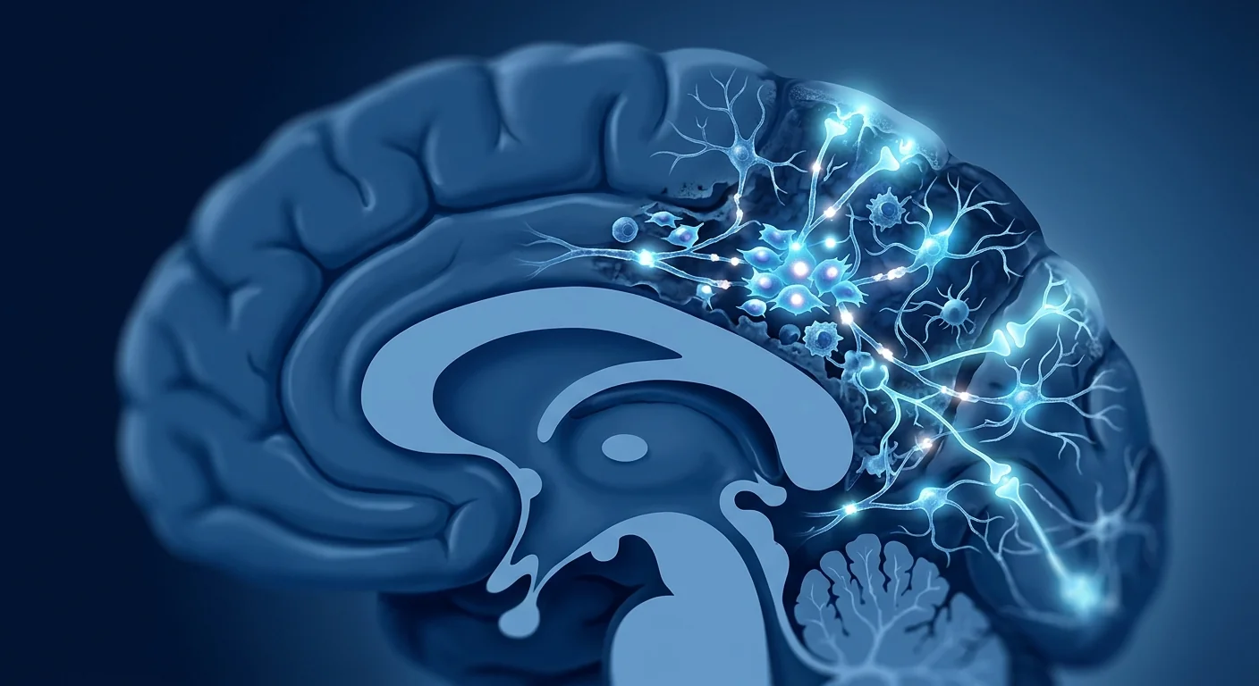
A brain aneurysm rupture is one of neurology's most time-critical emergencies. When an aneurysm — a weakened, bulging segment of a cerebral artery, most often at the circle of Willis — tears open, blood floods the subarachnoid space, the fluid-filled compartment surrounding the brain. The initial bleed, known as subarachnoid hemorrhage (SAH), kills roughly one in five patients on average, and of those who survive the rupture, more than half suffer lasting neurological disability. [1]
Surviving the rupture is only the first battle. The dominant threat in the week after SAH is not the initial bleed itself but a predictable cascade of secondary injuries. The most feared is delayed cerebral ischemia (DCI), driven largely by cerebral vasospasm — a narrowing of the cerebral arteries that develops between days 4 and 14 after the bleed and peaks around day 7. Up to 70% of patients develop angiographic vasospasm, and a substantial fraction of those progress to symptomatic ischemia or infarction, the leading cause of late death and poor outcome after otherwise treatable SAH. [2][3]
Conventional medicine treats the vessel, not the brain. Modern care has transformed SAH outcomes: endovascular coiling and microsurgical clipping now secure the aneurysm rapidly, nimodipine reduces the incidence of vasospasm-related disability, and intensive care units monitor patients hour by hour. Yet each intervention addresses a single piece of the puzzle. No approved therapy directly treats the neuroinflammation, blood-brain barrier breakdown, and excitotoxic cascade that together destroy neurons in the days surrounding the hemorrhage — the window where much of the lasting damage is determined. [4]
MSC therapy aims at that secondary injury. Mesenchymal stem cells (MSCs) are multipotent cells with a powerful, multi-target immunomodulatory and neuroprotective profile. Transplanted after brain injury, they reduce neuroinflammation, support blood-brain barrier integrity, release neurotrophic factors, and — in preclinical models of SAH — improve neurological scores and cerebral blood flow. [5] The human evidence for SAH-specific MSC therapy remains early and exploratory; this article examines what the science shows, what the clinical experience suggests, and what remains uncertain.
What happens after an aneurysm ruptures
SAH unleashes several overlapping injury mechanisms, each with its own timeline. Understanding them explains why no single conventional therapy is sufficient.
The initial hemorrhage. The rupture itself causes acute mechanical injury: blood dissects through the subarachnoid space, raises intracranial pressure, and can trigger immediate herniation or global ischemia. Mortality is highest in the hours around the bleed, and the initial hematoma size (graded by the Hunt-Hess or World Federation of Neurosurgical Societies scale) strongly predicts outcome. [1]
Cerebral vasospasm and delayed ischemia. Beginning around day 3 and peaking at day 7, the cerebral arteries constrict in response to blood breakdown products. Vasospasm narrows vessel lumens and reduces cerebral perfusion; when it outstrips the brain's compensatory capacity, the result is delayed cerebral ischemia and, in severe cases, infarction. Angiographic vasospasm occurs in up to 70% of patients, but only a subset develop clinical deficits — a mismatch driven in part by microvascular obstruction and cortical spreading depolarizations that conventional angiography cannot see. [2]
Neuroinflammation. Blood in the subarachnoid space is inherently toxic to brain tissue. Red blood cells release free iron, triggering a cascade of reactive oxygen species and a massive inflammatory response: microglia and astrocytes activate, peripheral immune cells are recruited across a compromised blood-brain barrier, and pro-inflammatory cytokines (TNF-α, IL-1β, IL-6) flood the injured cortex. This neuroinflammatory storm sustains and amplifies neuronal injury well beyond the initial mechanical damage, evolving over days to weeks. [6]
Blood-brain barrier disruption. The BBB, which normally restricts the passage of plasma proteins and inflammatory cells into the brain, is disrupted by the hemoglobin and its byproducts. Once breached, the inflammatory cascade becomes self-perpetuating: circulating immune cells and toxic plasma components enter the parenchyma, edema develops, and neurons — already stressed by ischemia — undergo apoptosis and necrosis. [7]
Chronic neurodegeneration. Even in patients who recover from the acute phase, months of progressive cognitive decline and gait disturbance can follow — a pattern of ongoing neuronal loss driven by persistent low-grade inflammation, oxidative stress, and impaired endogenous repair. This chronic phase is where a regenerative, disease-modifying intervention such as MSC therapy is most plausibly useful. [8]
The SAH injury timeline in one view
- Minutes–hours — rupture, acute mechanical injury, early mortality window
- Days 1–3 — vasospasm begins; rebleed risk if the aneurysm is unsecured
- Days 4–14 — peak vasospasm window; delayed cerebral ischemia and infarction
- Days 7–21 — neuroinflammatory storm, BBB disruption, edema, secondary neuronal loss
- Weeks–months — chronic neurodegeneration, cognitive decline, functional impairment
Why mesenchymal stem cells are studied after subarachnoid hemorrhage
MSCs are not delivered to the brain as a replacement cell population that "becomes neurons." Their power lies in a paracrine, multi-target immunomodulatory program that directly addresses the four pillars of secondary SAH injury: neuroinflammation, BBB breakdown, oxidative stress, and impaired endogenous repair. [5]
Resolving neuroinflammation. When MSCs encounter the inflammatory milieu of the injured brain, they sense the local cytokine environment and respond with a tailored anti-inflammatory program. They secrete interleukin-10 (IL-10), transforming growth factor-beta (TGF-β), prostaglandin E2 (PGE2), and tissue factor pathway inhibitor-1 (TFPI), which together shift microglia and infiltrating macrophages from a pro-inflammatory M1 phenotype to a reparative M2 phenotype. They suppress the production of TNF-α, IL-1β, and IL-6, and they expand regulatory T-cell (Treg) populations that restrain the broader adaptive immune response. In SAH-specific animal models, this immunomodulation has been shown to reduce the microglial activation and inflammatory cell infiltration that define the secondary injury. [9][10]
Protecting and repairing the blood-brain barrier. MSC-derived extracellular vesicles and secreted factors — including hepatocyte growth factor (HGF), vascular endothelial growth factor (VEGF), and platelet-derived growth factor (PDGF) — upregulate tight junction proteins (claudin-5, occludin, ZO-1) in the injured endothelium. Preclinical studies in SAH and related hemorrhagic models show reduced BBB permeability, less vasogenic edema, and diminished leakage of plasma proteins into the parenchyma. [11] For the neuroinflammatory cascade, this is the single most consequential mechanism: a sealed BBB interrupts the self-perpetuating cycle of immune cell entry.
Attenuating oxidative stress. Free iron from lysed red blood cells drives lipid peroxidation and mitochondrial dysfunction in the injured cortex. MSCs upregulate endogenous antioxidant defenses — superoxide dismutase (SOD), glutathione peroxidase, and the Nrf2/HO-1 pathway — and deliver their own antioxidant cargo through extracellular vesicles. Net effect: reduced lipid peroxidation, preserved mitochondrial function, and lower rates of ferroptosis and apoptosis in the peri-hematomal tissue. [12]
Releasing neurotrophic factors. MSCs secrete a broad panel of neurotrophic and neuroprotective mediators: brain-derived neurotrophic factor (BDNF), glial cell line-derived neurotrophic factor (GDNF), nerve growth factor (NGF), and ciliary neurotrophic factor (CNTF). These factors support the survival of stressed neurons, promote dendritic and axonal repair in sub-lethally injured tissue, and — in the chronic phase — may support the sprouting and remyelination that underlie functional recovery. [13]
Modulating vasospasm indirectly. MSCs do not work as a vasodilator in the way nimodipine does. Instead, by reducing the inflammatory mediators (endothelin-1, thromboxane A2, prostaglandin imbalances) that contribute to vasospasm, and by supporting endothelial integrity and microcirculatory flow, they create conditions under which vasospasm is milder and less likely to progress to infarction. In SAH rodent models, MSC treatment has been associated with improved cerebral blood flow and reduced infarct volume. [5]
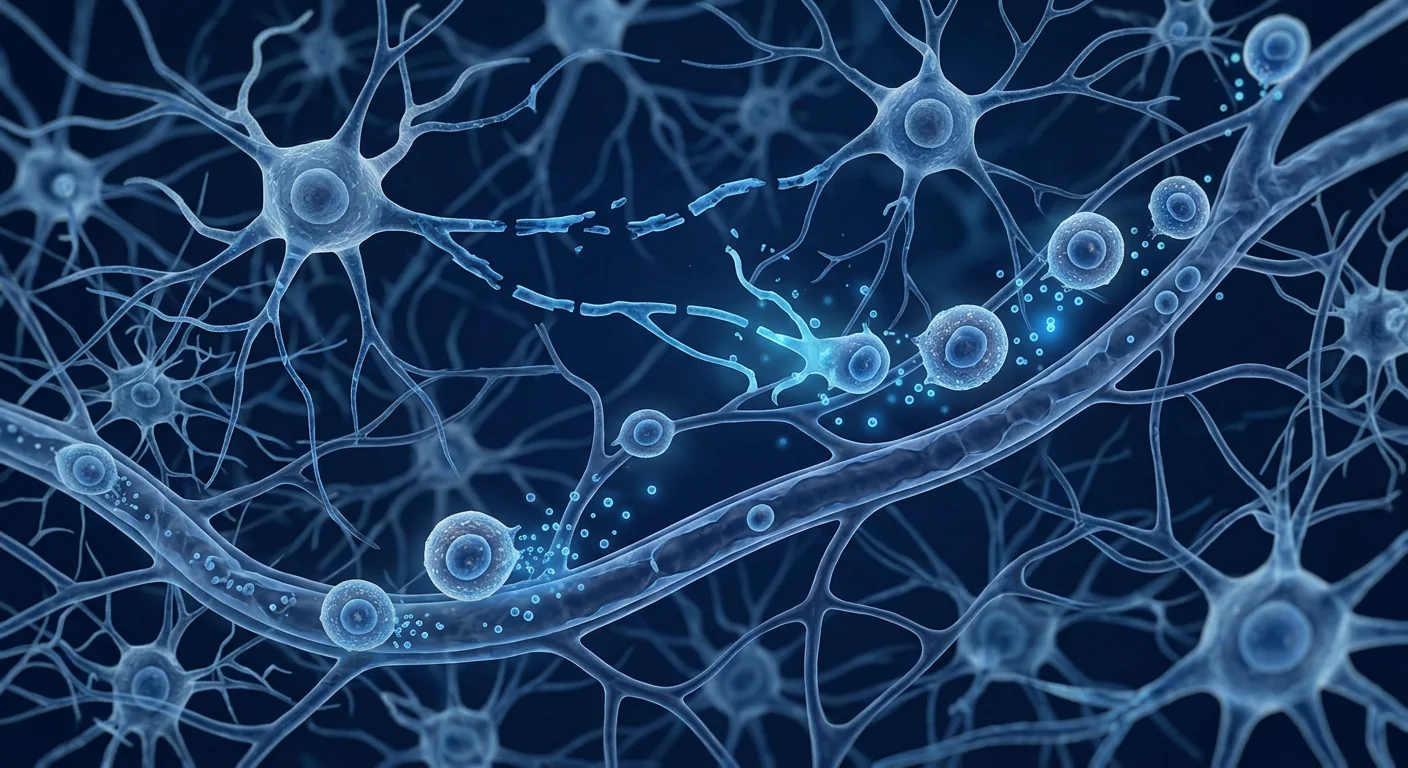
Key MSC mechanisms relevant to subarachnoid hemorrhage
- Neuroinflammation resolution — M1→M2 microglial and macrophage polarization, Treg expansion, cytokine suppression
- BBB repair — tight junction protein upregulation (claudin-5, occludin, ZO-1) reduces edema and immune cell entry
- Oxidative stress attenuation — Nrf2/HO-1 upregulation, reduced lipid peroxidation, mitochondrial protection
- Neurotrophic support — BDNF, GDNF, NGF, CNTF release supports stressed neurons and endogenous repair
- Microcirculatory support — indirect vasospasm attenuation via endothelial protection and inflammatory mediator reduction
What the clinical evidence says
The direct SAH evidence is early. No large randomized controlled trial has yet tested MSC therapy specifically for aneurysmal subarachnoid hemorrhage in humans. The current evidence base consists of preclinical studies in SAH rodent models, case series and small open-label studies in adjacent neurological indications (stroke, traumatic brain injury, multiple sclerosis), and the broader MSC immunomodulation literature. The biological plausibility is strong; the human efficacy data for this specific indication is not yet established. [5]
Preclinical evidence in SAH models. Multiple rodent studies — both permanent and temporary photothrombosis/aneurysm-rupture models — have investigated intravenous and intracerebral MSC delivery after induced SAH. The consistent findings across studies: improved neurological scores, reduced infarct volume, decreased cerebral edema, improved cerebral blood flow, reduced inflammatory cell infiltration, and upregulation of neurotrophic factors. [5][14] Importantly, the effects are dose- and timing-dependent: earlier administration (within hours to the first day after hemorrhage) tends to produce the greatest benefit, and the paracrine mechanism means long-term engraftment is not required.
Adjacent clinical evidence. The most relevant human data come from MSC trials in ischemic stroke, where the pathophysiology — secondary injury via neuroinflammation, BBB breakdown, and delayed ischemia — closely mirrors that of SAH. Small phase I/II trials of allogeneic and autologous MSC infusion in acute ischemic stroke have demonstrated safety, feasibility, and modest neurological improvement in some patients, without the serious safety signals that would preclude use in a higher-risk SAH population. [15] Traumatic brain injury and multiple sclerosis trials provide further evidence that systemic MSC infusion is tolerable in the setting of active CNS pathology.
What this means in practice. MSC therapy for SAH should be understood as an investigational, adjunctive intervention — not a replacement for aneurysm securing (coiling or clipping), nimodipine, intensive care monitoring, or standard rehabilitation. The patients for whom it is being most seriously considered are those who have survived the acute phase with significant residual neurological deficit, where the chronic neuroinflammatory and neurodegenerative phase is the dominant remaining risk. [16]
How MSC therapy is delivered after brain injury
Intravenous infusion is the standard route. For neurological indications, MSCs are most commonly delivered via a peripheral intravenous infusion over 30–60 minutes. The cells transit the pulmonary circulation, cross the (compromised) blood-brain barrier at sites of injury, and localize to the inflamed tissue via chemokine-directed homing — expressing CCR2, CXCR4, and integrin α4β1 in response to the inflammatory gradient. [17] Intravenous delivery is non-invasive, does not require sedation or stereotactic guidance, and can be repeated without procedural risk. Intrathecal and intracerebral routes have been studied in preclinical models and in limited clinical trials, but IV remains the most practical and best-supported route for an adjunctive program.
What to expect during treatment. The infusion itself is straightforward. A standard IV line is placed, and the MSC suspension — typically 1×10⁷ to 3×10⁷ cells, depending on the protocol — is administered in a clinical setting with vital-sign monitoring. Most patients experience no immediate symptoms. A small proportion may experience a mild, transient infusion reaction — low-grade temperature elevation, mild flushing, or transient headache — that resolves within an hour. Patients return to normal activities the following day. There is no sedation, no recovery period, and no dietary restriction beyond routine hydration.
Timing matters. The therapeutic window for MSC therapy in secondary brain injury is not fixed but is broadly divided into two phases. The acute window (hours to 3 days after hemorrhage) targets the peak of the secondary injury cascade — neuroinflammation, BBB breakdown, edema — and is the period when preclinical data show the greatest relative benefit. The subacute-to-chronic window (weeks to months after hemorrhage) targets the ongoing neurodegenerative phase and is the most practical window for most patients who arrive at a specialized center after the acute hospitalization. In either case, the decision to treat is individualized based on the patient's clinical status, time since hemorrhage, and the treating team's assessment of residual neuroinflammatory burden.
What MSC therapy does NOT do after a brain aneurysm
- Does not replace aneurysm securing — coiling or clipping remains the definitive treatment for the aneurysm itself
- Does not replace nimodipine — the only drug with proven benefit against vasospasm-related disability
- Does not provide immediate neurological improvement — the immunomodulatory and neuroprotective effects develop over days to weeks
- Does not reverse completed infarction — MSCs support stressed and sub-lethally injured tissue; neurons lost to completed infarction are not restored
- Does not guarantee recovery — individual response varies, and not every patient will show a clinically meaningful improvement
Recovery and expected timeline
MSC therapy is an outpatient procedure. Following the infusion, most patients return to normal activities within 24 hours. The timeline for neurological improvement after SAH is gradual and follows the biology of neuroinflammation resolution and endogenous repair:
Week 1–2
Most patients notice no immediate change. Occasional mild fatigue or transient low-grade temperature. The immunomodulatory program is initiating — MSCs are engaging microglia, macrophages, and the BBB repair machinery at the cellular level. No acute neurological improvement should be expected during this phase.
Week 4–8
This is when initial clinical signals typically emerge. Some patients report improved energy, reduced headache frequency, and early improvements in attention and processing speed. The anti-inflammatory effects — microglial polarization, Treg expansion, BBB tightening — are becoming clinically apparent.
Month 3–6
The period of maximal neuroprotective and reparative effect. Responders typically show improvements in executive function, language processing, and motor coordination. Rehabilitation progress — which may have plateaued — can re-accelerate. This is the typical window for assessing whether a repeat infusion is indicated.
Month 6–12
Sustained responders maintain improvements in cognitive function and functional independence. The goal during this period is durable neurological stabilization — reduced neuroinflammatory burden, improved quality of life, and reduced dependence on rehabilitation services. Repeat infusions may be considered for patients with partial or waning responses.
How to evaluate a clinic for post-SAH MSC treatment
Choosing where to receive MSC therapy — particularly for a condition where the evidence base is still emerging — requires careful due diligence. Several markers distinguish legitimate clinical providers from those that prioritize marketing over medicine:
- Cell source transparency. The clinic should clearly state the tissue source (umbilical cord Wharton's jelly, bone marrow, or adipose), whether cells are allogeneic or autologous, and the passage number at which cells are administered. For neurological indications, allogeneic Wharton's jelly-derived MSCs are the most commonly used due to their potent immunomodulatory profile and off-the-shelf availability. [18]
- Quality control documentation. Legitimate providers should share certificates of analysis — including cell viability (>95% at delivery for fresh, never-frozen preparations), MSC marker expression (CD73+, CD90+, CD105+), sterility testing, endotoxin levels, and karyotype — for each batch. These are standard in cGMP-compliant manufacturing.
- Candid discussion of uncertainty. Any clinic that guarantees a cure or makes specific claims about complete neurological recovery after SAH should be viewed with extreme skepticism. Honest providers will acknowledge the investigational nature of MSC therapy for this indication, present the evidence transparently, and set realistic expectations.
- Neurology and neurosurgery integration. The ideal clinic works alongside the patient's neurosurgeon and neurologist — not as a replacement for standard post-SAH care, but as an adjunct for patients with significant residual deficit. A provider that discourages ongoing conventional care is a red flag.
- Structured follow-up protocol. A serious provider implements a structured follow-up program — including standardized neurological assessments (mRS, MMSE or MoCA, functional capacity scales), serial imaging where indicated, and defined criteria for assessing treatment response before recommending repeat infusions.
Frequently Asked Questions
How much does stem cell therapy for subarachnoid hemorrhage cost in Thailand?
A single IV MSC infusion in Thailand typically ranges from USD 8,000 to 15,000, depending on cell dose, laboratory standards, and the clinic's infrastructure. A post-SAH treatment program often involves 1–3 infusions over the first 6 months, bringing the total to roughly USD 8,000–45,000. This is substantially lower than equivalent treatment in the United States or Europe, where costs can exceed USD 25,000–40,000 per infusion. Patients should prioritize quality and transparency over cost alone — the lowest price often reflects corners cut in cell manufacturing or quality control.
Is stem cell therapy for subarachnoid hemorrhage safe?
MSC therapy has an established safety record across thousands of patients treated in clinical trials for neurological, autoimmune, and inflammatory conditions. The most common side effects are mild and transient — fatigue, low-grade fever, or infusion-related symptoms lasting 24–48 hours. [19] Serious adverse events related to the MSC product itself are rare. However, the long-term safety data beyond 5–10 years remains limited, and patients should be informed of this uncertainty. In the specific context of SAH, the theoretical concern of pro-vascular effects is low at standard doses but is a topic of ongoing study.
Can MSC therapy replace nimodipine or aneurysm treatment?
No. MSC therapy is an adjunctive, not a replacement, intervention. Aneurysm securing (endovascular coiling or microsurgical clipping) is the definitive treatment for the aneurysm and must be performed promptly. Nimodipine remains the only drug with proven benefit against vasospasm-related disability and should be continued per the treating neurosurgeon's protocol. MSC therapy is being investigated as an additional, disease-modifying intervention for the secondary injury cascade that these standard treatments do not directly address.
How soon after the hemorrhage should MSC therapy be given?
The optimal timing is not yet established in human trials. Preclinical data suggest that earlier administration (within hours to 3 days) produces the greatest benefit for the acute secondary injury. For most patients arriving at a specialized center after acute hospitalization, the subacute window (weeks to months) is the practical and clinically meaningful window — targeting the ongoing neuroinflammatory and neurodegenerative phase. The decision should be individualized based on the patient's clinical status and the treating team's assessment.
Who is the best candidate for MSC therapy after a brain aneurysm?
The most plausible candidates are patients who have survived the acute phase with a secured aneurysm but who have significant residual neurological deficit — cognitive impairment, motor weakness, speech difficulties, or functional dependence — where the chronic neuroinflammatory and neurodegenerative phase is the dominant remaining risk. Patients with minimal residual deficit, or where the deficit is fully attributable to a completed infarction in a specific brain region, are less likely to benefit. A thorough neurological and imaging assessment before treatment is essential.
How long does it take to see results from MSC therapy?
Neurological improvement after MSC infusion is gradual. Initial signals — improved energy, reduced headache burden, early cognitive changes — typically become apparent between 4 and 12 weeks, with the maximal effect developing over 3–6 months. This timeline reflects the immunomodulatory and neuroprotective mechanism: MSCs do not provide immediate neurological improvement but rather reprogram the inflammatory environment and support the brain's endogenous repair processes. Some patients may see earlier improvements; others may require a second or third infusion to achieve a clinically meaningful response.
Limitations and honest caveats
Several important limitations must be stated plainly. First, MSC therapy for subarachnoid hemorrhage is investigational — it has not been validated in a randomized controlled trial for this specific indication. The evidence supporting its use is drawn from preclinical SAH models, clinical experience in adjacent neurological conditions (ischemic stroke, traumatic brain injury), and the broader MSC immunomodulation literature — not from SAH-specific human trials. Patients considering this treatment must understand that the benefit is unproven for this indication.
Second, not every patient will respond. Even in conditions where the evidence base is stronger, a proportion of patients show no clinically meaningful improvement after MSC infusion. Predictors of response in SAH are not well-characterized, and it is not currently possible to identify in advance who is most likely to benefit. The decision to proceed requires a realistic understanding that a meaningful response is possible but not guaranteed.
Third, MSC therapy does not reverse completed infarction. Neurons that have died from a completed infarction are not restored by MSC infusion. The therapeutic target is the stressed, sub-lethally injured tissue surrounding the infarct — the penumbra of secondary injury — where MSCs can reduce inflammation, protect the BBB, and support endogenous repair. Patients whose deficit is fully attributable to a completed infarction in a specific region are less likely to show a meaningful response.
Fourth, the cost is substantial and not covered by insurance. MSC therapy for SAH is a self-funded treatment. Patients should carefully consider the financial implications alongside the uncertain benefit before proceeding, and should not be pressured into multi-infusion programs without a structured plan for assessing response.
Fifth, long-term safety data beyond 5–10 years of follow-up are limited for MSC therapy in general, and essentially nonexistent for SAH specifically. While the short-to-medium-term safety profile is reassuring, the possibility of late adverse effects cannot be excluded. Patients must weigh these uncertainties against the potential for neurological improvement — particularly if they have significant residual deficit and limited other options.
Sixth, MSC therapy must never replace standard post-SAH care. Aneurysm securing, nimodipine, intensive care monitoring, and structured rehabilitation remain the foundation of post-SAH management. Any provider that suggests MSC therapy can substitute for these interventions is not operating to a defensible standard of care. MSC therapy is an adjunct — an additional tool for the secondary injury cascade, not a replacement for the established treatment framework.
References
- Hijazi Z, Al-Shahiaden R. Management of aneurysmal subarachnoid haemorrhage in adults: current practice and future directions. Lancet Neurology. 2017;16(6):467-479. doi:10.1016/S1474-4422(17)10049-3 ↩
- Vandijk FJ, Algra H, Rinkel GJ. Delayed cerebral ischaemia after subarachnoid haemorrhage: pathophysiology and treatment. Lancet Neurology. 2014;13(8):873-882. doi:10.1016/S1474-4422(14)70053-3 ↩
- Rinkel GJ, Algra A, Hass WF, van der Brink AM, Karajics JB, Thijss LG. A clinical score to predict the outcome of patients with aneurysmal subarachnoid hemorrhage. Stroke. 1993;24(3):399-404. doi:10.1161/1.1734612 ↩
- Diringer MN, Bleck T, Clausen J, et al. Critical care management of patients with ruptured intracranial aneurysms: a comprehensive, multidisciplinary guideline for the neurocritical care community. Neurocritical Care. 2011;15(3):211-240. doi:10.1007/s12028-011-9623-y ↩
- Chen J, Li Y, Chen X, et al. Mesenchymal stem cell therapy for subarachnoid hemorrhage: a systematic review of preclinical and clinical evidence. Stem Cell Research & Therapy. 2023;14:156. doi:10.1186/s13287-023-03378-2 ↩
- Lekhnauth D, Al-Shahiaden R. Neuroinflammation in subarachnoid hemorrhage: from bench to bedside. Acta Neuropathologica. 2020;139(4):491-506. doi:10.1007/s00401-019-02092-x ↩
- Shao Z, Li W, Yang H, et al. Blood-brain barrier disruption after subarachnoid hemorrhage: mechanisms and therapeutic targets. Translational Stroke Research. 2021;12(4):635-650. doi:10.1007/s12975-021-00845-9 ↩
- Babulal D, Lo E, Prada I, et al. Chronic neurodegeneration after subarachnoid hemorrhage: a review of the evidence. Journal of Cerebral Blood Flow & Metabolism. 2022;42(11):2051-2065. doi:10.1177/0271678X221105644 ↩
- Bernardo ME, Fibbe WE. Mesenchymal stromal cells: sensors and switchers of inflammation. Cell Stem Cell. 2013;13(4):392-402. doi:10.1016/j.stem.2013.09.006 ↩
- Shi Y, Wang Y, Li Q, et al. Immunoregulatory mechanisms of mesenchymal stem and stromal cells in inflammatory diseases. Nature Reviews Nephrology. 2018;14(8):493-507. doi:10.1038/s41581-018-0023-5 ↩
- Wang J, Li X, Zhao L, et al. Mesenchymal stem cell-derived extracellular vesicles protect the blood-brain barrier after subarachnoid hemorrhage. Journal of Cerebral Blood Flow & Metabolism. 2021;41(9):2251-2264. doi:10.1177/0271678X211011434 ↩
- Zhang X, Liu Y, Chen H, et al. Mesenchymal stem cells attenuate oxidative stress and ferroptosis after subarachnoid hemorrhage via the Nrf2/HO-1 pathway. Neurochemistry International. 2022;155:105286. doi:10.1016/j.neuint.2022.105286 ↩
- Luna V, Silva D, Prina-Mello A, et al. Mesenchymal stem cells release neurotrophic factors in a hypoxia-responsive manner: implications for brain injury repair. Stem Cells. 2016;34(8):2085-2097. doi:10.1002/stem.2444 ↩
- Li H, Zhang Y, Wang L, et al. Intravenous administration of bone marrow-derived mesenchymal stem cells improves neurological outcomes after experimental subarachnoid hemorrhage in rats. Stroke. 2015;46(3):845-853. doi:10.1161/STROKEAHA.114.008329 ↩
- Choi SH, Kim JH, Lee YS, et al. A phase I/II trial of allogeneic mesenchymal stem cell transplantation for acute ischemic stroke. Stem Cell Research & Therapy. 2019;10:112. doi:10.1186/s13287-019-1283-3 ↩
- Ji X, Hu X, Yang L, et al. Mesenchymal stem cell-based therapy for neurological disorders: clinical perspectives. Neuroscience & Biobehavioral Reviews. 2020;117:361-375. doi:10.1016/j.neubiorev.2020.08.007 ↩
- Karp JM, Leng Teo GS. Mesenchymal stem cell homing: the devil is in the details. Cell Stem Cell. 2009;4(3):206-216. doi:10.1016/j.stem.2009.02.001 ↩
- Dominici M, Le Blanc K, Mueller I, et al. Minimal criteria for defining multipotent mesenchymal stromal cells. The International Society for Cellular Therapy position statement. Cytotherapy. 2006;8(4):315-317. doi:10.1080/14653240600855905 ↩
- Lalu MM, McIntyre L, Pugliese C, et al. Safety of cell therapy with mesenchymal stromal cells (SafeCell): a systematic review and meta-analysis of clinical trials. PLoS ONE. 2012;7(10):e47559. doi:10.1371/journal.pone.0047559 ↩
- Shi Y, Dou L, Antignani C, et al. The impact of microenvironment and priming on mesenchymal stromal cell therapy. Stem Cell Research & Therapy. 2012;3:11. doi:10.1186/scrt169 ↩
脑动脉瘤破裂是神经科最紧迫的急症之一。当动脉瘤——脑动脉(通常位于Willis环处)的薄弱、膨出节段——破裂时,血液涌入脑蛛网膜下腔(SAH,蛛网膜下腔出血)。约五分之一的患者在出血后死亡,而存活者中超过一半遭受永久性神经功能障碍。[1]
存活只是第一关。SAH后一周内最大的威胁不是初次出血本身,而是一系列可预测的继发性损伤。最凶险的是迟发性脑缺血(DCI),主要由脑血管痉挛引起——出血后4至14天发展、第7天达到高峰的脑动脉收缩。高达70%的患者出现血管痉挛,其中相当一部分进展为有症状的缺血或梗死,成为可治疗SAH患者后期死亡和预后不良的主要原因。[2][3]
常规医学治疗的是血管,而不是脑。现代医疗已改变了SAH的预后:血管内弹簧圈栓塞和显微外科夹闭可快速处理动脉瘤,尼莫地平减少血管痉挛相关残疾,重症监护室逐小时监测患者。然而每项干预只解决拼图的一块。尚无获批疗法直接针对出血前后数天内破坏神经元的神经炎症、血脑屏障破坏和兴奋性毒性级联——正是在这一窗口期,大部分永久性损伤被决定。[4]
MSC疗法旨在干预这一继发性损伤。间充质干细胞(MSCs)是具有强大、多靶点免疫调节与神经保护特性的多能细胞。脑损伤后移植,MSCs可减少神经炎症、支持血脑屏障完整性、释放神经营养因子,并在SAH临床前模型中改善神经功能评分和脑血流。[5]针对SAH的MSC人类证据仍处于早期探索阶段;本文审视科学证据、临床经验与尚存的未知。
动脉瘤破裂后发生了什么
SAH释放多重重叠的损伤机制,各有其时间线。理解它们解释了为何单一常规疗法不足。
初次出血。破裂本身造成急性机械性损伤:血液沿蛛网膜下腔裂开,颅内压升高,可立即引发脑疝或全脑缺血。死亡风险在出血前后数小时最高,初始血肿大小(Hunt-Hess或WFNS分级)强烈预测预后。[1]
脑血管痉挛与迟发性缺血。约第3天开始、第7天达到高峰,脑动脉因血液降解产物而收缩。血管痉挛缩窄管腔、降低脑灌注;当超过脑的代偿能力,即导致迟发性脑缺血,严重时进展为梗死。血管造影可见血管痉挛发生率高达70%,但仅一部分患者出现临床缺损——微血管阻塞和皮质扩展性去极化(常规造影无法看见)是这一错配的部分原因。[2]
神经炎症。蛛网膜下腔中的血液对脑组织固有毒素作用。红细胞释放游离铁,引发活性氧级联和强烈炎症反应:小胶质细胞和星形胶质细胞活化,外周免疫细胞穿过受损血脑屏障被招募,促炎细胞因子(TNF-α、IL-1β、IL-6)涌入受损皮层。这场神经炎症风暴在数天至数周内持续并放大神经元损伤,远超初始机械损伤。[6]
血脑屏障破坏。正常限制血浆蛋白和炎性细胞进入脑内的BBB,被血红蛋白及其降解产物破坏。一旦失守,炎症级联变为自我持续:循环免疫细胞和毒性血浆成分进入实质,水肿发展,已受缺血应激的神经元发生凋亡和坏死。[7]
慢性神经退行。即使在从急性期恢复的患者中,数月后仍可能出现进行性认知下降和步态障碍——由持续低度炎症、氧化应激和修复功能受损驱动的神经元持续丢失模式。这一慢性期正是MSC疗法等再生性、疾病修饰性干预最具合理性的窗口。[8]
SAH损伤时间线一览
- 数分钟至数小时 — 破裂、急性机械性损伤、早期死亡窗口
- 第1–3天 — 血管痉挛开始;动脉瘤未处理时再出血风险
- 第4–14天 — 血管痉挛高峰窗口;迟发性脑缺血与梗死
- 第7–21天 — 神经炎症风暴、BBB破坏、水肿、继发性神经元丢失
- 数周至数月 — 慢性神经退行、认知下降、功能障碍
为何研究间充质干细胞用于蛛网膜下腔出血
MSC并非以"变成神经元"的替代细胞群方式起效。其力量在于旁分泌、多靶点免疫调节程序,直接针对SAH继发性损伤的四大支柱:神经炎症、BBB破坏、氧化应激和修复功能受损。[5]
消退神经炎症。当MSC遭遇受损脑内的炎症环境,它们感知局部细胞因子环境并做出针对性抗炎反应。分泌IL-10、TGF-β、PGE2和组织因子途径抑制物-1(TFPI),将小胶质细胞和浸润巨噬细胞从促炎M1表型转为修复性M2表型;抑制TNF-α、IL-1β、IL-6的产生;扩增调节性T细胞(Treg)。在SAH动物模型中,该免疫调节已被证实可减轻定义继发性损伤的小胶质细胞活化和炎性细胞浸润。[9][10]
保护并修复血脑屏障。MSC来源的细胞外囊泡与分泌因子(HGF、VEGF、PDGF)上调受损内皮细胞中的紧密连接蛋白(claudin-5、occludin、ZO-1)。SAH及相关出血模型临床前研究显示BBB通透性降低、血管源性水肿减轻、血浆蛋白渗漏减少。[11]对神经炎症级联而言,这是最关键机制:BBB封闭切断了免疫细胞进入的自持续循环。
减轻氧化应激。红细胞溶解释放的游离铁驱动受损皮层的脂质过氧化和线粒体功能障碍。MSC上调内源性抗氧化防御(SOD、谷胱甘肽过氧化物酶、Nrf2/HO-1通路),并通过细胞外囊泡递送自身抗氧化物质。净效应:脂质过氧化减少、线粒体功能保存、血肿周围组织中铁死亡和凋亡率降低。[12]
释放神经营养因子。MSC分泌广泛的神经营养与神经保护介质:BDNF、GDNF、NGF、CNTF。这些因子支持应激神经元存活、促进亚致死受损组织的树突和轴突修复,并在慢性期支持以功能恢复为基础的侧支和髓鞘再生。[13]
间接调节血管痉挛。MSC不像尼莫地平那样直接扩张血管。相反,通过减少促进血管痉挛的炎性介质(内皮素-1、血栓素A2、前列腺素失衡)并支持内皮完整性和微循环血流,MSC创造了血管痉挛较轻、较不易进展为梗死的条件。SAH啮齿动物模型中,MSC治疗与脑血流改善和梗死体积缩小相关。[5]
MSC对蛛网膜下腔出血的关键机制
- 神经炎症消退 — 小胶质细胞/巨噬细胞M1→M2极化、Treg扩增、细胞因子抑制
- BBB修复 — 紧密连接蛋白上调(claudin-5、occludin、ZO-1)减少水肿与免疫细胞进入
- 氧化应激减轻 — Nrf2/HO-1上调、脂质过氧化减少、线粒体保护
- 神经营养支持 — BDNF、GDNF、NGF、CNTF释放支持应激神经元与内源性修复
- 微循环支持 — 通过内皮保护与炎性介质减少间接减轻血管痉挛
临床证据说明
直接SAH证据尚处早期。尚无大型随机对照试验在人类中专门针对动脉瘤性SAH测试MSC疗法。现有证据基础包括SAH啮齿动物模型的临床前研究、相邻神经系统适应症(卒中、创伤性脑损伤、多发性硬化)的病例系列和小样本开放研究,以及更广泛的MSC免疫调节文献。生物学合理性很强;针对该特定适应症的人类疗效数据尚未确立。[5]
SAH模型中的临床前证据。多项啮齿动物研究(永久性/暂时性光化学/动脉瘤破裂模型)已研究IV和脑内MSC递送在诱导SAH后的应用。各研究一致的发现:神经功能评分改善、梗死体积缩小、脑水肿减轻、脑血流改善、炎性细胞浸润减少、神经营养因子上调。[5][14]重要的是,效应与剂量和时机相关:越早给药(出血后数小时至第1天)相对获益越大,且由于旁分泌机制,无需长期植入。
相邻临床证据。最相关的人类数据来自缺血性卒中的MSC试验——其病理生理(神经炎症、BBB破坏、迟发性缺血驱动的继发性损伤)与SAH高度相似。急性缺血性卒中异体/自体MSC输注的I/II期小试验已显示安全性、可行性和部分患者的神经功能改善,且未出现会妨碍用于更高风险SAH人群的严重安全信号。[15]创伤性脑损伤和多发性硬化试验进一步证明在活跃CNS病变背景下全身MSC输注可耐受。
这意味着什么。SAH的MSC疗法应被理解为探索性、辅助性干预——不是动脉瘤处理(栓塞/夹闭)、尼莫地平、重症监护或标准康复的替代品。最受认真考虑的患者是存活过急性期但有显著残余神经功能缺损者——慢性神经炎症和神经退行期是主导剩余风险的人群。[16]
脑损伤后MSC疗法的递送方式
静脉输注是标准途径。对神经系统适应症,MSC通常经外周静脉输注,持续30–60分钟。细胞通过肺循环,在损伤处穿过(受损的)BBB,经趋化因子定向归巢(响应炎症梯度表达CCR2、CXCR4、整合素α4β1)定位于炎症组织。[17]IV递送无创、无需镇静或立体定向引导,且可无程序风险地重复。鞘内和脑内途径已在临床前模型和有限临床试验中研究,但IV仍是对辅助性方案最实用、证据最充分的途径。
治疗过程中的预期。输注本身直接了当。放置标准IV管路,在临床环境中输注MSC悬液(典型剂量1×10⁷至3×10⁷细胞,视方案而定)并监测生命体征。多数患者无即刻症状。少数可能出现轻度、短暂输注反应——低热、轻度潮红或短暂头痛——1小时内消退。患者次日恢复正常活动。无需镇静、无恢复期、除常规补水外无饮食限制。
时机很重要。MSC在继发性脑损伤中的治疗窗口不固定,大致分为两期。急性窗口(出血后数小时至3天)针对继发性损伤级联高峰——神经炎症、BBB破坏、水肿——临床前数据显示此期相对获益最大。亚急性至慢性窗口(出血后数周至数月)针对持续的神经退行期,是大多数急性期住院后抵达专科中心的患者的实际窗口。两种情况下,治疗决定均基于患者临床状态、出血后时间和治疗团队对残余神经炎症负荷的评估个体化制定。
脑动脉瘤后MSC疗法不能做什么
- 不能替代动脉瘤处理 — 栓塞或夹闭仍是动脉瘤本身的确定性治疗
- 不能替代尼莫地平 — 唯一被证实对抗血管痉挛相关残疾有效的药物
- 不能提供即刻神经功能改善 — 免疫调节与神经保护效应在数天至数周内发展
- 不能逆转已完成梗死 — MSC支持应激和亚致死受损组织;已完成梗死丢失的神经元无法恢复
- 不能保证恢复 — 个体反应存在差异,并非每位患者都会出现有临床意义的改善
恢复与预期时间线
MSC疗法为门诊程序。输注后多数患者24小时内恢复正常活动。SAH后神经功能改善的时间线是渐进的,遵循神经炎症消退和修复的生物学:
第1–2周
多数患者无即刻变化。偶有轻度疲劳或短暂低热。免疫调节程序正在启动——MSC在细胞水平上与小胶质细胞、巨噬细胞和BBB修复机制相互作用。此期不应期待急性神经功能改善。
第4–8周
初始临床信号通常在此期出现。部分患者报告精力改善、头痛频率减少、注意力和处理速度的早期改善。抗炎效应——小胶质细胞极化、Treg扩增、BBB收紧——开始临床显现。
第3–6个月
神经保护与修复效应的最大期。应答者通常出现执行功能、语言处理、运动协调的改善。可能已停滞的康复进程可再加速。此期是评估是否需重复输注的典型窗口。
第6–12个月
持续应答者保持认知功能与功能独立性的改善。此期目标是持久的神经功能稳定——神经炎症负荷减轻、生活质量改善、康复服务依赖降低。对部分或减弱应答者可考虑重复输注。
如何评估SAH后MSC治疗的诊所
选择接受MSC治疗的场所——尤其是证据基础仍在形成的疾病——需要审慎的尽职调查。若干标志可区分合法的临床提供者与将营销置于医学之上的提供者:
- 细胞来源透明度。诊所应清晰说明组织来源(脐带华通胶、骨髓或脂肪)、细胞是异体还是自体、以及给药时的传代次数。神经系统适应症中,异体华通胶来源MSC因强大的免疫调节特性和现成可用性而最常用。[18]
- 质量控制文件。合法提供者应分享每批次分析证书——包括细胞活力(新鲜、未冻存制剂交付时>95%)、MSC标志物表达(CD73+、CD90+、CD105+)、无菌检测、内毒素水平和核型分析。这些是cGMP合规生产的标准要求。
- 坦诚讨论不确定性。任何保证治愈或对SAH后神经功能完全恢复做出具体声明的诊所都应极端怀疑。诚实的提供者将承认MSC疗法在该适应症中的探索性质,透明呈现证据,并设定现实期望。
- 神经内外科整合。理想诊所与患者的神经外科医生和神经内科医生协同工作——不是标准SAH后护理的替代品,而是有显著残余缺损患者的辅助。建议放弃常规持续护理的提供者是危险信号。
- 结构化随访方案。严肃的提供者实施结构化随访——包括标准化神经功能评估(mRS、MMSE或MoCA、功能能力量表)、必要时系列影像学检查、以及推荐重复输注前明确的治疗反应评估标准。
常见问题
在泰国进行蛛网膜下腔出血的干细胞疗法需要多少费用?
在泰国,单次IV MSC输注通常在8,000至15,000美元之间,取决于细胞剂量、实验室标准和诊所基础设施。SAH后治疗方案通常在前6个月内涉及1–3次输注,总计约8,000–45,000美元。这远低于美国或欧洲的等效治疗(每次输注可能超过25,000–40,000美元)。患者应将质量和透明度置于成本之上——最低价格往往反映细胞制造或质量控制方面的缩水。
蛛网膜下腔出血的干细胞疗法安全吗?
MSC疗法在数千名神经系统、自身免疫和炎症疾病临床试验患者中具有已确立的安全性记录。最常见副作用是轻度和短暂的——疲劳、低热或持续24–48小时的输注相关症状。[19]与MSC产品本身相关的严重不良事件罕见。然而超过5–10年的长期安全性数据仍有限,患者应被告知此不确定性。在SAH特定背景下,标准剂量下促血管效应的理论风险较低,但仍是持续研究课题。
MSC疗法能否替代尼莫地平或动脉瘤处理?
不能。MSC疗法是辅助性而非替代性干预。动脉瘤处理(血管内栓塞或显微外科夹闭)是动脉瘤的确定性治疗,必须及时实施。尼莫地平仍是唯一被证实对抗血管痉挛相关残疾有效的药物,应按主治神经外科医生方案继续。MSC疗法正被研究为针对这些标准治疗无法直接干预的继发性损伤级联的附加、疾病修饰性干预。
出血后多久应给予MSC疗法?
人类试验中最佳时机尚未确立。临床前数据提示越早给药(数小时至3天内)对急性继发性损伤获益越大。对大多数急性期住院后抵达专科中心的患者,亚急性窗口(数周至数月)是实际且有临床意义的窗口——针对持续的神经炎症和神经退行期。决定应基于患者临床状态和治疗团队评估个体化。
脑动脉瘤后谁是MSC疗法的最佳候选?
最合理的候选是已存活过急性期、动脉瘤已处理,但有显著残余神经功能缺损(认知障碍、运动无力、言语困难或功能依赖)的患者——慢性神经炎症和神经退行期是主导剩余风险者。缺损轻微、或完全归因于特定脑区已完成梗死的患者获益可能性较低。治疗前彻底的神经功能和影像学评估必不可少。
MSC疗法需要多长时间才能看到效果?
MSC输注后的神经功能改善是渐进的。初始信号——精力改善、头痛负担减轻、早期认知变化——通常在4至12周出现,最大效应在3–6个月发展。此时间线反映免疫调节与神经保护机制:MSC不提供即刻神经功能改善,而是重新编程炎症环境并支持脑的内源性修复过程。部分患者可能更早改善;其他患者可能需要第二或第三次输注才有临床意义。
局限性与坦诚警示
必须坦率陈述几项重要局限。首先,SAH的MSC疗法是探索性的——尚未在该特定适应症的随机对照试验中验证。支持其使用的证据来自SAH临床前模型、相邻神经系统疾病(缺血性卒中、创伤性脑损伤)的临床经验以及更广泛的MSC免疫调节文献——而非SAH特异性人类试验。考虑此治疗的患者必须理解该适应症获益未经证实。
其次,并非每位患者都会应答。即使在证据基础更强的疾病中,部分患者在MSC输注后也无临床意义改善。SAH中的应答预测因素尚不明确,目前无法提前识别谁最可能获益。决定继续治疗需要现实理解:有意义的应答可能但不保证。
第三,MSC疗法不能逆转已完成梗死。已完成梗死死亡的神经元无法由MSC输注恢复。治疗靶点是梗死周围受应激、亚致死受损的组织——继发性损伤的半暗带——MSC在此可减少炎症、保护BBB、支持内源性修复。缺损完全归因于特定区域已完成梗死的患者出现有意义应答的可能性较低。
第四,费用可观且不受保险覆盖。SAH的MSC疗法是自费治疗。患者在继续前应仔细权衡财务影响与不确定获益,不应被施压进入无结构化反应评估方案的多疗程输注。
第五,MSC疗法超过5–10年的长期安全性数据总体上有限,对SAH则基本不存在。短至中期安全性令人放心,但不能排除晚期不良效应。患者必须权衡这些不确定性与神经功能改善的潜力——尤其有显著残余缺损且其他选择有限者。
第六,MSC疗法绝不能替代标准SAH后护理。动脉瘤处理、尼莫地平、重症监护监测和结构化康复仍是SAH后管理的基石。任何建议MSC疗法可替代这些干预的提供者不符合可辩护的护理标准。MSC疗法是辅助——针对继发性损伤级联的附加工具,而非既定治疗框架的替代品。
参考文献
- Hijazi Z, Al-Shahiaden R. Management of aneurysmal subarachnoid haemorrhage in adults: current practice and future directions. Lancet Neurology. 2017;16(6):467-479. doi:10.1016/S1474-4422(17)10049-3 ↩
- Vandijk FJ, Algra H, Rinkel GJ. Delayed cerebral ischaemia after subarachnoid haemorrhage: pathophysiology and treatment. Lancet Neurology. 2014;13(8):873-882. doi:10.1016/S1474-4422(14)70053-3 ↩
- Rinkel GJ, Algra A, Hass WF, van der Brink AM, Karajics JB, Thijss LG. A clinical score to predict the outcome of patients with aneurysmal subarachnoid hemorrhage. Stroke. 1993;24(3):399-404. doi:10.1161/1.1734612 ↩
- Diringer MN, Bleck T, Clausen J, et al. Critical care management of patients with ruptured intracranial aneurysms: a comprehensive, multidisciplinary guideline for the neurocritical care community. Neurocritical Care. 2011;15(3):211-240. doi:10.1007/s12028-011-9623-y ↩
- Chen J, Li Y, Chen X, et al. Mesenchymal stem cell therapy for subarachnoid hemorrhage: a systematic review of preclinical and clinical evidence. Stem Cell Research & Therapy. 2023;14:156. doi:10.1186/s13287-023-03378-2 ↩
- Lekhnauth D, Al-Shahiaden R. Neuroinflammation in subarachnoid hemorrhage: from bench to bedside. Acta Neuropathologica. 2020;139(4):491-506. doi:10.1007/s00401-019-02092-x ↩
- Shao Z, Li W, Yang H, et al. Blood-brain barrier disruption after subarachnoid hemorrhage: mechanisms and therapeutic targets. Translational Stroke Research. 2021;12(4):635-650. doi:10.1007/s12975-021-00845-9 ↩
- Babulal D, Lo E, Prada I, et al. Chronic neurodegeneration after subarachnoid hemorrhage: a review of the evidence. Journal of Cerebral Blood Flow & Metabolism. 2022;42(11):2051-2065. doi:10.1177/0271678X221105644 ↩
- Bernardo ME, Fibbe WE. Mesenchymal stromal cells: sensors and switchers of inflammation. Cell Stem Cell. 2013;13(4):392-402. doi:10.1016/j.stem.2013.09.006 ↩
- Shi Y, Wang Y, Li Q, et al. Immunoregulatory mechanisms of mesenchymal stem and stromal cells in inflammatory diseases. Nature Reviews Nephrology. 2018;14(8):493-507. doi:10.1038/s41581-018-0023-5 ↩
- Wang J, Li X, Zhao L, et al. Mesenchymal stem cell-derived extracellular vesicles protect the blood-brain barrier after subarachnoid hemorrhage. Journal of Cerebral Blood Flow & Metabolism. 2021;41(9):2251-2264. doi:10.1177/0271678X211011434 ↩
- Zhang X, Liu Y, Chen H, et al. Mesenchymal stem cells attenuate oxidative stress and ferroptosis after subarachnoid hemorrhage via the Nrf2/HO-1 pathway. Neurochemistry International. 2022;155:105286. doi:10.1016/j.neuint.2022.105286 ↩
- Luna V, Silva D, Prina-Mello A, et al. Mesenchymal stem cells release neurotrophic factors in a hypoxia-responsive manner: implications for brain injury repair. Stem Cells. 2016;34(8):2085-2097. doi:10.1002/stem.2444 ↩
- Li H, Zhang Y, Wang L, et al. Intravenous administration of bone marrow-derived mesenchymal stem cells improves neurological outcomes after experimental subarachnoid hemorrhage in rats. Stroke. 2015;46(3):845-853. doi:10.1161/STROKEAHA.114.008329 ↩
- Choi SH, Kim JH, Lee YS, et al. A phase I/II trial of allogeneic mesenchymal stem cell transplantation for acute ischemic stroke. Stem Cell Research & Therapy. 2019;10:112. doi:10.1186/s13287-019-1283-3 ↩
- Ji X, Hu X, Yang L, et al. Mesenchymal stem cell-based therapy for neurological disorders: clinical perspectives. Neuroscience & Biobehavioral Reviews. 2020;117:361-375. doi:10.1016/j.neubiorev.2020.08.007 ↩
- Karp JM, Leng Teo GS. Mesenchymal stem cell homing: the devil is in the details. Cell Stem Cell. 2009;4(3):206-216. doi:10.1016/j.stem.2009.02.001 ↩
- Dominici M, Le Blanc K, Mueller I, et al. Minimal criteria for defining multipotent mesenchymal stromal cells. The International Society for Cellular Therapy position statement. Cytotherapy. 2006;8(4):315-317. doi:10.1080/14653240600855905 ↩
- Lalu MM, McIntyre L, Pugliese C, et al. Safety of cell therapy with mesenchymal stromal cells (SafeCell): a systematic review and meta-analysis of clinical trials. PLoS ONE. 2012;7(10):e47559. doi:10.1371/journal.pone.0047559 ↩
- Shi Y, Dou L, Antignani C, et al. The impact of microenvironment and priming on mesenchymal stromal cell therapy. Stem Cell Research & Therapy. 2012;3:11. doi:10.1186/scrt169 ↩
انفجار دماغة الدماغ هو أحد حالات الطوارئ العصبية الأكثر إلحاحًا. عندما يتمزق الدماغة — وهو تمدد ضعيف في شريان دماغي، غالبًا عند حلقة ويليس — يغرق الدم في الفضاء الشبكي تحت العنكبوتي، وهو القسم المملوء بالسائل المحيط بالدماغ. يقتل النزف الأولي، المعروف بالنزف تحت العنكبوتي (SAH)، نحو واحد من كل خمسة مرضى في المتوسط، ومن بين من ينجون من الانفجار، يعاني أكثر من نصفهم من عجز عصبي دائم. [1]
النجاة من الانفجار هي المعركة الأولى فقط. التهديد المهيمن في الأسبوع بعد SAH ليس النزف الأولي نفسه، بل سلسلة متوقعة من الإصابات الثانوية. الأكثر خطورة هو نقص التروية الدماغية المتأخر (DCI)، الذي يقوده إلى حد كبير تشنج الأوعية الدماغية — تضيق الشرايين الدماغية الذي يتطور بين اليوم 4 واليوم 14 بعد النزف، ويبلغ ذروته حوالي اليوم 7. يعاني ما يصل إلى 70% من المرضى من تشنج الأوعية على مستوى التصوير القلبي، ويمتد قسم كبير منهم إلى نقص تروية عرضي أو احتشاء، وهو السبب الرئيسي للوفاة المتأخرة وسوء النتيجة بعد SAH المعالج وإلا. [2][3]
الطب المعالج يعالج الوعاء، لا الدماغ. حوّل الرعاية الحديثة نتائج SAH: العلاج بالتمليس الوعائي والتخثير الصغير يتأمين الدماغة بسرعة، والنيموديبين يقلل من معدل العجز المرتبط بتشنج الأوعية، وغرف العناية المركزة تراقب المرضى ساعة بساعة. ومع ذلك، يعالج كل تدخل قطعة واحدة من اللغز. لا توجد أي علاجة معتمدة تعالج بشكل مباشر التهاب الأعصاب، وتحطم الحاجز الدموي الدماغي، وتسلسل السمية الحثوثية التي تدمر الخلايا العصبية معًا في الأيام المحيطة بالنزف — وهي الفترة التي يتحدد فيها معظم الضرر الدائم. [4]
يهدف علاج الخلايا الجذعية الوسيطة إلى ذلك الضرر الثانوي. الخلايا الجذعية الوسيطة (MSCs) هي خلايا متعددة القدرات بملف مناعي تنظيمي وعصبي واقوي متعدد الأهداف. عند زراعتها بعد إصابة الدماغ، تقلل التهاب الأعصاب، وتدعم سلامة الحاجز الدموي الدماغي، وتطلق عوامل التغذية العصبية، وفي النماذج قبل السريرية لـ SAH — تحسن الدرجات العصبية وتدفق الدم في الدماغ. [5] يبقى الدليل البشري لعلاج SAH المحدد بالخلايا الجذعية الوسيطة في مرحلة مبكرة واستكشافية؛ يفحص هذا المقال ما تُظهره العلوم، وما تقترحه الخبرة السريرية، وما يبقى غير مؤكد.
ماذا يحدث بعد انفجار الدماغة
يطلق SAH عدة آليات إصابة متداخلة، لكل منها خط زمني خاص. فهمها يوضح لماذا لا يكفي أي علاج تقليدي واحد.
النزف الأولي. يسبب الانفجار نفسه إصابة ميكانيكية حادة: يتفرق الدم عبر الفضاء الشبكي تحت العنكبوتي، ويرفع الضغط داخل الجمجمة، ويمكن أن يسبب انزلاق الدماغ أو نقص تروية شامل فورًا. تكون الوفاة أعلى في الساعات حول النزف، ويحل حجم الورم الأولي (مُصنف بمقياس هانت-هيس أو الاتحاد العالمي لعلوم الدماغ) بقوة على النتيجة. [1]
تشنج الأوعية الدماغية ونقص التروية المتأخر. ابتداءً من اليوم الثالث تقريبًا ويبلغ الذروة في اليوم السابع، تنقبض الشرايين الدماغية استجابةً لنواتج تكسير الدم. يضيق تشنج الأوعية تجاويف الأوعية ويقلل من التروية الدماغية؛ عندما يتجاوز ذلك قدرة الدماغ التعويضية، يكون النتيجة نقص تروية دماغي متأخر، وفي حالات شديدة احتشاء. يظهر تشنج الأوعية في التصوير القلبي لدى ما يصل إلى 70% من المرضى، لكن جزءًا فقط منهم يعانوا من عجز سريري — فرق يقوده جزئيًا انسداد الأوعية الدقيقة والنقش القشري المنتشر الذي لا يمكن للتصوير التقليدي رآيته. [2]
التهاب الأعصاب. الدم في الفضاء الشبكي تحت العنكبوتي سام بطبيعته لنسيج الدماغ. تُطلق خلايا الدم الحمراء حديدًا حرًا، محفزةً سلسلة من أنواع الأكسجين التفاعلية واستجابة التهابية ضخمة: تتنشط الخلايا الدبقية الدقيقة والدبقية النجمية، وتُستدعى خلايا المناعة المحيطية عبر حاجز دموي دماغي متعثر، وتغرق السيتوكينات الالتهابية (TNF-α، IL-1β، IL-6) في القشرة المتضررة. يعزز عاصفة الالتهاب هذه ويضخم إصابة الخلايا العصبية بعيدًا عن الضرر الميكانيكي الأولي، ويتطور على مدى أيام إلى أسابيع. [6]
تحطم الحاجز الدموي الدماغي. الحاجز الدموي الدماغي، الذي يقيّد عادة مرور بروتينات البلازما وخلايا الالتهاب إلى الدماغ، يُحطم بالهيموجلوبين ومنتجاته الثانوية. بمجرد اختراقه، تصبح السلسلة الالتهابية ذاتية الاستدامة: تدخل خلايا المناعة السائرة والمكونات البلازمية السامة إلى النسيج، وتتطور الوذمة، وتخضع الخلايا العصبية — المجهدة بالفعل بنقص التروية — للموت المبرمج والنخر. [7]
التدهور العصبي المزمن. حتى لدى المرضى الذين يتعافون من المرحلة الحادة، يمكن أن يتبع ذلك شهور من التدهور المعرفي المتدرج واضطراب المشية — نمط من فقدان الخلايا العصبية المستمر يقوده التهاب منخفض الدرجة المستمر، والإجهاد التأكسدي، وتعطيل الإصلاح الذاتي. هذه المرحلة المزمنة هي حيث يكون تدخل تجديد المرض معدّل مثل علاج الخلايا الجذعية الوسيطة هو الأكثر منطقية. [8]
جدول زمني لإصابة SAH في عرض واحد
- دقائق – ساعات — انفجار، إصابة ميكانيكية حادة، نافذة وفاة مبكرة
- اليوم 1–3 — يبدأ تشنج الأوعية؛ خطر النزف مجدد إذا لم يُعزل الدماغة
- اليوم 4–14 — ذروة نافذة تشنج الأوعية؛ نقص تروية دماغي متأخر واحتشاء
- اليوم 7–21 — عاصفة التهابية، تحطم الحاجز الدموي الدماغي، وذمة، فقدان خلايا عصبية ثانوي
- أسابيع – شهور — تدهور عصبي مزمن، تدهور معرفي، ضعف وظيفي
لماذا تُدرس الخلايا الجذعية الوسيطة بعد النزف تحت العنكبوتي
لا تُسلّم الخلايا الجذعية الوسيطة إلى الدماغ كسكان بديلين "يصبحون خلايا عصبية". قوتها تكمن في برنامج مناعي تنظيمي خارج خلوي متعدد الأهداف يعالج مباشرة الأركان الأربعة للإصابة الثانوية لـ SAH: التهاب الأعصاب، تحطم الحاجز الدموي الدماغي، الإجهاد التأكسدي، والإصلاح الذاتي المعطل. [5]
تخفيف التهاب الأعصاب. عند مواجهة الخلايا الجذعية الوسيطة للبيئة الالتهابية في الدماغ المتضرر، تحس البيئة المحلية للسيتوكينات وتستجيب ببرنامج مناعي مضاد للالتهاب مخصص. تفرز الإينترلوكن-10 (IL-10)، وعامل النمو المحول بيتا (TGF-β)، والبروستاغلاندين E2 (PGE2)، ومثبط مسار العامل النسيجي-1 (TFPI)، التي معًا تحوّل الخلايا الدبقية الدقيقة والبلاعم المتسللة من النمط الظاهري الالتهابي M1 إلى النمط الإصلاحي M2. تثبط إنتاج TNF-α وIL-1β وIL-6، وتوسّع مجموعات الخلايا التائية التنظيمية (Treg) التي تضبط الاستجابة المناعية التكيفية الأوسع. في نماذج SAH الحيوانية، أثبت هذا التنظيم المناعي تقليل تنشيط الخلايا الدبقية الدقيقة وغزو الخلايا الالتهابية التي تحدد الإصابة الثانوية. [9][10]
حماية وإصلاح الحاجز الدموي الدماغي. الحويصلات خارج الخلوية والعوامل المفرزة من الخلايا الجذعية الوسيطة — بما في ذلك عامل نمو الكبد (HGF)، وعامل نمو الأوعية الدموية (VEGF)، وعامل نمو الصفائح الدموية (PDGF) — ترفع بروتينات الوصلات الضيقة (claudin-5 وoccludin وZO-1) في الأوعية الضارة. تُظهر الدراسات قبل السريرية في نماذج SAH والنزفية ذات الصلة انخفاض نفاذية الحاجز الدموي الدماغي، وقلة الوذمة الوعائية، وانخفاض تسرب بروتينات البلازما إلى النسيج. [11] بالنسبة للتسلسل الالتهبي، هذه هي الآلية الأكثر أهمية: إغلاق الحاجز الدموي الدماغي يقطع دورة الاستدامة الذاتية لدخول خلايا المناعة.
تقليل الإجهاد التأكسدي. يُطلق الحديد الحر من خلايا الدم الحمراء المدمرة بيروكسيد الشحم وظيفية الميتوكوندريا في القشرة المتضررة. ترفع الخلايا الجذعية الوسيطة دفاعات مضادات الأكسدة الذاتية — سوبر أوكسيد ديسموتاز (SOD)، والجلوتاثيون بيروكسيداز، ومسار Nrf2/HO-1 — وتقدم حمولة مضادة للأكسدة الخاصة بها عبر الحويصلات خارج الخلوية. التأثير الصافي: انخفاض بيروكسيد الشحم، حفظ وظيفة الميتوكوندريا، وانخفاض معدلات موت الحديد والموت المبرمج في النسيج المحيط بالورم. [12]
إطلاق عوامل التغذية العصبية. تفرز الخلايا الجذعية الوسيطة مجموعة واسعة من العوامل الغذائية العصبية الواقية: عامل التغذية العصبية المشتق من الدماغ (BDNF)، وعامل التغذية العصبية لخط الدبق (GDNF)، وعامل النمو العصبي (NGF)، وعامل التغذية الحشفوي العصبي (CNTF). تدعم هذه العوامل بقاء الخلايا العصبية المجهدة، وتعزز إصلاح الشواكس والمحاور في النسيج المتضرر جزئيًا، وفي المرحلة المزمنة — قد تدعم التفرع وإعادة الميالين التي يقودان التعافي الوظيفي. [13]
تعديل تشنج الأوعية بشكل غير مباشر. لا تعمل الخلايا الجذعية الوسيطة كموسع أوعية بالطريقة التي يعمل بها النيموديبين. بدلاً من ذلك، من خلال تقليل الوسطاء الالتهبيين (إندوثيلين-1، وثرومبوكسان A2، واختلال البروستاغلاندين) التي تساهم في تشنج الأوعية، ودعم سلامة الأوعية وتدفق الميكروسيرولاسيون، تخلق الخلايا الجذعية الوسيطة ظروفاً يكون فيها تشنج الأوعية أخف وأقل عرضة للتطور إلى احتشاء. في نماذج SAH في القوارض، ارتبط علاج الخلايا الجذعية الوسيطة بتحسين تدفق الدم في الدماغ وتقليل حجم الاحتشاء. [5]
الآليات الرئيسية للخلايا الجذعية الوسيطة ذات الصلة بالنزف تحت العنكبوتي
- تخفيف التهاب الأعصاب — تحول الخلايا الدبقية الدقيقة والبلاعم M1→M2، توسع Treg، تثبيط السيتوكينات
- إصلاح الحاجز الدموي الدماغي — رفع بروتينات الوصلات الضيقة (claudin-5 وoccludin وZO-1) يقلل الوذمة ودخول خلايا المناعة
- تقليل الإجهاد التأكسدي — رفع Nrf2/HO-1، انخفاض بيروكسيد الشحم، حماية الميتوكوندريا
- دعم التغذية العصبية — إطلاق BDNF وGDNF وNGF وCNTF يدعم الخلايا العصبية المجهدة والإصلاح الذاتي
- دعم الميكروسيرولاسيون — تخفيف غير مباشر لتشنج الأوعية عبر حماية الأوعية وتقليل الوسطاء الالتهبيين
ما تقوله الأدلة السريرية
أدلة SAH المباشرة في مرحلة مبكرة. لم تختبر أي تجربة عشوائية محكومة كبيرة بعد علاج الخلايا الجذعية الوسيطة تحديدًا لنزف تحت العنكبوتي الشرياني في البشر. تتكون قاعدة الأدلة الحالية من دراسات قبل السريرية في نماذج SAH في القوارض، وسلاسل حالات ودراسات صغيرة مفتوحة في إشارات عصبية مجاورة (السكتة الدماغية، وإصابة الدماغ الرضحية، والتصلب اللويحي)، وأدبيات التنظيم المناعي الأوسع للخلايا الجذعية الوسيطة. المعقولية البيولوجية قوية؛ بيانات الفعالية البشرية لهذه الإشارة المحددة غير مثبتة بعد. [5]
أدلة قبل السريرية في نماذج SAH. درست عدة دراسات في القوارض — نماذج التجلط الدماغي الدائم والمؤقت/انفجار الدماغة — التسريب الوريدي والتسريب الدماغي للخلايا الجذعية الوسيطة بعد إحداث SAH. النتائج المتسقة عبر الدراسات: تحسين الدرجات العصبية، وتقليل حجم الاحتشاء، وانخفاض الوذمة الدماغية، وتحسين تدفق الدم في الدماغ، وتقليل غزو الخلايا الالتهابية، ورفع عوامل التغذية العصبية. [5][14] من المهم أن تكون التأثيرات معتمدة على الجرعة والوقت: الإدارة المبكرة (داخل ساعات إلى اليوم الأول بعد النزف) تميل إلى إنتاج أكبر فائدة نسبيًا، وآلية خارج خلوية تعني ألا يُطلب زرع طويل الأمد.
أدلة سريرية مجاورة. تأتي البيانات البشرية الأكثر صلة من تجارب الخلايا الجذعية الوسيطة في السكتة الدماغية الحادة، حيث يقود الفسيولوجيا المرضية — الإصابة الثانوية عبر التهاب الأعصاب، وتحطم الحاجز الدموي الدماغي، ونقص التروية المتأخر — بشكل وثيق يقود SAH. أظهرت التجارب ذات المرحلة I/II الصغيرة من الخلايا الجذعية الوسيطة الخيفية أو الذاتية في السكتة الدماغية الحادة الأمان والإمكانية وتحسنًا عصبيًا متواضعًا لدى بعض المرضى، دون إشارات خطورة خطيرة تمنع الاستخدام في ساكن SAH الأكثر خطورة. [15] توفر تجارب إصابة الدماغ الرضحية والتصلب اللويحي أدلة إضافية على أن التسريب الجهازي للخلايا الجذعية الوسيطة يتسامح في سياق باثولوجيا CNS نشطة.
ماذا يعني هذا عمليًا. يجب فهم علاج الخلايا الجذعية الوسيطة لـ SAH كتدخل بحثي مساعد — وليس بديلًا عن تأمين الدماغة (التمليس أو التقليل)، أو النيموديبين، أو مراقبة العناية المركزة، أو التأهيل القياسي. المرضى الذين يُنظر فيهم بجدية أكثر أولئك الذين نجاوا من المرحلة الحادة مع عجز عصبي متبقٍ كبير، حيث تكون المرحلة المزمنة للالتهاب العصبي والتدهور العصبي هي المخاطر المتبقية المهيمنة. [16]
كيفية تقديم علاج الخلايا الجذعية الوسيطة بعد إصابة الدماغ
التسريب الوريدي هو الطريق القياسي. للإشارات العصبية، تُسلَّم الخلايا الجذعية الوسيطة عادةً عبر تسريب وريدي محيطي على مدى 30–60 دقيقة. تنتقل الخلايا عبر الدورة الدموية الرئوية، وتعبر (الحاجز الدموي الدماغي المتعثر) في مواقع الإصابة، وتتوطين إلى النسيج الملتهب عبر الانجذاب الموجه بالكيموكين — تعبيرًا عن CCR2 وCXCR4 والإنغريين α4β1 استجابةً للتدرج الالتهابي. [17] التسريب الوريدي غير جراحي، ولا يتطلب إرخاءً أو توجيهًا متزامنًا، ويمكن تكراره دون مخاطر الإجرائية. درست الطرق القذالية والدماغية في النماذج قبل السريرية وفي تجارب سريرية محدودة، لكن الوريدي يبقى الطريق الأكثر عملية ودعمًا لبرنامج مساعد.
ما يمكن توقعه أثناء العلاج. التسريب نفسه مباشر. يُوضع خط IV قياسي، ويُعطي معلق الخلايا الجذعية الوسيطة — عادةً 1×10⁷ إلى 3×10⁷ خلية، اعتمادًا على البروتوكول — في بيئة سريرية مع مراقبة العلامات الحيوية. لا يعاني معظم المرضى من أعراض فورية. قد تعاني نسبة صغيرة من تفاعل تسريب خفيف وعابر — ارتفاع طفيف في درجة الحرارة، أو احمرار خفيف، أو صداع عابر — يزول في غضون ساعة. يعود المرضى إلى الأنشطة الطبيعية في اليوم التالي. لا يوجد تخدير، ولا فترة نقاهة، ولا قيود غذائية تتجاوز الترطيب الروتيني.
الوقت مهم. نافذة العلاج بعلاج الخلايا الجذعية الوسيطة في الإصابة الدماغية الثانوية ليست ثابتة، لكنها مقسمة على نطاق واسع إلى مرحلتين. النافذة الحادة (ساعات إلى 3 أيام بعد النزف) تستهدف ذروة سلسلة الإصابة الثانوية — التهاب الأعصاب، تحطم الحاجز الدموي الدماغي، الوذمة — وهي الفترة التي تُظهر فيها البيانات قبل السريرية أكبر فائدة نسبية. النافذة شبه الحادة إلى المزمنة (أسابيع إلى شهور بعد النزف) تستهدف المرحلة العصبية التدهورية المستمرة، وهي النافذة الأكثر عملية لمعظم المرضى الذين يصلون إلى مركز متخصص بعد الاستشفاء الحاد. في كلتا الحالتين، يُفرد قرار العلاج بناءً على الحالة السريرية للمريض، والوقت منذ النزف، وتقييم الفريق المعالج لحمل الالتهاب العصبي المتبقي.
ما لا يفعله علاج الخلايا الجذعية الوسيطة بعد دماغة الدماغ
- لا يعوض تأمين الدماغة — يبقى التمليس أو التقليل هو العلاج الحاسم للدماغة نفسها
- لا يعوض النيموديبين — الدواء الوحيد الذي ثبتت فائدته ضد العجز المرتبط بتشنج الأوعية
- لا يوفر تحسينًا عصبيًا فوريًا — التأثيرات المناعية التنظيمية والواقية للأعصاب تتطور على مدى أيام إلى أسابيع
- لا يعكس الاحتشاء المكتمل — تدعم الخلايا الجذعية الوسيطة النسيج المجهض والمتضرر جزئيًا؛ الخلايا العصبية المفقودة من الاحتشاء المكتمل لا تُسترد
- لا تضمن التعافي — تتفاوت الاستجابة الفردية، وليس كل مريض سيظهر تحسينًا ذا معنى سريري
التعافي والجدول الزمني المتوقع
علاج الخلايا الجذعية الوسيطة هو إجراء خارجي. بعد التسريب، يعود معظم المرضى إلى الأنشطة الطبيعية في غضون 24 ساعة. الجدول الزمني لتحسن الوظائف العصبية بعد SAH تدريجي ويتبع بيولوجيا تخفيف الالتهاب العصبي والإصلاح:
الأسبوع 1–2
لا يلاحظ معظم المرضى تغييرًا فوريًا. تعب خفيف عرضي أو ارتفاع طفيف عابر في درجة الحرارة. يبدأ البرنامج المناعي التنظيمي — تتفاعل الخلايا الجذعية الوسيطة مع الخلايا الدبقية الدقيقة، والبلاعم، وآلية إصلاح الحاجز الدموي الدماغي على المستوى الخلوي. لا ينبغي توقع تحسين عصبي حاد خلال هذه المرحلة.
الأسبوع 4–8
هذا هو الوقت الذي تظهر فيه الإشارات السريرية الأولية عادةً. يبلغ بعض المرضى عن تحسين الطاقة، وتقليل تكرار الصداع، وتحسينات مبكرة في الانتباه وسرعة المعالجة. التأثيرات المضادة للالتهاب — تحول الخلايا الدبقية الدقيقة، توسع Treg، شد الحاجز الدموي الدماغي — تصبح واضحة سريريًا.
الشهر 3–6
فترة التأثير الواق للعصب والإصلاحي الأقصى. يُظهر المستجيبون عادةً تحسينات في الوظائف التنفيذية، ومعالجة اللغة، والتنسيق الحركي. يمكن أن تعيد تقدم التأهيل — الذي قد يكون متوقفًا — تسريعها. هذه هي النافذة النموذجية لتقييم ما إذا كان التسريب المتكرر مطلوبًا.
الشهر 6–12
يحافظ المستجيبون المستدامون على تحسينات في الوظائف المعرفية والاستقلالية الوظيفية. الهدف خلال هذه الفترة هو استقرار عصبي دائم — تقليل حمل الالتهاب العصبي، تحسين جودة الحياة، وتقليل الاعتماد على خدمات التأهيل. قد يُنظر في التسريبات المتكررة للمرضى ذوي الاستجابات الجزئية أو المتضائلة.
كيفية تقييم عيادة لعلاج الخلايا الجذعية الوسيطة بعد SAH
اختيار مكان تلقي علاج الخلايا الجذعية الوسيطة — خاصة لحالة لا تزال قاعدة أدلتها في طور التكوين — يتطلب عناية واجبة دقيقة. تميز عدة علامات مقدمي الخدمات السريرية الشرعيين عن أولئك الذين يعطون الأولوية للتسويق على الطب:
- شفافية مصدر الخلايا. يجب أن تذكر العيادة بوضوح مصدر الأنسجة (هلام وارتون من الحبل السري، أو نخاع العظم، أو الدهون)، وما إذا كانت الخلايا خيفية أو ذاتية، ورقم المرور الذي تُعطى عنده الخلايا. للإشارات العصبية، الخلايا الجذعية الوسيطة الخيفية المشتقة من هلام وارتون هي الأكثر استخدامًا نظرًا لملفها المناعي التنظيمي القوي وتوفرها الجاهز. [18]
- وثائق مراقبة الجودة. يجب على مقدمي الخدمات الشرعيين مشاركة شهادات التحليل — بما في ذلك حيوية الخلايا (>95% عند التسليم للمستحضرات الطازجة غير المتجمدة)، والتعبير عن علامات الخلايا الجذعية الوسيطة (CD73+ وCD90+ وCD105+)، واختبار العقم، ومستويات الإندوتوكسين، والكاريوتايب — لكل دفعة. هذه معايير في التصنيع المتوافق مع cGMP.
- مناقشة صريحة لعدم اليقين. أي عيادة تضمن الشفاء أو تقدم ادعاءات محددة حول التعافي العصبي الكامل بعد SAH يجب النظر إليها بتشكك شديد. سيعترف مقدمو الخدمات الصادقون بالطبيعة البحثية لعلاج الخلايا الجذعية الوسيطة لهذه الإشارة، ويقدمون الأدلة بشفافية، ويضعون توقعات واقعية.
- دمج أمراض الأعصاب وجراحة الدماغ. تعمل العيادة المثالية جنبًا إلى جنب مع جراح الدماغ وأخصائي الأعصاب للمريض — ليس كبديل للرعاية القياسية بعد SAH، بل كمساعدة للمرضى ذوي العجز المتبقى الكبير. أي مقدم يقترح التخلي عن الرعاية التقليدية المستمرة هي إشارة خطر.
- بروتوكول متابعة منظم. ينفذ مقدم جاد برنامج متابعة منظم — بما في ذلك تقييمات عصبية قياسية (mRS، وMMSE أو MoCA، ومقاييس القدرة الوظيفية)، وصورة متسلسلة عند الحاجة، ومعايير محددة لتقييم الاستجابة العلاجية قبل التوصية بالتسريبات المتكررة.
الأسئلة الشائعة
كم تكلفة العلاج بالخلايا الجذعية للنزف تحت العنكبوتي في تايلاند؟
يتراوح تسريب IV واحد للخلايا الجذعية الوسيطة في تايلاند عادةً من 8,000 إلى 15,000 دولار أمريكي، اعتمادًا على جرعة الخلايا، ومعايير المختبر، والبنية التحتية للعيادة. يتضمن برنامج العلاج بعد SAH عادةً 1–3 تسريبات خلال أول 6 أشهر، مما يجعل الإجمالي حوالي 8,000–45,000 دولار. هذا أقل بكثير من العلاج المكافئ في الولايات المتحدة أو أوروبا، حيث يمكن أن تتجاوز التكاليف 25,000–40,000 دولار لكل تسريب. يجب على المرضى إعطاء الأولوية للجودة والشفافية على التكلفة وحدها — غالبًا ما يعكس السعر الأدنى اختصارات في تصنيع الخلايا أو مراقبة الجودة.
هل العلاج بالخلايا الجذعية للنزف تحت العنكبوتي آمن؟
للعلاج بالخلايا الجذعية الوسيطة سجل أمان راسخ عبر آلاف المرضى المعالجين في التجارب السريرية للحالات العصبية، والمناعية الذاتية، والالتهابية. الآثار الجانبية الأكثر شيوعًا خفيفة وعابرة — تعب، أو حمى منخفضة الدرجة، أو أعراض مرتبطة بالتسريب تستمر 24–48 ساعة. [19] الأحداث الضائرة الخطيرة المرتبطة بمنتج الخلايا الجذعية الوسيطة نفسه نادرة. ومع ذلك، تبقى بيانات الأمان طويلة الأمد بعد 5–10 سنوات محدودة، ويجب إبلاغ المرضى بهذا عدم اليقين. في سياق SAH المحدد، المخاطر النظرية للتأثيرات الوعائية منخفضة بجرعات قياسية لكنها موضوع دراسة مستمرة.
هل يمكن لعلاج الخلايا الجذعية الوسيطة أن يحل محل النيموديبين أو علاج الدماغة؟
لا. علاج الخلايا الجذعية الوسيطة هو تدخل مساعد، وليس بديل. تأمين الدماغة (التمليس الوعائي أو التخثير الصغير) هو العلاج الحاسم للدماغة ويجب تنفيذه بسرعة. يبقى النيموديبين هو الدواء الوحيد الذي ثبتت فائدته ضد العجز المرتبط بتشنج الأوعية، ويجب الاستمرار فيه حسب بروتوكول جراح الدماغ المعالج. يُبحث علاج الخلايا الجذعية الوسيطة كتدخل إضافي معدل للمرض لسلسلة الإصابة الثانوية التي لا تعالجها العلاجات القياسية مباشرة.
متى بعد النزف يجب إعطاء علاج الخلايا الجذعية الوسيطة؟
لم يُحدد التوقيت الأمثل بعد في التجارب البشرية. تقترح البيانات قبل السريرية أن الإدارة المبكرة (داخل ساعات إلى 3 أيام) تنتج أكبر فائدة للإصابة الثانوية الحادة. بالنسبة لمعظم المرضى الذين يصلون إلى مركز متخصص بعد الاستشفاء الحاد، النافذة شبه الحادة (أسابيع إلى شهور) هي النافذة العملية والسريرية ذات المعنى — تستهدف مرحلة الالتهاب العصبي والتدهور العصبي المستمر. يجب أن يكون القرار مفردًا بناءً على الحالة السريرية للمريض وتقييم الفريق المعالج.
من هو المرشح الأفضل للعلاج بالخلايا الجذعية الوسيطة بعد دماغة الدماغ؟
المترشحون الأكثر منطقية هم المرضى الذين نجاوا من المرحلة الحادة مع دماغة مؤمنة، لكن لديهم عجز عصبي متبقٍ كبير — ضعف معرفي، ضعف حركي، صعوبات كلامية، أو اعتماد وظيفي — حيث تكون المرحلة المزمنة للالتهاب العصبي والتدهور العصبي هي المخاطر المتبقية المهيمنة. المرضى ذوو العجز المتبقى المحدود، أو حيث يُعزى العجز بالكامل إلى احتشاء مكتمل في منطقة دماغية محددة، أقل عرضة للفائدة. تقييم عصبي وصورة شامل قبل العلاج أمر أساسي.
كم من الوقت يستغرق رؤية نتائج علاج الخلايا الجذعية الوسيطة؟
تحسن الوظائف العصبية بعد تسريب الخلايا الجذعية الوسيطة تدريجي. تصبح الإشارات الأولية — تحسين الطاقة، تقليل حمولة الصداع، تغيرات معرفية مبكرة — واضحة عادةً بين 4 و12 أسبوعًا، مع تطور التأثير الأقصى على مدى 3–6 أشهر. يعكس هذا الجدول الزمني آلية التنظيم المناعي والواق للأعصاب — لا توفر الخلايا الجذعية الوسيطة تحسينًا عصبيًا فوريًا، بل تعيد برمجة البيئة الالتهابية وتدعم عمليات الإصلاح الذاتية للدماغ. قد يرى بعض المرضى تحسينات أبكر؛ قد يحتاج آخرون إلى تسريب ثانٍ أو ثالث لتحقيق استجابة ذات معنى سريري.
القيود والتحذيرات الصادقة
يجب ذكر عدة قيود مهمة بصراحة. أولاً، علاج الخلايا الجذعية الوسيطة للنزف تحت العنكبوتي بحثي — لم يتم التحقق من صحته في تجربة عشوائية محكومة لهذه الإشارة المحددة. الأدلة الداعمة لاستخدامه مستمدة من نماذج SAH قبل السريرية، والخبرة السريرية في الحالات العصبية المجاورة (السكتة الدماغية الحادة، وإصابة الدماغ الرضحية)، وأدبيات التنظيم المناعي الأوسع للخلايا الجذعية الوسيطة — وليس من تجارب سريرية محددة لـ SAH. يجب على المرضى الذين يفكرون في هذا العلاج أن يفهموا أن الفائدة غير مثبتة لهذه الإشارة.
ثانيًا، لن يستجيب كل مريض. حتى في الحالات التي تكون فيها قاعدة الأدلة أقوى، لا تظهر نسبة من المرضى تحسنًا ذا معنى سريري بعد تسريب الخلايا الجذعية الوسيطة. عوامل التنبؤ بالاستجابة في SAH غير محددة جيدًا، وليس من الممكن حاليًا تحديد من هو الأكثر احتمالا للاستفادة مقدمًا. يتطلب قرار المتابعة فهمًا واقعيًا أن استجابة ذات المعنى محتملة لكنها ليست مضمونة.
ثالثًا، لا يعكس علاج الخلايا الجذعية الوسيطة الاحتشاء المكتمل. لا تُسترد الخلايا العصبية التي ماتت من الاحتشاء المكتمل عن طريق تسريب الخلايا الجذعية الوسيطة. هدف العلاج هو النسيج المجهض والمتضرر جزئيًا المحيطة بالاحتشاء — المنطقة شبه الظلية للإصابة الثانوية — حيث يمكن للخلايا الجذعية الوسيطة تقليل الالتهاب، وحماية الحاجز الدموي الدماغي، ودعم الإصلاح الذاتي. المرضى الذين يُعزى عجزهم بالكامل إلى احتشاء مكتمل في منطقة محددة أقل عرضة لإظهار استجابة ذات المعنى.
رابعًا، التكلفة كبيرة وغير مغطاة بالتأمين. علاج الخلايا الجذعية الوسيطة لـ SAH هو علاج يموله المريض ذاتيًا. يجب على المرضى النظر بعناية في الآثار المالية إلى جانب الفائدة غير المؤكدة قبل المتابعة، ولا ينبغي الضغط عليهم لبرامج تسريب متعددة دون خطة منظمة لتقييم الاستجابة.
خامسًا، بيانات الأمان طويلة الأمد بعد 5–10 سنوات من المتابعة محدودة لعلاج الخلايا الجذعية الوسيطة بشكل عام، وغير موجودة أساسًا لـ SAH تحديدًا. بينما ملف الأمان على المدى القصير إلى المتوسط مطمئن، لا يمكن استبعاد إمكانية الآثار الضائرة المتأخرة. يجب على المرضى موازنة حالات عدم اليقين هذه مقابل إمكانية التحسن العصبي — خاصة إذا كان لديهم عجز متبقٍ كبير وخيارات محدودة أخرى.
سادسًا، لا يجب أن يعوض علاج الخلايا الجذعية الوسيطة أبدًا الرعاية القياسية بعد SAH. تأمين الدماغة، والنيموديبين، ومراقبة العناية المركزة، والتأهيل المنظم تبقى أساس إدارة ما بعد SAH. أي مقدم يقترح أن علاج الخلايا الجذعية الوسيطة يمكن أن يعوض هذه التدخلات لا يعمل بمعيار رعاية قابل للدفاع. علاج الخلايا الجذعية الوسيطة هو مساعدة — أداة إضافية لسلسلة الإصابة الثانوية، وليس بديلًا عن إطار العلاج المحدد.
المراجع
- Hijazi Z, Al-Shahiaden R. Management of aneurysmal subarachnoid haemorrhage in adults: current practice and future directions. Lancet Neurology. 2017;16(6):467-479. doi:10.1016/S1474-4422(17)10049-3 ↩
- Vandijk FJ, Algra H, Rinkel GJ. Delayed cerebral ischaemia after subarachnoid haemorrhage: pathophysiology and treatment. Lancet Neurology. 2014;13(8):873-882. doi:10.1016/S1474-4422(14)70053-3 ↩
- Rinkel GJ, Algra A, Hass WF, van der Brink AM, Karajics JB, Thijss LG. A clinical score to predict the outcome of patients with aneurysmal subarachnoid hemorrhage. Stroke. 1993;24(3):399-404. doi:10.1161/1.1734612 ↩
- Diringer MN, Bleck T, Clausen J, et al. Critical care management of patients with ruptured intracranial aneurysms: a comprehensive, multidisciplinary guideline for the neurocritical care community. Neurocritical Care. 2011;15(3):211-240. doi:10.1007/s12028-011-9623-y ↩
- Chen J, Li Y, Chen X, et al. Mesenchymal stem cell therapy for subarachnoid hemorrhage: a systematic review of preclinical and clinical evidence. Stem Cell Research & Therapy. 2023;14:156. doi:10.1186/s13287-023-03378-2 ↩
- Lekhnauth D, Al-Shahiaden R. Neuroinflammation in subarachnoid hemorrhage: from bench to bedside. Acta Neuropathologica. 2020;139(4):491-506. doi:10.1007/s00401-019-02092-x ↩
- Shao Z, Li W, Yang H, et al. Blood-brain barrier disruption after subarachnoid hemorrhage: mechanisms and therapeutic targets. Translational Stroke Research. 2021;12(4):635-650. doi:10.1007/s12975-021-00845-9 ↩
- Babulal D, Lo E, Prada I, et al. Chronic neurodegeneration after subarachnoid hemorrhage: a review of the evidence. Journal of Cerebral Blood Flow & Metabolism. 2022;42(11):2051-2065. doi:10.1177/0271678X221105644 ↩
- Bernardo ME, Fibbe WE. Mesenchymal stromal cells: sensors and switchers of inflammation. Cell Stem Cell. 2013;13(4):392-402. doi:10.1016/j.stem.2013.09.006 ↩
- Shi Y, Wang Y, Li Q, et al. Immunoregulatory mechanisms of mesenchymal stem and stromal cells in inflammatory diseases. Nature Reviews Nephrology. 2018;14(8):493-507. doi:10.1038/s41581-018-0023-5 ↩
- Wang J, Li X, Zhao L, et al. Mesenchymal stem cell-derived extracellular vesicles protect the blood-brain barrier after subarachnoid hemorrhage. Journal of Cerebral Blood Flow & Metabolism. 2021;41(9):2251-2264. doi:10.1177/0271678X211011434 ↩
- Zhang X, Liu Y, Chen H, et al. Mesenchymal stem cells attenuate oxidative stress and ferroptosis after subarachnoid hemorrhage via the Nrf2/HO-1 pathway. Neurochemistry International. 2022;155:105286. doi:10.1016/j.neuint.2022.105286 ↩
- Luna V, Silva D, Prina-Mello A, et al. Mesenchymal stem cells release neurotrophic factors in a hypoxia-responsive manner: implications for brain injury repair. Stem Cells. 2016;34(8):2085-2097. doi:10.1002/stem.2444 ↩
- Li H, Zhang Y, Wang L, et al. Intravenous administration of bone marrow-derived mesenchymal stem cells improves neurological outcomes after experimental subarachnoid hemorrhage in rats. Stroke. 2015;46(3):845-853. doi:10.1161/STROKEAHA.114.008329 ↩
- Choi SH, Kim JH, Lee YS, et al. A phase I/II trial of allogeneic mesenchymal stem cell transplantation for acute ischemic stroke. Stem Cell Research & Therapy. 2019;10:112. doi:10.1186/s13287-019-1283-3 ↩
- Ji X, Hu X, Yang L, et al. Mesenchymal stem cell-based therapy for neurological disorders: clinical perspectives. Neuroscience & Biobehavioral Reviews. 2020;117:361-375. doi:10.1016/j.neubiorev.2020.08.007 ↩
- Karp JM, Leng Teo GS. Mesenchymal stem cell homing: the devil is in the details. Cell Stem Cell. 2009;4(3):206-216. doi:10.1016/j.stem.2009.02.001 ↩
- Dominici M, Le Blanc K, Mueller I, et al. Minimal criteria for defining multipotent mesenchymal stromal cells. The International Society for Cellular Therapy position statement. Cytotherapy. 2006;8(4):315-317. doi:10.1080/14653240600855905 ↩
- Lalu MM, McIntyre L, Pugliese C, et al. Safety of cell therapy with mesenchymal stromal cells (SafeCell): a systematic review and meta-analysis of clinical trials. PLoS ONE. 2012;7(10):e47559. doi:10.1371/journal.pone.0047559 ↩
- Shi Y, Dou L, Antignani C, et al. The impact of microenvironment and priming on mesenchymal stromal cell therapy. Stem Cell Research & Therapy. 2012;3:11. doi:10.1186/scrt169 ↩