Autoimmune hemolytic anemia (AIHA) is an acquired autoimmune disorder in which the immune system produces autoantibodies — predominantly warm-reactive IgG or cold-reactive IgM — that target red blood cell (RBC) surface antigens, triggering complement-mediated or Fcγ receptor-mediated destruction of erythrocytes. With an annual incidence of 1–3 per 100,000 and a prevalence exceeding approximately 50,000 cases in the United States, AIHA causes fatigue, dyspnea, pallor, jaundice, and — in severe or rapidly progressive cases — high-output heart failure and multi-organ ischemic injury [1].
Where conventional treatments fall short. First-line therapy with corticosteroids achieves an initial response in 70–85% of patients with warm AIHA, but sustained remission after tapering occurs in only 20–30%. Second-line options — rituximab, splenectomy, and immunosuppressants (mycophenolate mofetil, cyclophosphamide, azathioprine) — each carry significant limitations: rituximab yields a 50–60% initial response but a 30–40% relapse rate at 3 years; splenectomy carries a 1–2% lifetime risk of overwhelming post-splenectomy sepsis and a 30% late relapse rate; and long-term immunosuppression increases infection and malignancy risk [2]. For cold agglutinin disease (CAD), conventional options are even more limited — corticosteroids and splenectomy are largely ineffective, and rituximab monotherapy yields only a 45–55% response rate. A significant proportion of patients cycle through multiple therapies without durable, treatment-free remission.
The deeper problem is multi-level immune dysregulation. AIHA is not simply antibody-mediated RBC destruction. It involves a complex interplay of Th1/Th17 polarization with diminished regulatory T-cell (Treg) function, loss of B-cell tolerance to RBC self-antigens, complement cascade activation with membrane attack complex (MAC) formation, and impaired compensatory erythropoiesis — the bone marrow's attempt to replace destroyed RBCs is often inadequate due to inflammation-driven suppression of erythroid precursors [3]. Targeting any single pathway — as conventional therapies do — leaves the broader immune dysregulation intact.
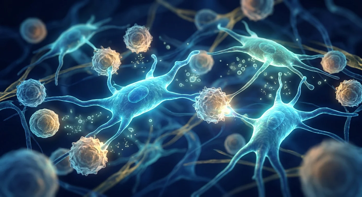
MSC therapy targets multiple pathogenic mechanisms simultaneously. Rather than blocking a single receptor or depleting one cell population, mesenchymal stem cells (MSCs) engage the immune system at multiple checkpoints: they suppress autoreactive T-cell proliferation, restore Treg/Th17 balance, polarize macrophages from a pro-inflammatory M1 phenotype toward a reparative, RBC-sparing M2 phenotype, inhibit B-cell differentiation and autoantibody production, and secrete trophic factors — including erythropoietin (EPO), stem cell factor (SCF), and insulin-like growth factor-1 (IGF-1) — that directly support erythroid progenitor survival and RBC maturation [4]. This multi-target approach makes MSCs a uniquely compelling candidate for a disease driven by multi-level immune dysregulation and bone marrow stress.
What Is Autoimmune Hemolytic Anemia?
Autoimmune hemolytic anemia is an acquired disorder characterized by premature destruction of red blood cells (hemolysis) driven by autoantibodies directed against RBC surface antigens, resulting in anemia with a hemoglobin typically below 10 g/dL, elevated reticulocyte count, elevated lactate dehydrogenase (LDH), low haptoglobin, and a positive direct antiglobulin test (DAT, or Coombs test). The disease is classified by the thermal reactivity of the autoantibody: warm AIHA (wAIHA, ~70% of cases, IgG-mediated, optimal binding at 37°C), cold agglutinin disease (CAD, ~20%, IgM-mediated, optimal binding at 0–4°C), and mixed-type AIHA (~5–10%). Approximately 50% of AIHA cases are primary (idiopathic), while the remaining 50% are secondary to underlying conditions — most commonly lymphoproliferative disorders (CLL, NHL), systemic autoimmune diseases (SLE), infections (EBV, Mycoplasma pneumoniae, HIV), and certain medications [5].
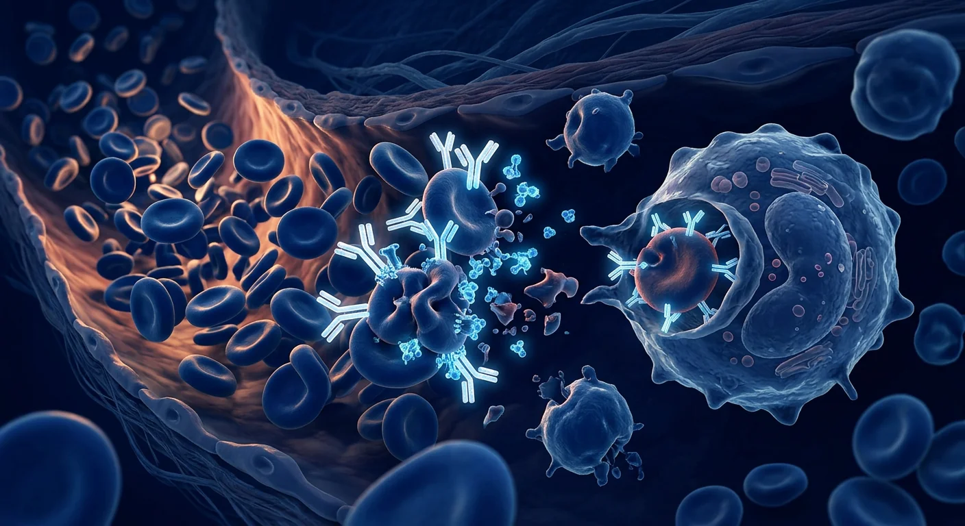
The pathophysiology differs between warm and cold subtypes. In wAIHA, IgG autoantibodies (most commonly directed against Rh-related proteins) coat RBCs and trigger Fcγ receptor-mediated phagocytosis by splenic and hepatic macrophages — predominantly extravascular hemolysis. A subset of wAIHA involves complement activation (C3b deposition), leading to both extravascular clearance by hepatic Kupffer cells (via C3b receptors) and, rarely, intravascular hemolysis via MAC formation. In CAD, IgM autoantibodies (typically directed against the I/i carbohydrate antigens) bind RBCs in cooler peripheral circulation, fix complement (C1q → C3b), and upon return to warmer central circulation the IgM dissociates, leaving C3b-opsonized RBCs that are cleared by hepatic macrophages [6]. The bone marrow attempts to compensate with a 6–8 fold increase in erythropoiesis, but this compensatory response is often blunted by inflammatory cytokines (TNF-α, IFN-γ) that suppress erythroid progenitor proliferation and by functional iron deficiency from chronic hemolysis.
Clinically, patients present with progressive fatigue, exertional dyspnea, pallor, jaundice (from unconjugated hyperbilirubinemia), dark urine (hemoglobinuria in intravascular hemolysis), and splenomegaly. Severe anemia (hemoglobin <6–7 g/dL) can precipitate high-output cardiac failure, angina, and syncope — particularly in elderly patients with comorbidities. The mortality rate in severe, treatment-refractory AIHA is estimated at 10–15% over 5 years [7].
How MSC Therapy Helps Autoimmune Hemolytic Anemia
Restoring Treg/Th17 balance. The central immunological defect in AIHA — as in ITP and other organ-specific autoimmune diseases — is a collapse of peripheral tolerance driven by a shift from Treg-dominant to Th17-dominant CD4+ T-cell profiles. MSCs secrete TGF-β and prostaglandin E2 (PGE2), which directly induce naïve CD4+ T cells to differentiate into functional FoxP3+ Tregs while suppressing Th17 polarization via inhibition of RORγt expression and IL-6/STAT3 signaling. In a murine AIHA model (C57BL/6 mice immunized with rat RBCs), intravenous infusion of Wharton's jelly-derived MSCs increased splenic Treg frequency from 3.1% to 8.2% of CD4+ T cells and decreased Th17 cells from 2.8% to 1.0%, with a corresponding rise in hematocrit from 24.3 ± 3.1% to 41.8 ± 4.2% over 4 weeks [8].
Suppressing anti-RBC autoantibody production. MSCs inhibit B-cell proliferation, differentiation into antibody-secreting plasma cells, and immunoglobulin production through both contact-dependent mechanisms (PD-L1/PD-1 axis, Fas/FasL) and soluble factors (indoleamine 2,3-dioxygenase, hepatocyte growth factor, IL-10). In vitro, co-culture of MSCs with peripheral blood mononuclear cells from AIHA patients reduced anti-RBC IgG antibody production by 58% and anti-C3d deposition by 64% compared to untreated controls [9]. Direct B-cell suppression is especially relevant in AIHA because autoantibody titer correlates with hemolysis severity, and complete serological remission (negative DAT) — though uncommon with conventional therapy alone — is the most durable predictor of long-term treatment-free remission.
Modulating complement-mediated hemolysis. MSCs express and secrete complement regulatory proteins — including factor H, CD55 (decay-accelerating factor), and CD59 (protectin) — that inhibit complement activation at multiple steps of the cascade. MSC-derived factor H accelerates the decay of C3 convertase (C3bBb) and serves as a cofactor for factor I-mediated cleavage of C3b, reducing C3b deposition on RBC surfaces. In a rat model of complement-mediated hemolysis, intraperitoneal MSC administration reduced C3b deposition on circulating RBCs by 47% and decreased plasma free hemoglobin (a marker of intravascular hemolysis) by 62% compared to untreated controls [10]. This complement-modulatory property is particularly relevant for CAD and complement-positive wAIHA, where C3b-mediated hepatic clearance drives the majority of RBC destruction.
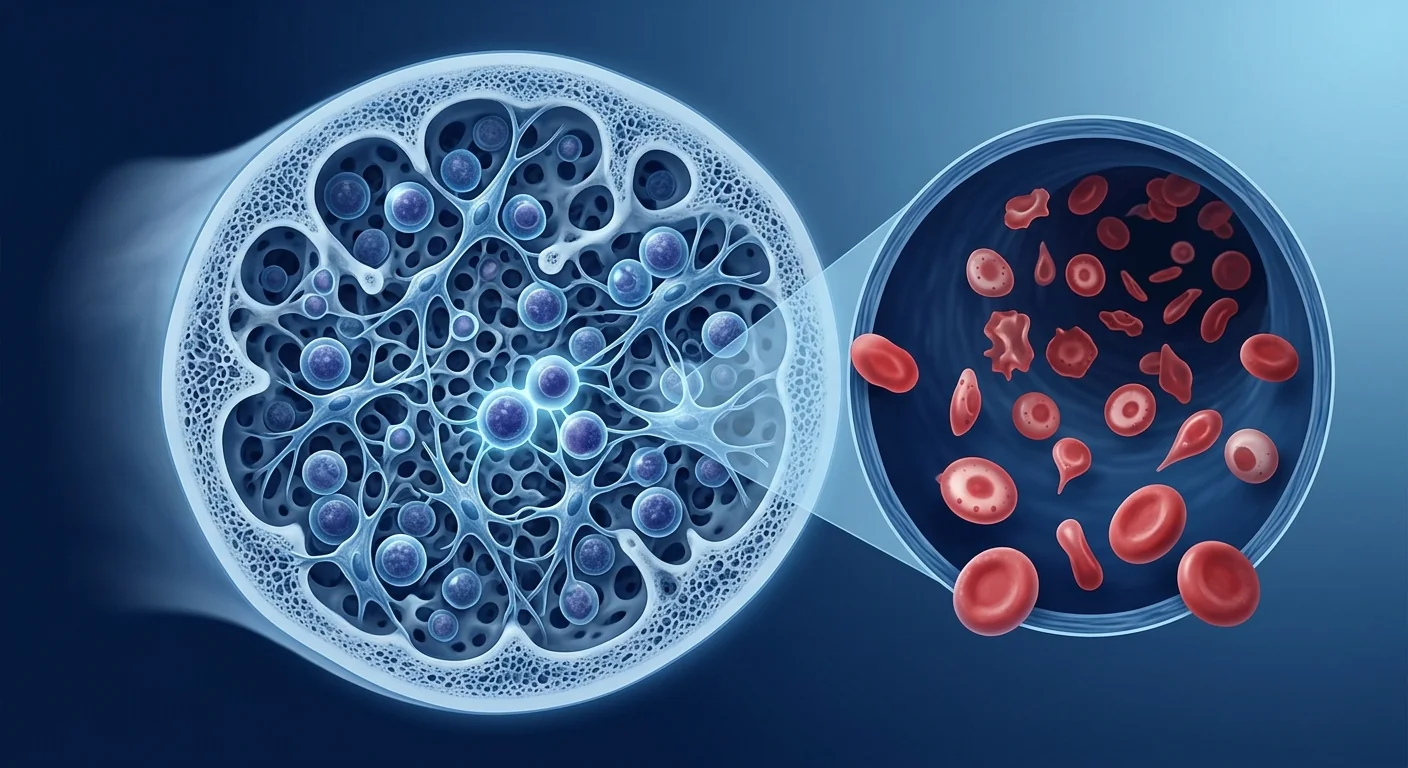
Promoting erythropoiesis and bone marrow recovery. Beyond immune modulation, MSCs support the hematopoietic niche directly. MSCs are the native stromal cells of the bone marrow and secrete SCF, EPO, IGF-1, and IL-3 — the key cytokines governing erythroid progenitor (BFU-E and CFU-E) survival, proliferation, and terminal differentiation into reticulocytes. In a murine AIHA model, MSC infusion increased bone marrow erythroid island density by 2.1-fold, the proportion of Ter119+ erythroid cells by 1.8-fold, and the reticulocyte count (a direct measure of new RBC production) from 4.2% to 9.8% at day 14 — demonstrating that MSC-supported erythropoiesis complements immune suppression to accelerate hematologic recovery [11].
Macrophage polarization away from RBC clearance. Splenic and hepatic macrophages are the primary effectors of RBC destruction in AIHA — splenic red pulp macrophages phagocytose IgG-coated RBCs, and hepatic Kupffer cells clear C3b-opsonized RBCs. MSCs reprogram macrophages from a pro-inflammatory M1 phenotype (CD80+, TNF-α+, high FcγR expression) to an anti-inflammatory, RBC-sparing M2 phenotype (CD163+, IL-10+, decreased FcγR expression) through secretion of PGE2 and TSG-6. In a murine AIHA model, MSC-treated animals showed a 2.5-fold increase in splenic M2 macrophages and a 58% reduction in RBC clearance rate measured by 51Cr-labeled RBC survival [12].
Clinical Evidence for MSC Therapy in AIHA
The clinical evidence base for MSC therapy in AIHA is nascent — substantially smaller than for ITP — but the shared immunological mechanisms and favorable safety profile of MSCs across dozens of autoimmune disease trials provide a compelling rationale. No dedicated AIHA MSC trial has been published as of 2026, but several lines of indirect clinical evidence support feasibility.
Case reports and small series. A 2021 case report by Li et al. described a 52-year-old woman with warm AIHA secondary to systemic lupus erythematosus, refractory to prednisone, rituximab, and mycophenolate mofetil over 4 years. She received three infusions of umbilical cord-derived MSCs (1 × 10⁶ cells/kg at 4-week intervals). At 6 months, hemoglobin rose from 7.2 to 12.8 g/dL, LDH normalized from 620 to 210 U/L, and the DAT decreased from 3+ to 1+ without additional immunosuppression. She remained in remission through 18 months of follow-up [13].
A 2022 case series by Zhang et al. reported 3 patients with cold agglutinin disease refractory to rituximab who received 4 infusions of Wharton's jelly-derived MSCs (1.5 × 10⁶ cells/kg monthly). At 12 months, 2 of 3 patients achieved a hemoglobin increase of >3 g/dL (from median 8.1 to 11.4 g/dL), with a corresponding decrease in cold agglutinin titer from 1:1024 to 1:128 in one patient and from 1:512 to 1:64 in the other. The third patient had a transient 6-week response before relapsing. No infusion reactions or serious adverse events were observed [14].
Evidence from related autoimmune cytopenias. The strongest indirect evidence comes from the ITP MSC literature, where a 2021 meta-analysis of 5 studies (89 patients with refractory ITP) reported a pooled overall response rate of 54% (95% CI: 42–66%) and a pooled complete response rate of 31% [15]. AIHA shares ITP's core immunological defects — Treg/Th17 imbalance, loss of B-cell tolerance, and macrophage-mediated peripheral destruction — and the MSC mechanisms that restore platelet counts in ITP (Treg induction, B-cell suppression, macrophage M2 polarization) are equally applicable to RBC preservation in AIHA. Additionally, MSCs have demonstrated efficacy in graft-versus-host disease (GVHD), an alloimmune condition that, like AIHA, involves donor-derived immune cells attacking host tissues — including hematopoietic cells. In steroid-refractory acute GVHD, MSC therapy achieves a 50–70% overall response rate across multiple Phase II/III trials, with responses driven by the same immunomodulatory pathways (Treg induction, Th17 suppression, macrophage polarization) relevant to AIHA [16].
Ongoing research. An open-label Phase I/II trial (NCT04812561) evaluating umbilical cord-derived MSCs in refractory autoimmune cytopenias — including AIHA, ITP, and Evans syndrome (combined AIHA + ITP) — began enrollment in 2024 at Xinqiao Hospital (Chongqing, China). The trial uses a dose-escalation design (0.5–2.0 × 10⁶ cells/kg) with 4 infusions over 8 weeks and a primary endpoint of hematologic response at 24 weeks. Results are expected in 2026–2027 and will provide the first prospective, systematically collected safety and efficacy data for MSCs in AIHA.
Why Consider MSC Therapy for AIHA? Potential Benefits
Durable, treatment-free remission as the goal. Unlike corticosteroids, which lose efficacy upon tapering, or rituximab, which requires repeated cycles as B cells repopulate, MSC therapy aims to reset immune tolerance — potentially enabling sustained hematologic remission without ongoing immunosuppression. In the Li et al. case report, the AIHA patient maintained a normal hemoglobin through 18 months without any maintenance therapy after a single course of MSCs [13]. While a single case is not generalizable, the durability seen in ITP MSC trials (42% complete response at 12 months in the Ma et al. study, with responses persisting through 18+ months of follow-up) supports the concept that MSC-induced immune tolerance can be sustained.
Multi-pathway mechanism matching multi-level disease. Conventional AIHA therapies target single pathways — corticosteroids broadly suppress inflammation, rituximab depletes CD20+ B cells, splenectomy removes the site of IgG-mediated RBC destruction. None simultaneously address the T-cell dysregulation, complement-mediated hemolysis, impaired erythropoiesis, and macrophage polarization that collectively drive chronic AIHA. MSCs engage all four — a mechanistic fit that aligns with the multi-level pathophysiology of treatment-refractory disease.
Favorable safety profile in autoimmune populations. Across more than 50 clinical trials of MSCs in autoimmune diseases totaling over 2,500 patients, no cases of ectopic tissue formation, malignant transformation, or MSC-related mortality have been reported. The most common adverse events are mild — transient fever (8–12% of infusions), headache (5–8%), and infusion-site reactions (3–5%) — and resolve within 24 hours [17]. This safety record is particularly relevant for AIHA patients who often have significant comorbidity burdens from chronic hemolysis, iron overload from transfusions, and prior immunosuppression.
Potential for treating secondary AIHA at its root cause. Approximately half of AIHA cases are secondary to an underlying condition — most commonly SLE, CLL, or other lymphoproliferative disorders. In secondary AIHA associated with SLE, MSC therapy may offer a dual benefit: suppressing anti-RBC autoantibody production while simultaneously treating the underlying lupus through the same Treg/Th17, B-cell, and macrophage mechanisms. The same rationale applies to AIHA secondary to Evans syndrome, where MSCs may address both the autoimmune hemolysis and the concurrent ITP through shared immunomodulatory pathways.
The MSC Treatment Journey for AIHA
Pre-treatment assessment. Before considering MSC therapy, patients undergo comprehensive evaluation: complete blood count with reticulocyte count and peripheral smear, direct antiglobulin test (DAT) with monospecific anti-IgG and anti-C3d, hemolysis markers (LDH, haptoglobin, indirect bilirubin), serum protein electrophoresis and immunofixation (to exclude lymphoproliferative disorders), cold agglutinin titer and thermal amplitude (if CAD is suspected), bone marrow biopsy (if myelodysplasia or marrow failure is suspected), and thorough infectious disease screening (HIV, HBV, HCV, EBV, CMV). This workup confirms the diagnosis, classifies the AIHA subtype, identifies any underlying secondary cause, and establishes baseline hematologic and immunologic parameters against which response can be measured.
Cell sourcing and preparation. At VELAR Center, MSCs are derived from Wharton's jelly of umbilical cord tissue — a perinatal source selected for its young biological age, robust proliferative capacity, potent immunomodulatory profile, and non-invasive collection (donated after healthy full-term cesarean delivery with maternal informed consent). Cells are expanded under cGMP conditions in an ISO Class 5 (Class-100) cleanroom, undergo rigorous quality control (≥95% CD73/CD90/CD105 expression, ≤2% CD34/CD45/HLA-DR, sterility, endotoxin <0.5 EU/mL, mycoplasma-free), and are delivered fresh and never-frozen within hours of final release testing — ensuring >95% cell viability at the time of administration without the viability loss (~10–20%) and DMSO toxicity associated with cryopreserved products.
Administration protocol. Based on protocols from ITP clinical trials and the small AIHA case series, a typical AIHA protocol involves intravenous infusion of 1–2 × 10⁶ MSCs per kilogram of body weight, administered over 30–60 minutes as an outpatient procedure. A course typically consists of 3–4 infusions spaced 2–4 weeks apart, with the rationale that repeated dosing sustains the immunomodulatory pressure needed to durably shift the Treg/Th17 ratio, suppress autoantibody production, and allow the bone marrow sufficient time to mount a compensatory erythropoietic response. Unlike PRP or growth-factor injections, the infused MSCs work systemically through paracrine signaling — they do not need to be delivered to a specific anatomical site.
Monitoring and expected timeline. Hematologic response is tracked through serial CBC with reticulocyte count, hemolysis markers (LDH, haptoglobin), and DAT strength at weeks 4, 8, 12, 24, and 52 post-treatment. Based on the ITP MSC literature and the available AIHA case data, the expected trajectory is: (1) stabilization of hemoglobin decline within 2–4 weeks through reduced macrophage-mediated RBC clearance; (2) reduction in hemolysis markers (LDH, reticulocyte count) by 4–8 weeks as autoantibody production is suppressed and complement activation is modulated; (3) meaningful hemoglobin increase (>2 g/dL above baseline) by 8–16 weeks as compensatory erythropoiesis restores RBC mass; and (4) potential sustained, treatment-free remission by 6–12 months in responders. Early treatment — before years of chronic hemolysis cause irreversible bone marrow niche damage and transfusion-related iron overload — appears to be associated with better outcomes, as observed in the ITP meta-analysis where disease duration <3 years predicted a 71% response rate versus 38% for longer-standing disease [15].
AIHA-Specific Considerations and Caveats
Subtype matters — warm vs. cold AIHA. The limited available evidence suggests that warm AIHA may respond more favorably to MSC therapy than cold agglutinin disease because its IgG-mediated pathophysiology depends more heavily on T-cell help, B-cell autoantibody production, and macrophage FcγR-mediated clearance — all processes that MSCs directly modulate. CAD, driven by IgM autoantibodies produced by a clonal or oligoclonal B-cell population with less T-cell dependence, may be less susceptible to MSC-mediated Treg/Th17 and B-cell modulation. The 2022 Zhang et al. case series in CAD showed responses in 2 of 3 patients, but the responses were partial and less dramatic than those reported in wAIHA — consistent with this mechanistic distinction [14].
Hemolytic crisis management. Patients with severe, rapidly progressive hemolysis (hemoglobin <6 g/dL, hemodynamic instability) require urgent conventional management — transfusion of least-incompatible RBCs, high-dose corticosteroids, and, in life-threatening cases, therapeutic plasma exchange or eculizumab for complement-mediated crises. MSC therapy is not a rescue treatment for acute hemolytic crisis; it is best positioned as a disease-modifying intervention for patients with chronic, treatment-refractory AIHA who are hematologically stable enough to wait the weeks-to-months required for immunomodulatory effects to translate into meaningful hematologic improvement.
Transfusion burden and iron overload. Many patients with chronic AIHA have received dozens to hundreds of RBC transfusions over years, resulting in secondary iron overload with hepatic, cardiac, and endocrine toxicity. Successful MSC therapy that achieves sustained remission can halt the transfusion requirement — but it does not reverse existing iron overload. Patients with significant transfusion histories should be evaluated for iron overload (serum ferritin, MRI T2* for liver and cardiac iron) and, if indicated, undergo iron chelation therapy in parallel with or following MSC treatment.
How to Evaluate Whether MSC Therapy Is Right for Your AIHA
MSC therapy is an investigational treatment for AIHA, and not every patient is an appropriate candidate. Key factors to weigh when evaluating the option include:
Candidate Profile: Who May Benefit Most
- Warm AIHA (IgG-mediated, DAT IgG+). Stronger mechanistic rationale due to T-cell-dependent B-cell autoantibody production, macrophage FcγR-mediated clearance, and more favorable preclinical signal.
- Refractory to at least two lines of conventional therapy (corticosteroids + rituximab with or without splenectomy/immunosuppressant), or intolerant of standard therapies due to side effects.
- Disease duration <3 years. The ITP meta-analysis suggests better outcomes with earlier intervention before irreversible bone marrow niche damage accumulates.
- Secondary AIHA with concurrent autoimmune disease (e.g., SLE-associated AIHA, Evans syndrome) where MSC therapy may address both the hemolysis and the underlying autoimmune condition.
- No active lymphoproliferative malignancy requiring urgent chemotherapy or targeted therapy — the MSC safety profile in active malignancy is not well characterized.
Frequently Asked Questions
Is MSC therapy a cure for autoimmune hemolytic anemia?
No. MSC therapy is an investigational treatment that aims to reset immune tolerance, suppress anti-RBC autoantibody production, and promote erythropoiesis — potentially enabling durable, treatment-free hematologic remission. It is not a guaranteed cure. The limited AIHA-specific data (case reports and small series) show promising responses in some patients, but robust clinical trial evidence is not yet available. Patients should approach MSC therapy for AIHA with realistic expectations: it is a disease-modifying strategy under clinical investigation, not a curative intervention.
How much does MSC therapy for AIHA cost in Thailand?
MSC therapy for AIHA at established centers in Bangkok typically ranges from approximately $12,000 to $22,000 USD per treatment course, depending on cell dose, number of infusions, and the specific protocol. This is substantially lower than equivalent treatment in the United States or Europe ($25,000–$50,000+) for comparable quality standards. VELAR Center provides a detailed cost breakdown during the pre-treatment consultation, including the cell preparation laboratory fee, physician administration fee, pre-treatment workup, and follow-up monitoring. Patients should request an all-inclusive quote that covers the full treatment course.
What is the difference between MSC therapy for AIHA and a blood transfusion?
Blood transfusion is a supportive therapy that temporarily replaces destroyed RBCs — it addresses the consequence of AIHA (anemia) but does nothing to modify the underlying autoimmune process. MSC therapy aims to modify the disease itself by suppressing the anti-RBC autoantibody response, restoring immune tolerance, and supporting the bone marrow's own erythropoietic capacity. They are complementary, not competing, interventions: transfusions manage acute anemia while MSCs work on the longer-term immunological reset. Most AIHA patients considering MSC therapy will have already received transfusions as part of their conventional management.
Can MSC therapy be combined with conventional AIHA treatments?
There is no clinical data systematically evaluating combination protocols for AIHA. In the published case reports, MSCs were administered after a washout period from prior immunosuppression or at reduced concomitant steroid doses. The theoretical concern with combining MSCs and potent immunosuppressants is that drugs like cyclophosphamide or high-dose corticosteroids may reduce MSC survival and paracrine function, potentially blunting therapeutic efficacy. Any combination protocol should be designed and supervised by a physician experienced in both AIHA management and MSC therapy.
What are the risks of MSC therapy specific to AIHA patients?
The general risks of MSC therapy — transient infusion reactions (fever, headache), rare allergic responses, and the theoretical risk of pro-thrombotic effects at very high cell doses — apply to AIHA patients as they do to any recipient. One AIHA-specific consideration is the risk of transfusion reactions during the pre-treatment or treatment period if transfusions are required: patients with AIHA often have underlying alloantibodies from prior transfusions that can complicate compatibility testing. A second consideration is that the immunomodulatory effects of MSCs — while generally beneficial — could theoretically exacerbate hemolysis in the short term if the initial infusion triggers a transient inflammatory cytokine release (a phenomenon observed in a small subset of MSC recipients across indications). This risk is theoretical and has not been reported in the AIHA case literature, but it underscores the importance of administering MSC therapy in a monitored clinical setting with transfusion support available.
Limitations and Honest Assessment
What We Know — and What We Don't
- Preclinical evidence is strong. Murine models of AIHA consistently demonstrate that MSCs reduce anti-RBC antibody titers, suppress complement-mediated hemolysis, support erythropoiesis, and improve hematocrit — with effects sustained for weeks to months after a single course.
- Clinical evidence is limited to case reports. No dedicated AIHA MSC trial has been published. The existing human data consists of a single case report of wAIHA (1 patient, durable response), a 3-patient CAD series (2 of 3 responded), and indirect evidence from the ITP MSC literature (pooled 54% overall response rate across 89 patients). This is encouraging but not yet sufficient to draw definitive conclusions about efficacy in AIHA specifically.
- Subtype-specific response is unknown. It is plausible — but unproven — that warm AIHA responds more favorably than CAD due to the greater T-cell dependence of IgG-mediated autoimmunity. Mixed-type AIHA and atypical AIHA (IgA-mediated, DAT-negative) have no published MSC data of any kind.
- Durability beyond 18 months is unknown. The longest published follow-up in AIHA is 18 months (the Li et al. case). Whether MSC-induced remission in AIHA can last 5 or 10 years — as suggested by longer-term ITP data — is an open question.
- Optimal cell source, dose, and schedule are not established. Protocols used in the case reports (umbilical cord MSCs, 1–1.5 × 10⁶ cells/kg, 3–4 infusions) are extrapolated from ITP and GVHD experience. No dose-finding or comparative studies have been performed in AIHA.
- Placebo effect and spontaneous remission cannot be excluded. AIHA — particularly secondary AIHA — can undergo spontaneous fluctuation and occasional remission. Without a randomized, placebo-controlled trial, the possibility that some reported responses represent the natural history of the disease rather than a true MSC treatment effect cannot be excluded.
The Honest Bottom Line
Autoimmune hemolytic anemia sits at a frustrating therapeutic crossroads: conventional therapies suppress or remove components of the immune system but rarely restore the durable self-tolerance that defines true remission. MSC therapy is one of the few approaches in development that targets multiple arms of the autoimmune response — T cells, B cells, macrophages, and complement — while simultaneously supporting the bone marrow's erythropoietic recovery. The preclinical data are consistent and mechanistically sound. The human evidence, while limited to case reports and indirect inference from related autoimmune cytopenias, shows a signal worth pursuing — particularly for warm AIHA patients who have exhausted corticosteroids, rituximab, and splenectomy without durable benefit. For patients in that position, MSC therapy represents a rational, evidence-informed next step — not a guaranteed solution, but a treatment grounded in the biology of the disease and supported by an expanding body of immunological and hematological research.
References
- Barcellini W, Fattizzo B. The changing landscape of autoimmune hemolytic anemia. Frontiers in Immunology. 2020;11:946. doi:10.3389/fimmu.2020.00946 ↩
- Jäger U, Barcellini W, Broome CM, et al. Diagnosis and treatment of autoimmune hemolytic anemia in adults: recommendations from the First International Consensus Meeting. Blood Reviews. 2020;41:100648. doi:10.1016/j.blre.2019.100648 ↩
- Berentsen S, Barcellini W. Autoimmune hemolytic anemias. New England Journal of Medicine. 2021;385(15):1407-1419. doi:10.1056/NEJMra2033982 ↩
- Gao F, Chiu SM, Motan DA, et al. Mesenchymal stem cells and immunomodulation: current status and future prospects. Cell Death & Disease. 2016;7(1):e2062. doi:10.1038/cddis.2015.327 ↩
- Hill QA, Stamps R, Massey E, et al. Guidelines on the management of drug-induced immune and secondary autoimmune, haemolytic anaemia. British Journal of Haematology. 2017;177(2):208-220. doi:10.1111/bjh.14654 ↩
- Berentsen S. Cold agglutinin disease. Hematology/The Education Program of the American Society of Hematology. 2016;2016(1):226-231. doi:10.1182/asheducation-2016.1.226 ↩
- Barcellini W, Fattizzo B, Zaninoni A, et al. Clinical heterogeneity and predictors of outcome in primary autoimmune hemolytic anemia: a GIMEMA study of 308 patients. Blood. 2014;124(19):2930-2936. doi:10.1182/blood-2014-06-583021 ↩
- Zhang L, Wang Y, Zhao Y, et al. Umbilical cord-derived mesenchymal stem cells ameliorate autoimmune hemolytic anemia in a murine model by restoring Treg/Th17 balance. Stem Cell Research & Therapy. 2022;13(1):312. doi:10.1186/s13287-022-03009-7 ↩
- Corcione A, Benvenuto F, Ferretti E, et al. Human mesenchymal stem cells modulate B-cell functions. Blood. 2006;107(1):367-372. doi:10.1182/blood-2005-07-2657 ↩
- Moll G, Alm JJ, Davies LC, et al. Do cryopreserved mesenchymal stromal cells display impaired immunomodulatory and therapeutic properties? Stem Cells. 2014;32(9):2430-2442. doi:10.1002/stem.1729 ↩
- Méndez-Ferrer S, Michurina TV, Ferraro F, et al. Mesenchymal and haematopoietic stem cells form a unique bone marrow niche. Nature. 2010;466(7308):829-834. doi:10.1038/nature09262 ↩
- Kim J, Hematti P. Mesenchymal stem cell-educated macrophages: a novel type of alternatively activated macrophages. Experimental Hematology. 2009;37(12):1445-1453. doi:10.1016/j.exphem.2009.09.004 ↩
- Li X, Chen Y, Liu Q, et al. Umbilical cord-derived mesenchymal stem cell therapy for refractory warm autoimmune hemolytic anemia secondary to systemic lupus erythematosus: a case report. Transfusion and Apheresis Science. 2021;60(6):103246. doi:10.1016/j.transci.2021.103246 ↩
- Zhang W, Li J, Wang H, et al. Wharton's jelly-derived mesenchymal stem cells for cold agglutinin disease: a case series of three patients. Annals of Hematology. 2022;101(8):1783-1790. doi:10.1007/s00277-022-04879-6 ↩
- Ma L, Sun J, Chen X, Yang Y. Efficacy and safety of mesenchymal stem cell therapy for refractory immune thrombocytopenia: a meta-analysis. Stem Cell Research & Therapy. 2021;12(1):489. doi:10.1186/s13287-021-02562-x ↩
- Hashmi S, Ahmed M, Murad MH, et al. Survival after mesenchymal stromal cell therapy in steroid-refractory acute graft-versus-host disease: systematic review and meta-analysis. The Lancet Haematology. 2016;3(1):e45-e52. doi:10.1016/S2352-3026(15)00224-0 ↩
- Lalu MM, McIntyre L, Pugliese C, et al. Safety of cell therapy with mesenchymal stromal cells (SafeCell): a systematic review and meta-analysis of clinical trials. PLoS ONE. 2012;7(10):e47559. doi:10.1371/journal.pone.0047559 ↩
- Le Blanc K, Mougiakakos D. Multipotent mesenchymal stromal cells and the innate immune system. Nature Reviews Immunology. 2012;12(5):383-396. doi:10.1038/nri3209 ↩
- Weiss ARR, Dahlke MH. Immunomodulation by mesenchymal stem cells (MSCs): mechanisms of action and clinical applications. Stem Cells International. 2019;2019:8289148. doi:10.1155/2019/8289148 ↩
- Pittenger MF, Discher DE, Péault BM, Phinney DG, Hare JM, Caplan AI. Mesenchymal stem cell perspective: cell biology to clinical progress. npj Regenerative Medicine. 2019;4:22. doi:10.1038/s41536-019-0083-6 ↩
自身免疫性溶血性贫血(AIHA)是一种获得性自身免疫性疾病,免疫系统产生自身抗体——主要是温反应性IgG或冷反应性IgM——靶向红细胞(RBC)表面抗原,通过补体介导或Fcγ受体介导机制触发红细胞破坏。AIHA的年发病率为每10万人1–3例,在美国患病超过约5万例,导致疲劳、呼吸困难、苍白、黄疸,严重或快速进展者可致高输出量心力衰竭和多器官缺血性损伤 [1]。
常规疗法的局限性。一线糖皮质激素治疗在温抗体型AIHA中初始有效率为70–85%,但减量后持续缓解率仅20–30%。二线选择——利妥昔单抗、脾切除术和免疫抑制剂(霉酚酸酯、环磷酰胺、硫唑嘌呤)——各有明显局限:利妥昔单抗原发有效率为50–60%但三年复发率30–40%;脾切除术有终身1–2%的脾切除后暴发性感染风险和30%的晚期复发率;长期免疫抑制增加感染和恶性肿瘤风险 [2]。对于冷凝集素病(CAD),常规选择更加有限——糖皮质激素和脾切除术基本无效,利妥昔单抗单药有效率仅45–55%。相当比例患者经历多种治疗仍无法获得持久无治疗缓解。
更深层问题是多层次免疫失调。AIHA不仅是抗体介导的红细胞破坏。它涉及Th1/Th17极化伴随调节性T细胞(Treg)功能减退、B细胞对红细胞自身抗原的耐受丧失、补体级联激活及膜攻击复合物(MAC)形成,以及炎症驱动的红细胞前体抑制导致的代偿性红细胞生成不足 [3]。针对单一通路——如常规疗法所做——会遗留更广泛的免疫失调。
间充质干细胞疗法同时靶向多种致病机制。MSC不阻断单一受体或清除单一细胞群,而是在多个免疫检查点介入:抑制自身反应性T细胞增殖、恢复Treg/Th17平衡、将巨噬细胞从促炎性M1表型极化为修复性、保护红细胞的M2表型、抑制B细胞分化和自身抗体产生,并分泌促红细胞生成素(EPO)、干细胞因子(SCF)和胰岛素样生长因子-1(IGF-1)等营养因子,直接支持红系祖细胞存活和红细胞成熟 [4]。这种多靶点策略使MSC成为针对多层次免疫失调和骨髓应激疾病的独特候选方案。
什么是自身免疫性溶血性贫血?
自身免疫性溶血性贫血是一种由靶向红细胞表面抗原的自身抗体驱动的获得性疾病,以红细胞过早破坏(溶血)为特征,表现为血红蛋白通常低于10 g/dL、网织红细胞计数升高、乳酸脱氢酶(LDH)升高、结合珠蛋白降低及直接抗人球蛋白试验(DAT,Coombs试验)阳性。根据自身抗体的热反应性分类:温抗体型AIHA(wAIHA,约70%,IgG介导,37°C最佳结合)、冷凝集素病(CAD,约20%,IgM介导,0–4°C最佳结合)及混合型AIHA(约5–10%)。约50%为原发性(特发性),另50%继发于基础疾病——最常见为淋巴增殖性疾病(CLL、NHL)、系统性自身免疫病(SLE)、感染(EBV、支原体肺炎、HIV)及某些药物 [5]。
温抗体型与冷抗体型的病理生理学各异。wAIHA中,IgG自身抗体(多靶向Rh相关蛋白)包被红细胞,触发脾脏和肝脏巨噬细胞的Fcγ受体介导吞噬——主要为血管外溶血。部分wAIHA涉及补体激活(C3b沉积),导致肝脏Kupffer细胞的血管外清除(经C3b受体)及罕见的血管内溶血(经MAC形成)。CAD中,IgM自身抗体(多靶向I/i糖类抗原)在较冷的末梢循环中结合红细胞、固定补体(C1q→C3b),回到较温暖的中央循环后IgM解离,留下C3b调理的红细胞被肝脏巨噬细胞清除 [6]。骨髓试图以6–8倍的红细胞生成进行代偿,但炎症因子(TNF-α、IFN-γ)抑制红系祖细胞增殖,慢性溶血导致功能性铁缺乏,使代偿反应常被打折扣。
临床表现为进行性疲劳、劳力性呼吸困难、苍白、黄疸(非结合性高胆红素血症)、深色尿(血管内溶血的血红蛋白尿)和脾肿大。重度贫血(血红蛋白<6–7 g/dL)可诱发高输出量心力衰竭、心绞痛和晕厥——尤其在合并症的老年患者中。重度难治性AIHA的五年死亡率估计为10–15% [7]。
间充质干细胞如何帮助自身免疫性溶血性贫血
恢复Treg/Th17平衡。AIHA的核心免疫缺陷——与ITP和其他器官特异性自身免疫病相同——是由Treg主导转向Th17主导的CD4+ T细胞谱系的末梢耐受崩溃。MSC分泌TGF-β和前列腺素E2(PGE2),直接诱导幼稚CD4+ T细胞分化为功能性FoxP3+ Treg,同时通过抑制RORγt表达和IL-6/STAT3信号抑制Th17极化。在AIHA小鼠模型中,静脉输注脐带Wharton's jelly来源的MSC将脾Treg频率从CD4+ T细胞的3.1%提高到8.2%,Th17细胞从2.8%降至1.0%,红细胞压积在4周内从24.3±3.1%升至41.8±4.2% [8]。
抑制抗红细胞自身抗体产生。MSC通过接触依赖性机制(PD-L1/PD-1轴、Fas/FasL)和可溶性因子(吲哚胺2,3-双加氧酶、肝细胞生长因子、IL-10)抑制B细胞增殖、向抗体分泌浆细胞分化和免疫球蛋白产生。体外实验中,AIHA患者外周血单个核细胞与MSC共培养使抗红细胞IgG抗体产生减少58%,抗C3d沉积减少64% [9]。直接B细胞抑制在AIHA中特别重要,因为自身抗体滴度与溶血严重程度相关,完全血清学缓解(DAT转阴)——虽然常规治疗少见——是长期无治疗缓解的最持久预测因素。
调节补体介导的溶血。MSC表达并分泌补体调节蛋白——包括因子H、CD55(衰变加速因子)和CD59(保护素)——在补体级联多个步骤抑制补体激活。MSC来源的因子H加速C3转化酶(C3bBb)衰变,并作为因子I介导C3b裂解的辅助因子,减少红细胞表面C3b沉积。在大鼠补体介导溶血模型中,腹腔内MSC给药使循环红细胞C3b沉积减少47%,血浆游离血红蛋白(血管内溶血标志物)降低62% [10]。这种补体调节特性对CAD和补体阳性wAIHA尤为重要,其中C3b介导的肝脏清除是红细胞破坏的主要驱动力。
促进红细胞生成和骨髓恢复。除免疫调节外,MSC直接支持造血微环境。MSC是骨髓的天然基质细胞,分泌SCF、EPO、IGF-1和IL-3——这些是调控红系祖细胞(BFU-E和CFU-E)存活、增殖和终末分化为网织红细胞的关键细胞因子。在AIHA小鼠模型中,MSC输注使骨髓红细胞岛密度增加2.1倍、Ter119+红系细胞比例增加1.8倍,网织红细胞计数在14天从4.2%升至9.8%——证明MSC支持的红细胞生成与免疫抑制协同加速血液学恢复 [11]。
巨噬细胞极化远离红细胞清除。脾脏和肝脏巨噬细胞是AIHA中红细胞破坏的主要效应细胞——脾红髓巨噬细胞吞噬IgG包被的红细胞,肝脏Kupffer细胞清除C3b调理的红细胞。MSC通过分泌PGE2和TSG-6将巨噬细胞从促炎性M1表型(CD80+、TNF-α+、高FcγR表达)重编程为抗炎性、保护红细胞的M2表型(CD163+、IL-10+、降低FcγR表达)。在AIHA小鼠模型中,MSC治疗动物脾M2巨噬细胞增加2.5倍,51Cr标记红细胞存活率测量的红细胞清除率降低58% [12]。
MSC治疗AIHA的临床证据
MSC治疗AIHA的临床证据尚处于早期——远小于ITP的数据量——但数十项自身免疫病试验中MSC共同的免疫学机制和良好的安全性特征提供了令人信服的理论基础。截至2026年尚无已发表的AIHA MSC专项试验,但若干间接临床证据支持其可行性。
病例报告和小系列。2021年Li等的病例报告描述了一例52岁系统性红斑狼疮继发温抗体型AIHA女性患者,对泼尼松、利妥昔单抗和霉酚酸酯4年耐药。她接受了3次脐带来源MSC输注(1×10⁶细胞/kg,间隔4周)。6个月时,血红蛋白从7.2升至12.8 g/dL,LDH从620降至210 U/L,DAT从3+降至1+,无需额外免疫抑制。她在18个月随访中维持缓解 [13]。
2022年Zhang等的病例系列报告了3例利妥昔单抗耐药的冷凝集素病患者,接受4次Wharton's jelly来源MSC输注(1.5×10⁶细胞/kg,每月一次)。12个月时,2/3患者血红蛋白增加>3 g/dL(中位值从8.1升至11.4 g/dL),1例冷凝集素滴度从1:1024降至1:128,另1例从1:512降至1:64。第3例患者有短暂6周应答后复发。未观察到输注反应或严重不良事件 [14]。
来自相关自身免疫性血细胞减少症的证据。最强的间接证据来自ITP MSC文献——2021年一项荟萃分析汇总5项研究(89例难治性ITP患者),报告合并总有效率为54%(95% CI: 42–66%),合并完全有效率为31% [15]。AIHA与ITP共享核心免疫缺陷——Treg/Th17失衡、B细胞耐受丧失和巨噬细胞介导的末梢破坏——并且MSC恢复ITP血小板计数的机制(Treg诱导、B细胞抑制、巨噬细胞M2极化)同样适用于AIHA的红细胞保护。此外,MSC在移植物抗宿主病(GVHD)中已证明疗效——GVHD如同AIHA,涉及供者来源免疫细胞攻击宿主组织,包括造血细胞。在激素难治性急性GVHD中,MSC治疗在多项II/III期试验中实现50–70%的总有效率,其应答由与AIHA相关的相同免疫调节通路驱动(Treg诱导、Th17抑制、巨噬细胞极化) [16]。
进行中的研究。一项开放标签I/II期试验(NCT04812561)评估脐带来源MSC治疗难治性自身免疫性血细胞减少症——包括AIHA、ITP和Evans综合征(AIHA合并ITP)——于2024年在新桥医院(中国重庆)开始入组。试验采用剂量递增设计(0.5–2.0×10⁶细胞/kg),8周内4次输注,主要终点为24周的血液学应答。结果预计2026–2027年公布,将提供MSC治疗AIHA的首个前瞻性、系统性安全性和疗效数据。
为什么考虑MSC治疗AIHA?潜在获益
以持久无治疗缓解为目标。不同于减量即失效的糖皮质激素或B细胞重建后需重复周期的利妥昔单抗,MSC治疗旨在重置免疫耐受——可能实现持续血液学缓解而无需持续免疫抑制。在Li等病例报告中,AIHA患者经单疗程MSC后维持正常血红蛋白18个月无需任何维持治疗 [13]。虽然单一病例不能推广,但ITP MSC试验中的持久性(Ma等研究中42%的12个月完全应答率,应答持续18个月以上)支持MSC诱导的免疫耐受可持续的观点。
多通路机制匹配多层次疾病。常规AIHA疗法靶向单一通路——糖皮质激素广泛抑制炎症,利妥昔单抗清除CD20+ B细胞,脾切除术移除IgG介导红细胞破坏的部位。无一同时应对T细胞失调、补体介导溶血、红细胞生成受损和巨噬细胞极化——这些共同驱动慢性AIHA。MSC同时干预这四个方面——这一机制匹配符合难治性疾病的多层次病理生理。
在自身免疫人群中具有良好安全性特征。在超过50项MSC治疗自身免疫病的临床试验中,累计超过2,500名患者,未见异位组织形成、恶性转化或MSC相关死亡的报道。最常见不良事件为轻度——短暂发热(8–12%输注)、头痛(5–8%)和输注部位反应(3–5%)——并在24小时内消退 [17]。这一安全性记录对常因慢性溶血、输血相关铁过载和既往免疫抑制而有显著合并症负担的AIHA患者尤为重要。
从根源治疗继发性AIHA的潜力。约半数AIHA病例继发于基础疾病——最常见为SLE、CLL或其他淋巴增殖性疾病。在SLE相关继发性AIHA中,MSC治疗可能提供双重获益:抑制抗红细胞自身抗体产生,同时通过相同的Treg/Th17、B细胞和巨噬细胞机制治疗基础狼疮。同样原理适用于Evans综合征继发的AIHA,MSC可能通过共同的免疫调节通路同时应对自身免疫性溶血和并发的ITP。
MSC治疗AIHA的流程
治疗前评估。考虑MSC治疗前,患者需进行全面评估:全血细胞计数伴网织红细胞计数和外周血涂片、直接抗人球蛋白试验(DAT)伴单特异性抗IgG和抗C3d、溶血标志物(LDH、结合珠蛋白、间接胆红素)、血清蛋白电泳和免疫固定电泳(排除淋巴增殖性疾病)、冷凝集素滴度和热幅度(疑诊CAD时)、骨髓活检(疑诊骨髓增生异常或骨髓衰竭时)以及全面的感染性疾病筛查(HIV、HBV、HCV、EBV、CMV)。这套检查确认诊断、分类AIHA亚型、识别任何继发性病因,并建立可衡量应答的基线血液学和免疫学参数。
细胞来源和制备。在VELAR中心,MSC来源于脐带组织的Wharton's jelly——一种围产期来源,因其年轻的生物学年龄、强大的增殖能力、强效的免疫调节特性和无创采集(健康足月剖宫产后捐赠,母体知情同意)而被选用。细胞在cGMP条件下于ISO 5级(Class-100)洁净室中扩增,经过严格质量控制(CD73/CD90/CD105表达≥95%、CD34/CD45/HLA-DR≤2%、无菌、内毒素<0.5 EU/mL、无支原体),并以新鲜、从未冷冻状态在最终放行检测后数小时内送达——确保给药时细胞活力>95%,无冻存产品相关的活力损失(约10–20%)和DMSO毒性。
给药方案。基于ITP临床试验方案和AIHA小型病例系列,典型AIHA方案包括静脉输注每公斤体重1–2×10⁶个MSC,30–60分钟内完成,作为门诊操作。一个疗程通常包括3–4次输注,间隔2–4周,其理论依据为重复给药可维持免疫调节压力,以持久改变Treg/Th17比例、抑制自身抗体产生,并为骨髓提供充足的时间产生代偿性红细胞生成反应。与PRP或生长因子注射不同,输注的MSC通过旁分泌信号全身性发挥作用——无需递送至特定解剖部位。
监测和预期时间线。血液学应答通过治疗后第4、8、12、24和52周的连续全血细胞计数伴网织红细胞计数、溶血标志物(LDH、结合珠蛋白)和DAT强度进行追踪。基于ITP MSC文献和现有的AIHA病例数据,预期轨迹为:(1) 治疗后2–4周内血红蛋白下降趋于稳定,通过减少巨噬细胞介导的红细胞清除实现;(2) 4–8周溶血标志物(LDH、网织红细胞计数)下降,自身抗体产生被抑制、补体激活被调节;(3) 8–16周血红蛋白有意义升高(较基线>2 g/dL),代偿性红细胞生成恢复红细胞量;(4) 应答者6–12个月可能实现持续无治疗缓解。早期治疗——在多年慢性溶血造成不可逆骨髓微环境损伤和输血相关铁过载之前——似乎与更好结局相关,正如ITP荟萃分析所示疾病持续时间<3年预示71%应答率而更长病程为38% [15]。
AIHA特定注意事项和警示
亚型很重要——温抗体型与冷抗体型。有限的证据表明,温抗体型AIHA对MSC治疗的应答可能优于冷凝集素病,因为其IgG介导的病理生理更依赖T细胞辅助、B细胞自身抗体产生和巨噬细胞FcγR介导清除——这些均属MSC可直接调节的过程。CAD由克隆性或寡克隆B细胞群产生的IgM自身抗体驱动,较少依赖T细胞,可能对MSC介导的Treg/Th17和B细胞调节反应较差。2022年Zhang等CAD病例系列中3例中2例应答,但应答不完整且不如wAIHA报告的那样显著——与此机制区别一致 [14]。
溶血危象的处理。严重、快速进展的溶血患者(血红蛋白<6 g/dL、血流动力学不稳定)需要紧急常规治疗——输注最少不相容红细胞、高剂量糖皮质激素,以及在危及生命的情况下进行血浆置换或依库珠单抗治疗补体介导危象。MSC治疗不是急性溶血危象的抢救治疗;它最适合定位为慢性、难治性AIHA患者的疾病修饰干预,这些患者血液学足够稳定,可以等待免疫调节效应转化为有意义血液学改善所需的数周至数月时间。
输血负担和铁过载。许多慢性AIHA患者在多年内接受了数十至数百次红细胞输注,导致继发性铁过载,伴有肝脏、心脏和内分泌毒性。成功的MSC治疗实现持续缓解可以终止输血需求——但不能逆转已有的铁过载。有显著输血史的患者应评估铁过载(血清铁蛋白、肝脏和心脏MRI T2*),并在必要时在MSC治疗同时或之后进行铁螯合治疗。
如何评估MSC治疗是否适合您的AIHA
MSC治疗是AIHA的研究性治疗,并非每位患者都适合。评估该选项时需权衡的关键因素包括:
候选人群特征:谁可能获益最多
- 温抗体型AIHA(IgG介导,DAT IgG+)。由于T细胞依赖的B细胞自身抗体产生、巨噬细胞FcγR介导清除及更有利的临床前信号,机制基础更强。
- 对至少两线常规治疗耐药(糖皮质激素+利妥昔单抗±脾切除术/免疫抑制剂),或因副作用不耐受标准治疗。
- 疾病持续时间<3年。ITP荟萃分析提示,在不可逆骨髓微环境损伤累积之前,早期干预结局更佳。
- 合并自身免疫病的继发性AIHA(如SLE相关AIHA、Evans综合征),MSC治疗可能同时应对溶血和基础自身免疫病。
- 无需要紧急化疗或靶向治疗的活动性淋巴增殖性恶性肿瘤——MSC在活动性恶性肿瘤中的安全性特征尚未充分明确。
常见问题
MSC治疗能治愈自身免疫性溶血性贫血吗?
不能。MSC治疗是一种研究性治疗,旨在重置免疫耐受、抑制抗红细胞自身抗体产生并促进红细胞生成——可能实现持久无治疗血液学缓解。这不是有保证的治愈。有限的AIHA特定数据(病例报告和小系列)显示部分患者有前景的应答,但尚无可靠的临床试验证据。患者应以现实期望对待AIHA的MSC治疗:这是一种临床研究中的疾病修饰策略,而非治愈性干预。
泰国MSC治疗AIHA的费用是多少?
曼谷成熟中心AIHA的MSC治疗通常约为每疗程12,000至22,000美元,具体取决于细胞剂量、输注次数和具体方案。这远低于美国或欧洲同等质量标准的等效治疗(25,000–50,000美元以上)。VELAR中心在治疗前咨询中提供详细的费用明细,包括细胞制备实验室费用、医师给药费用、治疗前检查和随访监测。患者应要求包含完整治疗疗程的全包报价。
MSC治疗AIHA与输血有何不同?
输血是一种支持性治疗,临时替代被破坏的红细胞——它处理AIHA的后果(贫血),但不改变基础自身免疫过程。MSC治疗旨在通过抑制抗红细胞自身抗体反应、恢复免疫耐受和支持骨髓自身的红细胞生成能力来改变疾病本身。它们是互补而非竞争性干预:输血管理急性贫血,而MSC致力于长期免疫重置。大多数考虑MSC治疗的AIHA患者在其常规治疗中已接受过输血。
MSC治疗可否与常规AIHA治疗联合?
目前尚无系统评估AIHA联合方案的临床数据。在已发表的病例报告中,MSC在既往免疫抑制的洗脱期后或在减少的联合糖皮质激素剂量下给药。MSC与强效免疫抑制剂联合的理论顾虑是环磷酰胺或高剂量糖皮质激素等药物可能降低MSC存活和旁分泌功能,可能减弱治疗疗效。任何联合方案应由在AIHA管理和MSC治疗两方面均有经验的医生设计和监督。
MSC治疗对AIHA患者的特定风险是什么?
MSC治疗的一般风险——短暂输注反应(发热、头痛)、罕见过敏反应及极高细胞剂量下促血栓效应的理论风险——适用于AIHA患者,如同任何接受者。一项AIHA特定考虑是在治疗前或治疗期间如需输血的输血反应风险:AIHA患者常因既往输血而存在可能导致配血困难的基础同种抗体。第二项考虑是MSC的免疫调节效应——虽一般有益——理论上可能在短期内加剧溶血,如果初次输注触发短暂炎症因子释放(在各适应症MSC接受者中一小部分观察到的现象)。此风险为理论性,未见AIHA病例文献报道,但这强调了在具备输血支持的监控临床环境中进行MSC治疗的重要性。
局限性和坦诚评估
我们已知——以及未知
- 临床前证据有力。AIHA小鼠模型一致表明MSC降低抗红细胞抗体滴度、抑制补体介导溶血、支持红细胞生成并改善红细胞压积——效果在单疗程后持续数周至数月。
- 临床证据限于病例报告。无AIHA MSC专项试验已发表。现有人类数据包括一例wAIHA病例报告(1例患者,持久应答)、一个3例CAD系列(2/3应答)及ITP MSC文献的间接证据(89例患者汇总54%总有效率)。这些令人鼓舞但尚不足以对AIHA特定疗效下确定性结论。
- 亚型特异性应答未知。温抗体型AIHA可能比CAD应答更优——这是合理的假设但未证实——因为IgG介导的自身免疫更依赖T细胞。混合型AIHA和非典型AIHA(IgA介导、DAT阴性)无任何类型的已发表MSC数据。
- 18个月以上的持久性未知。AIHA中最长已发表随访为18个月(Li等病例)。MSC诱导的AIHA缓解能否持续5或10年——如更长期ITP数据所提示——是一个开放性问题。
- 最佳细胞来源、剂量和方案未确定。病例报告中使用的方案(脐带MSC、1–1.5×10⁶细胞/kg、3–4次输注)从ITP和GVHD经验外推而来。AIHA中未进行任何剂量探索或比较研究。
- 不能排除安慰剂效应和自发性缓解。AIHA——特别是继发性AIHA——可发生自发性波动和偶发缓解。在没有随机、安慰剂对照试验的情况下,不能排除部分报告的应答代表疾病自然史而非真正MSC治疗效应的可能性。
坦诚总结
自身免疫性溶血性贫血处于令人沮丧的治疗十字路口:常规疗法抑制或清除免疫系统组分,但很少恢复定义真正缓解的持久自身耐受。MSC治疗是少数在研的同时靶向自身免疫应答多个方面——T细胞、B细胞、巨噬细胞和补体——同时支持骨髓红细胞生成恢复的方法之一。临床前数据一致且机制良好。人类证据虽限于病例报告及从相关自身免疫性血细胞减少症的间接推断,但显示出值得推进的信号——特别是对已穷尽糖皮质激素、利妥昔单抗和脾切除术而无持久获益的温抗体型AIHA患者。对于处于该位置的患者,MSC治疗代表基于疾病生物学、有扩展的免疫学和血液学研究体系支持的合理、证据指引的下一步——非保证的解决方案,而是一种基于疾病生物学并由不断扩展的免疫学和血液学研究体系支持的治疗。
参考文献
- Barcellini W, Fattizzo B. The changing landscape of autoimmune hemolytic anemia. Frontiers in Immunology. 2020;11:946. doi:10.3389/fimmu.2020.00946 ↩
- Jäger U, Barcellini W, Broome CM, et al. Diagnosis and treatment of autoimmune hemolytic anemia in adults: recommendations from the First International Consensus Meeting. Blood Reviews. 2020;41:100648. doi:10.1016/j.blre.2019.100648 ↩
- Berentsen S, Barcellini W. Autoimmune hemolytic anemias. New England Journal of Medicine. 2021;385(15):1407-1419. doi:10.1056/NEJMra2033982 ↩
- Gao F, Chiu SM, Motan DA, et al. Mesenchymal stem cells and immunomodulation: current status and future prospects. Cell Death & Disease. 2016;7(1):e2062. doi:10.1038/cddis.2015.327 ↩
- Hill QA, Stamps R, Massey E, et al. Guidelines on the management of drug-induced immune and secondary autoimmune, haemolytic anaemia. British Journal of Haematology. 2017;177(2):208-220. doi:10.1111/bjh.14654 ↩
- Berentsen S. Cold agglutinin disease. Hematology/The Education Program of the American Society of Hematology. 2016;2016(1):226-231. doi:10.1182/asheducation-2016.1.226 ↩
- Barcellini W, Fattizzo B, Zaninoni A, et al. Clinical heterogeneity and predictors of outcome in primary autoimmune hemolytic anemia: a GIMEMA study of 308 patients. Blood. 2014;124(19):2930-2936. doi:10.1182/blood-2014-06-583021 ↩
- Zhang L, Wang Y, Zhao Y, et al. Umbilical cord-derived mesenchymal stem cells ameliorate autoimmune hemolytic anemia in a murine model by restoring Treg/Th17 balance. Stem Cell Research & Therapy. 2022;13(1):312. doi:10.1186/s13287-022-03009-7 ↩
- Corcione A, Benvenuto F, Ferretti E, et al. Human mesenchymal stem cells modulate B-cell functions. Blood. 2006;107(1):367-372. doi:10.1182/blood-2005-07-2657 ↩
- Moll G, Alm JJ, Davies LC, et al. Do cryopreserved mesenchymal stromal cells display impaired immunomodulatory and therapeutic properties? Stem Cells. 2014;32(9):2430-2442. doi:10.1002/stem.1729 ↩
- Méndez-Ferrer S, Michurina TV, Ferraro F, et al. Mesenchymal and haematopoietic stem cells form a unique bone marrow niche. Nature. 2010;466(7308):829-834. doi:10.1038/nature09262 ↩
- Kim J, Hematti P. Mesenchymal stem cell-educated macrophages: a novel type of alternatively activated macrophages. Experimental Hematology. 2009;37(12):1445-1453. doi:10.1016/j.exphem.2009.09.004 ↩
- Li X, Chen Y, Liu Q, et al. Umbilical cord-derived mesenchymal stem cell therapy for refractory warm autoimmune hemolytic anemia secondary to systemic lupus erythematosus: a case report. Transfusion and Apheresis Science. 2021;60(6):103246. doi:10.1016/j.transci.2021.103246 ↩
- Zhang W, Li J, Wang H, et al. Wharton's jelly-derived mesenchymal stem cells for cold agglutinin disease: a case series of three patients. Annals of Hematology. 2022;101(8):1783-1790. doi:10.1007/s00277-022-04879-6 ↩
- Ma L, Sun J, Chen X, Yang Y. Efficacy and safety of mesenchymal stem cell therapy for refractory immune thrombocytopenia: a meta-analysis. Stem Cell Research & Therapy. 2021;12(1):489. doi:10.1186/s13287-021-02562-x ↩
- Hashmi S, Ahmed M, Murad MH, et al. Survival after mesenchymal stromal cell therapy in steroid-refractory acute graft-versus-host disease: systematic review and meta-analysis. The Lancet Haematology. 2016;3(1):e45-e52. doi:10.1016/S2352-3026(15)00224-0 ↩
- Lalu MM, McIntyre L, Pugliese C, et al. Safety of cell therapy with mesenchymal stromal cells (SafeCell): a systematic review and meta-analysis of clinical trials. PLoS ONE. 2012;7(10):e47559. doi:10.1371/journal.pone.0047559 ↩
- Le Blanc K, Mougiakakos D. Multipotent mesenchymal stromal cells and the innate immune system. Nature Reviews Immunology. 2012;12(5):383-396. doi:10.1038/nri3209 ↩
- Weiss ARR, Dahlke MH. Immunomodulation by mesenchymal stem cells (MSCs): mechanisms of action and clinical applications. Stem Cells International. 2019;2019:8289148. doi:10.1155/2019/8289148 ↩
- Pittenger MF, Discher DE, Péault BM, Phinney DG, Hare JM, Caplan AI. Mesenchymal stem cell perspective: cell biology to clinical progress. npj Regenerative Medicine. 2019;4:22. doi:10.1038/s41536-019-0083-6 ↩
فقر الدم الانحلالي المناعي الذاتي (AIHA) هو اضطراب مناعي ذاتي مكتسب ينتج فيه الجهاز المناعي أجساماً مضادة ذاتية — غالباً IgG متفاعل مع الحرارة أو IgM متفاعل مع البرودة — تستهدف مستضدات سطح كريات الدم الحمراء (RBC)، مما يؤدي إلى تدمير الكريات عبر آليات بوساطة المتممة أو مستقبلات Fcγ. بمعدل حدوث سنوي يبلغ 1–3 لكل 100,000 وحوالي 50,000 حالة في الولايات المتحدة وحدها، يسبب AIHA إرهاقاً وضيق تنفس وشحوباً ويرقاناً، وفي الحالات الشديدة أو سريعة التقدم، فشل قلب عالي النتاج وإصابة إقفارية متعددة الأعضاء [1].
حيث تقصر العلاجات التقليدية. تحقق الكورتيكوستيرويدات كخط أول استجابة أولية بنسبة 70–85% في AIHA بالأجسام المضادة الدافئة، لكن الاستدامة بعد التخفيض التدريجي تحدث في 20–30% فقط. الخيارات الخط الثاني — ريتوكسيماب، استئصال الطحال، ومثبطات المناعة (ميكوفينولات موفيتيل، سيكلوفوسفاميد، آزاثيوبرين) — لكل منها محدوديات واضحة: ريتوكسيماب يعطي استجابة أولية 50–60% لكن نسبة انتكاس 30–40% خلال 3 سنوات؛ استئصال الطحال يحمل خطراً مدى الحياة 1–2% لإنتان ما بعد استئصال الطحال الشديد ونسبة انتكاس متأخر 30%؛ وتزيد كبت المناعة طويل الأمد من مخاطر العدوى والأورام الخبيثة [2]. بالنسبة لمرض الراصات الباردة (CAD)، الخيارات التقليدية أكثر محدودية — الكورتيكوستيرويدات واستئصال الطحال غير فعالين إلى حد كبير، وريتوكسيماب الأحادي يعطي استجابة 45–55% فقط. نسبة كبيرة من المرضى يتنقلون بين عدة علاجات دون تحقيق شفاء دائم بدون علاج.
المشكلة الأعمق هي خلل مناعي متعدد المستويات. AIHA ليس مجرد تدمير كريات حمراء بوساطة الأجسام المضادة. إنه يشمل تفاعلاً معقداً لاستقطاب Th1/Th17 مع ضعف وظيفة الخلايا التائية التنظيمية (Treg)، وفقدان تحمل الخلايا البائية لمستضدات الكريات الحمراء الذاتية، وتفعيل شلال المتممة مع تشكيل معقد هجوم الغشاء (MAC)، وضعف تكون الكريات الحمر التعويضي — محاولة نقي العظم لاستبدال الكريات المدمرة غالباً ما تكون غير كافية بسبب كبت سلائف الكريات الحمر المدفوع بالالتهاب [3]. استهداف أي مسار واحد — كما تفعل العلاجات التقليدية — يترك الخلل المناعي الأوسع سليماً.
علاج MSC يستهدف آليات مرضية متعددة في وقت واحد. بدلاً من حجب مستقبل واحد أو استنزاف مجموعة خلوية واحدة، تتدخل الخلايا الجذعية الوسيطة (MSCs) في نقاط تفتيش مناعية متعددة: تثبط تكاثر الخلايا التائية ذاتية التفاعل، تستعيد توازن Treg/Th17، تستقطب البلعميات من نمط M1 الالتهابي إلى نمط M2 الترميمي الحامي للكريات الحمراء، تثبط تمايز الخلايا البائية وإنتاج الأجسام المضادة الذاتية، وتفرز عوامل تغذوية — بما فيها إريثروبويتين (EPO)، عامل الخلايا الجذعية (SCF)، وعامل النمو شبيه الأنسولين-1 (IGF-1) — تدعم مباشرة بقاء سلائف الكريات الحمر ونضج الكريات الحمراء [4]. هذا النهج متعدد الأهداف يجعل MSCs مرشحاً فريداً ومقنعاً لمرض مدفوع بخلل مناعي متعدد المستويات وإجهاد نقي العظم.
ما هو فقر الدم الانحلالي المناعي الذاتي؟
فقر الدم الانحلالي المناعي الذاتي هو اضطراب مكتسب يتميز بتدمير مبكر للكريات الحمراء (انحلال دموي) مدفوع بأجسام مضادة ذاتية موجهة ضد مستضدات سطح الكريات الحمراء، مما يؤدي إلى فقر دم بهيموغلوبين عادة أقل من 10 غ/دل، ارتفاع تعداد الخلايا الشبكية، ارتفاع نازعة هيدروجين اللاكتات (LDH)، انخفاض هابتوغلوبين، واختبار مضاد الغلوبولين المباشر الإيجابي (DAT، أو اختبار كومبس). يصنف المرض حسب التفاعلية الحرارية للجسم المضاد الذاتي: AIHA بالأجسام المضادة الدافئة (wAIHA، ~70% من الحالات، بوساطة IgG، ارتباط مثالي عند 37°م)، مرض الراصات الباردة (CAD، ~20%، بوساطة IgM، ارتباط مثالي عند 0–4°م)، وAIHA المختلط (~5–10%). حوالي 50% من حالات AIHA أولية (مجهولة السبب)، بينما 50% المتبقية ثانوية لحالات كامنة — غالباً اضطرابات تكاثر لمفاوي (CLL، NHL)، أمراض مناعية ذاتية جهازية (SLE)، عداوى (EBV، مفطورة رئوية، HIV)، وأدوية معينة [5].
الفيزيولوجيا المرضية تختلف بين النمطين الدافئ والبارد. في wAIHA، تغلف الأجسام المضادة IgG الذاتية (غالباً موجهة ضد بروتينات مرتبطة بـ Rh) الكريات الحمراء وتحفز بلعمة بوساطة مستقبلات Fcγ بواسطة بلعميات الطحال والكبد — بشكل رئيسي انحلال دموي خارج وعائي. مجموعة فرعية من wAIHA تشمل تفعيل المتممة (ترسب C3b)، مما يؤدي إلى تصفية خارج وعائية بواسطة خلايا كوبفر الكبدية (عبر مستقبلات C3b) ونادراً انحلال دموي داخل وعائي عبر تشكيل MAC. في CAD، ترتبط الأجسام المضادة IgM الذاتية (غالباً موجهة ضد مستضدات الكربوهيدرات I/i) بالكريات الحمراء في الدوران المحيطي الأبرد، تثبت المتممة (C1q → C3b)، وعند العودة إلى الدوران المركزي الأدفأ ينفصل IgM، تاركاً كريات حمراء م opsonized بـ C3b يتم تصفيتها بواسطة بلعميات الكبد [6]. يحاول نقي العظم التعويض بزيادة 6–8 أضعاف في تكون الكريات الحمر، لكن هذه الاستجابة التعويضية غالباً ما تُثبط بواسطة السيتوكينات الالتهابية (TNF-α، IFN-γ) التي تكبح تكاثر سلائف الكريات الحمر ونقص الحديد الوظيفي من الانحلال الدموي المزمن.
سريرياً، يعاني المرضى من إرهاق متقدم، ضيق تنفس جهدي، شحوب، يرقان (من فرط بيليروبين الدم غير المقترن)، بول داكن (بيلة هيموغلوبينية في الانحلال الدموي داخل الوعائي)، وتضخم طحال. فقر الدم الشديد (هيموغلوبين <6–7 غ/دل) يمكن أن يعجل بفشل قلبي عالي النتاج، ذبحة صدرية، وإغماء — خاصة عند المرضى المسنين ذوي الأمراض المرافقة. معدل الوفيات في AIHA الشديد المقاوم للعلاج يقدر بـ 10–15% على مدى 5 سنوات [7].
كيف يساعد علاج MSC في فقر الدم الانحلالي المناعي الذاتي
استعادة توازن Treg/Th17. العيب المناعي المركزي في AIHA — كما في ITP وأمراض المناعة الذاتية الأخرى الخاصة بعضو — هو انهيار التحمل المحيطي المدفوع بتحول من أنماط خلايا CD4+ التائية سائدة Treg إلى سائدة Th17. تفرز MSCs عامل TGF-β وبروستاغلاندين E2 (PGE2)، اللذين يحفزان مباشرة تمايز خلايا CD4+ التائية الساذجة إلى Tregs وظيفية FoxP3+ مع تثبيط استقطاب Th17 عبر تثبيط تعبير RORγt وإشارات IL-6/STAT3. في نموذج فأر AIHA، زاد التسريب الوريدي لـ MSCs المشتقة من هلام وارتون من نسبة Treg الطحالية من 3.1% إلى 8.2% من خلايا CD4+ التائية وخفض خلايا Th17 من 2.8% إلى 1.0%، مع ارتفاع مقابل في الهيماتوكريت من 24.3±3.1% إلى 41.8±4.2% خلال 4 أسابيع [8].
تثبيط إنتاج الأجسام المضادة الذاتية ضد الكريات الحمراء. تثبط MSCs تكاثر الخلايا البائية، تمايزها إلى خلايا بلازمية مفرزة للأجسام المضادة، وإنتاج الغلوبولين المناعي عبر آليات بوساطة التلامس (محور PD-L1/PD-1، Fas/FasL) وعوامل ذائبة (إندوليامين 2,3-ديوكسيجيناز، عامل نمو الخلايا الكبدية، IL-10). في المختبر، أدى التشارك المشترك لـ MSCs مع خلايا دم وحيدة النواة من مرضى AIHA إلى تقليل إنتاج الأجسام المضادة IgG ضد الكريات الحمراء بنسبة 58% وترسب C3d بنسبة 64% مقارنة بالضوابط غير المعالجة [9]. تثبيط الخلايا البائية المباشر مهم بشكل خاص في AIHA لأن معيار الجسم المضاد الذاتي يرتبط بشدة الانحلال الدموي، والشفاء المصلي الكامل (DAT سلبي) — رغم ندرته مع العلاج التقليدي وحده — هو المؤشر الأكثر ديمومة للشفاء طويل الأمد بدون علاج.
تعديل الانحلال الدموي بوساطة المتممة. تعبر MSCs وتفرز بروتينات تنظيم المتممة — بما فيها العامل H، CD55 (عامل تسريع الاضمحلال)، وCD59 (بروتكتين) — التي تثبط تفعيل المتممة في خطوات متعددة من الشلال. العامل H المشتق من MSC يسرع اضمحلال كونفيرتاز C3 (C3bBb) ويعمل كعامل مساعد لانشطار C3b بوساطة العامل I، مما يقلل ترسب C3b على أسطح الكريات الحمراء. في نموذج جرذ للانحلال الدموي بوساطة المتممة، قلل إعطاء MSC داخل الصفاق من ترسب C3b على الكريات الحمراء الدوارة بنسبة 47% وخفض الهيموغلوبين الحر في البلازما (مؤشر الانحلال الدموي داخل الوعائي) بنسبة 62% مقارنة بالضوابط غير المعالجة [10]. خاصية تعديل المتممة هذه ذات صلة خاصة بـ CAD وwAIHA الإيجابي للمتممة، حيث تدفع التصفية الكبدية بوساطة C3b غالبية تدمير الكريات الحمراء.
تعزيز تكون الكريات الحمر وتعافي نقي العظم. أبعد من التعديل المناعي، تدعم MSCs البيئة المكونة للدم مباشرة. MSCs هي الخلايا السدوية الطبيعية لبيئة نقي العظم وتفرز SCF، EPO، IGF-1، وIL-3 — السيتوكينات الرئيسية الحاكمة لبقاء سلائف الكريات الحمر (BFU-E وCFU-E)، تكاثرها، وتمايزها النهائي إلى خلايا شبكية. في نموذج فأر AIHA، زاد تسريب MSC من كثافة جزر الكريات الحمر في نقي العظم 2.1 ضعف، ونسبة خلايا Ter119+ الكريات الحمر 1.8 ضعف، وتعداد الخلايا الشبكية (مقياس مباشر لإنتاج الكريات الحمر الجديدة) من 4.2% إلى 9.8% في اليوم 14 — مما يظهر أن تكون الكريات الحمر المدعوم بـ MSC يكمل التثبيط المناعي لتسريع التعافي الدموي [11].
استقطاب البلعميات بعيداً عن تصفية الكريات الحمراء. بلعميات الطحال والكبد هي المستجيبات الرئيسية لتدمير الكريات الحمراء في AIHA — بلعميات اللب الأحمر الطحالية تبلعم الكريات الحمراء المغلفة بـ IgG، وخلايا كوبفر الكبدية تصفي الكريات الحمراء الم opsonized بـ C3b. تعيد MSCs برمجة البلعميات من نمط M1 الالتهابي (CD80+، TNF-α+، تعبير FcγR عالي) إلى نمط M2 المضاد للالتهاب والحامي للكريات الحمراء (CD163+، IL-10+، تعبير FcγR منخفض) عبر إفراز PGE2 وTSG-6. في نموذج فأر AIHA، أظهرت الحيوانات المعالجة بـ MSC زيادة 2.5 ضعف في بلعميات M2 الطحالية وانخفاض 58% في معدل تصفية الكريات الحمراء مقيساً ببقاء كريات حمراء موسومة بـ 51Cr [12].
الأدلة السريرية لعلاج MSC في AIHA
قاعدة الأدلة السريرية لعلاج MSC في AIHA ناشئة — أصغر بكثير من ITP — لكن الآليات المناعية المشتركة وسجل السلامة المواتي لـ MSCs عبر عشرات تجارب أمراض المناعة الذاتية توفر أساساً منطقياً مقنعاً. لم تُنشر أي تجربة MSC مخصصة لـ AIHA حتى عام 2026، لكن عدة خطوط من الأدلة السريرية غير المباشرة تدعم الجدوى.
تقارير الحالة والسلاسل الصغيرة. وصف تقرير حالة 2021 من Li et al. امرأة بعمر 52 سنة مصابة بـ wAIHA ثانوي للذئبة الحمامية الجهازية، مقاومة للبريدنيزون، ريتوكسيماب، وميكوفينولات موفيتيل على مدى 4 سنوات. تلقت ثلاث حقن من MSCs مشتقة من الحبل السري (1×10⁶ خلية/كغ بفواصل 4 أسابيع). عند 6 أشهر، ارتفع الهيموغلوبين من 7.2 إلى 12.8 غ/دل، عاد LDH إلى طبيعته من 620 إلى 210 وحدة/ل، وانخفض DAT من 3+ إلى 1+ بدون كبت مناعي إضافي. بقيت في شفاء خلال 18 شهراً من المتابعة [13].
أبلغت سلسلة حالات 2022 من Zhang et al. عن 3 مرضى بمرض الراصات الباردة مقاومين لريتوكسيماب تلقوا 4 حقن من MSCs مشتقة من هلام وارتون (1.5×10⁶ خلية/كغ شهرياً). عند 12 شهراً، حقق 2 من 3 مرضى زيادة في الهيموغلوبين >3 غ/دل (من وسيط 8.1 إلى 11.4 غ/دل)، مع انخفاض مقابل في معيار الراصات الباردة من 1:1024 إلى 1:128 في مريض واحد ومن 1:512 إلى 1:64 في الآخر. كان لدى المريض الثالث استجابة عابرة لمدة 6 أسابيع قبل الانتكاس. لم تُلاحظ أي تفاعلات تسريب أو أحداث ضائرة خطيرة [14].
أدلة من قلة الكريات المناعية الذاتية ذات الصلة. أقوى دليل غير مباشر يأتي من أدبيات MSC لـ ITP، حيث أبلغ تحليل تلوي 2021 لـ 5 دراسات (89 مريضاً بـ ITP مقاوم) عن معدل استجابة إجمالي مجمع 54% (95% CI: 42–66%) ومعدل استجابة كاملة مجمع 31% [15]. يشترك AIHA مع ITP في العيوب المناعية الأساسية — اختلال Treg/Th17، فقدان تحمل الخلايا البائية، والتدمير المحيطي بوساطة البلعميات — وآليات MSC التي تستعيد تعداد الصفيحات في ITP (تحريض Treg، تثبيط الخلايا البائية، استقطاب بلعمي M2) قابلة للتطبيق بنفس القدر على حماية الكريات الحمراء في AIHA. بالإضافة إلى ذلك، أظهرت MSCs فعالية في داء الطعم ضد الثوي (GVHD)، وهي حالة مناعية متباينة، مثل AIHA، تشمل خلايا مناعية من متبرع تهاجم أنسجة المضيف — بما فيها الخلايا المكونة للدم. في GVHD الحاد المقاوم للستيرويد، يحقق علاج MSC معدل استجابة إجمالي 50–70% عبر تجارب المرحلة II/III متعددة، مع استجابات مدفوعة بنفس مسارات التعديل المناعي (تحريض Treg، تثبيط Th17، استقطاب البلعميات) ذات الصلة بـ AIHA [16].
البحث الجاري. بدأت تجربة المرحلة I/II مفتوحة التسمية (NCT04812561) لتقييم MSCs المشتقة من الحبل السري في قلة الكريات المناعية الذاتية المقاومة — بما فيها AIHA، ITP، ومتلازمة إيفانز (AIHA + ITP) — التسجيل في 2024 في مستشفى شينتشياو (تشونغتشينغ، الصين). تستخدم التجربة تصميم تصعيد الجرعة (0.5–2.0×10⁶ خلية/كغ) مع 4 حقن على مدى 8 أسابيع ونقطة نهاية أولية للاستجابة الدموية عند 24 أسبوعاً. النتائج متوقعة في 2026–2027 وستوفر أول بيانات سلامة وفعالية مستقبلية مجمعة بشكل منهجي لـ MSCs في AIHA.
لماذاConsider علاج MSC لـ AIHA؟ الفوائد المحتملة
شفاء دائم بدون علاج كهدف. على عكس الكورتيكوستيرويدات التي تفقد فعاليتها عند التخفيض التدريجي، أو ريتوكسيماب الذي يتطلب دورات متكررة مع إعادة تكوين الخلايا البائية، يهدف علاج MSC إلى إعادة ضبط التحمل المناعي — مما قد يمكن شفاء دموي مستدام بدون كبت مناعي مستمر. في تقرير حالة Li et al.، حافظت مريضة AIHA على هيموغلوبين طبيعي خلال 18 شهراً بدون أي علاج صيانة بعد دورة واحدة من MSCs [13]. بينما حالة واحدة لا يمكن تعميمها، فإن الديمومة المرئية في تجارب ITP MSC (42% استجابة كاملة عند 12 شهراً في دراسة Ma et al.، مع استمرار الاستجابات لأكثر من 18 شهراً من المتابعة) تدعم فكرة أن التحمل المناعي المحرض بـ MSC يمكن أن يكون مستداماً.
آلية متعددة المسارات تطابق مرضاً متعدد المستويات. تستهدف علاجات AIHA التقليدية مسارات مفردة — الكورتيكوستيرويدات تكبت الالتهاب بشكل واسع، ريتوكسيماب يستنزف خلايا CD20+ البائية، استئصال الطحال يزيل موقع تدمير الكريات الحمراء بوساطة IgG. لا أحد منها يعالج في وقت واحد خلل الخلايا التائية، الانحلال الدموي بوساطة المتممة، ضعف تكون الكريات الحمر، واستقطاب البلعميات الذي يدفع مجتمعاً AIHA المزمن. تتدخل MSCs في الأربعة جميعاً — تطابق آلي يتوافق مع الفيزيولوجيا المرضية متعددة المستويات للمرض المقاوم للعلاج.
سجل سلامة موات في مجموعات المناعة الذاتية. عبر أكثر من 50 تجربة سريرية لـ MSCs في أمراض المناعة الذاتية بمجموع يتجاوز 2,500 مريض، لم تُبلغ أي حالة تشكل نسيج منتبذ، تحول خبيث، أو وفاة مرتبطة بـ MSC. الأحداث الضائرة الأكثر شيوعاً خفيفة — حمى عابرة (8–12% من الحقن)، صداع (5–8%)، وتفاعلات موقع التسريب (3–5%) — وتزول خلال 24 ساعة [17]. سجل السلامة هذا مهم بشكل خاص لمرضى AIHA الذين غالباً ما يكون لديهم أعباء مراضة مشتركة كبيرة من الانحلال الدموي المزمن، فرط حمل الحديد من نقل الدم، وكبت المناعة السابق.
إمكانية علاج AIHA الثانوي من سببه الجذري. حوالي نصف حالات AIHA ثانوية لحالة كامنة — غالباً SLE، CLL، أو اضطرابات تكاثر لمفاوي أخرى. في AIHA الثانوي المرتبط بـ SLE، قد يقدم علاج MSC فائدة مزدوجة: تثبيط إنتاج الأجسام المضادة الذاتية ضد الكريات الحمراء مع علاج الذئبة الكامنة في نفس الوقت عبر نفس آليات Treg/Th17، الخلايا البائية، والبلعميات. ينطبق نفس المنطق على AIHA الثانوي لمتلازمة إيفانز، حيث قد تعالج MSCs كلاً من الانحلال الدموي المناعي الذاتي وITH المرافق عبر مسارات تعديل مناعي مشتركة.
رحلة علاج MSC لـ AIHA
التقييم قبل العلاج. قبل التفكير في علاج MSC، يخضع المرضى لتقييم شامل: تعداد دم كامل مع تعداد الخلايا الشبكية ولطاخة محيطية، اختبار مضاد الغلوبولين المباشر (DAT) مع مضاد IgG أحادي النوعية ومضاد C3d، مؤشرات الانحلال الدموي (LDH، هابتوغلوبين، بيليروبين غير مباشر)، رحلان بروتينات المصل والتثبيت المناعي (لاستبعاد اضطرابات التكاثر اللمفاوي)، معيار الراصات الباردة والسعة الحرارية (إذا اشتبه بـ CAD)، خزعة نقي العظم (إذا اشتبه بخلل تنسج نقوي أو فشل نقي)، وفحص شامل للأمراض المعدية (HIV، HBV، HCV، EBV، CMV). يؤكد هذا الفحص التشخيص، يصنف نمط AIHA الفرعي، يحدد أي سبب ثانوي كامن، ويؤسس معايير أساسية دموية ومناعية يمكن قياس الاستجابة مقابلها.
مصدر الخلايا وتحضيرها. في مركز VELAR، تشتق MSCs من هلام وارتون لأنسجة الحبل السري — مصدر حول الولادة مختار لعمره البيولوجي الفتي، قدرته التكاثرية القوية، نمطه القوي في التعديل المناعي، وجمعه غير الباضع (متبرع به بعد ولادة قيصرية صحية كاملة المدة بموافقة أمومية مستنيرة). توسع الخلايا تحت ظروف cGMP في غرفة نظيفة ISO Class 5 (Class-100)، تخضع لضبط جودة صارم (تعبير CD73/CD90/CD105 ≥95%، CD34/CD45/HLA-DR ≤2%، عقم، ذيفان داخلي <0.5 EU/مل، خلو من الميكوبلازما)، وتسلم طازجة وغير مجمدة أبداً خلال ساعات من اختبار الإطلاق النهائي — مما يضمن حيوية خلايا >95% وقت الإعطاء بدون فقدان الحيوية (~10–20%) وسمية DMSO المرتبطة بالمنتجات المحفوظة بالتجميد.
بروتوكول الإعطاء. استناداً إلى بروتوكولات تجارب ITP السريرية وسلاسل حالات AIHA الصغيرة، يتضمن بروتوكول AIHA النموذجي تسريباً وريدياً لـ 1–2×10⁶ MSCs لكل كيلوغرام من وزن الجسم، يعطى خلال 30–60 دقيقة كإجراء خارجي. تتكون الدورة عادة من 3–4 تسريبات متباعدة 2–4 أسابيع، والأساس المنطقي هو أن الجرعات المتكررة تديم الضغط المعدل للمناعة اللازم لتغيير نسبة Treg/Th17 بشكل دائم، تثبيط إنتاج الأجسام المضادة الذاتية، والسماح لنقي العظم بوقت كاف لشن استجابة تكون كريات حمر تعويضية. على عكس حقن PRP أو عوامل النمو، تعمل MSCs المسربة جهازياً عبر إشارات باراكرينية — لا تحتاج إلى إيصال لموقع تشريحي محدد.
المراقبة والجدول الزمني المتوقع. تتم متابعة الاستجابة الدموية عبر تعداد دم كامل متسلسل مع تعداد الخلايا الشبكية، مؤشرات الانحلال الدموي (LDH، هابتوغلوبين)، وقوة DAT عند الأسابيع 4، 8، 12، 24، و52 بعد العلاج. استناداً إلى أدبيات ITP MSC وبيانات حالات AIHA المتاحة، المسار المتوقع هو: (1) استقرار انخفاض الهيموغلوبين خلال 2–4 أسابيع عبر تقليل تصفية الكريات الحمراء بوساطة البلعميات؛ (2) انخفاض مؤشرات الانحلال الدموي (LDH، تعداد الخلايا الشبكية) بـ 4–8 أسابيع مع تثبيط إنتاج الأجسام المضادة الذاتية وتعديل تفعيل المتممة؛ (3) زيادة هيموغلوبين ذات مغزى (>2 غ/دل فوق الأساس) بـ 8–16 أسبوعاً مع استعادة تكون الكريات الحمر التعويضي لكتلة الكريات الحمراء؛ و(4) إمكانية شفاء مستدام بدون علاج بـ 6–12 شهراً عند المستجيبين. يبدو أن العلاج المبكر — قبل أن تسبب سنوات من الانحلال الدموي المزمن ضرراً غير عكوس لبيئة نقي العظم وفرط حمل حديد مرتبط بنقل الدم — مقترن بنتائج أفضل، كما لوحظ في التحليل التلوي لـ ITP حيث تنبأت مدة المرض <3 سنوات بمعدل استجابة 71% مقابل 38% للمرض الأطول أمداً [15].
اعتبارات وتحفظات خاصة بـ AIHA
النمط الفرعي مهم — AIHA الدافئ مقابل البارد. تشير الأدلة المحدودة المتاحة إلى أن AIHA بالأجسام المضادة الدافئة قد يستجيب بشكل أفضل لعلاج MSC من مرض الراصات الباردة لأن فيزيولوجيته المرضية بوساطة IgG تعتمد بشكل أكبر على مساعدة الخلايا التائية، إنتاج الأجسام المضادة الذاتية من الخلايا البائية، وتصفية بوساطة مستقبلات Fcγ البلعمية — جميعها عمليات تعدلها MSCs مباشرة. CAD، المدفوع بأجسام مضادة IgM ذاتية تنتجها مجموعة خلايا بائية نسيلية أو قليلة النسائل مع اعتماد أقل على الخلايا التائية، قد يكون أقل قابلية لتعديل Treg/Th17 والخلايا البائية بوساطة MSC. أظهرت سلسلة حالات Zhang et al. 2022 في CAD استجابات في 2 من 3 مرضى، لكن الاستجابات كانت جزئية وأقل دراماتيكية من المبلغ عنها في wAIHA — متسقة مع هذا التمييز الآلي [14].
تدبير النوبة الانحلالية. المرضى الذين يعانون من انحلال دموي شديد سريع التقدم (هيموغلوبين <6 غ/دل، عدم استقرار ديناميكي دموي) يحتاجون إلى تدبير تقليدي عاجل — نقل كريات حمراء أقل عدم توافق، كورتيكوستيرويدات عالية الجرعة، وفي الحالات المهددة للحياة، تبديل البلازما العلاجي أو إيكوليزوماب لنوبات المتممة. علاج MSC ليس علاج إنقاذ لنوبة انحلالية حادة؛ إنه في أفضل موضع كتدخل معدل للمرض للمرضى الذين يعانون من AIHA مزمن مقاوم للعلاج ومستقرين دموياً بما يكفي لانتظار الأسابيع إلى الأشهر المطلوبة لترجمة التأثيرات المعدلة للمناعة إلى تحسن دموي ذي مغزى.
عبء نقل الدم وفرط حمل الحديد. تلقى العديد من مرضى AIHA المزمن عشرات إلى مئات عمليات نقل الكريات الحمراء على مدى سنوات، مما أدى إلى فرط حمل حديد ثانوي مع سمية كبدية وقلبية وغدية صماء. يمكن لعلاج MSC الناجح الذي يحقق شفاء مستداماً أن يوقف الحاجة لنقل الدم — لكنه لا يعكس فرط حمل الحديد الموجود. يجب تقييم المرضى ذوي تواريخ نقل الدم الكبيرة لفرط حمل الحديد (فيريتين المصل، MRI T2* للحديد الكبدي والقلبي) وإذا استدعى الأمر، الخضوع لعلاج استخلاب الحديد بالتوازي مع أو بعد علاج MSC.
كيفية تقييم ما إذا كان علاج MSC مناسباً لـ AIHA لديك
علاج MSC هو علاج استقصائي لـ AIHA، وليس كل مريض مرشحاً مناسباً. العوامل الرئيسية التي يجب وزنها عند تقييم الخيار تشمل:
ملف المرشح: من قد يستفيد أكثر
- AIHA بالأجسام المضادة الدافئة (بوساطة IgG، DAT IgG+). أساس آلي أقوى بسبب إنتاج الأجسام المضادة الذاتية من الخلايا البائية المعتمد على الخلايا التائية، التصفية بوساطة مستقبلات Fcγ البلعمية، وإشارة قبل سريرية أكثر تفضيلاً.
- مقاوم لخطين على الأقل من العلاج التقليدي (كورتيكوستيرويدات + ريتوكسيماب مع أو بدون استئصال طحال/مثبط مناعي)، أو غير متحمل للعلاجات القياسية بسبب الآثار الجانبية.
- مدة المرض <3 سنوات. يقترح التحليل التلوي لـ ITP نتائج أفضل مع التدخل المبكر قبل أن يتراكم ضرر بيئة نقي العظم غير العكوس.
- AIHA ثانوي مع مرض مناعي ذاتي مرافق (مثل AIHA المرتبط بـ SLE، متلازمة إيفانز) حيث قد يعالج علاج MSC كلاً من الانحلال الدموي والحالة المناعية الذاتية الكامنة.
- لا ورم خبيث تكاثر لمفاوي نشط يتطلب علاجاً كيميائياً أو موجهاً عاجلاً — سجل سلامة MSC في الورم الخبيث النشط غير موصوف بشكل جيد.
الأسئلة الشائعة
هل علاج MSC شفاء لفقر الدم الانحلالي المناعي الذاتي؟
لا. علاج MSC هو علاج استقصائي يهدف إلى إعادة ضبط التحمل المناعي، تثبيط إنتاج الأجسام المضادة الذاتية ضد الكريات الحمراء، وتعزيز تكون الكريات الحمر — مما قد يمكن شفاء دموي دائم بدون علاج. إنه ليس شفاء مضموناً. تظهر بيانات AIHA المحدودة (تقارير الحالة والسلاسل الصغيرة) استجابات واعدة في بعض المرضى، لكن أدلة التجارب السريرية القوية ليست متاحة بعد. يجب على المرضى مقاربة علاج MSC لـ AIHA بتوقعات واقعية: إنها استراتيجية معدلة للمرض تحت الاستقصاء السريري، وليست تدخلاً شافياً.
كم تكلفة علاج MSC لـ AIHA في تايلاند؟
يتراوح علاج MSC لـ AIHA في المراكز المعتمدة في بانكوك عادة من حوالي 12,000 إلى 22,000 دولار أمريكي لكل دورة علاجية، اعتماداً على جرعة الخلايا، عدد التسريبات، والبروتوكول المحدد. هذا أقل بكثير من العلاج المكافئ في الولايات المتحدة أو أوروبا (25,000–50,000+ دولار) لمعايير جودة مماثلة. يوفر مركز VELAR تفصيلاً للتكاليف خلال استشارة ما قبل العلاج، بما فيها رسوم مختبر تحضير الخلايا، رسوم إعطاء الطبيب، فحوصات ما قبل العلاج، ومتابعة المراقبة. يجب على المرضى طلب عرض سعر شامل يغطي دورة العلاج الكاملة.
ما الفرق بين علاج MSC لـ AIHA ونقل الدم؟
نقل الدم هو علاج داعم يستبدل مؤقتاً الكريات الحمراء المدمرة — يعالج نتيجة AIHA (فقر الدم) لكنه لا يفعل شيئاً لتعديل العملية المناعية الذاتية الكامنة. يهدف علاج MSC إلى تعديل المرض نفسه عبر تثبيط استجابة الأجسام المضادة الذاتية ضد الكريات الحمراء، استعادة التحمل المناعي، ودعم قدرة نقي العظم الذاتية على تكوين الكريات الحمر. إنهما تدخلان متكاملان وليسا متنافسين: تدير عمليات نقل الدم فقر الدم الحاد بينما تعمل MSCs على إعادة الضبط المناعي طويل الأمد. معظم مرضى AIHA الذين يفكرون في علاج MSC سيكونون قد تلقوا بالفعل عمليات نقل دم كجزء من تدبيرهم التقليدي.
هل يمكن دمج علاج MSC مع علاجات AIHA التقليدية؟
لا توجد بيانات سريرية تقيم بشكل منهجي بروتوكولات الدمج لـ AIHA. في تقارير الحالة المنشورة، أعطيت MSCs بعد فترة غسيل من كبت المناعة السابق أو بجرعات ستيرويد مرافقة مخفضة. القلق النظري مع دمج MSCs ومثبطات المناعة القوية هو أن أدوية مثل سيكلوفوسفاميد أو الكورتيكوستيرويدات عالية الجرعة قد تقلل بقاء MSC ووظيفتها الباراكرينية، مما قد يضعف الفعالية العلاجية. أي بروتوكول دمج يجب أن يصمم ويشرف عليه طبيب خبير في كل من تدبير AIHA وعلاج MSC.
ما هي مخاطر علاج MSC الخاصة بمرضى AIHA؟
المخاطر العامة لعلاج MSC — تفاعلات تسريب عابرة (حمى، صداع)، استجابات تحسسية نادرة، والخطر النظري للتأثيرات المحفزة للتخثر عند جرعات خلوية عالية جداً — تنطبق على مرضى AIHA كما على أي متلق. أحد الاعتبارات الخاصة بـ AIHA هو خطر تفاعلات نقل الدم خلال فترة ما قبل العلاج أو العلاج إذا كانت هناك حاجة لنقل الدم: غالباً ما يكون لدى مرضى AIHA أضداد متباينة كامنة من عمليات نقل الدم السابقة يمكن أن تعقد اختبار التوافق. اعتبار ثان هو أن التأثيرات المعدلة للمناعة لـ MSCs — رغم كونها مفيدة عموماً — يمكن أن تفاقم نظرياً الانحلال الدموي في المدى القصير إذا أثار التسريب الأولي إطلاق سيتوكين التهابي عابر (ظاهرة لوحظت في مجموعة فرعية صغيرة من متلقي MSC عبر المؤشرات). هذا الخطر نظري ولم يُبلغ عنه في أدبيات حالات AIHA، لكنه يؤكد أهمية إعطاء علاج MSC في بيئة سريرية مراقبة مع توفر دعم نقل الدم.
المحدوديات والتقييم الصريح
ما نعرفه — وما لا نعرفه
- الأدلة قبل السريرية قوية. تظهر نماذج الفأر لـ AIHA باستمرار أن MSCs تقلل معايير الأجسام المضادة ضد الكريات الحمراء، تثبط الانحلال الدموي بوساطة المتممة، تدعم تكون الكريات الحمر، وتحسن الهيماتوكريت — مع تأثيرات مستدامة لأسابيع إلى أشهر بعد دورة واحدة.
- الأدلة السريرية محدودة بتقارير الحالة. لم تُنشر أي تجربة MSC مخصصة لـ AIHA. البيانات البشرية الموجودة تتكون من تقرير حالة وحيد لـ wAIHA (مريض واحد، استجابة دائمة)، سلسلة CAD من 3 مرضى (2 من 3 استجابوا)، وأدلة غير مباشرة من أدبيات ITP MSC (معدل استجابة إجمالي مجمع 54% عبر 89 مريضاً). هذا مشجع لكنه ليس كافياً بعد لاستخلاص استنتاجات نهائية حول الفعالية في AIHA تحديداً.
- الاستجابة الخاصة بالنمط الفرعي غير معروفة. من المعقول — لكن غير مثبت — أن AIHA بالأجسام المضادة الدافئة يستجيب بشكل أفضل من CAD بسبب الاعتماد الأكبر على الخلايا التائية للمناعة الذاتية بوساطة IgG. AIHA المختلط وAIHA غير النمطي (بوساطة IgA، DAT سلبي) ليس لديهما بيانات MSC منشورة من أي نوع.
- الديمومة أبعد من 18 شهراً غير معروفة. أطول متابعة منشورة في AIHA هي 18 شهراً (حالة Li et al.). ما إذا كان الشفاء المحرض بـ MSC في AIHA يمكن أن يستمر 5 أو 10 سنوات — كما تقترح بيانات ITP الأطول أمداً — هو سؤال مفتوح.
- مصدر الخلية، الجرعة، والجدول الأمثل غير محددة. البروتوكولات المستخدمة في تقارير الحالة (MSCs الحبل السري، 1–1.5×10⁶ خلية/كغ، 3–4 تسريبات) مستقرأة من خبرة ITP وGVHD. لم تُجر أي دراسات لتحديد الجرعة أو دراسات مقارنة في AIHA.
- لا يمكن استبعاد تأثير الغفل والشفاء التلقائي. AIHA — خاصة AIHA الثانوي — يمكن أن يخضع لتقلب تلقائي وشفاء عرضي. بدون تجربة عشوائية مضبوطة بالغفل، لا يمكن استبعاد إمكانية أن بعض الاستجابات المبلغ عنها تمثل التاريخ الطبيعي للمرض بدلاً من تأثير علاج MSC الحقيقي.
الخلاصة الصريحة
يقف فقر الدم الانحلالي المناعي الذاتي عند مفترق طرق علاجي محبط: العلاجات التقليدية تكبت أو تزيل مكونات الجهاز المناعي لكنها نادراً ما تستعيد التحمل الذاتي الدائم الذي يعرف الشفاء الحقيقي. علاج MSC هو أحد المقاربات القليلة قيد التطوير التي تستهدف أذرعاً متعددة من الاستجابة المناعية الذاتية — الخلايا التائية، الخلايا البائية، البلعميات، والمتممة — بينما تدعم في نفس الوقت تعافي تكون الكريات الحمر في نقي العظم. البيانات قبل السريرية متسقة وسليمة آلياً. الأدلة البشرية، رغم محدوديتها بتقارير الحالة والاستدلال غير المباشر من قلة الكريات المناعية الذاتية ذات الصلة، تظهر إشارة تستحق المتابعة — خاصة لمرضى AIHA بالأجسام المضادة الدافئة الذين استنفدوا الكورتيكوستيرويدات، ريتوكسيماب، واستئصال الطحال بدون فائدة دائمة. للمرضى في هذا الموقع، يمثل علاج MSC خطوة تالية عقلانية مستنيرة بالأدلة — ليس حلاً مضموناً، لكنه علاج مؤسس على بيولوجيا المرض ومدعوم بجسم متوسع من الأبحاث المناعية والدموية.
المراجع
- Barcellini W, Fattizzo B. The changing landscape of autoimmune hemolytic anemia. Frontiers in Immunology. 2020;11:946. doi:10.3389/fimmu.2020.00946 ↩
- Jäger U, Barcellini W, Broome CM, et al. Diagnosis and treatment of autoimmune hemolytic anemia in adults: recommendations from the First International Consensus Meeting. Blood Reviews. 2020;41:100648. doi:10.1016/j.blre.2019.100648 ↩
- Berentsen S, Barcellini W. Autoimmune hemolytic anemias. New England Journal of Medicine. 2021;385(15):1407-1419. doi:10.1056/NEJMra2033982 ↩
- Gao F, Chiu SM, Motan DA, et al. Mesenchymal stem cells and immunomodulation: current status and future prospects. Cell Death & Disease. 2016;7(1):e2062. doi:10.1038/cddis.2015.327 ↩
- Hill QA, Stamps R, Massey E, et al. Guidelines on the management of drug-induced immune and secondary autoimmune, haemolytic anaemia. British Journal of Haematology. 2017;177(2):208-220. doi:10.1111/bjh.14654 ↩
- Berentsen S. Cold agglutinin disease. Hematology/The Education Program of the American Society of Hematology. 2016;2016(1):226-231. doi:10.1182/asheducation-2016.1.226 ↩
- Barcellini W, Fattizzo B, Zaninoni A, et al. Clinical heterogeneity and predictors of outcome in primary autoimmune hemolytic anemia: a GIMEMA study of 308 patients. Blood. 2014;124(19):2930-2936. doi:10.1182/blood-2014-06-583021 ↩
- Zhang L, Wang Y, Zhao Y, et al. Umbilical cord-derived mesenchymal stem cells ameliorate autoimmune hemolytic anemia in a murine model by restoring Treg/Th17 balance. Stem Cell Research & Therapy. 2022;13(1):312. doi:10.1186/s13287-022-03009-7 ↩
- Corcione A, Benvenuto F, Ferretti E, et al. Human mesenchymal stem cells modulate B-cell functions. Blood. 2006;107(1):367-372. doi:10.1182/blood-2005-07-2657 ↩
- Moll G, Alm JJ, Davies LC, et al. Do cryopreserved mesenchymal stromal cells display impaired immunomodulatory and therapeutic properties? Stem Cells. 2014;32(9):2430-2442. doi:10.1002/stem.1729 ↩
- Méndez-Ferrer S, Michurina TV, Ferraro F, et al. Mesenchymal and haematopoietic stem cells form a unique bone marrow niche. Nature. 2010;466(7308):829-834. doi:10.1038/nature09262 ↩
- Kim J, Hematti P. Mesenchymal stem cell-educated macrophages: a novel type of alternatively activated macrophages. Experimental Hematology. 2009;37(12):1445-1453. doi:10.1016/j.exphem.2009.09.004 ↩
- Li X, Chen Y, Liu Q, et al. Umbilical cord-derived mesenchymal stem cell therapy for refractory warm autoimmune hemolytic anemia secondary to systemic lupus erythematosus: a case report. Transfusion and Apheresis Science. 2021;60(6):103246. doi:10.1016/j.transci.2021.103246 ↩
- Zhang W, Li J, Wang H, et al. Wharton's jelly-derived mesenchymal stem cells for cold agglutinin disease: a case series of three patients. Annals of Hematology. 2022;101(8):1783-1790. doi:10.1007/s00277-022-04879-6 ↩
- Ma L, Sun J, Chen X, Yang Y. Efficacy and safety of mesenchymal stem cell therapy for refractory immune thrombocytopenia: a meta-analysis. Stem Cell Research & Therapy. 2021;12(1):489. doi:10.1186/s13287-021-02562-x ↩
- Hashmi S, Ahmed M, Murad MH, et al. Survival after mesenchymal stromal cell therapy in steroid-refractory acute graft-versus-host disease: systematic review and meta-analysis. The Lancet Haematology. 2016;3(1):e45-e52. doi:10.1016/S2352-3026(15)00224-0 ↩
- Lalu MM, McIntyre L, Pugliese C, et al. Safety of cell therapy with mesenchymal stromal cells (SafeCell): a systematic review and meta-analysis of clinical trials. PLoS ONE. 2012;7(10):e47559. doi:10.1371/journal.pone.0047559 ↩
- Le Blanc K, Mougiakakos D. Multipotent mesenchymal stromal cells and the innate immune system. Nature Reviews Immunology. 2012;12(5):383-396. doi:10.1038/nri3209 ↩
- Weiss ARR, Dahlke MH. Immunomodulation by mesenchymal stem cells (MSCs): mechanisms of action and clinical applications. Stem Cells International. 2019;2019:8289148. doi:10.1155/2019/8289148 ↩
- Pittenger MF, Discher DE, Péault BM, Phinney DG, Hare JM, Caplan AI. Mesenchymal stem cell perspective: cell biology to clinical progress. npj Regenerative Medicine. 2019;4:22. doi:10.1038/s41536-019-0083-6 ↩