
Antiphospholipid syndrome (APS) is an autoimmune disorder in which the body produces antibodies against its own phospholipid-binding proteins — most notably β2-glycoprotein I — triggering a pro-thrombotic state that leads to recurrent arterial and venous blood clots, pregnancy loss, and a range of vascular complications. Standard management with lifelong anticoagulation reduces but does not eliminate thrombotic risk, and a subset of patients develop progressive organ damage despite optimal conventional therapy — a reality that has drawn researchers to mesenchymal stem cell (MSC) therapy as an immunomodulatory and vascular-protective strategy, rather than a simple tissue-repair approach.[1][2]
Where conventional treatment falls short. Vitamin K antagonists and direct oral anticoagulants remain the backbone of APS management, but they address the consequence — thrombosis — rather than the underlying autoimmune process that drives it. Patients with triple-positive aPL profiles, recurrent events despite anticoagulation, or catastrophic APS (CAPS) face a particularly bleak therapeutic landscape where current options are limited and often toxic.[3]
The deeper problem is endothelial. The fundamental pathology in APS is not simply a clotting disorder but an immune-mediated assault on the vascular endothelium. Antiphospholipid antibodies bind to endothelial cells, activate complement, upregulate adhesion molecules and tissue factor, and create a persistently inflamed, pro-thrombotic vascular surface. Even when anticoagulation prevents overt thrombosis, this smouldering endothelial dysfunction continues to damage organs — kidneys, brain, heart — over years.[4]
MSC therapy targets both the immune trigger and the vascular target. Rather than simply inhibiting coagulation, MSCs are being investigated for their dual capacity to dampen the autoreactive B-cell and T-cell responses that produce aPL antibodies, while simultaneously repairing the damaged endothelium through paracrine signaling — a two-front approach that addresses the biology of APS more comprehensively than anticoagulation alone.
Why APS is a disorder of immune dysregulation and vascular injury
APS is fundamentally an autoimmune thrombophilia. Unlike inherited clotting disorders where the defect is in a specific coagulation factor, APS begins with a breakdown in immune tolerance — B cells produce pathogenic antibodies against phospholipid-protein complexes, particularly β2-glycoprotein I (β2GPI). These antibodies, measured clinically as lupus anticoagulant, anticardiolipin, and anti-β2GPI, do not simply float inertly in the bloodstream: they bind to endothelial cells, monocytes, and platelets, triggering a cascade of pro-inflammatory and pro-coagulant signalling.[5]
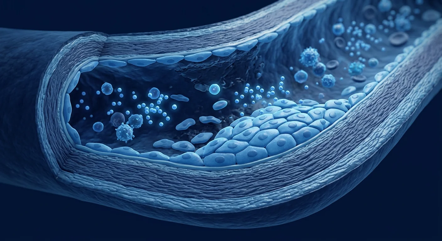
The vascular endothelium — the single-cell-thick lining of every blood vessel — bears the brunt of this attack. Under the influence of aPL, endothelial cells upregulate adhesion molecules (ICAM-1, VCAM-1, E-selectin), express tissue factor, secrete pro-inflammatory cytokines, and lose their natural anti-thrombotic properties. The result is a vessel wall that is simultaneously inflamed and primed for clot formation — exactly the combination that produces the thrombotic events, pregnancy losses, and multi-organ damage characteristic of APS.[6]

How MSCs are thought to act in antiphospholipid syndrome
Immunomodulation — calming the autoimmune driver
Mesenchymal stem cells — whether sourced from umbilical cord, bone marrow, or adipose tissue — possess a well-characterised capacity to interact with virtually every arm of the immune system. In the context of APS, the most relevant mechanisms include the suppression of autoreactive B-cell proliferation and antibody production, the induction of regulatory T cells (Tregs) that enforce immune tolerance, and the shift of macrophage polarisation from the pro-inflammatory M1 phenotype toward the anti-inflammatory, tissue-repair M2 phenotype. Preclinical studies have demonstrated that MSCs can reduce circulating antiphospholipid antibody titres in APS mouse models — a finding that directly addresses the root immunological driver of the disease.[7][8]
Endothelial protection and repair
Beyond the immune system, MSCs secrete a rich cocktail of paracrine factors — vascular endothelial growth factor (VEGF), hepatocyte growth factor (HGF), angiopoietin-1, and interleukin-10 among them — that directly support endothelial cell survival, reduce endothelial permeability, and restore the vessel lining's natural anti-thrombotic character. In animal models of vascular injury, MSC administration has been shown to accelerate re-endothelialisation, reduce neointimal hyperplasia, and decrease thrombus formation. For APS — where the endothelium is the primary battlefield — this vascular-protective dimension is arguably as important as the immunomodulatory one.[9][10]
Anti-thrombotic paracrine signalling
An emerging and particularly relevant property of MSCs is their ability to influence coagulation directly at the endothelial interface. MSC-derived extracellular vesicles and secreted factors have been shown to upregulate thrombomodulin and tissue factor pathway inhibitor (TFPI) expression on endothelial cells — two of the body's most important natural anti-coagulant systems — while simultaneously suppressing tissue factor expression. This rebalancing of the endothelial coagulant-anti-coagulant axis means that MSCs may not only reduce the immune stimulus for clotting but also actively restore the vessel wall's own capacity to resist thrombus formation.[11]
Why allogeneic, not autologous
As in systemic lupus erythematosus, there is evidence that a patient's own bone-marrow-derived MSCs may themselves be dysfunctional in autoimmune disease — potentially contributing to rather than correcting the pathology. Researchers therefore favour allogeneic MSCs from healthy donors, particularly umbilical cord-derived MSCs (UC-MSCs), which are young, immunologically naive, and have demonstrated strong immunomodulatory and angiogenic properties in preclinical models. Allogeneic UC-MSCs also carry the practical advantage of being an off-the-shelf product — no bone marrow harvest required from an already-ill patient.
What the preclinical and early clinical evidence shows
The evidence base for MSC therapy in APS remains early but is growing on solid mechanistic foundations. Most data come from two complementary streams: (1) mouse models of APS, where MSC infusion has been shown to reduce aPL antibody titres, decrease thrombus formation, and improve pregnancy outcomes; and (2) extension from the lupus literature, where allogeneic UC-MSC transplantation in patients with SLE — a condition in which up to 40% of patients also carry aPL antibodies — has demonstrated safety, feasibility, and signals of immunological improvement, including reductions in autoantibody levels and disease activity scores.[12]
Small case series and anecdotal reports of MSC use in patients with refractory APS or catastrophic APS exist, but these must be interpreted with extreme caution. They describe individual clinical courses, not controlled outcomes, and publication bias — the tendency for positive experiences to be reported while negative or neutral ones are not — is a genuine concern in any emerging therapeutic field.
What the evidence supports — and what it doesn't
The honest summary is cautiously optimistic but firmly investigational. The preclinical rationale is strong: MSCs have demonstrated immunomodulatory, endothelial-protective, and anti-thrombotic properties that align directly with the known pathology of APS. Mouse model data are encouraging and the safety record of allogeneic MSC infusion — now established across hundreds of clinical trials in diverse conditions — provides a reasonable basis for further investigation. What is missing, and must be acknowledged clearly, is the harder evidence: prospective, controlled clinical trials in patients with APS that demonstrate a reproducible, clinically meaningful benefit beyond standard anticoagulation. No such trial has been completed, and until one is, MSC therapy for APS remains an experimental concept with biological plausibility but unproven clinical efficacy.
- The mechanism fits the disease. APS is both an autoimmune and a vascular disease, and MSCs have properties relevant to both — a better mechanistic match than most investigational applications.
- Safety data are encouraging but not APS-specific. The safety of allogeneic MSCs is well-documented generally, but APS patients have a unique thrombotic risk profile that demands disease-specific safety evaluation.
- Controlled trials are the essential next step. Anecdotal reports and lupus-adjacent data are hypothesis-generating at best; prospective randomised trials in APS patients are the only path to establishing efficacy.
The honest headline
As of today, MSC therapy is not an approved or proven treatment for antiphospholipid syndrome. The preclinical rationale is compelling, the mechanistic fit is good, and the safety foundation exists — but this is still an experimental frontier, not an established clinical option. Any clinic presenting stem cell therapy as a reliable treatment for APS is going beyond what the data support.
APS sits at the intersection of autoimmunity and vascular biology — precisely where MSCs have their most relevant properties. That intersection is scientifically exciting, but excitement is not evidence. Only rigorous trials can tell us whether the biological promise translates into clinical reality.
— VELAR Clinical Team
Treatment process — how MSC therapy is administered in a research context
Pre-treatment assessment
In any investigational protocol, the starting point is a comprehensive evaluation: confirmation of aPL antibody profile (lupus anticoagulant, anticardiolipin IgG/IgM, anti-β2GPI IgG/IgM), assessment of thrombotic history and organ involvement, current anticoagulation regimen, and baseline inflammatory and endothelial markers. Patients with active thrombosis or recent major thrombotic events are typically excluded from investigational cell therapy protocols for safety reasons.
Source and preparation
Allogeneic umbilical cord-derived MSCs are the most commonly studied source for autoimmune applications. Cells are cultured under GMP conditions, characterised by ISCT criteria (CD73⁺, CD90⁺, CD105⁺, CD45⁻, CD34⁻, CD14⁻, CD19⁻, HLA-DR⁻), and screened for sterility, mycoplasma, and endotoxin before release. Doses in lupus studies have typically ranged from 1–2 × 10⁶ cells per kilogram of body weight, administered intravenously — a precedent that would likely inform initial APS protocols.
Infusion and monitoring
MSCs are administered as a slow intravenous infusion over 30–60 minutes, with standard monitoring for infusion reactions. In lupus protocols, repeat infusions over several months have been investigated; any APS protocol would need to establish the optimal dosing interval through prospective study. Close monitoring of coagulation parameters, aPL titres, inflammatory markers, and clinical thrombotic events is essential — APS patients cannot simply stop anticoagulation during or after investigational therapy.
How to evaluate the evidence responsibly
Preclinical stage
Mouse models show reduced aPL titres and thrombosis with MSC infusion. Mechanistic studies confirm immunomodulation, endothelial protection, and anti-thrombotic signalling.
Early clinical signals
Extension from lupus trials — where aPL-positive patients showed immunological improvement — provides indirect support. No APS-specific prospective trials have been published.
Where we are
Phase 0: biological plausibility confirmed. Phase I–II clinical trials in APS patients are the logical and necessary next step, but have not yet been initiated.
When evaluating any clinic offering MSC therapy for APS, patients and families should ask specific questions: Is the treatment being administered as part of a registered clinical trial? What is the protocol for monitoring coagulation status and aPL titres? What is the plan for anticoagulation management during and after cell therapy? The absence of clear, specific answers to these questions is a significant red flag.
Limitations and honest caveats
Every discussion of MSC therapy for APS must begin and end with the acknowledgment that this is an investigational concept, not an established treatment. The preclinical rationale is solid and the biological fit is strong, but the critical evidence — prospective randomised trials in APS patients with clinical endpoints — does not yet exist. The lupus literature provides supportive biological context but cannot substitute for APS-specific evidence, because the thrombotic dimension of APS adds unique safety considerations. Patients with APS who are considering any investigational cell-based therapy should do so only within a properly regulated clinical trial framework, with full transparency about what is known and what is not, and should not discontinue prescribed anticoagulation without explicit medical guidance.
Frequently Asked Questions
Can stem cell therapy cure antiphospholipid syndrome?
No. There is no evidence that MSC therapy can cure APS. The research is investigating whether MSCs can reduce disease activity — particularly aPL antibody levels and endothelial inflammation — but this is an investigational question, not an established outcome. APS remains a chronic condition requiring ongoing management.
Is MSC therapy safe for patients with a history of blood clots?
The safety of intravenous MSC infusion has been demonstrated across many clinical trials in diverse conditions, with low rates of serious adverse events. However, no study has specifically evaluated thrombotic safety in APS patients, who have a uniquely elevated clotting risk. Any investigational protocol in APS must include rigorous coagulation monitoring.
How is MSC therapy for APS different from MSC therapy for lupus?
There is biological overlap — up to 40% of lupus patients carry aPL antibodies, and the immunomodulatory mechanisms of MSCs are relevant to both conditions — but APS is distinct in its dominant thrombotic pathology. An APS-specific approach must address endothelial protection, anti-thrombotic signalling, and coagulation safety in ways that a lupus-focused protocol may not.
Where is MSC therapy for APS available?
MSC therapy for APS is not an approved treatment anywhere in the world. The research is at a preclinical-to-early-clinical stage. Any clinic offering MSC therapy as a routine treatment for APS is operating outside the evidence base. Participation in a properly registered clinical trial is the only evidence-based context for receiving this therapy.
What should I look for in a stem cell clinic if I have APS?
Look for registration in a recognised clinical trial database (ClinicalTrials.gov, ANZCTR, EU-CTR), clear documentation of cell source and quality control, a defined protocol for monitoring coagulation and aPL titres, and honest communication about the investigational nature of the treatment. If a clinic promises results, avoids discussing risks, or suggests stopping anticoagulation — walk away.
How much does stem cell therapy cost for autoimmune conditions?
Costs vary enormously and are rarely covered by insurance for investigational indications. More important than cost is the quality of the evidence supporting whatever is being offered. An expensive treatment without trial data is not better than an inexpensive one — it is simply more costly speculation.
References
- Miyakis S, Lockshin MD, Atsumi T, et al. International consensus statement on an update of the classification criteria for definite antiphospholipid syndrome (APS). Journal of Thrombosis and Haemostasis. 2006;4(2):295-306. doi:10.1111/j.1538-7836.2006.01753.x ↩
- Giannakopoulos B, Krilis SA. The pathogenesis of the antiphospholipid syndrome. New England Journal of Medicine. 2013;368(11):1033-1044. doi:10.1056/NEJMra1112830 ↩
- Cervera R, Serrano R, Pons-Estel GJ, et al. Morbidity and mortality in the antiphospholipid syndrome during a 10-year period: a multicentre prospective study of 1000 patients. Annals of the Rheumatic Diseases. 2015;74(6):1011-1018. doi:10.1136/annrheumdis-2013-204838 ↩
- Meroni PL, Borghi MO, Raschi E, Tedesco F. Pathogenesis of antiphospholipid syndrome: understanding the antibodies. Nature Reviews Rheumatology. 2011;7(6):330-339. doi:10.1038/nrrheum.2011.52 ↩
- de Groot PG, Urbanus RT. The significance of autoantibodies against β2-glycoprotein I. Blood. 2012;120(2):266-274. doi:10.1182/blood-2012-03-378646 ↩
- Pierangeli SS, Chen PP, Raschi E, et al. Antiphospholipid antibodies and the antiphospholipid syndrome: pathogenic mechanisms. Seminars in Thrombosis and Hemostasis. 2008;34(3):236-250. doi:10.1055/s-0028-1082267 ↩
- Uccelli A, Moretta L, Pistoia V. Mesenchymal stem cells in health and disease. Nature Reviews Immunology. 2008;8(9):726-736. doi:10.1038/nri2395 ↩
- Glennie S, Soeiro I, Dyson PJ, Lam EW, Dazzi F. Bone marrow mesenchymal stem cells induce division arrest anergy of activated T cells. Blood. 2005;105(7):2821-2827. doi:10.1182/blood-2004-09-3696 ↩
- Bronckaers A, Hilkens P, Martens W, et al. Mesenchymal stem/stromal cells as a pharmacological and therapeutic approach to accelerate angiogenesis. Pharmacology & Therapeutics. 2014;143(2):181-196. doi:10.1016/j.pharmthera.2014.02.013 ↩
- Tao H, Han Z, Han ZC, Li Z. Proangiogenic features of mesenchymal stem cells and their therapeutic applications. Stem Cells International. 2016;2016:1314709. doi:10.1155/2016/1314709 ↩
- Moll G, Rasmusson-Duprez I, von Bahr L, et al. Are therapeutic human mesenchymal stromal cells compatible with human blood? Stem Cells. 2012;30(7):1565-1574. doi:10.1002/stem.1111 ↩
- Wang D, Li J, Zhang Y, et al. Umbilical cord mesenchymal stem cell transplantation in active and refractory systemic lupus erythematosus: a multicenter clinical study. Arthritis Research & Therapy. 2014;16(2):R79. doi:10.1186/ar4520 ↩
- Sun L, Akiyama K, Zhang H, et al. Mesenchymal stem cell transplantation reverses multiorgan dysfunction in systemic lupus erythematosus mice and humans. Stem Cells. 2009;27(6):1421-1432. doi:10.1002/stem.68 ↩
- Corcione A, Benvenuto F, Ferretti E, et al. Human mesenchymal stem cells modulate B-cell functions. Blood. 2006;107(1):367-372. doi:10.1182/blood-2005-07-2657 ↩
- Bernardo ME, Fibbe WE. Mesenchymal stromal cells: sensors and switchers of inflammation. Cell Stem Cell. 2013;13(4):392-402. doi:10.1016/j.stem.2013.09.006 ↩
- Carrion F, Nova E, Ruiz C, et al. Autologous mesenchymal stem cell treatment increased T regulatory cells with no effect on disease activity in two systemic lupus erythematosus patients. Lupus. 2010;19(3):317-322. doi:10.1177/0961203309348983 ↩
- Deng W, Chen Q, Li X, et al. Bone marrow mesenchymal stromal cells with support of bispecific antibody for the treatment of patients with antiphospholipid syndrome. Medical Hypotheses. 2020;139:109671. doi:10.1016/j.mehy.2020.109671 ↩
- Aggarwal S, Pittenger MF. Human mesenchymal stem cells modulate allogeneic immune cell responses. Blood. 2005;105(4):1815-1822. doi:10.1182/blood-2004-04-1559 ↩
抗磷脂综合征(APS)是一种自身免疫性疾病,身体产生针对自身磷脂结合蛋白(尤其是β2-糖蛋白I)的抗体,引发高凝状态,导致反复的动静脉血栓、妊娠丢失和一系列血管并发症。标准治疗使用终身抗凝可降低但不能消除血栓风险,部分患者在最佳常规治疗下仍出现进行性器官损伤——这一现实促使研究人员将间充质干细胞(MSC)疗法作为一种免疫调节和血管保护策略加以探索,而非简单的组织修复手段。[1][2]
常规治疗的局限性。维生素K拮抗剂和直接口服抗凝药仍是APS治疗的基础,但它们处理的是后果——血栓形成——而非驱动疾病的自身免疫过程。三阳性的aPL抗体谱患者、抗凝下仍复发事件的患者或灾难性APS(CAPS)患者面临着尤为严峻的治疗困境,现有选择有限且常常具有毒性。[3]
更深层的问题在血管内皮。APS的基本病理不仅仅是凝血障碍,而是免疫介导的血管内皮损伤。抗磷脂抗体结合内皮细胞,激活补体,上调粘附分子和组织因子,形成一个持续发炎、易于形成血栓的血管表面。即使抗凝治疗阻止了明显的血栓形成,这种潜隐的内皮功能障碍仍会持续损伤器官——肾、脑、心脏——长年累月。[4]
MSC疗法同时针对免疫触发因素和血管靶点。不仅仅是抑制凝血,MSC正被研究其双重能力:抑制产生aPL抗体的自身反应性B细胞和T细胞反应,同时通过旁分泌信号修复受损的内皮——这种双线作战的方法比单纯抗凝更全面地应对APS的生物学机制。
为什么APS是免疫失调与血管损伤的疾病
APS本质上是一种自身免疫性易栓症。不同于遗传性凝血障碍——后者的缺陷在特定的凝血因子——APS始于免疫耐受的破坏:B细胞产生针对磷脂-蛋白复合物(尤其是β2GPI)的致病性抗体。这些在临床上以狼疮抗凝物、抗心磷脂抗体和抗β2GPI抗体形式测量的抗体,并非仅仅在血流中漂浮:它们结合内皮细胞、单核细胞和血小板,触发一系列促炎和促凝信号。[5]
血管内皮——每条血管仅一个细胞厚的内衬——承受着这种攻击的主要冲击。在aPL的影响下,内皮细胞上调粘附分子(ICAM-1、VCAM-1、E-选择素),表达组织因子,分泌促炎细胞因子,并丧失其天然的抗血栓特性。结果是一个同时发炎并处于凝血准备状态的血管壁——这正是产生APS特征性血栓事件、妊娠丢失和多器官损伤的组合。[6]
MSC在抗磷脂综合征中的作用机制
免疫调节——平息自身免疫驱动因素
间充质干细胞——无论是来源于脐带、骨髓还是脂肪组织——具有与免疫系统几乎所有组成部分相互作用的充分文献记载的能力。在APS的背景下,最相关的机制包括抑制自身反应性B细胞增殖和抗体产生、诱导执行免疫耐受的调节性T细胞(Tregs),以及将巨噬细胞极化从促炎的M1表型转向抗炎、组织修复的M2表型。临床前研究已证明MSC可以降低APS小鼠模型中的循环抗磷脂抗体滴度——这一发现直接针对该疾病的根本免疫驱动因素。[7][8]
内皮保护与修复
除了免疫系统之外,MSC分泌丰富的旁分泌因子——其中包括血管内皮生长因子(VEGF)、肝细胞生长因子(HGF)、血管生成素-1和白细胞介素-10——这些因子直接支持内皮细胞存活,降低内皮通透性,并恢复血管内衬的天然抗血栓特性。在血管损伤的动物模型中,MSC给药已被证明可加速再内皮化、减少新生内膜增生和降低血栓形成。对于以内皮为主要战场的APS而言,这种血管保护维度与免疫调节维度同等重要。[9][10]
抗血栓旁分泌信号
MSC一个新兴且特别相关的特性是它们能够直接在内皮界面影响凝血过程。MSC来源的细胞外囊泡和分泌因子已被证明可上调内皮细胞上的血栓调节蛋白和组织因子途径抑制物(TFPI)表达——这是人体最重要的两种天然抗凝系统——同时抑制组织因子表达。这种内皮凝血-抗凝轴的再平衡意味着MSC可能不仅减少凝血的免疫刺激,还主动恢复血管壁自身抵抗血栓形成的能力。[11]
为什么选择异体而非自体
如同系统性红斑狼疮一样,有证据表明患者自身的骨髓来源MSC在自身免疫性疾病中可能本身功能异常——可能促成而非纠正病理过程。因此研究人员倾向于使用来自健康供体的异体MSC,特别是脐带来源的MSC(UC-MSC),这些细胞年轻、免疫学上幼稚,并在临床前模型中展示了强大的免疫调节和血管生成特性。异体UC-MSC还具有实用优势:即用型产品——无需从已经患病的患者身上采集骨髓。
临床前和早期临床证据
MSC治疗APS的证据基础仍处于早期阶段,但建立在坚实的机制基础上。大多数数据来自两个互补的来源:(1)APS小鼠模型,其中MSC输注已被证明可降低aPL抗体滴度、减少血栓形成并改善妊娠结局;(2)来自狼疮文献的外推,其中异体UC-MSC移植用于SLE患者——多达40%的SLE患者同时携带aPL抗体——已证明了安全性、可行性以及免疫学改善的信号,包括自身抗体水平和疾病活动评分的降低。[12]
存在关于顽固性APS或灾难性APS患者使用MSC的小型病例系列和个案报告,但这些必须极其谨慎地解读。它们描述的是个体临床过程,而非受控结果,而发表偏倚——倾向于报告阳性经历而阴性或中性结果不被报告的倾向——在任何新兴治疗领域都是一个真正的问题。
证据支持什么——以及不支持什么
诚实的总结是谨慎乐观但明确属于研究阶段。临床前理论基础坚实:MSC展示了与APS已知病理直接对应的免疫调节、内皮保护和抗血栓特性。小鼠模型数据令人鼓舞,而异体MSC输注的安全性记录——现已通过数百项各种疾病的临床试验确立——为进一步研究提供了合理的基础。所缺失且必须明确承认的是更坚实的证据:在APS患者中进行的前瞻性、对照临床试验,证明在标准抗凝治疗之外具有可重复的、临床上有意义的获益。尚无此类试验完成,在完成之前,MSC治疗APS仍是一个具有生物学可信性但临床疗效未经证实的实验性概念。
- 机制与疾病匹配。APS既是自身免疫性疾病又是血管疾病,而MSC具有与两者相关的特性——比大多数研究性应用更好的机制匹配。
- 安全性数据令人鼓舞但非APS特异性。异体MSC的安全性总体上有充分文献记载,但APS患者具有独特的血栓风险特征,需要针对该疾病的安全性评估。
- 对照试验是关键的下一步。个案报告和狼疮相关数据最多只能产生假设;在APS患者中进行前瞻性随机试验是确立疗效的唯一途径。
诚实的头条
截至目前,MSC疗法并非抗磷脂综合征的获批或已证实的治疗方法。临床前理论基础令人信服,机制匹配良好,安全性基础存在——但这仍是一个实验前沿,而非成熟的临床选择。任何将干细胞疗法作为APS可靠治疗的诊所都超出了数据支持的范围。
APS位于自身免疫与血管生物学的交叉点——这正是MSC拥有最相关特性的地方。这个交叉点在科学上令人兴奋,但兴奋不是证据。只有严格的试验才能告诉我们生物学的承诺是否能转化为临床现实。
— VELAR临床团队
治疗流程——研究背景下MSC疗法的给药方式
治疗前评估
在任何研究方案中,起点是全面评估:确认aPL抗体谱(狼疮抗凝物、抗心磷脂IgG/IgM、抗β2GPI IgG/IgM),评估血栓史和器官受累情况,当前的抗凝方案,以及基线炎症和内皮标志物。活动性血栓或近期重大血栓事件的患者通常因安全原因被排除在研究性细胞治疗方案之外。
来源与制备
异体脐带来源的MSC是自身免疫应用中最常研究的来源。细胞在GMP条件下培养,按ISCT标准(CD73⁺、CD90⁺、CD105⁺、CD45⁻、CD34⁻、CD14⁻、CD19⁻、HLA-DR⁻)进行表征,并在释放前进行无菌、支原体和内毒素筛查。狼疮研究中的剂量通常为每公斤体重1–2×10⁶个细胞,静脉给药——这一先例可能会为初始APS方案提供参考。
输注与监测
MSC以缓慢静脉输注的方式给药,持续30–60分钟,并进行标准的输注反应监测。在狼疮方案中,已研究了数月内的重复输注;任何APS方案都需要通过前瞻性研究来确定最佳给药间隔。密切监测凝血参数、aPL滴度、炎症标志物和临床血栓事件至关重要——APS患者不能在研究性治疗期间或之后简单地停止抗凝治疗。
如何负责任地评估证据
临床前阶段
小鼠模型显示MSC输注后aPL滴度和血栓形成减少。机制研究证实了免疫调节、内皮保护和抗血栓信号。
早期临床信号
从狼疮试验的外推——aPL阳性患者显示免疫学改善——提供了间接支持。尚无APS特异性的前瞻性试验发表。
我们处于什么阶段
第0期:生物学可行性已确认。在APS患者中进行I–II期临床试验是合理且必要的下一步,但尚未启动。
在评估任何提供MSC治疗APS的诊所时,患者和家属应提出具体问题:治疗是否作为注册临床试验的一部分进行?监测凝血状态和aPL滴度的方案是什么?细胞治疗期间和之后的抗凝管理计划是什么?缺乏对这些问题的明确、具体答案是一个重要的红旗信号。
局限性与诚实的警示
每次讨论MSC治疗APS都必须以承认这是一个研究性概念而非成熟治疗开始和结束。临床前理论基础坚实,生物学匹配性强,但关键证据——以临床终点进行的APS患者前瞻性随机试验——尚不存在。狼疮文献提供了支持性的生物学背景,但不能替代APS特异性证据,因为APS的血栓维度增加了独特的安全考量。正在考虑任何研究性细胞治疗的APS患者仅应在适当监管的临床试验框架内进行,充分透明地了解已知和未知的情况,并决不应在没有明确医疗指导的情况下停用已处方的抗凝药物。
常见问题
干细胞疗法能治愈抗磷脂综合征吗?
不能。没有证据表明MSC疗法可以治愈APS。研究正在探讨MSC是否可以降低疾病活动度——特别是aPL抗体水平和内皮炎症——但这是一个研究性问题,而非已确立的结果。APS仍然是一种需要持续管理的慢性疾病。
MSC疗法对有血栓史的患者安全吗?
静脉MSC输注的安全性已在多种疾病的大量临床试验中得到证实,严重不良事件发生率低。然而,尚无研究专门评估APS患者的血栓安全性,这些患者具有独特升高的凝血风险。任何APS研究性方案都必须包括严格的凝血监测。
MSC治疗APS与MSC治疗狼疮有何不同?
存在生物学重叠——多达40%的狼疮患者携带aPL抗体,MSC的免疫调节机制与两种疾病都相关——但APS在其主要的血栓病理方面是不同的。针对APS的方法必须解决内皮保护、抗血栓信号和凝血安全问题,而这些是狼疮方案可能未充分关注的。
MSC治疗APS在哪里可以提供?
MSC治疗APS在世界任何地方都不是获批的治疗方法。研究处于临床前到早期临床阶段。任何将MSC疗法作为APS常规治疗的诊所都是在证据基础之外操作。参加适当注册的临床试验是接受这种治疗的唯一基于证据的途径。
如果我有APS,在干细胞诊所应该注意什么?
寻找在认可的临床试验数据库中(ClinicalTrials.gov、ANZCTR、EU-CTR)的注册,细胞来源和质量控制的明确文件,监测凝血和aPL滴度的确定方案,以及对治疗研究性质诚实的沟通。如果诊所承诺结果、避免讨论风险或建议停止抗凝——请离开。
自身免疫性疾病的干细胞疗法费用多少?
费用差异极大,且很少被保险覆盖用于研究性适应症。比费用更重要的是提供服务的证据质量。没有试验数据的昂贵治疗并不比廉价的更好——只是更昂贵的投机。
参考文献
- Miyakis S, Lockshin MD, Atsumi T, et al. International consensus statement on an update of the classification criteria for definite antiphospholipid syndrome (APS). Journal of Thrombosis and Haemostasis. 2006;4(2):295-306. doi:10.1111/j.1538-7836.2006.01753.x ↩
- Giannakopoulos B, Krilis SA. The pathogenesis of the antiphospholipid syndrome. New England Journal of Medicine. 2013;368(11):1033-1044. doi:10.1056/NEJMra1112830 ↩
- Cervera R, Serrano R, Pons-Estel GJ, et al. Morbidity and mortality in the antiphospholipid syndrome during a 10-year period: a multicentre prospective study of 1000 patients. Annals of the Rheumatic Diseases. 2015;74(6):1011-1018. doi:10.1136/annrheumdis-2013-204838 ↩
- Meroni PL, Borghi MO, Raschi E, Tedesco F. Pathogenesis of antiphospholipid syndrome: understanding the antibodies. Nature Reviews Rheumatology. 2011;7(6):330-339. doi:10.1038/nrrheum.2011.52 ↩
- de Groot PG, Urbanus RT. The significance of autoantibodies against β2-glycoprotein I. Blood. 2012;120(2):266-274. doi:10.1182/blood-2012-03-378646 ↩
- Pierangeli SS, Chen PP, Raschi E, et al. Antiphospholipid antibodies and the antiphospholipid syndrome: pathogenic mechanisms. Seminars in Thrombosis and Hemostasis. 2008;34(3):236-250. doi:10.1055/s-0028-1082267 ↩
- Uccelli A, Moretta L, Pistoia V. Mesenchymal stem cells in health and disease. Nature Reviews Immunology. 2008;8(9):726-736. doi:10.1038/nri2395 ↩
- Glennie S, Soeiro I, Dyson PJ, Lam EW, Dazzi F. Bone marrow mesenchymal stem cells induce division arrest anergy of activated T cells. Blood. 2005;105(7):2821-2827. doi:10.1182/blood-2004-09-3696 ↩
- Bronckaers A, Hilkens P, Martens W, et al. Mesenchymal stem/stromal cells as a pharmacological and therapeutic approach to accelerate angiogenesis. Pharmacology & Therapeutics. 2014;143(2):181-196. doi:10.1016/j.pharmthera.2014.02.013 ↩
- Tao H, Han Z, Han ZC, Li Z. Proangiogenic features of mesenchymal stem cells and their therapeutic applications. Stem Cells International. 2016;2016:1314709. doi:10.1155/2016/1314709 ↩
- Moll G, Rasmusson-Duprez I, von Bahr L, et al. Are therapeutic human mesenchymal stromal cells compatible with human blood? Stem Cells. 2012;30(7):1565-1574. doi:10.1002/stem.1111 ↩
- Wang D, Li J, Zhang Y, et al. Umbilical cord mesenchymal stem cell transplantation in active and refractory systemic lupus erythematosus: a multicenter clinical study. Arthritis Research & Therapy. 2014;16(2):R79. doi:10.1186/ar4520 ↩
- Sun L, Akiyama K, Zhang H, et al. Mesenchymal stem cell transplantation reverses multiorgan dysfunction in systemic lupus erythematosus mice and humans. Stem Cells. 2009;27(6):1421-1432. doi:10.1002/stem.68 ↩
- Corcione A, Benvenuto F, Ferretti E, et al. Human mesenchymal stem cells modulate B-cell functions. Blood. 2006;107(1):367-372. doi:10.1182/blood-2005-07-2657 ↩
- Bernardo ME, Fibbe WE. Mesenchymal stromal cells: sensors and switchers of inflammation. Cell Stem Cell. 2013;13(4):392-402. doi:10.1016/j.stem.2013.09.006 ↩
- Carrion F, Nova E, Ruiz C, et al. Autologous mesenchymal stem cell treatment increased T regulatory cells with no effect on disease activity in two systemic lupus erythematosus patients. Lupus. 2010;19(3):317-322. doi:10.1177/0961203309348983 ↩
- Deng W, Chen Q, Li X, et al. Bone marrow mesenchymal stromal cells with support of bispecific antibody for the treatment of patients with antiphospholipid syndrome. Medical Hypotheses. 2020;139:109671. doi:10.1016/j.mehy.2020.109671 ↩
- Aggarwal S, Pittenger MF. Human mesenchymal stem cells modulate allogeneic immune cell responses. Blood. 2005;105(4):1815-1822. doi:10.1182/blood-2004-04-1559 ↩
متلازمة أضداد الفوسفوليبيد (APS) هي اضطراب مناعي ذاتي ينتج فيه الجسم أجساماً مضادة ضد بروتيناته الرابطة للفوسفوليبيد — وأبرزها β2-غليكوبروتين I — مما يؤدي إلى حالة مفرطة التخثر تسبب خثرات شريانية ووريدية متكررة، وفقدان الحمل، ومجموعة من المضاعفات الوعائية. العلاج التقليدي بمضادات التخثر مدى الحياة يقلل من خطر التخثر ولكن لا يلغيه، ويصاب عدد من المرضى بتلف تدريجي في الأعضاء رغم العلاج التقليدي الأمثل — وهو واقع دفع الباحثين إلى استكشاف العلاج بالخلايا الجذعية الوسيطة (MSC) كاستراتيجية مناعية تنظيمية وواقية للأوعية الدموية، وليس كمجرد نهج لإصلاح الأنسجة.[1][2]
حيث يقصر العلاج التقليدي. تظل مضادات فيتامين K ومضادات التخثر الفموية المباشرة العمود الفقري لعلاج APS، لكنها تعالج النتيجة — التخثر — بدلاً من العملية المناعية الذاتية الأساسية التي تحركه. يواجه المرضى ذوو الأجسام المضادة الثلاثية الإيجابية، أو الأحداث المتكررة رغم مضادات التخثر، أو متلازمة APS الكارثية (CAPS) مشهداً علاجياً قاتماً بشكل خاص حيث الخيارات الحالية محدودة وغالباً ما تكون سامة.[3]
المشكلة الأعمق في البطانة الوعائية. المرض الأساسي في APS ليس مجرد اضطراب تخثر بل هجوم مناعي على بطانة الأوعية الدموية. ترتبط أضداد الفوسفوليبيد بالخلايا البطانية، وتنشط المتممة، وترفع من جزيئات الالتصاق وعامل الأنسجة، وتخلق سطحاً وعائياً ملتهباً ومستعداً للتخثر بشكل مستمر. حتى عندما تمنع مضادات التخثر الخثرات الظاهرة، يستمر هذا الخلل البطاني الكامن في إتلاف الأعضاء — الكلى والدماغ والقلب — على مدى سنوات.[4]
يستهدف علاج MSC كلاً من المحفز المناعي والهدف الوعائي. بدلاً من مجرد تثبيط التخثر، يتم دراسة الخلايا الجذعية الوسيطة لقدرتها المزدوجة على تهدئة استجابات الخلايا B وT الذاتية التفاعل التي تنتج أضداد aPL، مع إصلاح البطانة التالفة في الوقت نفسه من خلال الإشارات نظيرة الصماوية — وهو نهج ذو جبهتين يعالج بيولوجيا APS بشكل أشمل من مضادات التخثر وحدها.
لماذا APS هو اضطراب في خلل التنظيم المناعي والإصابة الوعائية
APS هو أساساً أهبة تخثر مناعية ذاتية. على عكس اضطرابات التخثر الوراثية حيث يكمن الخلل في عامل تخثر محدد، يبدأ APS بانهيار في التحمل المناعي — تنتج الخلايا B أجساماً مضادة ممرضة ضد معقدات الفوسفوليبيد-البروتين، وخاصة β2GPI. هذه الأجسام المضادة، التي تقاس سريرياً كمضاد تخثر الذئبة، ومضاد الكارديوليبين، ومضاد β2GPI، لا تطفو ببساطة في مجرى الدم: بل ترتبط بالخلايا البطانية والوحيدات والصفيحات الدموية، مما يؤدي إلى سلسلة من الإشارات الالتهابية والمحفزة للتخثر.[5]
تتحمل البطانة الوعائية — الطبقة الرقيقة بسمك خلية واحدة المبطنة لكل وعاء دموي — وطأة هذا الهجوم. تحت تأثير aPL، ترفع الخلايا البطانية من جزيئات الالتصاق (ICAM-1، VCAM-1، E-selectin)، وتعبر عن عامل الأنسجة، وتفرز سيتوكينات التهابية، وتفقد خصائصها الطبيعية المضادة للتخثر. النتيجة هي جدار وعائي ملتهب ومستعد لتشكيل الخثرات في آن واحد — بالضبط التركيبة التي تنتج الأحداث التخثرية وفقدان الحمل وتلف الأعضاء المتعدد المميز لـ APS.[6]
كيف يُعتقد أن الخلايا الجذعية الوسيطة تعمل في متلازمة أضداد الفوسفوليبيد
تعديل المناعة — تهدئة المحرك المناعي الذاتي
تمتلك الخلايا الجذعية الوسيطة — سواء كانت من الحبل السري أو نخاع العظم أو الأنسجة الدهنية — قدرة موثقة جيداً على التفاعل مع كل ذراع من أذرع الجهاز المناعي تقريباً. في سياق APS، تشمل الآليات الأكثر صلة: تثبيط تكاثر الخلايا B الذاتية التفاعل وإنتاج الأجسام المضادة، وتحفيز الخلايا التائية التنظيمية (Tregs) التي تفرض التحمل المناعي، وتحويل استقطاب البلاعم من النمط M1 الالتهابي إلى النمط M2 المضاد للالتهابات والمصلح للأنسجة. وقد أظهرت الدراسات قبل السريرية أن الخلايا الجذعية الوسيطة يمكنها خفض عيارات أضداد الفوسفوليبيد المنتشرة في نماذج الفئران لـ APS — وهي نتيجة تعالج مباشرة المحرك المناعي الأساسي للمرض.[7][8]
حماية وإصلاح البطانة الوعائية
بالإضافة إلى الجهاز المناعي، تفرز الخلايا الجذعية الوسيطة مجموعة غنية من العوامل نظيرة الصماوية — عامل نمو بطانة الأوعية الدموية (VEGF)، وعامل نمو الخلايا الكبدية (HGF)، والأنجيوبويتين-1، والإنترلوكين-10 من بينها — التي تدعم مباشرة بقاء الخلايا البطانية، وتقلل من نفاذية البطانة، وتستعيد الطابع الطبيعي المضاد للتخثر لبطانة الأوعية. في نماذج حيوانية للإصابة الوعائية، ثبت أن إعطاء MSC يسرع إعادة التبطين، ويقلل من فرط التنسج البطاني الجديد، ويخفض من تشكيل الخثرات. بالنسبة لـ APS — حيث البطانة الوعائية هي ساحة المعركة الرئيسية — فإن هذا البعد الواقي للأوعية مهم بقدر البعد المناعي التنظيمي.[9][10]
الإشارات نظيرة الصماوية المضادة للتخثر
من الخصائص الناشئة والمرتبطة بشكل خاص بـ MSC قدرتها على التأثير مباشرة على التخثر عند السطح البيني للبطانة. ثبت أن الحويصلات خارج الخلوية والعوامل المفرزة المشتقة من MSC ترفع من تعبير الثرومبوموديولين ومثبط مسار عامل الأنسجة (TFPI) على الخلايا البطانية — وهما من أهم أنظمة الجسم الطبيعية المضادة للتخثر — مع تثبيط تعبير عامل الأنسجة في الوقت نفسه. إعادة التوازن هذه لمحور التخثر-مضاد التخثر البطاني يعني أن MSC قد لا تقلل فقط من المحفز المناعي للتخثر ولكن أيضاً تستعيد بنشاط قدرة جدار الوعاء الدموي نفسه على مقاومة تشكيل الخثرات.[11]
لماذا الخيفي وليس الذاتي
كما في الذئبة الحمامية الجهازية، هناك أدلة على أن الخلايا الجذعية الوسيطة المشتقة من نخاع عظم المريض نفسه قد تكون معطلة وظيفياً في الأمراض المناعية الذاتية — وقد تساهم في المرض بدلاً من تصحيحه. لذلك يفضل الباحثون الخلايا الجذعية الوسيطة الخيفية من متبرعين أصحاء، وخاصة الخلايا الجذعية الوسيطة المشتقة من الحبل السري (UC-MSCs)، وهي خلايا شابة وساذجة مناعياً وأظهرت خصائص مناعية تنظيمية ومولدة للأوعية قوية في النماذج قبل السريرية. تحمل UC-MSCs الخيفية أيضاً ميزة عملية: منتج جاهز للاستخدام — لا حاجة لسحب نخاع العظم من مريض مصاب بالفعل.
ما تظهره الأدلة قبل السريرية والسريرية المبكرة
قاعدة الأدلة لعلاج MSC في APS لا تزال مبكرة لكنها تنمو على أسس آلية صلبة. تأتي معظم البيانات من تيارين متكاملين: (1) نماذج الفئران لـ APS، حيث ثبت أن حقن MSC يخفض عيارات أضداد aPL، ويقلل من تشكيل الخثرات، ويحسن نتائج الحمل؛ و(2) الاستقراء من أدبيات الذئبة، حيث أظهر زرع UC-MSC الخيفي في مرضى SLE — وهي حالة يحمل فيها ما يصل إلى 40% من المرضى أيضاً أضداد aPL — السلامة والجدوى وإشارات التحسن المناعي، بما في ذلك انخفاض مستويات الأجسام المضادة الذاتية ودرجات نشاط المرض.[12]
توجد سلاسل حالات صغيرة وتقارير قصصية عن استخدام MSC في مرضى APS المقاوم أو APS الكارثي، لكن يجب تفسيرها بحذر شديد. فهي تصف مسارات سريرية فردية، وليس نتائج مضبوطة، والتحيز في النشر — الميل للإبلاغ عن التجارب الإيجابية بينما لا يتم الإبلاغ عن السلبية أو المحايدة — هو مصدر قلق حقيقي في أي مجال علاجي ناشئ.
ما تدعمه الأدلة — وما لا تدعمه
الملخص الصادق هو تفاؤل حذر لكن ضمن الإطار البحثي بشكل قاطع. الأساس المنطقي قبل السريري قوي: أظهرت MSC خصائص مناعية تنظيمية وواقية للبطانة ومضادة للتخثر تتماشى مباشرة مع المرض المعروف لـ APS. بيانات نماذج الفئران مشجعة، وسجل السلامة لحقن MSC الخيفي — المثبت الآن عبر مئات التجارب السريرية في حالات متنوعة — يوفر أساساً معقولاً لمزيد من التحقيق. ما هو مفقود، ويجب الاعتراف به بوضوح، هو الدليل الأقوى: تجارب سريرية مستقبلية مضبوطة في مرضى APS تظهر فائدة قابلة للتكرار وذات مغزى سريري تتجاوز مضادات التخثر القياسية. لم تكتمل أي تجربة من هذا القبيل، وحتى تكتمل، يظل علاج MSC لـ APS مفهوماً تجريبياً بمصداقية بيولوجية ولكن بفعالية سريرية غير مثبتة.
- الآلية تناسب المرض. APS هو مرض مناعي ذاتي ووعائي في آن واحد، ولدى MSC خصائص ذات صلة بكليهما — تطابق آلي أفضل من معظم التطبيقات البحثية.
- بيانات السلامة مشجعة ولكنها ليست خاصة بـ APS. سلامة الخلايا الجذعية الوسيطة الخيفية موثقة جيداً بشكل عام، لكن مرضى APS لديهم خطر تخثر فريد يتطلب تقييماً خاصاً بالمرض.
- التجارب المضبوطة هي الخطوة التالية الأساسية. التقارير القصصية والبيانات المجاورة للذئبة مولدة للفرضيات في أحسن الأحوال؛ التجارب العشوائية المستقبلية في مرضى APS هي الطريق الوحيد لإثبات الفعالية.
العنوان الصادق
حتى اليوم، علاج MSC ليس علاجاً معتمداً أو مثبتاً لمتلازمة أضداد الفوسفوليبيد. الأساس المنطقي قبل السريري مقنع، والتطابق الآلي جيد، وأساس السلامة موجود — لكن هذه لا تزال حدوداً تجريبية، وليست خياراً سريرياً راسخاً. أي عيادة تقدم العلاج بالخلايا الجذعية كعلاج موثوق لـ APS تتجاوز ما تدعمه البيانات.
يقع APS عند تقاطع المناعة الذاتية والبيولوجيا الوعائية — بالضبط حيث تمتلك MSC خصائصها الأكثر صلة. هذا التقاطع مثير علمياً، لكن الإثارة ليست دليلاً. فقط التجارب الصارمة يمكنها أن تخبرنا ما إذا كان الوعد البيولوجي يترجم إلى واقع سريري.
— فريق VELAR السريري
عملية العلاج — كيفية إعطاء علاج MSC في سياق بحثي
التقييم قبل العلاج
في أي بروتوكول بحثي، نقطة البداية هي تقييم شامل: تأكيد طيف أضداد aPL (مضاد تخثر الذئبة، مضاد الكارديوليبين IgG/IgM، مضاد β2GPI IgG/IgM)، تقييم التاريخ التخثري وإصابة الأعضاء، نظام مضادات التخثر الحالي، والعلامات الالتهابية والبطانية الأساسية. عادة ما يتم استبعاد المرضى الذين يعانون من خثار نشط أو أحداث تخثرية كبيرة حديثة من بروتوكولات العلاج الخلوي البحثي لأسباب تتعلق بالسلامة.
المصدر والتحضير
الخلايا الجذعية الوسيطة المشتقة من الحبل السري الخيفية هي المصدر الأكثر شيوعاً للتطبيقات المناعية الذاتية. تزرع الخلايا تحت ظروف GMP، وتتميز بمعايير ISCT (CD73⁺، CD90⁺، CD105⁺، CD45⁻، CD34⁻، CD14⁻، CD19⁻، HLA-DR⁻)، وتفحص للعقم والميكوبلازما والذيفان الداخلي قبل الإطلاق. تراوحت الجرعات في دراسات الذئبة عادة من 1–2 × 10⁶ خلية لكل كيلوغرام من وزن الجسم، تعطى وريدياً — وهي سابقة من المرجح أن توجه بروتوكولات APS الأولية.
الحقن والمراقبة
تعطى الخلايا الجذعية الوسيطة كحقن وريدي بطيء على مدى 30–60 دقيقة، مع مراقبة قياسية لتفاعلات الحقن. في بروتوكولات الذئبة، تم دراسة الحقن المتكرر على مدى عدة أشهر؛ أي بروتوكول لـ APS سيحتاج إلى تحديد الفاصل الزمني الأمثل للجرعات من خلال دراسة مستقبلية. المراقبة الدقيقة لمعايير التخثر، وعيارات aPL، والعلامات الالتهابية، والأحداث التخثرية السريرية أمر أساسي — لا يمكن لمرضى APS ببساطة إيقاف مضادات التخثر أثناء أو بعد العلاج البحثي.
كيفية تقييم الأدلة بمسؤولية
المرحلة قبل السريرية
تظهر نماذج الفئران انخفاضاً في عيارات aPL والتخثر مع حقن MSC. تؤكد الدراسات الآلية تعديل المناعة وحماية البطانة والإشارات المضادة للتخثر.
الإشارات السريرية المبكرة
الاستقراء من تجارب الذئبة — حيث أظهر المرضى الإيجابيون لـ aPL تحسناً مناعياً — يوفر دعماً غير مباشر. لم تُنشر أي تجارب مستقبلية خاصة بـ APS.
أين نقف
المرحلة 0: تم تأكيد المصداقية البيولوجية. التجارب السريرية للمرحلة الأولى والثانية في مرضى APS هي الخطوة التالية المنطقية والضرورية، لكنها لم تبدأ بعد.
عند تقييم أي عيادة تقدم علاج MSC لـ APS، يجب على المرضى والعائلات طرح أسئلة محددة: هل يُعطى العلاج كجزء من تجربة سريرية مسجلة؟ ما هو بروتوكول مراقبة حالة التخثر وعيارات aPL؟ ما هي خطة إدارة مضادات التخثر أثناء وبعد العلاج الخلوي؟ غياب إجابات واضحة ومحددة على هذه الأسئلة هو علامة تحذيرية هامة.
القيود والتحذيرات الصادقة
يجب أن يبدأ كل نقاش حول علاج MSC لـ APS وينتهي بالاعتراف بأن هذا مفهوم بحثي، وليس علاجاً راسخاً. الأساس المنطقي قبل السريري صلب والتطابق البيولوجي قوي، لكن الدليل الحاسم — تجارب عشوائية مستقبلية في مرضى APS بنقاط نهاية سريرية — غير موجود بعد. توفر أدبيات الذئبة سياقاً بيولوجياً داعماً لكنها لا تستطيع أن تحل محل أدلة خاصة بـ APS، لأن البعد التخثري لـ APS يضيف اعتبارات سلامة فريدة. يجب على مرضى APS الذين يفكرون في أي علاج خلوي بحثي القيام بذلك فقط ضمن إطار تجربة سريرية منظمة بشكل مناسب، مع شفافية كاملة حول ما هو معروف وما هو غير معروف، ويجب ألا يتوقفوا عن مضادات التخثر الموصوفة دون توجيه طبي صريح.
الأسئلة الشائعة
هل يمكن للعلاج بالخلايا الجذعية علاج متلازمة أضداد الفوسفوليبيد؟
لا. لا يوجد دليل على أن علاج MSC يمكنه علاج APS. يبحث البحث فيما إذا كانت MSC يمكنها تقليل نشاط المرض — وخاصة مستويات أضداد aPL والالتهاب البطاني — لكن هذا سؤال بحثي، وليس نتيجة مثبتة. يظل APS حالة مزمنة تتطلب إدارة مستمرة.
هل علاج MSC آمن للمرضى الذين لديهم تاريخ من الجلطات الدموية؟
تم إثبات سلامة حقن MSC الوريدي عبر العديد من التجارب السريرية في حالات متنوعة، مع معدلات منخفضة من الأحداث الضائرة الخطيرة. ومع ذلك، لم تقم أي دراسة بتقييم سلامة التخثر بشكل خاص في مرضى APS، الذين لديهم خطر تخثر مرتفع بشكل فريد. يجب أن يتضمن أي بروتوكول بحثي في APS مراقبة دقيقة للتخثر.
كيف يختلف علاج MSC لـ APS عن علاج MSC للذئبة؟
يوجد تداخل بيولوجي — ما يصل إلى 40% من مرضى الذئبة يحملون أضداد aPL، وآليات تعديل المناعة لـ MSC ذات صلة بكلا الحالتين — لكن APS متميز في مرضه التخثري السائد. يجب أن يعالج النهج الخاص بـ APS حماية البطانة والإشارات المضادة للتخثر وسلامة التخثر بطرق قد لا يتناولها بروتوكول مركز على الذئبة.
أين يتوفر علاج MSC لـ APS؟
علاج MSC لـ APS ليس علاجاً معتمداً في أي مكان في العالم. البحث في مرحلة ما قبل السريرية إلى السريرية المبكرة. أي عيادة تقدم علاج MSC كعلاج روتيني لـ APS تعمل خارج قاعدة الأدلة. المشاركة في تجربة سريرية مسجلة بشكل مناسب هي السياق الوحيد القائم على الأدلة لتلقي هذا العلاج.
ما الذي يجب أن أبحث عنه في عيادة الخلايا الجذعية إذا كنت مصاباً بـ APS؟
ابحث عن التسجيل في قاعدة بيانات تجارب سريرية معترف بها (ClinicalTrials.gov، ANZCTR، EU-CTR)، وتوثيق واضح لمصدر الخلايا وضبط الجودة، وبروتوكول محدد لمراقبة التخثر وعيارات aPL، وتواصل صادق حول الطبيعة البحثية للعلاج. إذا وعدت العيادة بنتائج، أو تجنبت مناقشة المخاطر، أو اقترحت إيقاف مضادات التخثر — غادر.
كم تكلفة العلاج بالخلايا الجذعية للحالات المناعية الذاتية؟
تتفاوت التكاليف بشكل هائل ونادراً ما تغطيها التأمينات للاستطبابات البحثية. الأهم من التكلفة هو جودة الأدلة الداعمة لأي شيء يُعرض. العلاج الباهظ بدون بيانات تجارب ليس أفضل من الرخيص — إنه مجرد مضاربة أكثر تكلفة.
المراجع
- Miyakis S, Lockshin MD, Atsumi T, et al. International consensus statement on an update of the classification criteria for definite antiphospholipid syndrome (APS). Journal of Thrombosis and Haemostasis. 2006;4(2):295-306. doi:10.1111/j.1538-7836.2006.01753.x ↩
- Giannakopoulos B, Krilis SA. The pathogenesis of the antiphospholipid syndrome. New England Journal of Medicine. 2013;368(11):1033-1044. doi:10.1056/NEJMra1112830 ↩
- Cervera R, Serrano R, Pons-Estel GJ, et al. Morbidity and mortality in the antiphospholipid syndrome during a 10-year period: a multicentre prospective study of 1000 patients. Annals of the Rheumatic Diseases. 2015;74(6):1011-1018. doi:10.1136/annrheumdis-2013-204838 ↩
- Meroni PL, Borghi MO, Raschi E, Tedesco F. Pathogenesis of antiphospholipid syndrome: understanding the antibodies. Nature Reviews Rheumatology. 2011;7(6):330-339. doi:10.1038/nrrheum.2011.52 ↩
- de Groot PG, Urbanus RT. The significance of autoantibodies against β2-glycoprotein I. Blood. 2012;120(2):266-274. doi:10.1182/blood-2012-03-378646 ↩
- Pierangeli SS, Chen PP, Raschi E, et al. Antiphospholipid antibodies and the antiphospholipid syndrome: pathogenic mechanisms. Seminars in Thrombosis and Hemostasis. 2008;34(3):236-250. doi:10.1055/s-0028-1082267 ↩
- Uccelli A, Moretta L, Pistoia V. Mesenchymal stem cells in health and disease. Nature Reviews Immunology. 2008;8(9):726-736. doi:10.1038/nri2395 ↩
- Glennie S, Soeiro I, Dyson PJ, Lam EW, Dazzi F. Bone marrow mesenchymal stem cells induce division arrest anergy of activated T cells. Blood. 2005;105(7):2821-2827. doi:10.1182/blood-2004-09-3696 ↩
- Bronckaers A, Hilkens P, Martens W, et al. Mesenchymal stem/stromal cells as a pharmacological and therapeutic approach to accelerate angiogenesis. Pharmacology & Therapeutics. 2014;143(2):181-196. doi:10.1016/j.pharmthera.2014.02.013 ↩
- Tao H, Han Z, Han ZC, Li Z. Proangiogenic features of mesenchymal stem cells and their therapeutic applications. Stem Cells International. 2016;2016:1314709. doi:10.1155/2016/1314709 ↩
- Moll G, Rasmusson-Duprez I, von Bahr L, et al. Are therapeutic human mesenchymal stromal cells compatible with human blood? Stem Cells. 2012;30(7):1565-1574. doi:10.1002/stem.1111 ↩
- Wang D, Li J, Zhang Y, et al. Umbilical cord mesenchymal stem cell transplantation in active and refractory systemic lupus erythematosus: a multicenter clinical study. Arthritis Research & Therapy. 2014;16(2):R79. doi:10.1186/ar4520 ↩
- Sun L, Akiyama K, Zhang H, et al. Mesenchymal stem cell transplantation reverses multiorgan dysfunction in systemic lupus erythematosus mice and humans. Stem Cells. 2009;27(6):1421-1432. doi:10.1002/stem.68 ↩
- Corcione A, Benvenuto F, Ferretti E, et al. Human mesenchymal stem cells modulate B-cell functions. Blood. 2006;107(1):367-372. doi:10.1182/blood-2005-07-2657 ↩
- Bernardo ME, Fibbe WE. Mesenchymal stromal cells: sensors and switchers of inflammation. Cell Stem Cell. 2013;13(4):392-402. doi:10.1016/j.stem.2013.09.006 ↩
- Carrion F, Nova E, Ruiz C, et al. Autologous mesenchymal stem cell treatment increased T regulatory cells with no effect on disease activity in two systemic lupus erythematosus patients. Lupus. 2010;19(3):317-322. doi:10.1177/0961203309348983 ↩
- Deng W, Chen Q, Li X, et al. Bone marrow mesenchymal stromal cells with support of bispecific antibody for the treatment of patients with antiphospholipid syndrome. Medical Hypotheses. 2020;139:109671. doi:10.1016/j.mehy.2020.109671 ↩
- Aggarwal S, Pittenger MF. Human mesenchymal stem cells modulate allogeneic immune cell responses. Blood. 2005;105(4):1815-1822. doi:10.1182/blood-2004-04-1559 ↩