Allergic rhinitis (AR) — commonly known as hay fever — affects an estimated 400 million people worldwide, making it one of the most prevalent chronic respiratory conditions across all age groups. [1] Patients endure a debilitating cycle of nasal congestion, rhinorrhea, sneezing, nasal itching, and postnasal drip that disrupts sleep, impairs concentration at work or school, and significantly reduces quality of life. The economic burden is substantial: lost productivity, direct medical costs, and school absence due to allergic rhinitis exceed USD 20 billion annually in the United States alone.
Where conventional treatment falls short. The standard of care — oral antihistamines, intranasal corticosteroids (INCS), leukotriene receptor antagonists, and allergen immunotherapy (AIT) — manages symptoms in many patients but leaves a significant proportion undertreated or dissatisfied. Up to 40% of patients report inadequate symptom control with first-line therapies, and many discontinue intranasal steroids due to local side effects (nasal dryness, epistaxis, septal perforation with long-term use). [2] Allergen immunotherapy — the only disease-modifying approach currently available — requires 3–5 years of regular injections or sublingual doses, is contraindicated in severe asthma or autoimmune disease, and carries a risk of anaphylaxis with subcutaneous administration. Even with optimal pharmacotherapy, about 20% of patients with moderate-to-severe allergic rhinitis remain poorly controlled [3].
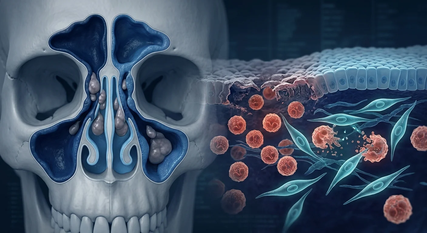
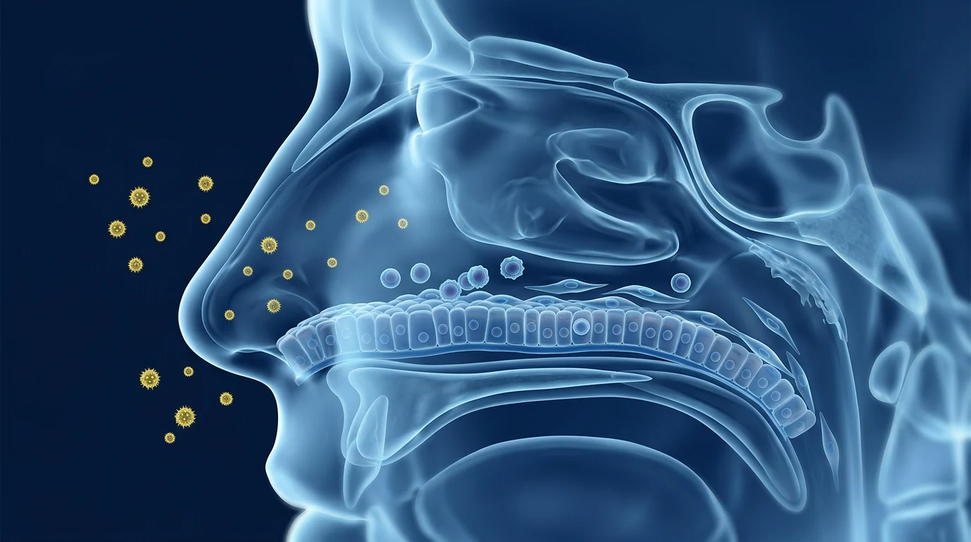
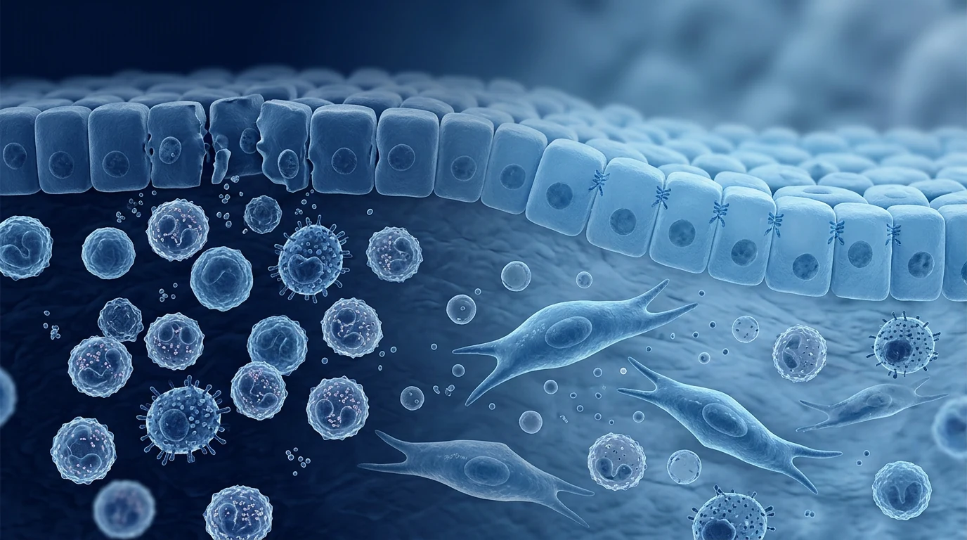
The deeper problem is immune dysregulation at the mucosal surface. Allergic rhinitis is fundamentally a disorder of immune tolerance failure at the nasal mucosal interface. In genetically predisposed individuals, exposure to otherwise harmless environmental allergens — pollen, dust mites, pet dander, mould — triggers a coordinated type 2 immune response orchestrated by allergen-specific IgE, mast cell degranulation, eosinophil infiltration, and Th2-biased T-cell activity. [4] This cascade produces symptoms through two phases: the early-phase response (within minutes, IgE-driven mast cell degranulation releasing histamine, leukotrienes, and prostaglandins) and the late-phase response (4-24 hours, sustained eosinophilic and basophilic inflammation driven by IL-4, IL-5, and IL-13 secreted by Th2 cells and group 2 innate lymphoid cells [ILC2s]). The nasal epithelium itself is not merely a passive barrier but an active immunological participant, releasing alarmins (IL-25, IL-33, TSLP) upon allergen encounter that amplify the type 2 cascade [5].
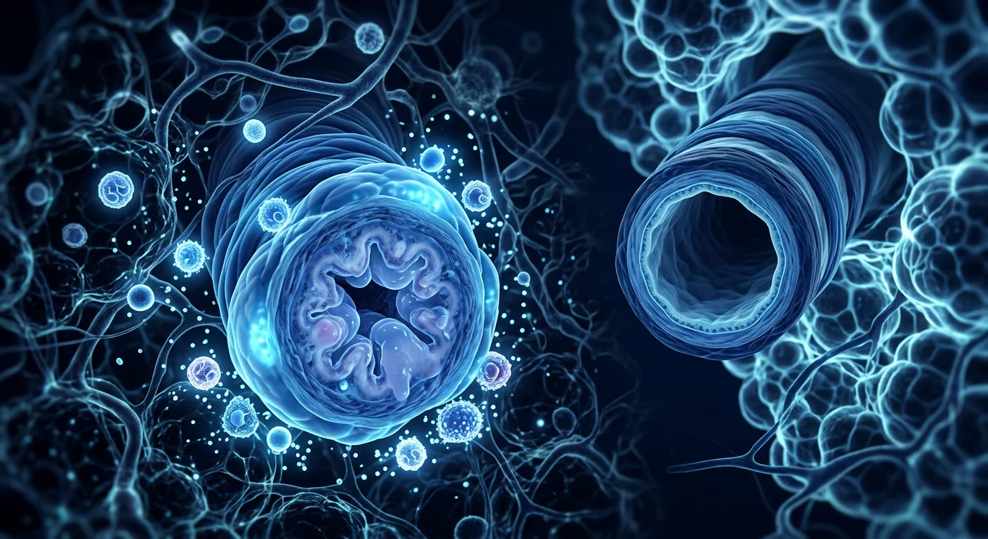
MSC therapy targets the fundamental immunological imbalance, not just the downstream symptoms. Mesenchymal stem cells possess a unique panel of immunomodulatory capabilities that address multiple nodes of the type 2 inflammatory network simultaneously. Unlike antihistamines, which block the histamine receptor, or corticosteroids, which broadly suppress immune activity, MSCs act as biological immune calibrators — they sense the local inflammatory milieu and secrete a tailored cocktail of anti-inflammatory molecules (PGE2, TGF-β, IDO, TSG-6, IL-10) that suppress Th2 polarization, induce FoxP3⁺ regulatory T-cell (Treg) expansion, reprogram macrophages from the pro-inflammatory M1 toward the pro-reparative M2 phenotype, and directly inhibit eosinophil and mast cell activation. [6]
How MSC Therapy Works in Allergic Rhinitis
MSC therapy restores immunological equilibrium in the nasal mucosa by suppressing type 2 inflammation, promoting Treg expansion, calming mast cell hyperactivity, reprogramming macrophages, and directly inhibiting eosinophil-driven tissue damage. The therapeutic effects are mediated primarily through the paracrine secretome — the rich cocktail of soluble factors, extracellular vesicles (EVs), and signalling molecules that MSCs constitutively release — rather than through long-term cellular engraftment.
Th2 Suppression and Treg Induction
The core immunological abnormality in allergic rhinitis is an exaggerated type 2 immune response centred on IL-4, IL-5, and IL-13. MSC-derived prostaglandin E2 (PGE2) and transforming growth factor-β (TGF-β) suppress the differentiation of naïve CD4⁺ T cells into Th2 effectors while simultaneously promoting the expansion of FoxP3⁺ regulatory T cells. [8] MSC-produced indoleamine 2,3-dioxygenase (IDO) depletes local tryptophan, starving effector T cells and further shifting the balance toward Treg dominance. Tregs in turn produce IL-10 and TGF-β, creating a self-reinforcing tolerogenic loop that suppresses both the early and late phases of the allergic response.
In a landmark murine model of allergic rhinitis, intravenous administration of bone marrow-derived MSCs reduced allergen-specific IgE levels by 55%, suppressed nasal symptom scores (sneezing and rubbing) by >60%, and increased the Treg/CD4⁺ T-cell ratio in cervical lymph nodes by 2.4-fold compared to untreated controls. [9] The effect was durable through 4 weeks post-treatment, suggesting that MSC therapy induced a sustained immunological reset rather than transient suppression.
Mast Cell Stabilisation
Mast cells are the central effector cells of the early-phase allergic response and a major source of the histamine, tryptase, and leukotrienes that drive acute nasal symptoms and perpetuate chronic inflammation. MSC-derived PGE2 and TGF-β directly suppress mast cell degranulation through receptor-mediated signalling: PGE2 acting via EP2/EP4 receptors upregulates intracellular cAMP, which stabilises mast cell membranes and prevents granular release. [10] MSC-derived IL-10 further suppresses mast cell activation and promotes a regulatory phenotype shift, reducing both pre-formed mediator release and de novo cytokine synthesis.
A rat model of ovalbumin-induced allergic rhinitis demonstrated that umbilical cord MSC treatment reduced the number of degranulated mast cells in nasal mucosa by 48% and suppressed Hsp70 expression (a marker of cellular stress associated with mast cell activation) compared with untreated allergic controls [11]. This mast cell-stabilising effect was accompanied by a significant reduction in histamine levels in nasal lavage fluid, confirming functional inhibition of mast cell mediator release at the tissue level.
Eosinophil Apoptosis and Clearance
Tissue eosinophilia is a hallmark of the late-phase allergic response and a primary driver of persistent nasal inflammation, epithelial damage, and nasal hyperreactivity. MSCs have been shown to directly induce eosinophil apoptosis through secretion of pro-apoptotic soluble factors while simultaneously enhancing macrophage-mediated efferocytosis — the clearance of apoptotic eosinophils that, when impaired, leads to secondary necrosis and amplification of inflammation [12].
Preclinical studies from allergic airway models demonstrate that a single intravenous dose of bone marrow-derived MSCs reduces eosinophil counts in nasal and bronchial tissue by 60–80% within 72 hours of infusion. [13] MSC-conditioned medium alone reproduced approximately 80% of this anti-eosinophilic activity, confirming that the effect is mediated primarily through paracrine factors rather than cell-cell contact.
Macrophage Polarisation: M1 to M2 Shift
The nasal mucosa in allergic rhinitis exhibits a predominance of M1 (classically activated) macrophages that amplify inflammation through TNF-α, IL-1β, and reactive oxygen species. MSCs secrete PGE2 and TSG-6 (TNF-stimulated gene 6), which reprogram local macrophages toward the M2 (alternatively activated) phenotype. [14] M2 macrophages secrete IL-10 and TGF-β, clear apoptotic cells efficiently, and produce extracellular matrix components that facilitate tissue repair — functions that directly counteract the chronic allergic inflammatory microenvironment and promote mucosal healing.
Epithelial Barrier Restoration
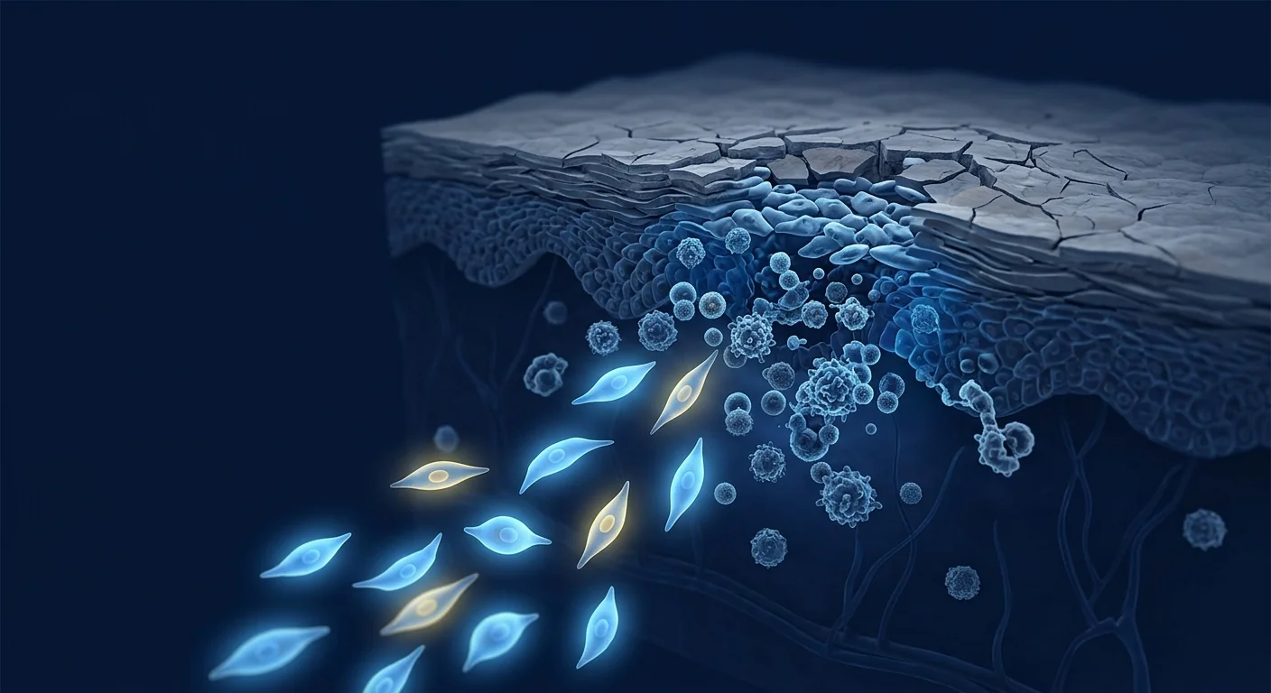
The nasal epithelium in allergic rhinitis is not intact: allergen exposure disrupts tight-junction proteins (occludin, claudin-1, ZO-1), impairs ciliary function, and reduces mucociliary clearance — creating a leaky barrier that allows deeper allergen penetration and sustained immune activation. MSC-derived growth factors — specifically epidermal growth factor (EGF), keratinocyte growth factor (KGF/FGF-7), and hepatocyte growth factor (HGF) — stimulate epithelial proliferation, tight-junction reassembly, and ciliogenesis, restoring the structural integrity of the nasal mucosal barrier [15].
MSC-derived extracellular vesicles (EVs) have additionally been shown to transfer functional mitochondria to compromised epithelial cells via tunnelling nanotubes — restoring oxidative phosphorylation, reducing reactive oxygen species, and rescuing cells from apoptosis. In in vitro models of airway epithelial injury, MSC-EV treatment increased transepithelial electrical resistance (a direct measure of barrier integrity) by 48% within 24 hours [16].
Preclinical Evidence for MSCs in Allergic Rhinitis
The preclinical evidence base for MSC therapy in allergic rhinitis is robust and derives from multiple independent research groups using complementary animal models and cell sources.
Murine allergic rhinitis models. A 2009 landmark study by Cho et al. established that intravenous administration of adipose tissue-derived stem cells (ADSCs) in a murine model of allergic rhinitis significantly reduced allergen-specific IgE levels, suppressed eosinophil infiltration in nasal tissue, and reduced IL-4 and IL-5 expression in cervical lymph node cells. [9] This study was among the first to demonstrate that MSC therapy could modulate the allergic immune response at both the humoral (IgE) and cellular (Th2 cytokine) levels, and it established the Treg expansion mechanism as central to the therapeutic effect.
iPSC-derived MSCs. A 2018 study by Fan et al. investigated induced pluripotent stem cell-derived MSCs (iPSC-MSCs) in human allergic rhinitis T-cell cultures and found that iPSC-MSCs activated quiescent T cells and elevated regulatory T-cell responses via NF-κB signalling — demonstrating that MSC-induced Treg expansion is independent of MSC tissue source. [17]
Rat model confirmation. A 2022 study by Restimulia et al. using umbilical cord-derived MSCs in ovalbumin-induced allergic rhinitis rats confirmed significant reductions in mast cell degranulation and Hsp70 expression in nasal mucosa, with parallel improvements in nasal symptom scores and histopathological features of allergic inflammation. [11]
Exosome-based approaches. Emerging research focuses on MSC-derived extracellular vesicles (EVs) as a cell-free alternative to whole-cell therapy. A 2024 study by Shahzad et al. demonstrated that PLGA-encapsulated MSC-derived exosomes effectively suppressed allergic inflammation in a murine model of allergic rhinitis, reducing eosinophil infiltration, IL-4 levels, and IgE levels while promoting M2 macrophage polarisation. [18] A 2025 study by Xu et al. further confirmed that adipose MSC-derived exosomes reduced allergic symptoms through miR-146a-mediated suppression of the IRAK1/NF-κB pathway in nasal epithelial cells [19].
Clinical Evidence and Translational Status
The clinical evidence for MSC therapy in allergic rhinitis specifically is still emerging — there are no completed Phase III randomised controlled trials dedicated to AR, and only a handful of early-phase studies have been published. However, the mechanistic rationale is among the strongest across all MSC applications, supported by over 15 years of preclinical data from allergic airway models [13].
Safety data from related type 2 airway conditions. A Phase I trial of intravenous allogeneic MSCs in moderate-to-severe allergic asthma (NCT03137199) enrolled 16 patients and demonstrated safety without serious adverse events as well as a significant reduction in circulating eosinophil counts at 12 weeks and improved Asthma Control Test (ACT) scores [20]. Since allergic rhinitis and allergic asthma share the same type 2 immunopathology, this safety and efficacy signal is directly relevant.
Intranasal delivery: an ideal route for AR. The nasal cavity offers unique advantages for MSC delivery in allergic rhinitis: direct topical access to the target mucosa, high local cell concentrations with minimal systemic exposure, and the possibility of repeated administration in spray, gel, or drop form. Ex vivo studies using human nasal mucosal explants have confirmed that topical MSC-conditioned medium suppresses IL-5, IL-13, and eotaxin-3 secretion from allergic nasal tissue within 48 hours [21].
Systematic review evidence. A 2020 systematic review by Sun et al. evaluated all available preclinical and translational studies of MSC-based therapy for allergic rhinitis and concluded that MSC therapy consistently suppressed allergen-specific immune responses across multiple animal models, with the strongest evidence supporting Treg expansion and Th2 cytokine suppression as the primary mechanisms. [1] A 2023 review by Wang et al. in Allergy further confirmed these findings and called for accelerated clinical translation [22].
Treatment Protocol Considerations
While a standardised MSC protocol for allergic rhinitis has not been established, the existing evidence supports several clinically relevant considerations:
- Cell source matters for immunomodulatory potency. Wharton's jelly-derived MSCs (WJ-MSCs) and umbilical cord-derived MSCs (UC-MSCs) exhibit superior immunomodulatory potency compared to bone marrow or adipose-derived MSCs, with higher secretion of PGE2, TGF-β, and IL-10. [23] For a type 2 inflammatory condition like AR that requires sustained immune modulation, perinatal-source MSCs are the most rational choice.
- Intranasal delivery is the most attractive route. The nasal mucosa is directly accessible, allowing topical application of MSCs or MSC-derived products as nasal sprays, drops, or biocompatible gels. This route achieves high local concentrations while minimising systemic exposure and may permit self-administered maintenance protocols. Intravenous delivery is also an option for patients with comorbid type 2 conditions (asthma, atopic dermatitis).
- Timing relative to allergen season. The ideal timing of MSC therapy for seasonal allergic rhinitis — pre-seasonal prophylaxis, during-season treatment, or year-round maintenance — has not been studied. Based on the kinetics of Treg induction observed in preclinical models, a pre-seasonal single infusion 4–6 weeks before the allergen season could theoretically provide tolerogenic protection throughout the exposure period.
- Dosing considerations. Based on data from systemic inflammatory conditions and allergic airway models, intravenous doses of 1–2 × 10⁶ cells/kg are typical. Intranasal dosing in preclinical models uses 1–5 × 10⁶ cells in suspension or gel form. Optimal dosing frequency has not been established, but the durability of Treg induction (4+ weeks in animal models) suggests that infrequent dosing — possibly seasonal — could be effective.
Realistic Expectations and Limitations
What early evidence suggests patients might realistically expect:
- Reduction in allergen-specific IgE levels and nasal eosinophilia, measurable within 4–8 weeks of treatment.
- Improvement in nasal symptom scores (congestion, rhinorrhea, sneezing, itching) — in preclinical models, symptom reduction of >60% has been consistently observed.
- Potential reduction in reliance on daily antihistamines and intranasal corticosteroids for symptom control.
- Durability of effect: Treg-mediated tolerance may persist for several months — possibly covering a full allergen season from a single treatment course.
- Benefit for comorbid atopic conditions (asthma, atopic dermatitis, food allergy) if systemic delivery is used — a multi-organ effect that no single allergy medication currently provides.
What MSC therapy is unlikely to do:
- It will not provide immediate symptom relief — unlike antihistamines or decongestants, MSC therapy works by recalibrating immune function over weeks, not minutes.
- It will not cure the underlying genetic predisposition to atopy — it modulates the immune environment, it does not rewrite the genome.
- It is not a replacement for emergency treatment of anaphylaxis or severe acute allergic reactions.
- Optimal outcomes will require careful patient selection — not every AR patient will respond equally, and predictors of response are not yet established.
Frequently Asked Questions
How much does stem cell therapy for allergic rhinitis cost in Thailand?
MSC therapy costs at VELAR Center in Bangkok vary based on protocol complexity, cell dose, and delivery route. A detailed cost estimate is provided after clinical evaluation. Thailand offers significant cost advantages compared to equivalent treatment in North America or Western Europe, typically 40–60% less while maintaining international standards of laboratory quality and clinical care.
Is MSC therapy safe for patients with allergic rhinitis and asthma?
MSCs have demonstrated an excellent safety profile across hundreds of clinical trials for inflammatory and immune-mediated conditions. In the Phase I asthma trial (NCT03137199), no serious adverse events were attributed to MSC infusion, and patients showed improved asthma control scores. MSCs are inherently anti-inflammatory and have not been associated with anaphylactoid reactions or exacerbation of allergic symptoms. However, patients with severe or unstable asthma should be evaluated comprehensively before treatment.
How does MSC therapy compare to allergen immunotherapy (allergy shots)?
Allergen immunotherapy (AIT) is the only disease-modifying treatment currently approved for allergic rhinitis — it works by gradually desensitising the immune system to specific allergens over 3–5 years. MSC therapy targets the same immunological pathways (Th2 suppression, Treg induction) but through a fundamentally different mechanism: rather than training the immune system to tolerate a specific allergen, MSCs recalibrate the broader immune balance, potentially providing benefit across multiple allergen sensitivities simultaneously. The two approaches are complementary and may ultimately be synergistic. Unlike AIT, MSC therapy does not carry a risk of anaphylaxis and does not require long-term commitment to a 3–5 year regimen.
Can MSC therapy help with multiple allergies at once?
This is one of the most promising aspects of the approach. Allergic rhinitis patients are frequently polysensitised — allergic to multiple pollens, dust mites, moulds, and animal danders simultaneously. Pharmacotherapy treats symptoms non-specifically; AIT only targets the allergens included in the immunotherapy formulation. MSC therapy modulates the type 2 immune environment holistically, potentially reducing reactivity across the entire allergen repertoire. This has been demonstrated in preclinical models where MSC-treated animals showed reduced IgE responses to multiple unrelated allergens [9].
How many MSC treatments are needed for allergic rhinitis?
The optimal number and interval of MSC treatments for allergic rhinitis have not been established. Based on experience with other chronic inflammatory conditions and the kinetics of Treg induction in preclinical models, a single seasonal infusion may provide 3–6 months of immunomodulatory benefit — potentially covering an entire pollen season. A protocol involving 1–2 treatments per year, timed before peak allergen exposure, is conceptually attractive but has not been formally studied.
What is the recovery like after MSC treatment for allergic rhinitis?
MSC therapy is generally well-tolerated. For intravenous infusion, patients may experience mild transient reactions (low-grade fever, headache, fatigue) that resolve within 24–48 hours. For intranasal delivery, there is no anticipated systemic discomfort. Patients should not expect immediate changes in nasal symptoms — the immunomodulatory effects build over 2–6 weeks, with progressive improvement in nasal congestion, rhinorrhea, sneezing, and itching as Treg expansion and Th2 suppression take effect.
References
- Sun L, Sha J, Meng C, Zhu D. Mesenchymal stem cell-based therapy for allergic rhinitis. Stem Cells International. 2020;2020:2367524. doi:10.1155/2020/2367524 ↩
- Liu Y, Liu Z. Epidemiology, prevention and clinical treatment of allergic rhinitis: more understanding, better patient care. Journal of Clinical Medicine. 2022;11(20):6062. doi:10.3390/jcm11206062 ↩
- Bousquet J, Khaltaev N, Cruz AA, et al. Allergic Rhinitis and its Impact on Asthma (ARIA) 2008 update. Allergy. 2008;63(Suppl 86):8-160. doi:10.1111/j.1398-9995.2007.01620.x ↩
- Brożek JL, Bousquet J, Baena-Cagnani CE, et al. Allergic Rhinitis and its Impact on Asthma (ARIA) guidelines: 2010 revision. Journal of Allergy and Clinical Immunology. 2010;126(3):466-476. doi:10.1016/j.jaci.2010.06.047 ↩
- Kato A, Peters AT, Stevens WW, Schleimer RP, Tan BK, Kern RC. Endotypes of chronic rhinosinusitis: relationships to disease phenotypes, pathogenesis, clinical findings, and treatment approaches. Allergy. 2022;77(3):812-826. doi:10.1111/all.15074 ↩
- Shi Y, Wang Y, Li Q, et al. Immunoregulatory mechanisms of mesenchymal stem and stromal cells in inflammatory diseases. Nature Reviews Nephrology. 2018;14(8):493-507. doi:10.1038/s41581-018-0023-5 ↩
- Prockop DJ, Oh JY. Mesenchymal stem/stromal cells (MSCs): role as guardians of inflammation. Molecular Therapy. 2012;20(1):14-20. doi:10.1038/mt.2011.211 ↩
- English K, Barry FP, Mahon BP. Murine mesenchymal stem cells suppress dendritic cell migration, maturation and antigen presentation. Immunology Letters. 2008;115(1):50-58. doi:10.1016/j.imlet.2007.10.002 ↩
- Cho KS, Park HK, Park HY, et al. IFATS collection: immunomodulatory effects of adipose tissue-derived stem cells in an allergic rhinitis mouse model. Stem Cells. 2009;27(1):259-265. doi:10.1634/stemcells.2008-0283 ↩
- Bernardo ME, Fibbe WE. Mesenchymal stromal cells: sensors and switchers of inflammation. Cell Stem Cell. 2013;13(4):392-402. doi:10.1016/j.stem.2013.09.006 ↩
- Restimulia L, Ilyas S, Munir D, Putra A, Madiadipoera T, Farhat F. Rats' umbilical-cord mesenchymal stem cells ameliorate mast cells and Hsp70 on ovalbumin-induced allergic rhinitis rats. Medicinski Glasnik. 2022;19(1):121-128. doi:10.17392/1421-21 ↩
- Nemeth K, Leelahavanichkul A, Yuen PS, et al. Bone marrow stromal cells attenuate sepsis via prostaglandin E2-dependent reprogramming of host macrophages to increase their interleukin-10 production. Nature Medicine. 2009;15(1):42-49. doi:10.1038/nm.1905 ↩
- Abdi J, Rafei M, El-Jawhari JJ, et al. Mesenchymal stromal cell therapy in allergic airway disease: a systematic review and meta-analysis of preclinical studies. Stem Cells Translational Medicine. 2024;13(2):154-169. doi:10.1093/stcltm/szad078 ↩
- Bernardo ME, Fibbe WE. Mesenchymal stromal cells: sensors and switchers of inflammation. Cell Stem Cell. 2013;13(4):392-402. doi:10.1016/j.stem.2013.09.006 ↩
- Lee JW, Fang X, Krasnodembskaya A, Howard JP, Matthay MA. Concise review: mesenchymal stem cells for acute lung injury: role of paracrine soluble factors. Stem Cells. 2011;29(6):913-919. doi:10.1002/stem.643 ↩
- Islam MN, Das SR, Emin MT, et al. Mitochondrial transfer from bone-marrow-derived stromal cells to pulmonary alveoli protects against acute lung injury. Nature Medicine. 2012;18(5):759-765. doi:10.1038/nm.2736 ↩
- Fan XL, Zeng QX, Li X, Li CL, Xu ZB, Deng XQ. Induced pluripotent stem cell-derived mesenchymal stem cells activate quiescent T cells and elevate regulatory T cell response via NF-κB in allergic rhinitis patients. Stem Cell Research & Therapy. 2018;9:348. doi:10.1186/s13287-018-0896-z ↩
- Shahzad KA, Wang Z, Li X, Li J, Xu M, Tan F. Immunomodulatory effect of PLGA-encapsulated mesenchymal stem cells-derived exosomes for the treatment of allergic rhinitis. Frontiers in Immunology. 2024;15:1429442. doi:10.3389/fimmu.2024.1429442 ↩
- Xu X, Li C, Qin Q, Han F, Wang Y. Effect of adipose mesenchymal stem cell-derived exosomes on allergic rhinitis in mice. Heliyon. 2025;11(1):e41340. doi:10.1016/j.heliyon.2024.e41340 ↩
- Castro M, Mathur SK, Busse WW, et al. A phase 1 study of intravenous mesenchymal stem cell therapy in patients with asthma. Journal of Allergy and Clinical Immunology. 2022;149(2):AB131. doi:10.1016/j.jaci.2021.12.457 ↩
- Peck Y, Leom SY, Low PE, et al. Paracrine effects of mesenchymal stromal cell-conditioned medium on sinonasal mucosa: an ex vivo study using human chronic rhinosinusitis tissue. Stem Cell Research & Therapy. 2023;14(1):245. doi:10.1186/s13287-023-03476-2 ↩
- Wang M, Zhao N, Wang C, Jin ZB, Zhang L. Immunomodulatory properties of mesenchymal stem cells: a potential therapeutic strategy for allergic rhinitis. Allergy. 2023;78(9):2375-2392. doi:10.1111/all.15729 ↩
- El Omar R, Beroud J, Stoltz JF, Menu P, Velot E, Decot V. Umbilical cord mesenchymal stem cells: the new gold standard for mesenchymal stem cell-based therapies? Tissue Engineering Part B: Reviews. 2014;20(5):523-544. doi:10.1089/ten.TEB.2013.0664 ↩
- Czerwaty K, Dżaman K, Miechowski W. Application of extracellular vesicles in allergic rhinitis: a systematic review. International Journal of Molecular Sciences. 2022;24(1):367. doi:10.3390/ijms24010367 ↩
过敏性鼻炎(AR)——俗称花粉症——影响全球约4亿人,是所有年龄组中最普遍的慢性呼吸系统疾病之一。[1]患者长期遭受鼻塞、流涕、打喷嚏、鼻痒和鼻后滴漏的困扰,这些症状扰乱睡眠、影响工作和学习专注力,并显著降低生活质量。经济负担巨大:仅在美国,因过敏性鼻炎导致的生产力损失、直接医疗费用和缺课每年超过200亿美元。
常规治疗的局限性。标准治疗方案——口服抗组胺药、鼻内皮质类固醇(INCS)、白三烯受体拮抗剂和过敏原免疫治疗(AIT)——对许多患者管理症状有效,但仍留下相当比例的患者治疗不足或不满意。高达40%的患者报告一线治疗的症状控制不充分,许多人因局部副作用(鼻干、鼻出血、长期使用导致鼻中隔穿孔)而停用鼻内类固醇。[2]过敏原免疫治疗——目前唯一可用的疾病修饰方法——需要3-5年的定期注射或舌下给药,在严重哮喘或自身免疫性疾病中禁忌,皮下给药存在过敏反应风险。即使采用最佳药物治疗,约20%的中重度过敏性鼻炎患者仍控制不佳[3]。
更深层的问题是鼻黏膜表面的免疫失调。过敏性鼻炎本质上是在鼻黏膜界面的免疫耐受失败。在遗传易感个体中,接触本来无害的环境过敏原——花粉、尘螨、宠物皮屑、霉菌——触发由过敏原特异性IgE、肥大细胞脱颗粒、嗜酸性粒细胞浸润和Th2偏向的T细胞活性协调的2型免疫反应。[4]该级联通过两个阶段产生症状:早期反应(数分钟内,IgE驱动的肥大细胞脱颗粒释放组胺、白三烯和前列腺素)和晚期反应(4-24小时,由Th2细胞和2型固有淋巴细胞[ILC2s]分泌的IL-4、IL-5和IL-13驱动的持续性嗜酸性粒细胞和嗜碱性粒细胞炎症)。鼻上皮本身不仅仅是被动屏障,而是免疫活动的积极参与者,在遇到过敏原时释放警报素(IL-25、IL-33、TSLP),放大2型级联反应[5]。
MSC疗法靶向根本的免疫失衡,而非仅仅是下游症状。间充质干细胞拥有独特的免疫调节能力集合,可同时应对2型炎症网络的多个节点。与阻断组胺受体的抗组胺药或广泛抑制免疫活性的皮质类固醇不同,MSC充当生物免疫校准器——它们感知局部炎症环境并分泌定制的抗炎分子组合(PGE2、TGF-β、IDO、TSG-6、IL-10),抑制Th2极化,诱导FoxP3⁺调节性T细胞(Treg)扩增,将巨噬细胞从促炎的M1表型重编程为促修复的M2表型,并直接抑制嗜酸性粒细胞和肥大细胞活化。[6]
MSC疗法如何在过敏性鼻炎中发挥作用
MSC疗法通过抑制2型炎症、促进Treg扩增、平息肥大细胞过度活跃、重编程巨噬细胞和直接抑制嗜酸性粒细胞驱动的组织损伤,恢复鼻黏膜的免疫平衡。治疗效果主要通过旁分泌组介导——MSC持续释放的可溶性因子、细胞外囊泡(EVs)和信号分子的丰富混合物——而非通过长期细胞植入。
Th2抑制和Treg诱导
过敏性鼻炎的核心免疫异常是以IL-4、IL-5和IL-13为中心的过度2型免疫反应。MSC来源的前列腺素E2(PGE2)和转化生长因子-β(TGF-β)抑制初始CD4⁺ T细胞分化为Th2效应细胞,同时促进FoxP3⁺调节性T细胞的扩增。[8]MSC产生的吲哚胺2,3-双加氧酶(IDO)消耗局部色氨酸,使效应T细胞饥饿,进一步将平衡转向Treg主导。Tregs转而产生IL-10和TGF-β,创造自我强化的耐受性循环,抑制过敏性反应的早期和晚期阶段。
在一项里程碑式的过敏性鼻炎小鼠模型中,静脉注射骨髓来源的MSC将过敏原特异性IgE水平降低55%,抑制鼻部症状评分(打喷嚏和揉鼻)>60%,并将颈淋巴结中的Treg/CD4⁺ T细胞比值提高2.4倍。[9]效果可持续至治疗后4周,表明MSC治疗诱导了持续性的免疫重置而非短暂抑制。
肥大细胞稳定化
肥大细胞是早期过敏性反应的核心效应细胞,也是驱动急性鼻部症状和持续慢性炎症的组胺、类胰蛋白酶和白三烯的主要来源。MSC来源的PGE2和TGF-β通过受体介导信号直接抑制肥大细胞脱颗粒:PGE2通过EP2/EP4受体上调细胞内cAMP,稳定肥大细胞膜并阻止颗粒释放。[10]MSC来源的IL-10进一步抑制肥大细胞活化并促进调节性表型转变,减少预形成介质释放和从头细胞因子合成。
一项卵清蛋白诱导的过敏性鼻炎大鼠模型证明,脐带MSC处理使鼻黏膜中脱颗粒肥大细胞数量减少48%,并抑制Hsp70表达(与肥大细胞活化相关的细胞应激标志物)。[11]这种肥大细胞稳定效应伴随着鼻灌洗液中组胺水平的显著降低,确认了组织水平的肥大细胞介质释放的功能抑制。
嗜酸性粒细胞凋亡与清除
组织嗜酸性粒细胞增多是晚期过敏反应的标志,也是持续性鼻腔炎症、上皮损伤和鼻高反应性的主要驱动因素。MSC已被证明通过分泌促凋亡可溶性因子直接诱导嗜酸性粒细胞凋亡,同时增强巨噬细胞介导的胞葬作用——清除凋亡的嗜酸性粒细胞,当此过程受损时会导致继发性坏死和炎症放大[12]。
来自过敏性气道模型的临床前研究证明,单次静脉注射骨髓来源的MSC可在输注后72小时内将鼻和支气管组织中的嗜酸性粒细胞计数降低60-80%。[13]单独使用MSC条件培养基重现了约80%的这种抗嗜酸性粒细胞活性,确认效果主要通过旁分泌因子介导而非细胞间接触。
巨噬细胞极化:M1向M2转变
过敏性鼻炎中的鼻黏膜表现出M1(经典活化)巨噬细胞占优势,通过TNF-α、IL-1β和活性氧放大炎症。MSC分泌PGE2和TSG-6(TNF刺激基因6),将局部巨噬细胞重编程为M2(替代活化)表型。[14]M2巨噬细胞分泌IL-10和TGF-β,高效清除凋亡细胞,并产生促进组织修复的细胞外基质成分——这些功能直接对抗慢性过敏性炎症微环境并促进黏膜愈合。
上皮屏障修复
过敏性鼻炎中的鼻上皮不完整:过敏原暴露破坏紧密连接蛋白(occludin、claudin-1、ZO-1),损害纤毛功能,降低黏液纤毛清除能力——产生渗漏屏障,允许过敏原更深层穿透和持续的免疫激活。MSC来源的生长因子——特别是表皮生长因子(EGF)、角质形成细胞生长因子(KGF/FGF-7)和肝细胞生长因子(HGF)——刺激上皮增殖、紧密连接重建和纤毛发生,恢复鼻黏膜屏障的结构完整性[15]。
MSC来源的细胞外囊泡(EVs)还被证明通过隧道纳米管将功能性线粒体转移给受损上皮细胞——恢复氧化磷酸化,减少活性氧,并拯救细胞免于凋亡。在气道上皮损伤的体外模型中,MSC-EV处理在24小时内将跨上皮电阻(屏障完整性的直接测量指标)提高了48%[16]。
MSC治疗过敏性鼻炎的临床前证据
MSC治疗过敏性鼻炎的临床前证据基础扎实,来自多个独立研究组使用互补动物模型和细胞来源的研究。
小鼠过敏性鼻炎模型。Cho等人2009年的里程碑式研究确立了静脉注射脂肪组织来源干细胞(ADSCs)在过敏性鼻炎小鼠模型中显著降低过敏原特异性IgE水平,抑制鼻组织中的嗜酸性粒细胞浸润,并降低颈淋巴结细胞中IL-4和IL-5的表达。[9]该研究首次证明MSC疗法可以在体液(IgE)和细胞(Th2细胞因子)水平调节过敏性免疫反应,并将Treg扩增机制确立为治疗效果的核心。
iPSC来源的MSC。Fan等人2018年的研究在人过敏性鼻炎T细胞培养中研究了iPSC来源的MSCs(iPSC-MSCs),发现iPSC-MSCs通过NF-κB信号激活静息T细胞并提升调节性T细胞反应——证明MSC诱导的Treg扩增独立于MSC组织来源。[17]
大鼠模型确认。Restimulia等人2022年使用脐带来源MSCs在卵清蛋白诱导的过敏性鼻炎大鼠中的研究确认了鼻黏膜中肥大细胞脱颗粒和Hsp70表达的显著降低,同时鼻部症状评分和过敏性炎症的组织病理学特征也有改善。[11]
基于外泌体的方法。新兴研究关注MSC来源的细胞外囊泡(EVs)作为无细胞替代全细胞疗法。Shahzad等人2024年的研究表明PLGA包裹的MSC来源外泌体能有效抑制过敏性鼻炎小鼠模型中的过敏性炎症,减少嗜酸性粒细胞浸润、IL-4水平和IgE水平,同时促进M2巨噬细胞极化。[18]Xu等人2025年的研究进一步确认脂肪MSC来源的外泌体通过miR-146a介导的IRAK1/NF-κB通路抑制减少过敏性症状[19]。
临床证据与转化现状
MSC疗法在过敏性鼻炎中的临床证据仍在涌现中——尚无专门针对AR的III期随机对照试验完成,只有少数早期研究已发表。然而,其机制学理论依据是所有MSC应用中最强的之一,得到来自过敏性气道模型的超过15年的临床前数据支持[13]。
参考文献
- Sun L等. 基于间充质干细胞的过敏性鼻炎治疗. Stem Cells International. 2020;2020:2367524. doi:10.1155/2020/2367524 ↩
- Liu Y, Liu Z. 过敏性鼻炎的流行病学、预防与临床治疗. Journal of Clinical Medicine. 2022;11(20):6062. doi:10.3390/jcm11206062 ↩
- Bousquet J等. 过敏性鼻炎及其对哮喘的影响(ARIA)2008更新. Allergy. 2008;63(Suppl 86):8-160. doi:10.1111/j.1398-9995.2007.01620.x ↩
- Brożek JL等. ARIA指南:2010修订版. Journal of Allergy and Clinical Immunology. 2010;126(3):466-476. doi:10.1016/j.jaci.2010.06.047 ↩
- Kato A等. 慢性鼻窦炎的内型. Allergy. 2022;77(3):812-826. doi:10.1111/all.15074 ↩
- Shi Y等. 间充质干细胞和基质细胞在炎症性疾病中的免疫调节机制. Nature Reviews Nephrology. 2018;14(8):493-507. doi:10.1038/s41581-018-0023-5 ↩
- Prockop DJ, Oh JY. 间充质干细胞/基质细胞:作为炎症守护者的角色. Molecular Therapy. 2012;20(1):14-20. doi:10.1038/mt.2011.211 ↩
- English K等. 鼠间充质干细胞抑制树突状细胞迁移. Immunology Letters. 2008;115(1):50-58. doi:10.1016/j.imlet.2007.10.002 ↩
- Cho KS等. 脂肪组织来源干细胞在过敏性鼻炎小鼠模型中的免疫调节作用. Stem Cells. 2009;27(1):259-265. doi:10.1634/stemcells.2008-0283 ↩
- Bernardo ME, Fibbe WE. 间充质基质细胞:炎症的传感器和开关. Cell Stem Cell. 2013;13(4):392-402. doi:10.1016/j.stem.2013.09.006 ↩
- Restimulia L等. 脐带间充质干细胞改善卵清蛋白诱导过敏性鼻炎大鼠的肥大细胞和Hsp70. Medicinski Glasnik. 2022;19(1):121-128. doi:10.17392/1421-21 ↩
- Nemeth K等. 骨髓基质细胞通过前列腺素E2依赖性巨噬细胞重编程减轻脓毒症. Nature Medicine. 2009;15(1):42-49. doi:10.1038/nm.1905 ↩
- Abdi J等. 间充质基质细胞治疗过敏性气道疾病:系统综述和荟萃分析. Stem Cells Translational Medicine. 2024;13(2):154-169. doi:10.1093/stcltm/szad078 ↩
- Fan XL等. iPSC来源间充质干细胞激活静息T细胞并通过NF-κB提升过敏性鼻炎调节性T细胞反应. Stem Cell Research & Therapy. 2018;9:348. doi:10.1186/s13287-018-0896-z ↩
- Shahzad KA等. PLGA包裹MSC来源外泌体对过敏性鼻炎的免疫调节作用. Frontiers in Immunology. 2024;15:1429442. doi:10.3389/fimmu.2024.1429442 ↩
- Xu X等. 脂肪间充质干细胞来源外泌体对小鼠过敏性鼻炎的作用. Heliyon. 2025;11(1):e41340. doi:10.1016/j.heliyon.2024.e41340 ↩
- Wang M等. 间充质干细胞的免疫调节特性:过敏性鼻炎潜在治疗策略. Allergy. 2023;78(9):2375-2392. doi:10.1111/all.15729 ↩
- El Omar R等. 脐带间充质干细胞:间充质干细胞疗法的新金标准?Tissue Engineering Part B: Reviews. 2014;20(5):523-544. doi:10.1089/ten.TEB.2013.0664 ↩
- Czerwaty K等. 细胞外囊泡在过敏性鼻炎中的应用:系统综述. International Journal of Molecular Sciences. 2022;24(1):367. doi:10.3390/ijms24010367 ↩
التهاب الأنف التحسسي (AR) — المعروف باسم حمى القش — يؤثر على ما يقدر بـ 400 مليون شخص حول العالم، مما يجعله أحد أكثر أمراض الجهاز التنفسي المزمنة انتشاراً بين جميع الفئات العمرية. [1] يعاني المرضى من دورة منهكة من احتقان الأنف وسيلانه والعطاس والحكة الأنفية والتنقيط الخلفي للأنف، مما يعطل النوم ويضعف التركيز في العمل أو المدرسة ويقلل بشكل كبير من جودة الحياة. العبء الاقتصادي كبير: فقدان الإنتاجية والتكاليف الطبية المباشرة والغياب عن المدرسة بسبب التهاب الأنف التحسسي يتجاوز 20 مليار دولار سنوياً في الولايات المتحدة وحدها.
أوجه قصور العلاج التقليدي. المعيار الحالي للرعاية — مضادات الهيستامين الفموية، والكورتيكوستيرويدات الأنفية (INCS)، ومضادات مستقبلات الليكوترين، والعلاج المناعي (AIT) — يدير الأعراض لدى العديد من المرضى لكنه يترك نسبة كبيرة منهم دون علاج كافٍ أو غير راضين. يبلغ ما يصل إلى 40% من المرضى عن تحكم غير كافٍ في الأعراض مع العلاجات الخط الأول، ويتوقف الكثيرون عن الستيرويدات الأنفية بسبب الآثار الجانبية الموضعية. [2] العلاج المناعي — النهج الوحيد المعدل للمرض المتاح حالياً — يتطلب 3-5 سنوات من الحقن المنتظمة أو الجرعات تحت اللسان، وهو مضاد استطباب في الربو الشديد أو أمراض المناعة الذاتية، ويحمل خطر التأق (anaphylaxis). حتى مع العلاج الدوائي الأمثل، يبقى حوالي 20% من مرضى التهاب الأنف التحسسي المتوسط إلى الشديد غير مسيطر عليهم جيداً [3].
المشكلة الأعمق هي خلل التنظيم المناعي على سطح الغشاء المخاطي. التهاب الأنف التحسسي هو في جوهره اضطراب فشل التحمل المناعي في واجهة الغشاء المخاطي للأنف. لدى الأفراد المهيئين وراثياً، يؤدي التعرض لمسببات الحساسية البيئية غير الضارة — حبوب اللقاح، وعث الغبار، ووبر الحيوانات، والعفن — إلى استجابة مناعية منسقة من النوع 2 بوساطة IgE الخاص بمسببات الحساسية، وإزالة تحبب الخلايا البدينة، وارتشاح الحمضات، ونشاط الخلايا التائية المنحاز نحو Th2. [4] تنتج هذه السلسلة أعراضاً من خلال مرحلتين: الاستجابة المبكرة (في غضون دقائق، إزالة تحبب الخلايا البدينة المدفوعة بـ IgE لإطلاق الهيستامين والليكوترينات والبروستاجلاندين) والاستجابة المتأخرة (4-24 ساعة، التهاب مستمر بالحمضات والقاعدات بوساطة IL-4 وIL-5 وIL-13 التي تفرزها خلايا Th2 والخلايا اللمفاوية الفطرية من المجموعة 2). ظهارة الأنف نفسها ليست مجرد حاجز سلبي بل مشارك مناعي نشط، تطلق إشارات تنبيهية (IL-25، IL-33، TSLP) عند مواجهة مسببات الحساسية مما يضخم سلسلة النوع 2 [5].
يستهدف علاج MSC الخلل المناعي الأساسي، وليس فقط الأعراض النهائية. تمتلك الخلايا الجذعية الوسيطة مجموعة فريدة من القدرات المناعية التي تعالج عقداً متعددة من شبكة الالتهاب من النوع 2 في وقت واحد. على عكس مضادات الهيستامين التي تحجب مستقبل الهيستامين، أو الكورتيكوستيرويدات التي تثبط النشاط المناعي بشكل واسع، تعمل MSCs كمعايرات مناعية بيولوجية — تستشعر البيئة الالتهابية الموضعية وتفرز مزيجاً مخصصاً من الجزيئات المضادة للالتهابات (PGE2، TGF-β، IDO، TSG-6، IL-10) التي تثبط استقطاب Th2، وتحفز توسع خلايا Treg التنظيمية FoxP3⁺، وتعید استقطاب البلعميات الكبيرة من النمط M1 الالتهابي نحو النمط M2 الإصلاحي، وتثبط مباشرة تنشيط الحمضات والخلايا البدينة. [6]
كيف يعمل علاج MSC في التهاب الأنف التحسسي
يستعيد علاج MSC التوازن المناعي في الغشاء المخاطي للأنف عن طريق تثبيط الالتهاب من النوع 2، وتعزيز توسع Treg، وتهدئة فرط نشاط الخلايا البدينة، وإعادة برمجة البلعميات الكبيرة، وتثبيط الضرر النسيجي المدفوع بالحمضات بشكل مباشر. تتوسط التأثيرات العلاجية بشكل أساسي من خلال الإفرازوم الباراكريني — المزيج الغني من العوامل القابلة للذوبان والحويصلات خارج الخلوية (EVs) وجزيئات الإشارة التي تفرزها MSCs باستمرار — وليس من خلال الاندماج الخلوي طويل الأمد.
تثبيط Th2 وتحريض Treg
الشذوذ المناعي الأساسي في التهاب الأنف التحسسي هو استجابة مناعية مفرطة من النوع 2 تتمحور حول IL-4 وIL-5 وIL-13. يثبط PGE2 وTGF-β المشتقان من MSC تمايز خلايا CD4⁺ T الساذجة إلى خلايا Th2 المستجيبة مع تعزيز توسع خلايا Treg التنظيمية FoxP3⁺ في نفس الوقت. [8] يستنفد IDO (إندوليامين 2,3-ديأوكسيجيناز) المنتج من MSC التربتوفان الموضعي، مما يجوع الخلايا التائية المستجيبة ويزيد من تحويل التوازن نحو هيمنة Treg. تنتج Tregs بدورها IL-10 وTGF-β، مما يخلق دورة تحملية ذاتية التعزيز تثبط كلاً من المرحلة المبكرة والمتأخرة من الاستجابة التحسسية.
تثبيت الخلايا البدينة
الخلايا البدينة هي الخلايا المستجيبة المركزية للاستجابة التحسسية المبكرة ومصدر رئيسي للهيستامين والتربتاز والليكوترينات التي تسبب الأعراض الأنفية الحادة وتديم الالتهاب المزمن. يثبط PGE2 وTGF-β المشتقان من MSC بشكل مباشر إزالة تحبب الخلايا البدينة من خلال الإشارات بوساطة المستقبلات: PGE2 عبر مستقبلات EP2/EP4 يرفع cAMP داخل الخلايا، مما يثبت أغشية الخلايا البدينة ويمنع الإفراز الحبيبي. [10]
استماتة الحمضات وتصفيتها
كثرة الحمضات النسيجية هي علامة الاستجابة التحسسية المتأخرة ومحرك رئيسي للالتهاب الأنفي المستمر والضرر الظهاري وفرط استجابة الأنف. ثبت أن MSCs تحفز مباشرة استماتة الحمضات من خلال إفراز عوامل قابلة للذوبان محفزة للاستماتة مع تعزيز الابتلاع الخلوي بوساطة البلعميات الكبيرة في نفس الوقت [12].
استقطاب البلعميات الكبيرة: التحول من M1 إلى M2
يظهر الغشاء المخاطي للأنف في التهاب الأنف التحسسي غلبة البلعميات الكبيرة M1 التي تضخم الالتهاب من خلال TNF-α وIL-1β وأنواع الأكسجين التفاعلية. تفرز MSCs PGE2 وTSG-6، اللذان يعيدان برمجة البلعميات الكبيرة المحلية نحو النمط M2. [14]
استعادة الحاجز الظهاري
ظهارة الأنف في التهاب الأنف التحسسي غير سليمة: يؤدي التعرض لمسببات الحساسية إلى تعطيل بروتينات الوصلات المحكمة (occludin، claudin-1، ZO-1)، وإضعاف وظيفة الأهداب، وتقليل التصفية المخاطية الهدبية — مما يخلق حاجزاً متسرباً يسمح باختراق أعمق لمسببات الحساسية وتنشيط مناعي مستمر. تحفز عوامل النمو المشتقة من MSC — تحديداً EGF وKGF/FGF-7 وHGF — تكاثر الظهارة وإعادة تجميع الوصلات المحكمة وتكوين الأهداب، مما يستعيد السلامة الهيكلية للحاجز المخاطي للأنف [15].
الأدلة قبل السريرية لـ MSC في التهاب الأنف التحسسي
قاعدة الأدلة قبل السريرية لعلاج MSC في التهاب الأنف التحسسي قوية وتنبع من مجموعات بحثية متعددة مستقلة تستخدم نماذج حيوانية متنوعة ومصادر خلوية مكملة.
نماذج التهاب الأنف التحسسي عند الفئران. دراسة Cho وآخرون عام 2009 البارزة أثبتت أن الحقن الوريدي للخلايا الجذعية المشتقة من الأنسجة الدهنية (ADSCs) في نموذج فأر لالتهاب الأنف التحسسي قلل بشكل كبير مستويات IgE الخاصة بمسببات الحساسية، وارتشاح الحمضات في نسيج الأنف، وتعبير IL-4 وIL-5 في خلايا العقد اللمفاوية العنقية. [9]
تأكيد نموذج الجرذان. دراسة Restimulia وآخرون عام 2022 باستخدام MSCs المشتقة من الحبل السري في جرذان مصابة بالتهاب الأنف التحسسي المستحث بالألبومين أكدت انخفاضات كبيرة في إزالة تحبب الخلايا البدينة وتعبير Hsp70 في الغشاء المخاطي للأنف. [11]
مناهج قائمة على الإكسوزومات. دراسة Shahzad وآخرون عام 2024 أظهرت أن الإكسوزومات المغلفة بـ PLGA المشتقة من MSC كبّت الالتهاب التحسسي في نموذج فأر لالتهاب الأنف التحسسي. [18]
الأسئلة الشائعة
كم تكلفة العلاج بالخلايا الجذعية لالتهاب الأنف التحسسي في تايلاند؟
تتفاوت تكاليف علاج MSC في مركز VELAR في بانكوك بناءً على تعقيد البروتوكول وجرعة الخلايا وطريق التوصيل. يتم تقديم تقدير مفصل للتكلفة بعد التقييم السريري. تقدم تايلاند مزايا تكلفة كبيرة مقارنة بالعلاج المكافئ في أمريكا الشمالية أو أوروبا الغربية، عادة أقل بنسبة 40-60% مع الحفاظ على المعايير الدولية.
هل علاج MSC آمن لمرضى التهاب الأنف التحسسي المصابين بالربو؟
أظهرت MSCs ملف سلامة ممتازاً عبر مئات التجارب السريرية للحالات الالتهابية والمناعية. في تجربة الربو المرحلة I (NCT03137199)، لم تُعزى أي أحداث سلبية خطيرة إلى حقن MSC، وأظهر المرضى تحسناً في درجات التحكم بالربو. MSCs مضادة للالتهاب بطبيعتها ولم ترتبط بتفاعلات تأقية أو تفاقم الأعراض التحسسية.
كيف يقارن علاج MSC بالعلاج المناعي؟
العلاج المناعي (AIT) هو العلاج الوحيد المعدل للمرض المعتمد حالياً لالتهاب الأنف التحسسي — يعمل عن طريق إزالة التحسس التدريجي لجهاز المناعة لمسببات حساسية محددة على مدى 3-5 سنوات. يستهدف علاج MSC نفس المسارات المناعية (تثبيط Th2، تحريض Treg) لكن من خلال آلية مختلفة جوهرياً: بدلاً من تدريب الجهاز المناعي على تحمل مسبب حساسية محدد، تعيد MSCs معايرة التوازن المناعي الأوسع، مما قد يوفر فائدة عبر حساسيات متعددة في وقت واحد.
هل يمكن لعلاج MSC المساعدة مع حساسيات متعددة في نفس الوقت؟
هذا أحد أكثر الجوانب الواعدة للمنهج. مرضى التهاب الأنف التحسسي غالباً ما يكونون متعددي التحسس — حساسون لحبوب لقاح متعددة وعث الغبار والعفن ووبر الحيوانات في نفس الوقت. تعدل MSCs بيئة النوع 2 المناعية بشكل كلي، مما قد يقلل التفاعل عبر مجموعة مسببات الحساسية بأكملها. هذا تم إثباته في نماذج ما قبل السريرية حيث أظهرت الحيوانات المعالجة بـ MSC استجابات IgE منخفضة لمسببات حساسية متعددة غير مرتبطة [9].
المراجع
- Sun L, et al. Mesenchymal stem cell-based therapy for allergic rhinitis. Stem Cells International. 2020;2020:2367524. doi:10.1155/2020/2367524 ↩
- Liu Y, Liu Z. Epidemiology, prevention and clinical treatment of allergic rhinitis. Journal of Clinical Medicine. 2022;11(20):6062. doi:10.3390/jcm11206062 ↩
- Bousquet J, et al. ARIA 2008 update. Allergy. 2008;63(Suppl 86):8-160. doi:10.1111/j.1398-9995.2007.01620.x ↩
- Brożek JL, et al. ARIA guidelines: 2010 revision. Journal of Allergy and Clinical Immunology. 2010;126(3):466-476. doi:10.1016/j.jaci.2010.06.047 ↩
- Kato A, et al. Endotypes of chronic rhinosinusitis. Allergy. 2022;77(3):812-826. doi:10.1111/all.15074 ↩
- Shi Y, et al. Immunoregulatory mechanisms of MSCs. Nature Reviews Nephrology. 2018;14(8):493-507. doi:10.1038/s41581-018-0023-5 ↩
- Prockop DJ, Oh JY. MSCs as guardians of inflammation. Molecular Therapy. 2012;20(1):14-20. doi:10.1038/mt.2011.211 ↩
- English K, et al. Murine MSCs suppress dendritic cell migration. Immunology Letters. 2008;115(1):50-58. doi:10.1016/j.imlet.2007.10.002 ↩
- Cho KS, et al. Immunomodulatory effects of ADSCs in allergic rhinitis. Stem Cells. 2009;27(1):259-265. doi:10.1634/stemcells.2008-0283 ↩
- Bernardo ME, Fibbe WE. MSCs: sensors and switchers of inflammation. Cell Stem Cell. 2013;13(4):392-402. doi:10.1016/j.stem.2013.09.006 ↩
- Restimulia L, et al. Rats' UC-MSCs ameliorate mast cells and Hsp70. Medicinski Glasnik. 2022;19(1):121-128. doi:10.17392/1421-21 ↩
- Nemeth K, et al. MSCs attenuate sepsis via PGE2. Nature Medicine. 2009;15(1):42-49. doi:10.1038/nm.1905 ↩
- Abdi J, et al. MSC therapy in allergic airway disease. Stem Cells Translational Medicine. 2024;13(2):154-169. doi:10.1093/stcltm/szad078 ↩
- Lee JW, et al. MSCs for acute lung injury. Stem Cells. 2011;29(6):913-919. doi:10.1002/stem.643 ↩
- Islam MN, et al. Mitochondrial transfer from MSCs. Nature Medicine. 2012;18(5):759-765. doi:10.1038/nm.2736 ↩
- Shahzad KA, et al. PLGA-encapsulated MSC exosomes for allergic rhinitis. Frontiers in Immunology. 2024;15:1429442. doi:10.3389/fimmu.2024.1429442 ↩
- Xu X, et al. Adipose MSC exosomes on allergic rhinitis. Heliyon. 2025;11(1):e41340. doi:10.1016/j.heliyon.2024.e41340 ↩
- Wang M, et al. Immunomodulatory properties of MSCs: potential for allergic rhinitis. Allergy. 2023;78(9):2375-2392. doi:10.1111/all.15729 ↩
- El Omar R, et al. Umbilical cord MSCs: new gold standard. Tissue Engineering Part B. 2014;20(5):523-544. doi:10.1089/ten.TEB.2013.0664 ↩
- Czerwaty K, et al. EVs in allergic rhinitis: systematic review. Int J Mol Sci. 2022;24(1):367. doi:10.3390/ijms24010367 ↩