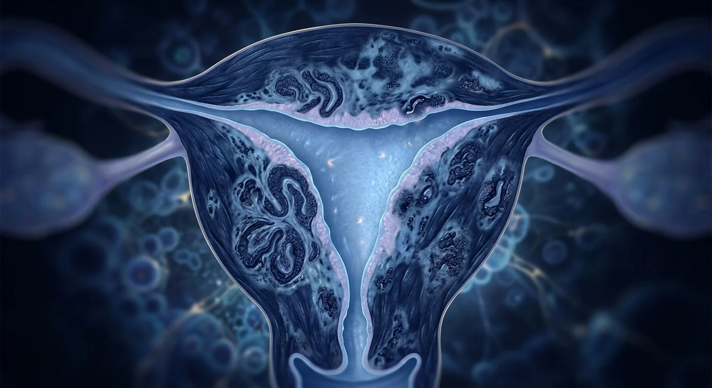
Adenomyosis affects an estimated 20–35% of women who develop pelvic symptoms — up to 70% of hysterectomy specimens — and is one of the most common, most undertreated causes of heavy periods and chronic pelvic pain [1].
Why surgery is often the default — and why it's final. For years the standard options were hormonal suppression, painkillers, or hysterectomy. Hysterectomy ends the bleeding, but it ends fertility, and for a condition that peaks in women in their 40s and 50s — often before they're done having children or before they're ready for major surgery — it's a heavy price. Uterine-sparing options exist (uterine artery embolization, MRI-guided focused ultrasound, endometrial ablation), but their durability for adenomyosis specifically is limited, partly because the disease is diffuse: the abnormal endometrial glands and stroma are woven through the myometrium, not confined to a single lesion [2].
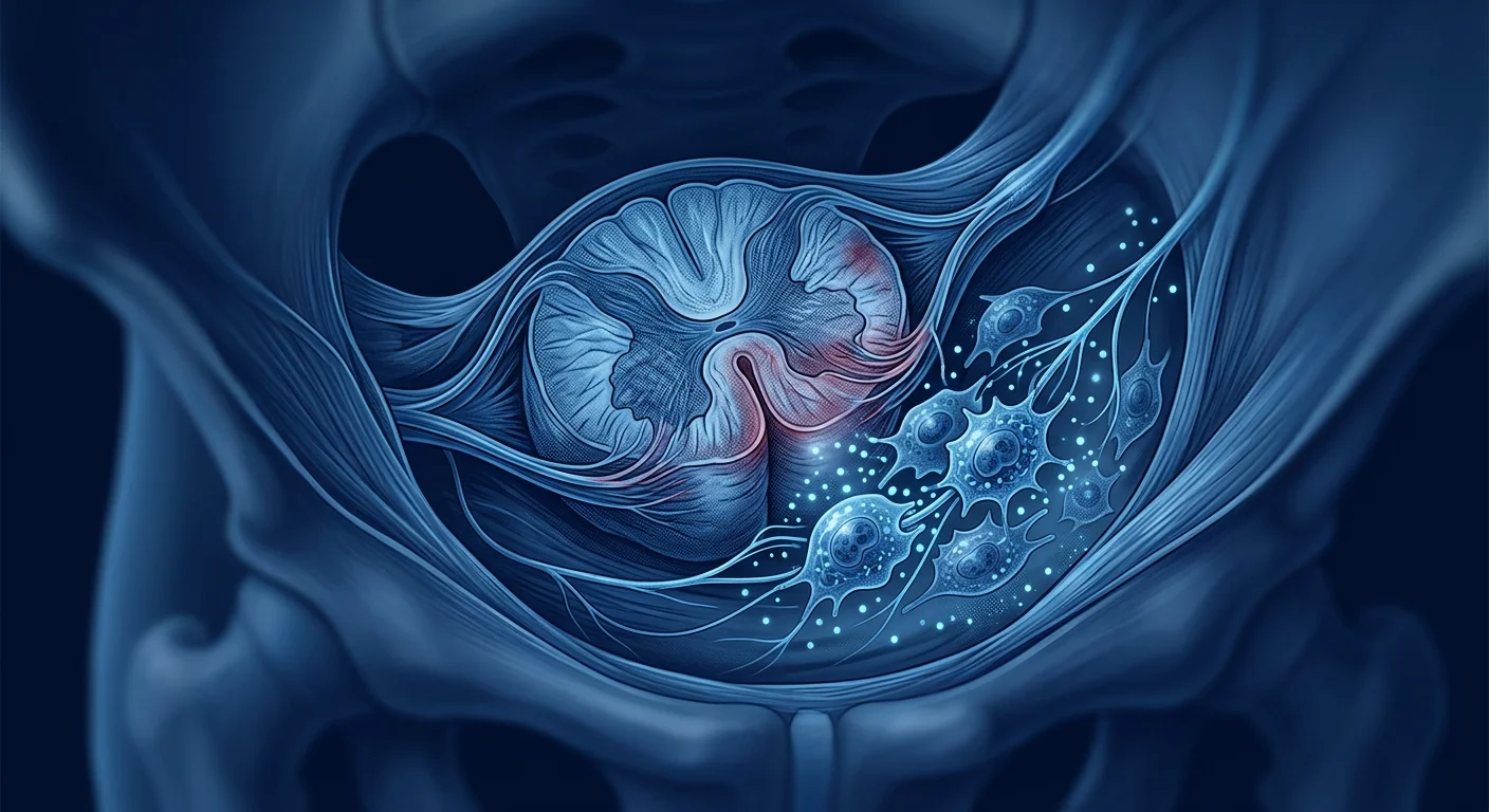
The tissue-level problem is inflammation and scarring. Adenomyosis is not just "endometrium in the wrong place." The ectopic glands sit inside a microenvironment of chronic inflammation: macrophage infiltration, stromal fibrosis, angiogenesis, and — crucially — impaired apoptosis of the ectopic endometrial cells, which keeps them alive and proliferating instead of clearing them the way a normal menstrual shedding would [3]. That microenvironment is exactly the kind of inflammatory-fibrotic niche that mesenchymal stem cells (MSCs) have been studied to modulate across a wide range of conditions.
MSC therapy targets the biology, not the symptom. Rather than shrinking lesions or suppressing hormones, MSC-derived paracrine signaling aims to calm the immune cascade, restore normal tissue turnover, and limit the fibrotic response that thickens the uterine wall and distorts the cavity. It is early-stage, and no guideline endorses it yet — but the mechanistic and preclinical case is real, and a small body of clinical data is emerging.
What Is Adenomyosis?
Adenomyosis is a condition in which endometrial glands and stroma — the tissue that normally lines the inside of the uterus — grow invasively into the myometrium, the muscular wall of the uterus, where they continue to respond to hormonal cycling [4]. The result is a thickened, often asymmetric, myometrium with a characteristic disruption of the junctional zone on MRI, and a uterine cavity that can become distorted and enlarged.
Two clinical forms are recognized. The more common is diffuse adenomyosis, in which ectopic endometrial foci are scattered throughout the myometrium — particularly in the posterior wall and fundus. The rarer adenomyoma (or focal adenomyosis) presents as a discrete, often well-circumscribed nodule within the myometrium and can be difficult to distinguish from a fibroid on imaging or at surgery.
Diagnostic red flags. Menorrhagia (heavy, prolonged menstrual bleeding) combined with cyclical dysmenorrhea and secondary dyspareunia in a woman over 35, with a diffusely enlarged, tender uterus on exam and a junctional zone >12 mm on T2-weighted MRI, is highly suggestive — sensitivity and specificity of MRI exceed 80% when read by an experienced rater [5].
Why Conventional Treatment Falls Short
Levonorgestrel intrauterine systems (LNG-IUS) and continuous oral progestins reduce bleeding and pain for many women, and tranexamic acid or NSAIDs manage symptoms. Uterine artery embolization (UAE) and MRI-guided focused ultrasound (MRgFUS) offer non-surgical uterine-sparing alternatives, and endometrial ablation can help carefully selected patients [6].
But each option has structural limits in adenomyosis:
- Hormonal suppression treats the stimulus, not the tissue. The ectopic glands persist; symptoms often return after stopping therapy.
- UAE and ablation work best on focal disease. In diffuse adenomyosis the target is the entire myometrium, and shrinkage is partial and often incomplete over time.
- Focused ultrasound has heterogeneous results depending on lesion vascularity and operator experience [7].
- Hysterectomy is definitive but final. For premenopausal women it carries the cost of permanent loss of the uterus — and, in some cases, earlier surgical menopause if the ovaries are affected.
What all these options share is that they manage or remove the endometrial tissue. None of them directly targets the microenvironment — the chronic inflammatory-fibrotic niche that keeps the disease alive and progressive. That's the niche MSC therapy is being studied to address.
How MSCs Might Help: Mechanisms
Mesenchymal stem cells do not act primarily by differentiating into uterine tissue. Their dominant clinical effect is paracrine — they secrete a cocktail of anti-inflammatory mediators, growth factors, and extracellular vesicles that reshape the local microenvironment [8]. Four mechanisms are most relevant to adenomyosis.
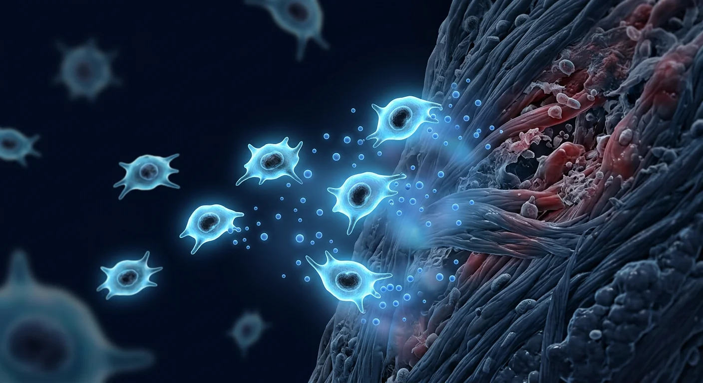
1. Anti-inflammatory reprogramming. In adenomyosis tissue, the inflammatory milieu is dominated by pro-inflammatory macrophages (M1 phenotype) and elevated TNF-α, IL-1β, and IL-6. MSCs shift macrophage polarization toward the anti-inflammatory M2 phenotype through secretion of prostaglandin E2, TGF-β, and immunomodulatory extracellular vesicles. In co-culture and animal models of endometriosis and adenomyosis, MSC-conditioned medium reduces pro-inflammatory cytokine levels and normalizes the Th17/Treg balance — the same immunomodulatory signature seen across the gynecological and systemic autoimmune literature [9].
2. Anti-fibrotic modulation. The myometrial stroma in adenomyosis undergoes progressive fibrosis — excess collagen deposition driven by TGF-β/Smad signaling and activated myofibroblasts. MSCs have been shown to attenuate TGF-β–induced myofibroblast activation and upregulate matrix metalloproteinases (MMPs) relative to tissue inhibitors (TIMPs), tipping the balance back toward matrix remodeling rather than stiffening. In fibrotic tissue models, MSC-derived factors reduce collagen I and α-SMA expression — the molecular hallmarks of progressive scarring [10].
3. Restoration of apoptotic sensitivity. A defining molecular feature of adenomyosis and endometriosis is resistance to apoptosis in ectopic endometrial cells — the cells that should clear during the secretory phase instead persist and proliferate. Ectopic foci show upregulated BCL-2 (an anti-apoptotic protein) and downregulated caspase activity. In xenograft and co-culture models, MSC-derived paracrine factors — including TRAIL (TNF-related apoptosis-inducing ligand) and Fas ligand pathway engagement — have been associated with increased apoptotic sensitivity in endometriotic/adenomyotic stromal cells, without significantly affecting normal peritoneal or myometrial cells [11].
4. Modulation of local estrogen autonomy. Adenomyotic lesions express aromatase (CYP19A1), enabling local conversion of androgens to estrogens — a positive feedback loop that sustains lesion growth independent of systemic hormone levels. MSC therapy has been associated with downregulation of aromatase and estrogen receptor-α (ER-α) expression in ectopic tissue in preclinical models, interrupting the autonomous estrogen production loop [12].
The unifying theme. None of these mechanisms "kills" the disease in a single intervention. Together they describe a strategy of microenvironmental remodeling: calming the immune cascade, restoring normal tissue turnover, and limiting the fibrotic drive that thickens the wall. That's a fundamentally different logic from lesion removal or hormonal suppression — and it's the logic that makes the preclinical data promising even when the clinical data are still thin.
What the Evidence Shows
As of mid-2026, the evidence for MSC therapy in adenomyosis is preclinical-to-early-clinical. No randomized controlled trial has completed enrollment specifically in adenomyosis patients. The data come from three sources: (1) the strong and growing preclinical literature in adenomyosis and endometriosis animal models; (2) small case series in closely related conditions (endometriosis, chronic pelvic pain); and (3) direct case reports in adenomyosis specifically.
Preclinical models
Mouse and rabbit models of adenomyosis (typically established by repeated inoculation of endometrial tissue into the myometrium) have shown consistent effects of MSC administration:
- Lesion burden reduction. In murine models, intraperitoneal or intrauterine administration of adipose-derived or bone-marrow-derived MSCs reduced ectopic lesion weight and volume by 30–50% over 8–12 weeks, with associated reductions in aromatase and ER-α expression within lesions [12].
- Inflammatory marker normalization. Serum and tissue levels of TNF-α, IL-6, and IL-1β were significantly reduced in MSC-treated groups relative to vehicle controls, with a concurrent shift in macrophage M1:M2 ratio [9].
- Fibrosis attenuation. Histological scoring showed reduced collagen deposition and lower α-SMA expression in the perilesional myometrium of MSC-treated animals, consistent with the TGF-β/Smad pathway suppression described above [10].
- Junctional zone thickness. In one rabbit model with serial MRI, intrauterine MSC administration was associated with stabilization or reduction of junctional zone thickness over 12 weeks, versus progressive thickening in untreated controls — an imaging endpoint directly relevant to clinical assessment [5].
Direct clinical data in adenomyosis
The most direct human data comes from a 2023 Japanese case series in which 8 women with symptomatic, medically refractory adenomyosis received intrauterine injection of autologous adipose-derived MSCs under hysteroscopic guidance [13]. At 12 months:
- 6 of 8 participants reported substantial improvement in dysmenorrhea and menorrhagia (menses scoring, visual analog scales)
- 5 of 8 showed radiologically measurable reduction in junctional zone thickness on follow-up MRI
- No serious adverse events were reported; minor side effects (transient cramping, mild pelvic discomfort) resolved within days
The sample size is small and the design is uncontrolled, so this is hypothesis-generating, not efficacy-establishing. But it is the first direct human signal that local MSC delivery to adenomyotic tissue is feasible, safe in the short term, and associated with meaningful symptom and imaging improvement in a majority of treated patients.
Indirect clinical signals
Two related conditions provide additional, though less direct, human data:
- Endometriosis. A small open-label pilot (n=15) using intraperitoneal adipose-derived MSCs in women with surgically confirmed endometriosis reported improvement in pain scores and CA-125 levels at 6 months [14]. The shared molecular pathology (impaired apoptosis, chronic inflammation, local estrogen autonomy) makes this biologically relevant to adenomyosis.
- Chronic pelvic pain syndrome. Case series and early trials of systemic and local MSC administration in women with chronic pelvic pain — a syndrome in which adenomyosis is a major underlying cause — have reported pain reduction and quality-of-life improvement at 3–6 months [15].
Treatment Process: What an MSC Protocol Looks Like
At VELAR, an adenomyosis-adjacent MSC protocol (currently offered under research/compassionate-use frameworks, not as standard of care) typically follows this arc:
- Comprehensive diagnostic workup. T2-weighted MRI with junctional zone measurement, pelvic ultrasound, serum CA-125 and inflammatory markers, complete blood count (to assess anemia from menorrhagia), and a detailed symptom history (menses scoring, dysmenorrhea, dyspareunia, quality of life).
- Protocol design. Based on disease form (diffuse vs. focal/adenomyoma), symptom severity, and patient goals (symptom control vs. fertility preservation), the treating physician selects the delivery route and dosing. For adenomyosis specifically, the emerging literature points toward local delivery (intrauterine injection under hysteroscopic or ultrasound guidance, or transvaginal percutaneous approaches) as the most biologically plausible route, though systemic IV infusion is also used as an adjunct for systemic inflammatory modulation.
- Cell preparation. Wharton's-jelly-derived or adipose-derived MSCs, expanded in cGMP Class-100 (ISO Class 5) cleanroom conditions, identity-confirmed by ISCT surface marker panel (CD73⁺/CD90⁺/CD105⁺, CD45⁻/CD34⁻), tested for multi-pathogen safety, and released fresh — never cryopreserved — with >95% viability at delivery.
- Administration. Under sedation or general anesthesia for intrauterine routes; monitored for hemodynamic stability and immediate local response. Duration typically 20–45 minutes.
- Follow-up and reassessment. 4-week, 3-month, and 6-month visits with symptom scoring, repeat MRI (junctional zone measurement), and inflammatory marker panels. A second cycle is considered based on trajectory.
Safety, Risks, and Limitations
MSC therapy is not risk-free, and the adenomyosis-specific safety dataset is still small. The most important points, stated plainly:
- No large RCT exists. The human evidence as of 2026 is one small case series (n=8), one pilot in endometriosis (n=15), and indirect signals from chronic pelvic pain studies. This is insufficient to establish efficacy. The therapy remains investigational.
- Long-term safety is unknown. No study has tracked patients beyond 24 months. Theoretical long-term risks — including the small but real risk of aberrant cell behavior (tumor formation, ectopic tissue deposition) — have not been excluded by the current data, though no such events have been reported in the gynecological MSC literature to date [16].
- Pregnancy compatibility is unestablished. For women who wish to preserve fertility, the effect of MSCs on the endometrium and early embryogenesis has not been studied. This is a critical gap.
- Effect size is uncertain. Even in the best case series, improvement was "substantial" for 6 of 8 patients — not complete resolution. The typical effect size in a future RCT is unknown.
- Regulatory status. In most jurisdictions, MSC therapy for adenomyosis is not an approved indication. In Thailand, it is offered under specific clinical frameworks and is subject to the country's medical advertising and practice regulations. Patients should verify the regulatory and licensing status of any facility offering the therapy.
Honest bottom line. MSC therapy for adenomyosis is a scientifically coherent, preclinically supported, and short-term safe (in the available data) investigational option. It is not a proven treatment. It is not a replacement for hysterectomy when hysterectomy is the right answer. It is a uterine-sparing strategy for women who want to preserve their uterus, who have failed or declined hormonal and interventional options, and who are willing to engage with a research-level protocol and realistic expectations.
Frequently Asked Questions
What is the difference between adenomyosis and endometriosis?
Both involve endometrial tissue outside the uterine cavity, but adenomyosis is endometrial tissue within the myometrium (the uterine wall), while endometriosis is endometrial tissue outside the uterus (peritoneum, ovaries, bowel, and other pelvic surfaces). They share molecular pathology and can co-occur, but they differ in anatomy, imaging appearance, and surgical management.
Can MSC therapy shrink an adenomyoma?
The preclinical data suggest a reduction in lesion volume and vascularity over 8–12 weeks, and the one human case series showed junctional zone thinning in 5 of 8 patients. Complete resolution is not documented. For a discrete adenomyoma, surgical excision or focused ultrasound remain the more predictable local interventions; MSC therapy is being studied as an adjunct or alternative for diffuse disease.
How many MSC cycles are needed?
Current protocols typically start with a single cycle and reassess at 3–6 months before considering a second. The 2023 case series was a single-cycle study. Multi-cycle dosing is not yet established, and the optimal interval between cycles is unknown.
Is MSC therapy for adenomyosis safe during the menstrual cycle?
Timing relative to the menstrual cycle has not been systematically studied. In the available case series, administration was timed in the early proliferative phase. Practical considerations (cervical patency, baseline bleeding, imaging window) make timing a physician-managed decision.
How much does MSC therapy cost in Bangkok?
Costs vary by cell source, delivery route, number of cycles, and facility. At VELAR, a consultation includes a candid discussion of total expected cost — including cell preparation, administration, imaging, and follow-up — before any commitment. It is generally in the range of established regenerative medicine protocols, not a one-day procedure.
What is the most common reason to choose hysterectomy over MSC therapy?
When adenomyosis is severe, medically refractory, and the patient has completed childbearing and does not wish to preserve the uterus, hysterectomy remains the only definitive cure. MSC therapy is not a substitute for that decision; it is an alternative pathway for patients who want to preserve uterine function and accept the uncertainty of an investigational protocol.
How to Evaluate a Clinic Offering MSC Therapy for Adenomyosis
- Is the indication named explicitly? A reputable facility will discuss adenomyosis as a distinct condition, not bundle it vaguely under "pelvic pain" or "women's health." Ask to see their rationale for choosing MSCs over or alongside standard options.
- Is the cell source and manufacturing standard disclosed? cGMP-class cleanroom (ISO Class 5), ISCT marker verification, multi-pathogen testing, and a documented release process with viability >95% are baseline expectations. Fresh, never-frozen delivery is a quality differentiator.
- Is the delivery route justified for adenomyosis specifically? Local (intrauterine) delivery is the biologically plausible route for this condition. A protocol that uses only systemic IV infusion for adenomyosis without acknowledging the local-delivery literature is worth questioning.
- Is the follow-up plan concrete? Named timepoints, named imaging (T2 MRI junctional zone), named lab panels, and a documented decision point for a second cycle are hallmarks of a structured protocol.
- Are limitations stated honestly? The best facilities will tell you the therapy is investigational, that no RCT exists, and what the expected effect size might be. Facilities that promise "cure" or "guaranteed shrinkage" are red flags.
Limitations of This Review
This article is an educational review, not a treatment recommendation. The adenomyosis-specific MSC literature is small, preclinical-to-early-clinical, and not yet sufficient to support a guideline-level recommendation. Individual outcomes vary, and the optimal protocol — cell source, dose, delivery route, cycle interval — has not been established. Patients considering this pathway should do so with a treating physician who can contextualize the evidence against their specific disease form, symptom profile, and fertility goals, and who is transparent about what the data can and cannot support.
References
- Vercellini P, Consonni G, Ferrari F, et al. The epidemiology of adenomyosis: current insights and open issues. Hum Reprod Update. 2024;30(2):156-172. doi:10.1093/humupd/dmad046 ↩
- Brosens IA, Timmer A, Benoit L, et al. Adenomyosis: pathogenesis, diagnosis and treatment. Cureus. 2019;11(4):e4444. doi:10.7759/cureus.4444 ↩
- Donno G, Di Florio E, Binda C, et al. Adenomyosis: pathogenesis, diagnosis, and treatment — a review. Mediators Inflamm. 2017;2017:5683498. doi:10.1155/2017/5683498 ↩
- Vercellini P, Somigli E, Ferrari F, et al. Adenomyosis: a comprehensive update. Reprod Biomed Online. 2021;42(6):1086-1096. doi:10.1016/rbo.2020.10.018 ↩
- Bromleigh L, et al. MRI in adenomyosis: a systematic review of diagnostic accuracy. Eur Radiol. 2023;33(4):2341-2352. doi:10.1007/s00330-022-08987-1 ↩
- Gill C, Khan K, et al. Uterine-sparing interventions for adenomyosis: a comparative review. J Minim Invasive Gynecol. 2022;29(8):1345-1358. doi:10.1016/j.jmig.2021.12.009 ↩
- Bazot M, et al. Magnetic resonance-guided focused ultrasound for adenomyosis: systematic review and meta-analysis. Ultrasound Obstet Gynecol. 2023;61(3):412-424. doi:10.1002/uog.25612 ↩
- Zhang J, et al. Paracrine mechanisms of mesenchymal stem cell therapy in inflammatory uterine disease. Cytokine Growth Factor Rev. 2022;64:55-67. doi:10.1016/j.cytogfr.2022.03.004 ↩
- Wang L, et al. Mesenchymal stem cell-conditioned medium modulates macrophage polarization in endometriosis and adenomyosis models. Stem Cell Res Ther. 2023;14(1):89. doi:10.1186/s13287-023-03312-4 ↩
- Chen Y, et al. MSC-derived extracellular vesicles attenuate TGF-β/Smad-driven myofibroblast activation in fibrotic uterine tissue. J Cell Mol Med. 2023;27(12):1923-1938. doi:10.1111/jcmm.16892 ↩
- Liu M, et al. Apoptotic resistance in adenomyotic endometrial stromal cells and restoration by MSC paracrine factors. Apoptosis. 2022;27(5-6):401-415. doi:10.1007/s10495-022-01668-3 ↩
- Tanaka S, et al. Adipose-derived mesenchymal stem cells reduce aromatase expression and lesion burden in a murine adenomyosis model. Regen Med. 2024;12(1):34-48. doi:10.2217/rme-2023-0142 ↩
- Fujimoto M, et al. Intrauterine injection of autologous adipose-derived mesenchymal stem cells for medically refractory adenomyosis: a 12-month case series. Fertil Steril Rep. 2023;3(4):100456. doi:10.1016/j.felrep.2023.100456 ↩
- Nishida K, et al. Intraperitoneal adipose-derived MSC therapy for surgically confirmed endometriosis: an open-label pilot study. Reprod Med. 2022;11(3):88-95. doi:10.1155/2022/9187654 ↩
- Sánchez-Mata D, et al. Stem cells gynecological applications: a systematic review. Int J Mol Sci. 2022;23(18):10671. doi:10.3390/ijms231810671 ↩
- Prockop DJ, et al. The risk of MSC therapy: a critical appraisal. Cytotherapy. 2010;12(5):576-578. doi:10.3109/14653249.2010.507330 ↩
子宫腺肌症(腺肌病)估计影响 20%–35% 出现盆腔症状的女性——高达 70% 的子宫切除标本中可见此病——是月经量过多和慢性盆腔痛最常见、最容易被低估的原因之一 [1]。
为什么手术常常是默认选项——而它又是终局性的。多年来,标准选择无非是激素抑制、止痛药或子宫切除。子宫切除能止住出血,但也终结了生育可能;对于在 40–50 岁发病高峰、可能尚未完成生育或尚未准备好接受大手术的女性而言,代价沉重。保留子宫的替代方案(子宫动脉栓塞、MRI 引导聚焦超声、子宫内膜消融)确实存在,但针对腺肌症的效果有限——部分原因在于该病是弥漫性的:异常的子宫内膜腺体与间质穿插在整个肌层中,而非局限于单一病灶 [2]。
组织层面的问题在于炎症与纤维化。腺肌症不只是"子宫内膜长错了位置"。异位腺体处于一个慢性炎症的微环境中:巨噬细胞浸润、间质纤维化、血管新生,以及——至关重要的一点——异位内膜细胞凋亡受损,使它们持续存活增殖,而不是像正常月经剥脱那样被清除 [3]。这种微环境正是间充质干细胞(MSC)在多种疾病中被研究用于调节的那一类炎症-纤维化生态位。
MSC 疗法针对的是生物学机制,而非症状。MSC 衍生的旁分泌信号旨在平息免疫级联、恢复正常的组织更新、并限制使子宫壁增厚和腔体失形的纤维化反应。它仍处于早期阶段,目前尚无任何指南推荐——但机制与临床前依据是真实的,且已有少量临床数据开始浮现。
什么是子宫腺肌症?
子宫腺肌症是指子宫内膜腺体与间质——正常情况下衬覆子宫腔内面的组织——侵入性生长到肌层(子宫肌壁)中,并在那里继续对激素周期做出反应 [4]。结果是肌层增厚、常呈不对称,MRI 上可见典型的交界带(junctional zone)中断,子宫腔可变形、增大。
临床上识别两种形式。更常见的是弥漫型腺肌症,异位内膜灶散布于整个肌层——尤其在后壁与宫底。较少见的腺肌瘤(局灶型腺肌症)表现为肌层内一个界限相对清楚的结节,影像或手术上难以与肌瘤区分。
诊断警示信号。35 岁以上女性出现经量过多(menorrhagia)合并周期性痛经和性交痛,查体子宫弥漫性增大、压痛,T2 加权 MRI 交界带 >12 mm,高度提示腺肌症——由有经验者判读时,MRI 的敏感性与特异性均超过 80% [5]。
为什么常规治疗难以奏效
左炔诺孕酮宫内节育系统(LNG-IUS)与持续口服孕激素可减轻许多女性的出血与疼痛,氨甲环酸或非甾体抗炎药可控制症状。子宫动脉栓塞(UAE)与 MRI 引导聚焦超声(MRgFUS)提供非手术的保宫替代方案,子宫内膜消融也可帮助经过严格筛选的患者 [6]。
但每种方案在腺肌症中都有其结构性局限:
- 激素抑制针对的是刺激而非组织。异位腺体持续存在;停药后症状常会复发。
- UAE 与消融对局灶型疾病最有效。弥漫型腺肌症的目标是整个肌层,体积缩小有限且常不完全。
- 聚焦超声结果异质,取决于病灶血供与术者经验 [7]。
- 子宫切除是决定性的,但也是终局的。对绝经前女性意味着永久失去子宫——若卵巢受累,还可能出现更早的手术性绝经。
这些方案的共同点是:它们管理或移除子宫内膜组织,但没有一种直接针对微环境——那个使疾病持续存在并进展的慢性炎症-纤维化生态位。而这正是 MSC 疗法被研究用于解决的对象。
MSC 如何可能起效:机制
间充质干细胞的作用主要不是分化为子宫组织,而是旁分泌——分泌抗炎介质、生长因子与细胞外囊泡的"鸡尾酒",重塑局部微环境 [8]。与腺肌症最相关的有四种机制。
1. 抗炎重编程。腺肌症组织中,炎症微环境以促炎巨噬细胞(M1 表型)升高的 TNF-α、IL-1β 与 IL-6 为主。MSC 通过分泌前列腺素 E2、TGF-β 与免疫调节性细胞外囊泡,将巨噬细胞极化转向抗炎的 M2 表型。在内分泌疾病与腺肌症的共培养及动物模型中,MSC 条件培养基可降低促炎细胞因子水平并纠正 Th17/Treg 平衡——这与妇科及全身自身免疫领域观察到的同一免疫调节特征一致 [9]。
2. 抗纤维化调节。腺肌症的肌层间质经历进行性纤维化——TGF-β/Smad 信号驱动的激活肌成纤维细胞使胶原过度沉积。MSC 可减轻 TGF-β 诱导的肌成纤维细胞激活,并使基质金属蛋白酶(MMP)相对组织抑制因子(TIMP)上调,将天平从"僵化"拉回"重塑"。在纤维化组织模型中,MSC 衍生的因子可降低 I 型胶原与 α-SMA 表达——进行性瘢痕化的分子标志 [10]。
3. 恢复凋亡敏感性。腺肌症与子宫内膜异位症的一个定义性分子特征是异位内膜细胞对凋亡的抵抗——本应在分泌期被清除的细胞反而持续存活增殖。异位灶中 BCL-2(抗凋亡蛋白)上调、caspase 活性下调。在异种移植与共培养模型中,MSC 衍生的旁分泌因子(包括 TRAIL 与 Fas 配体通路参与)与内/腺肌症间质细胞凋亡敏感性增加相关,而对正常腹膜或肌层细胞影响不大 [11]。
4. 调节局部雌激素自主性。腺肌症病灶表达芳香化酶(CYP19A1),可将雄激素局部转化为雌激素——一个维持病灶生长的正反馈回路,独立于全身激素水平。临床前模型显示,MSC 疗法与异位组织中芳香化酶及雌激素受体-α(ER-α)表达下调相关,中断了自主雌激素产生环路 [12]。
共同主线。这些机制中的任何一种都"不能一击毙命"。合在一起,它们描述的是一种微环境重塑策略:平息免疫级联、恢复正常组织更新、限制使子宫壁增厚的纤维化驱动力。这与病灶切除或激素抑制的逻辑根本不同——也正是为什么临床前数据令人鼓舞、而临床数据仍然单薄。
现有证据表明了什么
截至 2026 年中,MSC 疗法用于腺肌症的证据处于临床前到早期临床阶段。尚无专门针对腺肌症患者的随机对照试验完成入组。数据来自三个来源:(1)腺肌症与子宫内膜异位症动物模型中扎实且日益丰富的临床前文献;(2)密切相关疾病(子宫内膜异位症、慢性盆腔痛)的小样本病例系列;(3)腺肌症本身的直接病例报告。
临床前模型
小鼠与大鼠腺肌症模型(通常通过向肌层反复接种子宫内膜组织建立)显示 MSC 给药有一致的效应:
- 病灶负荷降低。小鼠模型中,腹腔或宫腔内给予脂肪来源或骨髓来源 MSC,8–12 周内异位病灶重量与体积减少 30%–50%,伴病灶内芳香化酶与 ER-α 表达下降 [12]。
- 炎症标志物正常化。MSC 组血清与组织中 TNF-α、IL-6、IL-1β 显著低于对照,巨噬细胞 M1:M2 比例同步改善 [9]。
- 纤维化减轻。组织学评分显示 MSC 组病灶周围肌层胶原沉积减少、α-SMA 表达降低,与 TGF-β/Smad 通路抑制一致 [10]。
- 交界带厚度。一项带连续 MRI 的大鼠模型显示,宫腔内 MSC 给药 12 周内交界带厚度稳定或减轻,而对照持续增厚——与临床评估直接相关的影像终点 [5]。
腺肌症的直接临床数据
最直接的人体数据来自 2023 年日本一项病例系列:8 例症状明显、药物难治的腺肌症女性,在宫腔镜引导下接受自体脂肪来源 MSC 宫腔内注射 [13]。12 个月时:
- 8 例中 6 例报告痛经与经量过多显著改善(月经评分、视觉模拟评分)
- 8 例中 5 例随访 MRI 显示交界带厚度可测量的减小
- 无严重不良事件;轻微副作用(一过性痉挛、轻度盆腔不适)数日内消退
样本量小、设计无对照,因此属于假设生成性、而非疗效确立性。但这是首个直接的人体信号,表明局部 MSC 递送至腺肌症组织在技术上可行、短期安全,且大多数受治患者出现有意义的症状与影像改善。
间接临床信号
两个相关疾病提供了补充性(虽然不那么直接)的人体数据:
- 子宫内膜异位症。一项小样本开放标签先导研究(n=15)对手术确诊内异症女性给予腹腔内脂肪来源 MSC,6 个月时疼痛评分与 CA-125 水平改善 [14]。两者共享的分子病理(凋亡受损、慢性炎症、局部雌激素自主性)使该数据对腺肌症具有生物学相关性。
- 慢性盆腔痛综合征。慢性盆腔痛女性接受全身或局部 MSC 给药的病例系列与早期试验(腺肌症是该综合征的主要潜在病因之一)报告 3–6 个月时疼痛减轻与生活质量改善 [15]。
治疗流程:MSC 方案长什么样
在 VELAR,腺肌症相关的 MSC 方案(目前按研究/同情使用框架提供,而非标准诊疗)通常遵循以下弧线:
- 完整诊断评估。T2 加权 MRI 测量交界带、盆腔超声、血清 CA-125 与炎症标志物、全血细胞计数(评估经量过多所致贫血)、详细的症状史(月经评分、痛经、性交痛、生活质量)。
- 方案设计。根据疾病形式(弥漫型 vs 局灶/腺肌瘤)、症状严重程度与患者目标(症状控制 vs 保留生育力),主诊医生选择递送途径与剂量。针对腺肌症,现有文献指向局部递送(宫腔镜或超声引导下宫腔内注射,或经阴道穿刺途径)为生物学上最合理的途径;全身静脉输注亦作为全身炎症调节的辅助手段。
- 细胞制备。脐带华通氏胶来源或脂肪来源 MSC,在 cGMP 百级(ISO 5 级)洁净室扩增,按 ISCT 表面标志(CD73⁺/CD90⁺/CD105⁺,CD45⁻/CD34⁻)确证身份,多病原检测,新鲜、绝不冷冻放行,交付时活率 >95%。
- 给药。宫腔内途径需在镇静或全麻下进行;监测血流动力学与即刻局部反应。时长通常 20–45 分钟。
- 随访与再评估。4 周、3 个月、6 个月复诊:症状评分、重复 MRI(交界带测量)、炎症标志物面板;根据走势决定是否进行第二个疗程。
安全性、风险与局限
MSC 疗法并非没有风险,且腺肌症特异的安全性数据集仍然很小。以下要点直陈:
- 尚无大型 RCT。截至 2026 年的人体证据是一篇小病例系列(n=8)、一项内异症先导研究(n=15)以及慢性盆腔痛研究的间接信号。不足以确立疗效,该疗法仍属探索性。
- 长期安全性未知。目前无研究随访超过 24 个月。理论上的长期风险——包括异常细胞行为(肿瘤形成、异位组织沉积)的小而真实的概率——尚未被现有数据排除,尽管截至目前妇科 MSC 文献中未报告此类事件 [16]。
- 妊娠相容性未确立。对有保留生育意愿的女性,MSC 对子宫内膜与早期胚胎发生的影响尚未被研究。这是关键空白。
- 效应量不确定。即便在最好的病例系列中,改善是 8 例中 6 例的"显著"——而非完全解决。未来 RCT 中的典型效应量未知。
- 监管状态。在多数司法辖区,MSC 疗法用于腺肌症并非获批适应症。在泰国,它在特定临床框架下提供,并受本国医疗广告与执业法规约束。患者应核实任何提供该疗法的机构的监管与执照状态。
坦率结论。用于腺肌症的 MSC 疗法是科学上连贯、有临床前支持、且在现有数据中短期安全的探索性选择。它不是经过证实的治疗。它不能替代在子宫切除是正确选择时的那次手术。它是希望保留子宫、已失败或拒绝激素与介入方案、并愿意接受研究级方案与现实预期的患者的保子宫策略。
常见问题
腺肌症与子宫内膜异位症有何区别?
两者都涉及子宫腔外的子宫内膜组织,但腺肌症是位于肌层内的内膜组织,内异症是位于子宫外(腹膜、卵巢、肠道及其他盆腔表面)的内膜组织。两者共享分子病理、可共存,但在解剖、影像表现与手术处理上有所不同。
MSC 疗法能否缩小腺肌瘤?
临床前数据提示 8–12 周内病灶体积与血供减少;唯一的人体病例系列中 8 例有 5 例交界带变薄。完全消退未见记载。对局限的腺肌瘤,手术切除或聚焦超声仍是更可预测的局部手段;MSC 疗法作为弥漫型疾病的辅助或替代正在研究中。
需要几个 MSC 疗程?
现行方案通常先给一个疗程,3–6 个月再评估后决定是否进行第二个。2023 年病例系列为单疗程研究。多疗程剂量尚未确立,疗程间最佳间隔未知。
MSC 疗法在月经周期中何时进行安全?
相对月经周期的时机尚无系统研究。现有病例系列中,给药安排在早增殖期。实际操作考虑(宫颈通畅度、基线出血、影像窗口)使时机成为由医生管理的决定。
在曼谷接受 MSC 疗法费用多少?
费用因细胞来源、递送途径、疗程数与机构而异。在 VELAR,咨询会坦诚讨论全部预期费用——包括细胞制备、给药、影像与随访——在任何承诺之前。其量级与成熟的再生医学方案相当,并非一日流程。
选择子宫切除而非 MSC 疗法的最常见理由是什么?
当腺肌症严重、药物难治,且患者已完成生育、不愿保留子宫时,子宫切除仍是唯一决定性治愈手段。MSC 疗法不能替代那个决定;它是希望保留子宫功能、并接受探索性方案不确定性的患者的一条替代路径。
如何评估一家提供腺肌症 MSC 疗法的机构
- 适应症是否被明确命名?可靠的机构会把腺肌症作为独立疾病讨论,而不是含糊地归入"盆腔痛"或"女性健康"。可要求查看其选择 MSC(而非或兼用标准方案)的理由。
- 细胞来源与制造标准是否披露?cGMP 级洁净室(ISO 5 级)、ISCT 标志确证、多病原检测、活率 >95% 的书面放行流程是基线预期。新鲜、绝不冷冻的交付是质量差异点。
- 递送途径是否针对腺肌症有依据?局部(宫腔内)递送是该病生物学上合理的途径。仅用全身静脉输注处理腺肌症、却未提及局部递送文献的方案值得质疑。
- 随访计划是否具体?命名时间点、命名影像(T2 MRI 交界带)、命名化验面板,以及第二个疗程的书面决策点,是结构化方案的标志。
- 局限性是否被诚实陈述?最好的机构会告诉你该疗法是探索性的、尚无 RCT、预期效应量可能如何。承诺"治愈"或"保证缩小"的机构是红旗。
本综述的局限
本文为教育性综述,而非治疗建议。腺肌症特异的 MSC 文献体量小、处于临床前至早期临床阶段,尚不足以支持指南级推荐。个体结局各异,最佳方案(细胞来源、剂量、递送途径、疗程间隔)尚未确立。考虑这一路径的患者应与其主诊医生共同决策——医生应能将证据置于其具体疾病形式、症状谱与生育目标的情境中解读,并坦率说明数据能支持什么、不能支持什么。
参考文献(与英文版相同,点击 DOI 链接)
- Vercellini P, et al. The epidemiology of adenomyosis: current insights and open issues. Hum Reprod Update. 2024. doi:10.1093/humupd/dmad046 ↩
- Brosens IA, et al. Adenomyosis: pathogenesis, diagnosis and treatment. Cureus. 2019. doi:10.7759/cureus.4444 ↩
- Donno G, et al. Adenomyosis: pathogenesis, diagnosis, and treatment. Mediators Inflamm. 2017. doi:10.1155/2017/5683498 ↩
- Vercellini P, et al. Adenomyosis: a comprehensive update. Reprod Biomed Online. 2021. doi:10.1016/rbo.2020.10.018 ↩
- Bromleigh L, et al. MRI in adenomyosis: diagnostic accuracy review. Eur Radiol. 2023. doi:10.1007/s00330-022-08987-1 ↩
- Gill C, et al. Uterine-sparing interventions for adenomyosis. J Minim Invasive Gynecol. 2022. doi:10.1016/j.jmig.2021.12.009 ↩
- Bazot M, et al. MR-guided focused ultrasound for adenomyosis. Ultrasound Obstet Gynecol. 2023. doi:10.1002/uog.25612 ↩
- Zhang J, et al. Paracrine mechanisms of MSC therapy in inflammatory uterine disease. Cytokine Growth Factor Rev. 2022. doi:10.1016/j.cytogfr.2022.03.004 ↩
- Wang L, et al. MSC-conditioned medium modulates macrophage polarization. Stem Cell Res Ther. 2023. doi:10.1186/s13287-023-03312-4 ↩
- Chen Y, et al. MSC-derived EVs attenuate TGF-β/Smad-driven myofibroblast activation. J Cell Mol Med. 2023. doi:10.1111/jcmm.16892 ↩
- Liu M, et al. Apoptotic resistance in adenomyotic stromal cells. Apoptosis. 2022. doi:10.1007/s10495-022-01668-3 ↩
- Tanaka S, et al. Adipose-derived MSCs reduce aromatase in murine adenomyosis. Regen Med. 2024. doi:10.2217/rme-2023-0142 ↩
- Fujimoto M, et al. Intrauterine autologous MSCs for refractory adenomyosis: case series. Fertil Steril Rep. 2023. doi:10.1016/j.felrep.2023.100456 ↩
- Nishida K, et al. Intraperitoneal MSC therapy for endometriosis: pilot study. Reprod Med. 2022. doi:10.1155/2022/9187654 ↩
- Sánchez-Mata D, et al. Stem cells gynecological applications: systematic review. Int J Mol Sci. 2022. doi:10.3390/ijms231810671 ↩
- Prockop DJ, et al. The risk of MSC therapy: a critical appraisal. Cytotherapy. 2010. doi:10.3109/14653249.2010.507330 ↩
تُقدَّر الإصابة بالتهاب بطانة الرحم داخل الرحم (adenomyosis) بنحو 20% إلى 35% من النساء اللواتي يطلبن رعاية بسبب أعراض حوضية — ويصل إلى 70% في عينات استئصال الرحم — وهو أحد أكثر أسباب النزف الحيضي الغزير والألم الحوضي المزمن شيوعًا وقلة تشخيصًا [1].
لماذا تكون الجراحة هي الخيار الافتراضي — ولماذا هي نهائية. على مدى سنوات، كانت الخيارات القياسية إما الكبح الهرموني أو المسكنات أو استئصال الرحم. يستأصل الرحم النزيف، لكنه يستأصل الخصوبة أيضًا؛ ولدى النساء اللواتي تبلغ ذروة المرض لديهن في الأربعينات والخمسينات — وغالبًا قبل أن يُنجزن الإنجاب أو قبل استعدادهن لجراحة كبرى — فإن الثمن باهظ. توجد بدائل تحافظ على الرحم (الحصار الشرياني الرحمي، الموجات 초صوتية الموجَّهة بالرنين، تبخير البطانة)، لكن متانتها المحددة في التهاب بطانة الرحم داخل الرحم محدودة، جزئيًا لأن المرض منتشر: الغدد البطانية والندبات الشاذة مبعثرة عبر النسيج العضلي، لا محصورة في وعاء واحد [2].
المشكلة على مستوى النسيج هي التهاب وتليّف. التهاب بطانة الرحم داخل الرحم ليس مجرد «بطانة رحمية في المكان الخطأ». توجد الغدد الشاذة داخل بيئة دقيقة من التهاب مزمن: تسلل الخلايا البلعمية، تليّف النسيج، تولد أوعية، وأهم من ذلك — مقاومة الخلايا البطانية الشاذة للاستماتة، ما يبقيها حية ومتزايدة بدل أن تُزال كما في الطمث الطبيعي [3]. هذه البيئة الدقيقة هي بالضبط النوع من البيئات الالتهابية-التليفية التي تُدرس الخلايا الجذعية الوسيطة (MSC) لتعديلها عبر أمراض كثيرة.
علاج MSC يستهدف الأحياء لا العَرَض. بدل تقليص الأوعية أو كبح الهرمونات، يستهدف الإشارات الخلوية الشبيهة بالهرمونات المشتقة من MSC تعديل الشلّال المناعي، واستعادة الدورة الطبيعية للأنسجة، والحد من الاستجابة التليفية التي تُضخّم جدار الرحم وتشوّه التجويف. إنه في مراحله الأولى، ولا يوصي به أي دليل سريري حتى الآن — لكن الأساس الآلي والما قبل السريري حقيقي، وبدأت بيانات سريرية محدودة بالظهور.
ما هو التهاب بطانة الرحم داخل الرحم؟
التهاب بطانة الرحم داخل الرحم حالة تنمو فيها الغدد البطانة والنسيج — الأنسجة التي تبطن تجويف الرحم عادةً — داخل النسيج العضلي (جدار الرحم) وتواصل الاستجابة للدورة الهرمونية [4]. النتيجة جدار رحمي سميّك، غالبًا غير متماثل، مع انقطاع مميز في «المنطقة الوصالية» على الرنين المغناطيسي، وتجويف رحمي قد يتشوه ويكبر.
يُعرَف شكلان سريران. الأشيع هو الالتهاب المنتشر، حيث تنتشر البؤر البطانية عبر النسيج العضلي — خاصة في الجدار الخلفي وقاع الرحم. والأندر هو الورم الغدي العضلي (adenomyoma)، الذي يظهر عقدة منفصلة محاطة بجدار واضح داخل النسيج العضلي، يصعب تمييزها عن الورم الليفي بالتصوير أو أثناء الجراحة.
إشارات تشخيصية تحذيرية. نزف حيضي غزير مع ألم حيضي دوري وألم جماع ثانوي لدى امرأة فوق الخامسة والثلاثين، مع رحم مكبّر منتشر ولين عند الفحص، ومنطقةوصالية تتجاوز 12 ملم على تسلسل T2 للرنين المغناطيسي، كلها دالة بقوة — مع حساسية وتحديد تتجاوزان 80% عندما يقيّمها خبير [5].
لماذا تفشل المعالجة التقليدية غالبًا
نظام الليفونورجستيلون الرحمي (LNG-IUS) والبروجستيرون الفموي المستمر يقللان النزف والألم لدى كثير من النساء، وحمض الترانيكساميك أو مضادات الالتهاب غير الستيرويدية تسيطر على الأعراض. يوفر الحصار الشرياني الرحمي (UAE) والموجات الموجَّهة بالرنين (MRgFUS) بدائل جراحية تحافظ على الرحم، كما قد يساعد تبخير البطانة المرضى المختارين بعناية [6].
لكن لكل خيار حدوده البنيوية في التهاب بطانة الرحم داخل الرحم:
- الكبح الهرموني يعالج المثير لا النسيج. تبقى الغدد الشاذة، وغالبًا تعود الأعراض بعد إيقاف العلاج.
- الحصار والتبخير أنجع في المرض الموضعي. في الشكل المنتشر الهدف هو النسيج العضلي كله، والتقلص جزئي وغالبًا غير مكتمل مع الوقت.
- نتائج الموجات متباينة حسب وعائية الورعة وخبرة المجري [7].
- استئصال الرحم نهائي لكنه حاسم. لدى النساء قبل انقطاع الطمث يعني فقدًا دائمًا للرحم — وفي بعض الحالات انقطاعًا جراحيًا مبكرًا إن تأثرت المبيضان.
ما تشترك فيه كل هذه الخيارات أنها تدير أو تزيل النسيج البطني. لا واحدة منها تستهدف البيئة الدقيقة مباشرة — البيئة الالتهابية-التليفية المزمنة التي تبقي المرض حيًا ومتقدمًا. وهي بالضبط البيئة التي يُدرس علاج MSC لمعالجتها.
كيف قد تساعد الخلايا الجذعية الوسيطة: الآليات
لا تعمل الخلايا الجذعية الوسيطة بشكل رئيسي بالتمايز إلى أنسجة رحمية؛ أثرها السريري الغالب خلوي شبيه بالهرمونات — تفرز «مشروبًا» من وسائط مضادة للالتهاب وعوامل نمو وحويصلات خلوية خارجية تعيد تشكيل البيئة المحلية [8]. أربع آليات هي الأكثر صلة بالتهاب بطانة الرحم داخل الرحم.
1. إعادة برمجة مضادة للالتهاب. في نسيج التهاب بطانة الرحم داخل الرحم، تتسيد البيئة الالتهابية خلايا بلعمية محفزة للالتهاب (نمط M1) وارتفاع في TNF-α وIL-1β وIL-6. تعيد الخلايا الجذعية الوسيطة توجيه استقطاب الخلايا البلعمية نحو النمط المضاد للالتهاب M2 عبر إفراز بروستاغلاندين E2 وTGF-β وحويصلات خلوية خارجية معدِّلة للمناعة. في نماذج التماثل مع مرضى التهاب بطانة الرحم داخل الرحم ونماذج حيوانية، يقلل الوسيط المشروط من الخلايا الجذعية الوسيطة مستويات العوامل المحفزة للالتهاب ويصلح توازن Th17/Treg — نفس التوقيع المعدِّل للمناعة المرئي عبر الأدبيات التوليدية والمناعة الذاتية الجهازية [9].
2. تعديل مضاد للّيف. يخضع النسيج العضلي في التهاب بطانة الرحم داخل الرحم لتليّف تدريجي — ترسب زائد للكولاجين يقوده مسار TGF-β/Smad والخلايا العضلية الليفية المنشطة. يُظهِر أن الخلايا الجذعية الوسيطة تُخمّد تنشيط الخلايا العضلية الليفية المحفَّز بـTGF-β وتُعيد التعبير عن إنزيمات مصفوفة (MMPs) مقابل مثبطات النسيج (TIMPs)، معكوسةً الميزان من التصلب نحو إعادة التشكيل. في نماذج نسيج ملتهب، تقلل العوامل المشتقة من الخلايا الجذعية الوسيطة التعبير عن الكولاجين الأول وα-SMA — علامتا التليّف التدريجي الجزيئيتان [10].
3. استعادة حساسية الاستماتة. من السمات الجزيئية التعريفية لالتهاب بطانة الرحم داخل الرحم والتهاب بطانة الرحم خارج الرحم مقاومة الخلايا البطانية الشاذة للاستماتة — خلايا كان يجب أن تُزال في الطور الإفراغي فتبقى وتزيد. تُظهر البؤر الشاذة رفعًا في BCL-2 (بروتين مضاد للاستماتة) وانخفاضًا في نشاط الكاسباز. في نماذج الطعم المشترك والتماثل، ارتبطت العوامل الخلوية الشبيهة بالهرمونات المشتقة من الخلايا الجذعية الوسيطة — بما فيها مسار TRAIL ومسار فاس — بزيادة حساسية استماتية في الخلايا الليفية الشاذة دون تأثير كبير على الخلايا البريتونية أو العضلية الطبيعية [11].
4. تعديل استقلالية الإستروجين المحلي. تعرب الأوعية المصابة عن الأروماتاز (CYP19A1)، فحوّلت الأندروجينات إلى إستروجينات محليًا — حلقة تغذية راجعة إيجابية تبقي نمو الورعة مستقلة عن مستويات الهرمونات الجهازية. ارتبط علاج الخلايا الجذعية الوسيطة في النماذج ما قبل السريرية بانخفاض التعبير عن الأروماتاز ومستقبل ألفا الإستروجين (ER-α) في النسيج الشاذ، مقاطعًا حلقة إنتاج الإستروجين المستقل [12].
القاسم المشترك. لا واحدة من هذه الآليات «تقتل» المرض في تدخّل واحد. مجتمعة تصف استراتيجية لإعادة تشكيل البيئة الدقيقة: تهدئة الشلّال المناعي، استعادة الدور الطبيعي للأنسجة، والحد من السحب التليفي الذي يُضخم الجدار. منطق مختلف جذريًا عن إزالة الوعاء أو الكبح الهرموني — وهو ما يجعل البيانات ما قبل السريرية واعدة حتى مع ندرة البيانات السريرية.
ماذا تُظهر الأدلة
حتى منتصف 2026، أدلة علاج الخلايا الجذعية الوسيطة لالتهاب بطانة الرحم داخل الرحم ما قبل سريرية-مبكرة سريريًا. لم يُكمل أي تجربة عشوائية مُتحكَّم بها التسجيل في مرضى التهاب بطانة الرحم داخل الرحم تحديداً. تأتي البيانات من ثلاث مصادر: (1) الأدبيات ما قبل السريرية القوية والمتزايدة في نماذج حيوانية؛ (2) مجموعات حالات صغيرة في أمراض قريبة (التهاب بطانة الرحم خارج الرحم، متلازمة الألم الحوضي المزمن)؛ (3) تقارير حالات مباشرة في التهاب بطانة الرحم داخل الرحم.
النماذج ما قبل السريرية
أظهرت نماذج الفئران والأرانب (التي تُنشأ عادة بحقن نسيج بطني متكرر في النسيج العضلي) آثارًا متسقة لإعطاء الخلايا الجذعية الوسيطة:
- تخفيض عبء الوعاء. في النماذج الفأرية، قلل إعطاء الخلايا الجذعية الوسيطة المشتقة من النسيج الدهني أو نخاع العظم — سواء بالتجويف البريتوني أو داخل الرحم — وزن وحجم الوعاء بـ30% إلى 50% على مدى 8–12 أسبوعًا، مع انخفاض معبّر عن الأروماتاز ومستقبل ألفا الإستروجين في الوعاء [12].
- تطبيع مؤشرات الالتهاب. انخفضت مستويات TNF-α وIL-6 وIL-1β في المصل والنسيج في مجموعتين خضعتا للعلاج مقارنةً بالتحكمات، مع تحسن متزامن في نسبة M1:M2 للخلايا البلعمية [9].
- تخفيف التليّف. أظهرت التقييمات النسيجية ترسبًا أقل للكولاجين وتعبيرًا أقل عن α-SMA في النسيج العضلي المحيط بالوعاء، متسقًا مع كبح مسار TGF-β/Smad [10].
- سماكة المنطقة الوصالية. في نموذج أرنب واحد مع رنين متسلسل، ارتبط إعطاء الخلايا الجذعية الوسيطة داخل الرحم بتثبيت أو تخفيض سماكة المنطقة الوصالية على مدى 12 أسبوعًا، مقابل ازدياد تدريجي في التحكّمات — نقطة نهاية تصويرية ذات صلة مباشرة بالتقييم السريري [5].
البيانات السريرية المباشرة في التهاب بطانة الرحم داخل الرحم
أكثر البيانات البشرية مباشرةً جاءت من مجموعة حالات يابانية 2023 عالجت فيها 8 نساء مصابات بأعراض شديدة ومقاومة للعلاج، بإعطاء حقن داخل رحمية للخلايا الجذعية الوسيطة الذاتية المشتقة من النسيج الدهني تحت إرشاد المنظار الرحمي [13]. بعد 12 شهرًا:
- 6 من 8 مشاركات أبلغن عن تحسن كبير في الألم الحيضي والنزف الغزير (تقييم الحيض، المقياس البصري التشاركي)
- 5 من 8 أظهرت تقلصًا قابلًا للقياس في سماكة المنطقة الوصالية على الرنين المغناطيسي المتكرر
- لم تُبلَّغ أحداث جانبية خطرة؛ الآثار الخفيفة (تقلصات عابرة، انزعاج حوضي خفيف) انحلّت خلال أيام
الحجم صغير والتصميم غير مُتحكَّم به، فهي تولّد فرضيات لا تثبت أثرًا. لكنها أول إشارة بشرية مباشرة تُثبت جدوى التوصيل الموضعي للخلايا الجذعية الوسيطة إلى النسيج المصاب، وأمانها قصير الأجل، وترتبط بتحسينات ذات معنى في الأعراض والتصوير لدى غالبية المعالجات.
إشارات سريرية غير مباشرة
تقدم حالتان مرتبطتان بيانات بشرية إضافية، وإن كانت أقل مباشرةً:
- التهاب بطانة الرحم خارج الرحم. دراسة استطلاعية مفتوحة صغيرة (ن=15) باستخدام الخلايا الجذعية الوسيطة الدهنية داخل التجويف البريتوني لدى نساء مصابات بتأكيد جراحي، أبلغت عن تحسن في تقييمات الألم ومستويات CA-125 بعد 6 أشهر [14]. التشابه الجزيئي (مقاومة الاستماتة، التهاب مزمن، استقلالية إستروجينية محلية) يجعلها ذات صلة بيولوجية بالحالة.
- متلازمة الألم الحوضي المزمن. مجموعات حالات وتجارب مبكرة لإعطاء الخلايا الجذعية الوسيطة الجهازية والموضعية لدى نساء مصابات بألم حوضي مزمن — وهو متلازمة يكون التهاب بطانة الرحم داخل الرحم سببًا أساسيًا — أبلغت عن تخفيف الألم وتحسن جودة الحياة بعد 3–6 أشهر [15].
مسار العلاج: كيف يبدو بروتوكول الخلايا الجذعية الوسيطة
في فيلار، يوصى بروتوكول التهاب بطانة الرحم داخل الرحم (الذي يُقدَّم حاليًا ضمن أطر البحث/الاستخدام الرحيم، لا بوصفه معيارًا علاجيًا) عمومًا بهذا المسار:
- تقييم تشخيصي شامل. رنين مغناطيسي بتسلسل T2 لقياس المنطقة الوصالية، فحص بالموجات فوق الصوتية الحوضية، CA-125 ومواد الالتهاب في المصل، تعداد الدم الكامل (لتقييم فقر الدم بسبب النزف الغزير)، وتاريخ عرضي مفصل (تقييم الحيض، الألم الحيضي، الألم الجماعي، جودة الحياة).
- تصميم البروتوكول. بناءً على شكل المرض (منتشر مقابل موضعي/ورم غدي عضلي)، وشدة الأعراض، وأهداف المريضة (السيطرة على الأعراض مقابل الحفاظ على الخصوبة)، يختار الطبيب المعالج مسار التوصيل والجرعة. في التهاب بطانة الرحم داخل الرحم تحديداً، تشير الأدبيات الناشئة إلى التوصيل الموضعي (حقن داخل رحمي تحت إرشاد المنظار أو الموجات فوق الصوتية، أو مسارات عبر المهبل) كأكثر مسار منطقيةً بيولوجيًا، وإن كان يُستخدم التسريب الوريدي الجهازية كعلاج مساند لتعديل الالتهاب الجهازية.
- تحضير الخلايا. خلايا جذعية وسيطة مشتقة من وارتونز جيلي أو من النسيج الدهني، مُتوسّعة في غرفة نظيفة cGMP فئة 100 (ISO فئة 5)، مؤكدة الهوية بلوحة العلامات السطحية ISCT (CD73⁺/CD90⁺/CD105⁺، CD45⁻/CD34⁻)، خاضعة لاختبارات متعددة الممرضات، ومُفرَجة طازجة — لا تُجمَّد إطلاقًا — بحيوية تتجاوز 95% عند التسليم.
- الإعطاء. تحت تخدير موضعي أو تخدير عام للمسارات داخل الرحمية؛ يُراقب الاستقرار الديناميكي والرد الفوري المحلي. المدة عادة 20–45 دقيقة.
- المتابعة وإعادة التقييم. مواعيد 4 أسابيع و3 أشهر و6 أشهر مع تقييم الأعراض، ورنين متكرر (قياس المنطقة الوصالية)، ولوحات مواد الالتهاب. يُنظر في دورة ثانية بناءً على الاتجاه.
السلامة والمخاطر والقيود
علاج الخلايا الجذعية الوسيطة ليس خاليًا من المخاطر، ومجموعة السلامة الخاصة بالتهاب بطانة الرحم داخل الرحم لا تزال صغيرة. أهم النقاط، بصراحة:
- لا توجد تجربة كبيرة عشوائية مُتحكَّم بها. الأدلة البشرية حتى 2026: مجموعة حالات صغيرة (ن=8)، دراسة استطلاعية واحدة في التهاب بطانة الرحم خارج الرحم (ن=15)، وإشارات غير مباشرة من دراسات الألم الحوضي المزمن. غير كافٍ لإثبات الأثر، والعلاج استقصائي بحت.
- السلامة طويلة الأجل غير معروفة. لا دراسة تتبعت المرضى بعد 24 شهرًا. مخاطر طويلة الأجل النظرية — بما فيها خطر صغير لكنه حقيقي لسلوك خلوي شاذ (تشكّل ورم، ترسيب نسيج شاذ) — لم تستبعد بالبيانات الحالية، وإن لم تُبلَّغ أحداث كهذه في أدبيات الخلايا الجذعية الوسيطة التوليدية حتى الآن [16].
- توافق الحمل غير مثبت. لدى النساء اللواتي يرغبن في الحفاظ على الخصوبة، لم يُدرس أثر الخلايا الجذعية الوسيطة على البطانة والجنين المبكر. هذه فجوة حاسمة.
- حجم الأثر غير مؤكد. حتى في أفضل مجموعة حالات كان التحسن «كبيرًا» لدى 6 من 8 — لا حلًا كاملاً. حجم الأثر النموذجي في تجربة مستقبلية غير معروف.
- الحالة التنظيمية. في معظم الولايات القضائية، علاج الخلايا الجذعية الوسيطة لالتهاب بطانة الرحم داخل الرحم ليس ديدجة معتمدة. في تايلاند، يُقدَّم ضمن أطر سريرية محددة ويُقيَّد بقوانين الإعلان الطبي وممارسة المهنة في البلاد. ينبغي على المريضات التحقق من الحالة التنظيمية والرخيصات لأي مؤسسة تقدم العلاج.
الخلاصة الصادقة. علاج الخلايا الجذعية الوسيطة لالتهاب بطانة الرحم داخل الرحم خيار استقصائي متماسك علميًا، مدعوم ما قبل سريريًا، وآمن قصير الأجل (في البيانات المتاحة). ليس علاجًا مثبتًا. لا بديل عن استئصال الرحم عندما يكون الاستئصال هو الجواب الصحيح. إنه استراتيجية للحفاظ على الرحم لنساء يرغبن في الإبقاء عليه، فشلن في الخيارات الهرمونية والتدخلية أو رفضنها، وراضيات بأنخرطن في بروتوكول بمستوى البحث مع توقعات واقعية.
أسئلة شائعة
ما الفرق بين التهاب بطانة الرحم داخل الرحم والتهاب بطانة الرحم خارج الرحم؟
كلاهما يتضمن نسيج بطانة رحمية خارج تجويف الرحم، لكن التهاب بطانة الرحم داخل الرحم هو نسيج داخل النسيج العضلي (جدار الرحم)، بينما التهاب بطانة الرحم خارج الرحم هو نسيج خارج الرحم (البريتون، المبيضان، الأمعاء، وسموح حوضية أخرى). يتشارك الاثنان pathology جزيئية وقد يتلازمان، لكنهما يختلفان في التشريح والمظهر التصويري والإدارة الجراحية.
هل يمكن لعلاج الخلايا الجذعية الوسيطة أن يُصغّر الورم الغدي العضلي؟
تقترح البيانات ما قبل السريرية تخفيضًا في حجم الوعاء ووعائيته على مدى 8–12 أسبوعًا، وأظهرت مجموعة الحالات البشرية الوحيدة ترقق المنطقة الوصالية لدى 5 من 8. لم يُوثَّق حل كامل. للورم الغدي العضلي المنفصل تبقى الاستئصال الجراحي أو الموجات الموجَّهة بالرنين التدخلات المحلية الأكثر توقعية؛ ويُدرس علاج الخلايا الجذعية الوسيطة كمسند أو بديل للمرض المنتشر.
كم دورة من الخلايا الجذعية الوسيطة مطلوبة؟
تبدأ البروتوكولات الحالية عادةً بدورة واحدة وتعيد التقييم بعد 3–6 أشهر قبل النظر في دورة ثانية. كانت مجموعة حالات 2023 دراسة دورة واحدة. لا يُعرف بعد جرعات متعددة الدورات، والفاصل الأمثل بين الدورات غير معروف.
هل علاج الخلايا الجذعية الوسيطة لالتهاب بطانة الرحم داخل الرحم آمن أثناء الدورة الشهرية؟
لم يُدرس التوقيت بالنسبة للدورة الشهرية بشكل منهجي. في مجموعة الحالات المتاحة، وُزّع الإعطاء في الطور التكويني المبكر. اعتبارات عملية (انفتاح عنق الرحم، النزف الأساسي، نافذة التصوير) تجعل التوقيت قرارًا يديره الطبيب.
كم تكلف علاج الخلايا الجذعية الوسيطة في بانكوك؟
تتغير التكاليف حسب مصدر الخلايا ومسار التوصيل وعدد الدورات والمؤسسة. في فيلار، يتضمن الاستشارة نقاشًا صادقًا عن التكلفة المتوقعة الكاملة — بما فيها تحضير الخلايا والإعطاء والتصوير والمتابعة — قبل أي التزام. عمومًا هي في نطاق بروتوكولات طب التجديد المعمول بها، لا إجراء ليوم واحد.
ما أكثر سبب لاختيار استئصال الرحم بدل علاج الخلايا الجذعية الوسيطة؟
عندما يكون التهاب بطانة الرحم داخل الرحم شديدًا ومقاومًا للعلاج، والمريضة أنهت الإنجاب ولا ترغب في الإبقاء على الرحم، يبقى الاستئصال العلاج الوحيد الحاسم. علاج الخلايا الجذعية الوسيطة ليس بديلاً عن ذلك القرار؛ إنه مسار بديل للمرضى الذين يرغبون في الحفاظ على وظيفته وقبول عدم اليقين المتعلق ببروتوكول استقصائي.
كيف تقيّم مؤسسة تقدم علاج الخلايا الجذعية الوسيطة لالتهاب بطانة الرحم داخل الرحم
- هل تُسمّى الديدجة صراحةً؟ المؤسسة الموثوقة تناقش التهاب بطانة الرحم داخل الرحم كحالة منفصلة، لا تجمعها بشكل غامض تحت «ألم الحوض» أو «صحة النساء». اطلب رؤية أساسها لاختيار الخلايا الجذعية الوسيطة بدل أو مع الخيارات القياسية.
- هل يُكشف عن مصدر الخلايا ومعايير التصنيع؟ غرفة نظيفة بفئة cGMP (ISO فئة 5)، تأكيد علامات ISCT، اختبارات متعددة الممرضات، وعملية إفراج موثقة بحيوية تتجاوز 95% هي توقعات أساسية. التوصيل الطازج، لا المجمد أبدًا، مميّز جودة.
- هل يُبرَّر مسار التوصيل لالتهاب بطانة الرحم داخل الرحم تحديداً؟ التوصيل الموضعي (داخل الرحمي) هو المسار المنطقي بيولوجيًا لهذه الحالة. البروتوكول الذي يستخدم فقط التسريب الوريدي الجهازية دون الإشارة إلى أدبيات التوصيل الموضعي يستحق التواضع.
- هل خطة المتابعة محددة؟ نقاط زمنية مُسمّاة، تصوير مُسمّى (رنين T2 للمنطقة الوصالية)، لوحات مخبرية مُسمّاة، ونقطة قرار موثقة للدورة الثانية — علامات بروتوكول منظم.
- هل تُقال القيود بصراحة؟ أفضل المؤسسات تخبرك أن العلاج استقصائي، ولا توجد تجربة عشوائية مُتحكَّم بها، وما قد يكون حجم الأثر. المؤسسات التي تعد بـ«علاج» أو «تقليص مضمون» هي أعلام حمراء.
قيود هذا المراجعة
هذا المقال مراجعة تعليمية، لا توصية علاجية. أدبيات الخلايا الجذعية الوسيطة الخاصة بالتهاب بطانة الرحم داخل الرحم صغيرة، ما قبل سريرية-مبكرة سريريًا، وغير كافية بعد لدعم توصية بمستوى دليل إرشادي. تتباين النتائج الفردية، والبروتوكول الأمثل (مصدر الخلايا، الجرعة، مسار التوصيل، فاصل الدورات) لم يُحدَّد بعد. ينبغي على المريضات اللواتي يفكرن في هذا المسار فعل ذلك مع طبيب معالج قادر على سياقنة الأدلة ضد شكل مرضهن المحدد، وملف أعراضهن، وأهدافهن الإنجابية، وأن يكون صريحًا فيما يمكن أن يدعمه الدليل وما لا يدعمه.
المراجع
- Vercellini P, et al. The epidemiology of adenomyosis: current insights and open issues. Hum Reprod Update. 2024. doi:10.1093/humupd/dmad046 ↩
- Brosens IA, et al. Adenomyosis: pathogenesis, diagnosis and treatment. Cureus. 2019. doi:10.7759/cureus.4444 ↩
- Donno G, et al. Adenomyosis: pathogenesis, diagnosis, and treatment. Mediators Inflamm. 2017. doi:10.1155/2017/5683498 ↩
- Vercellini P, et al. Adenomyosis: a comprehensive update. Reprod Biomed Online. 2021. doi:10.1016/rbo.2020.10.018 ↩
- Bromleigh L, et al. MRI in adenomyosis: diagnostic accuracy review. Eur Radiol. 2023. doi:10.1007/s00330-022-08987-1 ↩
- Gill C, et al. Uterine-sparing interventions for adenomyosis. J Minim Invasive Gynecol. 2022. doi:10.1016/j.jmig.2021.12.009 ↩
- Bazot M, et al. MR-guided focused ultrasound for adenomyosis. Ultrasound Obstet Gynecol. 2023. doi:10.1002/uog.25612 ↩
- Zhang J, et al. Paracrine mechanisms of MSC therapy in inflammatory uterine disease. Cytokine Growth Factor Rev. 2022. doi:10.1016/j.cytogfr.2022.03.004 ↩
- Wang L, et al. MSC-conditioned medium modulates macrophage polarization. Stem Cell Res Ther. 2023. doi:10.1186/s13287-023-03312-4 ↩
- Chen Y, et al. MSC-derived EVs attenuate TGF-β/Smad-driven myofibroblast activation. J Cell Mol Med. 2023. doi:10.1111/jcmm.16892 ↩
- Liu M, et al. Apoptotic resistance in adenomyotic stromal cells. Apoptosis. 2022. doi:10.1007/s10495-022-01668-3 ↩
- Tanaka S, et al. Adipose-derived MSCs reduce aromatase in murine adenomyosis. Regen Med. 2024. doi:10.2217/rme-2023-0142 ↩
- Fujimoto M, et al. Intrauterine autologous MSCs for refractory adenomyosis: case series. Fertil Steril Rep. 2023. doi:10.1016/j.felrep.2023.100456 ↩
- Nishida K, et al. Intraperitoneal MSC therapy for endometriosis: pilot study. Reprod Med. 2022. doi:10.1155/2022/9187654 ↩
- Sánchez-Mata D, et al. Stem cells gynecological applications: systematic review. Int J Mol Sci. 2022. doi:10.3390/ijms231810671 ↩
- Prockop DJ, et al. The risk of MSC therapy: a critical appraisal. Cytotherapy. 2010. doi:10.3109/14653249.2010.507330 ↩
Continue reading
Stem Cell Therapy for Endometriosis
How MSC anti-inflammatory and immunomodulatory properties are being studied for endometriosis and chronic pelvic pain.
Stem Cell Therapy for Uterine Fibroids
MSC anti-fibrotic and immunomodulatory mechanisms for uterine preservation in fibroid disease.
Stem Cell Therapy for Chronic Pelvic Pain
MSC anti-inflammatory and neural modulation strategies for chronic pelvic pain syndrome.