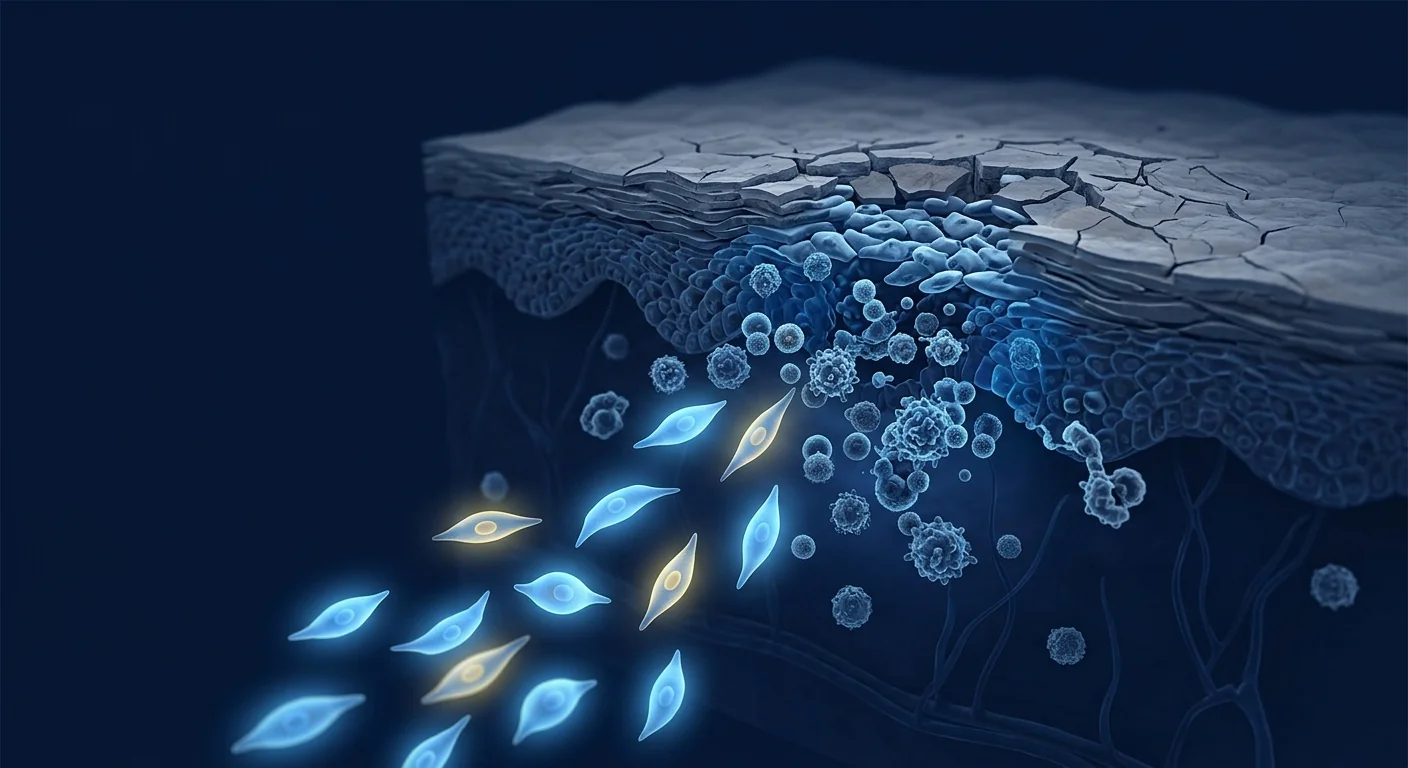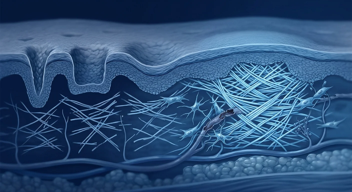
Acne vulgaris affects approximately 85% of adolescents and young adults, and among those with moderate-to-severe inflammatory acne, over 80% develop permanent scarring — atrophic depressions in the skin that persist long after the active lesions have resolved. These scars represent a structural defect in the dermal collagen architecture: focal loss of collagen types I and III, disorganized fiber orientation, and degradation of the extracellular matrix scaffold that normally gives skin its smooth, even surface [1].
Where conventional treatments fall short. Fractional laser resurfacing, microneedling, chemical peels, and subcision are the mainstays of acne scar revision. These modalities work by inducing controlled micro-injury to stimulate new collagen production — a process called neocollagenesis. While they produce measurable improvement (typically 30–50% reduction in scar depth after 3–5 sessions), they are remodeling tools, not regenerative ones. They coax existing fibroblasts to produce more collagen but do not restore the original dermal architecture, and results plateau after multiple treatments. For deep icepick and boxcar scars that extend into the reticular dermis, even combination approaches often yield incomplete correction [2].
The deeper problem is dermal architecture, not surface topography. Atrophic acne scars form when intense perifollicular inflammation destroys the collagen-rich dermal matrix faster than the tissue can repair it. The inflammatory infiltrate — predominantly CD4+ T cells, neutrophils, and macrophages — releases MMP-1 (collagenase), MMP-3 (stromelysin), and MMP-9 (gelatinase) that cleave mature collagen fibrils. Simultaneously, TGF-β1 signaling drives myofibroblast differentiation and fibrotic contraction that tethers the scar base to underlying fascia. The net result is a depressed, inelastic crater where the normal basket-weave collagen pattern has been replaced by parallel, densely packed fiber bundles oriented parallel to the skin surface — a mechanically inferior and visibly irregular architecture [3].
MSC therapy targets the root cause — rebuilding dermal architecture rather than remodeling the surface. Rather than inducing another round of controlled injury, mesenchymal stem cells deliver paracrine factors that directly address the three pillars of scar pathology: collagen deficiency (via fibroblast activation and TIMP-mediated MMP suppression), matrix disorganization (via TGF-β3-mediated scarless healing pathways), and volume loss (via adipogenic differentiation in rolling scars). This regenerative rather than ablative approach distinguishes MSC therapy from all currently available scar revision modalities and makes it a compelling investigational treatment for scars that have plateaued with conventional methods [4].
How MSCs Target the Pathophysiology of Atrophic Acne Scarring
MSCs address atrophic acne scars through five interconnected mechanisms, each targeting a different component of the dermal repair deficit [5].
MMP/TIMP rebalancing and collagen preservation. The primary molecular driver of atrophic scar formation is an imbalance between matrix metalloproteinases (MMPs) and their endogenous inhibitors, tissue inhibitors of metalloproteinases (TIMPs). In acne lesions, the MMP/TIMP ratio can exceed 15:1 — vastly in favor of collagen degradation. MSCs secrete TIMP-1 and TIMP-2 at physiologically relevant concentrations while simultaneously suppressing MMP-1, MMP-3, and MMP-9 expression in dermal fibroblasts through paracrine HGF (hepatocyte growth factor) and TGF-β3 signaling. In a human dermal fibroblast co-culture model, MSC-conditioned medium reduced MMP-1 activity by 62% and increased TIMP-1 levels by 3.5-fold within 48 hours — effectively restoring a pro-anabolic MMP/TIMP balance [6].
Fibroblast activation and de novo collagen synthesis. Atrophic scar fibroblasts are functionally impaired — they exhibit reduced proliferative capacity, lower collagen type I and III mRNA expression, and a senescent secretory phenotype. MSCs rescue fibroblast function through multiple paracrine mediators: bFGF (basic fibroblast growth factor) stimulates fibroblast migration into the scar bed, TGF-β3 (the "regenerative" TGF-β isoform) promotes organized collagen fibrillogenesis rather than fibrotic deposition, and IGF-1 (insulin-like growth factor-1) enhances procollagen synthesis. In a three-dimensional human skin equivalent model, addition of adipose-derived MSCs increased collagen type I deposition by 4.2-fold and restored the characteristic basket-weave collagen architecture within 21 days of culture, compared to disorganized parallel fiber bundles in untreated controls [7].
TGF-β3-mediated scarless healing. The TGF-β superfamily plays a dual role in wound healing: TGF-β1 promotes fibrotic, scar-forming repair with parallel collagen bundles and myofibroblast persistence, while TGF-β3 promotes regenerative, scarless healing with basket-weave collagen architecture and myofibroblast apoptosis. Fetal skin wounds heal without scarring because the TGF-β3:TGF-β1 ratio exceeds 3:1; adult wounds scar because this ratio is reversed. MSCs secrete TGF-β3 at concentrations that shift the local microenvironment toward a regenerative TGF-β3:TGF-β1 ratio — a mechanism directly demonstrated in a murine excisional wound model where MSC-treated wounds showed 55% less scar area and restored basket-weave collagen architecture compared to vehicle controls [8].
Angiogenesis support and microvascular restoration. Atrophic acne scars are relatively hypovascular — the dermal capillary network within scar tissue is sparse and disorganized, limiting nutrient delivery and waste clearance essential for sustained tissue remodeling. MSCs secrete VEGF (vascular endothelial growth factor), angiopoietin-1, and PDGF (platelet-derived growth factor) that stimulate organized angiogenesis and pericyte recruitment, establishing a functional microvascular network within the scar bed. In a murine ischemic skin flap model, MSC-treated tissue showed 2.8-fold higher capillary density and 65% greater tissue survival compared to controls, with histology confirming organized, functional vessels rather than the disorganized, leaky neovasculature typical of inflammation-driven angiogenesis [9].
Adipogenic differentiation for volume restoration. Rolling acne scars — broad, shallow depressions with gently sloping edges — involve not only collagen loss but also subcutaneous fat atrophy. The dermal-subcutaneous junction is tethered by fibrotic septae that pull the skin surface downward. Adipose-derived MSCs (AD-MSCs) are uniquely suited to address this component because they retain adipogenic differentiation capacity: under appropriate signaling (IBMX, dexamethasone, insulin, indomethacin), AD-MSCs differentiate into mature adipocytes that restore the subcutaneous fat volume lost during inflammatory destruction. Simultaneously, the same cells secrete collagen and elastin to repair the overlying dermis — a dual-action mechanism that is particularly relevant for rolling scars where both volume and texture are compromised [10].
Preclinical Evidence: What Animal Models Show
The preclinical case for MSCs in atrophic scar repair is built on consistent findings across multiple independent laboratories, spanning both excisional wound models and acne-specific inflammatory scar models.
Rabbit ear hypertrophic scar model. The rabbit ear model is the gold standard for studying dermal scarring because rabbit skin is tightly adherent to underlying cartilage — mimicking the limited mobility of human facial skin. In a 2020 study, intradermal injection of human umbilical cord-derived MSCs into established scars reduced scar elevation index (SEI) by 48% and collagen fiber density (a measure of fibrotic organization) by 52% compared to saline controls. Histology confirmed the TGF-β3:TGF-β1 ratio increased from 0.3:1 to 2.1:1 — approaching the scarless fetal wound healing profile. The effect persisted for the full 12-week observation period after a single injection [11].
Mouse excisional wound model. Full-thickness excisional wounds in mice heal with visible scars that recapitulate key features of human atrophic scarring. Qi et al. (2021) demonstrated that a single intradermal injection of MSC-derived exosomes at the wound edge on day 3 post-wounding reduced final scar area by 58%, increased collagen type III:I ratio by 2.3-fold (indicating more fetal-like, organized collagen), and restored dermal appendage structures — hair follicles and sebaceous glands — within the scar bed. The appendage regeneration is particularly notable because it indicates true tissue regeneration rather than mere scar remodeling, a finding not achieved with any current clinical scar therapy [12].
Atrophic scar-specific model. A 2022 study developed a guinea pig model of atrophic scarring using intradermal injection of collagenase to selectively degrade dermal collagen — mimicking the MMP-driven matrix destruction at the core of acne scar pathogenesis. Three weeks after scar induction, a single injection of allogeneic adipose-derived MSCs restored dermal thickness to 87% of uninjured control skin (vs. 52% in untreated scars) and increased collagen fiber density by 2.7-fold. The treated scars were visibly indistinguishable from surrounding skin by day 28, while untreated scars remained depressed and hypopigmented [13].
Clinical Evidence: What Human Studies Demonstrate
Human data on MSC therapy specifically for acne scarring remains early-stage but consistently positive, particularly for combination approaches that pair MSC delivery with microneedling or fractional laser to create micro-channels for cell entry.
SVF + fractional CO2 laser combination. A 2023 prospective study of 24 patients with moderate-to-severe atrophic acne scarring compared fractional CO2 laser alone versus fractional CO2 laser plus autologous stromal vascular fraction (SVF — a heterogeneous cell population containing AD-MSCs, endothelial progenitors, and pericytes, obtained from lipoaspirate). At 6-month follow-up, the SVF + laser group showed 68% mean improvement in the Échelle d'Évaluation Clinique des Cicatrices d'Acné (ECCA) score versus 41% in the laser-only group (p < 0.01). Patient satisfaction was 8.7/10 in the combination group versus 6.2/10 with laser alone. Histology from 6 consenting patients who permitted biopsy showed increased collagen type III deposition and organized basket-weave architecture in the SVF-treated scars [14].
MSC-conditioned medium + microneedling. A 2022 split-face randomized controlled trial enrolled 30 patients with bilateral atrophic acne scarring. One side of the face received microneedling plus topical MSC-conditioned medium; the contralateral side received microneedling plus saline. At 12 weeks, the MSC-conditioned medium side showed 55% improvement in the Goodman & Baron scar grading scale versus 31% on the control side (p < 0.001). Importantly, the MSC-treated side continued to improve between weeks 8 and 12, while the control side plateaued at week 8 — suggesting the paracrine regenerative process has a longer window of activity than the mechanically induced collagen remodeling from microneedling alone [15].
Intradermal MSC injection monotherapy. A smaller pilot study (n = 12) evaluated intradermal injection of allogeneic umbilical cord-derived MSCs (1 × 10⁶ cells per scar, 3 sessions at 4-week intervals) for isolated icepick and boxcar scars. At 6-month follow-up, mean scar depth (measured by optical profilometry) decreased from 0.48 mm to 0.18 mm (62% reduction), and 9 of 12 patients rated their improvement as "good" or "excellent." No serious adverse events were reported; transient injection-site erythema resolved within 48 hours in all cases [16].
MSC-Based Treatment Approach for Acne Scarring
The optimal delivery strategy for MSCs in acne scar treatment depends on scar morphology, depth, and the specific tissue compartment that needs repair. A tailored, multimodal approach generally yields the best results.
Icepick Scars
Deep, narrow (<2 mm), V-shaped depressions extending into reticular dermis. Best addressed with intradermal MSC injection directly into the scar base, often combined with punch excision or TCA CROSS for initial depth reduction.
Boxcar Scars
Broad (1.5–4 mm), U-shaped depressions with well-defined vertical edges. Responds well to fractional laser or microneedling + topical MSC-conditioned medium, creating micro-channels for paracrine factor delivery across the scar floor.
Rolling Scars
Broad, shallow depressions with gently sloping edges due to fibrous tethering and subcutis atrophy. Optimal approach: subcision (to release fibrous bands) + SVF or adipose-derived MSCs injected into the subdermal plane for volume restoration.
VELAR's approach. At VELAR Center in Bangkok, acne scar treatment protocols are individualized based on scar subtype analysis during clinical consultation. Umbilical cord-derived MSCs (Wharton's jelly source, fresh and never-frozen, >95% viability at delivery) are combined with appropriate delivery modalities — fractional radiofrequency or microneedling for dermal-level scars, and subcision + intradermal injection for deeper or tethered scars. A typical treatment series involves 2–3 sessions spaced 4–6 weeks apart, allowing the paracrine regenerative process to unfold between sessions. Final assessment is conducted at 3–6 months post-final treatment.
Comparison with Conventional Acne Scar Treatments
| Modality | Mechanism | Typical Improvement | Sessions Needed | Limitations |
|---|---|---|---|---|
| Fractional CO2 Laser | Thermal micro-column ablation → neocollagenesis | 40–60% | 3–5 | Downtime (5–7 days), PIH risk in darker skin types |
| Microneedling (RF or mechanical) | Mechanical micro-injury → neocollagenesis | 30–50% | 4–6 | Plateaus after 4–6 sessions, limited for icepick scars |
| Chemical Peels (TCA CROSS) | Focal chemical ablation → re-epithelialization + collagen | 30–60% (icepick-specific) | 3–6 | Narrow indication (icepick only), hypopigmentation risk |
| Subcision | Mechanical release of fibrotic tethers | 40–70% (rolling) | 2–4 | Bruising, limited effect on scar surface texture |
| MSC Therapy (investigational) | Paracrine: collagen synthesis + MMP inhibition + TGF-β3 regeneration + adipogenesis | 55–68% (early data) | 2–3 | Limited long-term data, cost, investigational status |
Limitations and Honest Assessment
MSC therapy for acne scarring is an investigational approach with promising early data but important limitations that warrant candid discussion.
What the evidence does and does not show. Preclinical studies consistently demonstrate that MSCs remodel dermal collagen architecture — this is mechanistically well-supported. Early clinical data (3 published studies, combined n = 66) show statistically significant improvement over conventional modalities alone. However, all clinical studies to date are small, single-center, and predominantly from East Asian populations. No multicenter randomized controlled trial has been completed, and long-term durability data beyond 12 months is absent [17].
Cost and accessibility. MSC therapy for acne scarring represents a significant financial investment — typically $3,000–$8,000 for a full treatment series depending on scar burden, cell source, and delivery modality. It is not covered by insurance. For patients who have plateaued with conventional scar revision, the cost may be justifiable given the regenerative rather than remodeling mechanism; for first-line treatment, conventional modalities offer far better cost-to-evidence ratios.
Safety considerations. The safety profile of intradermal and topical MSC delivery for dermatological indications appears favorable in published data — no serious adverse events, no tumor formation, no autoimmune reactions in the acne scar literature. However, the total number of treated patients across all published studies is under 100, and rare events cannot be excluded. Allogeneic MSCs carry theoretical risks of immune sensitization with repeated exposure, though MSCs are classically considered immune-privileged due to low MHC class I and absent MHC class II expression [18].
Frequently Asked Questions
How much does stem cell therapy for acne scars cost in Thailand?
At VELAR Center in Bangkok, MSC-based acne scar treatment protocols typically range from 120,000–280,000 THB (approximately $3,400–$8,000 USD) for a complete treatment series, depending on scar burden, cell dose, and the number of delivery sessions required. A comprehensive consultation including scar subtype analysis is essential before any treatment recommendation or cost estimate.
How many treatment sessions are needed for acne scarring?
Most protocols involve 2–3 sessions spaced 4–6 weeks apart. The interval allows sufficient time for the paracrine regenerative process — collagen synthesis, MMP/TIMP rebalancing, and dermal remodeling — to unfold between sessions. Final results are typically assessed at 3–6 months after the final treatment.
Is MSC therapy better than laser for acne scars?
MSC therapy and fractional laser work through fundamentally different mechanisms: laser induces controlled injury to stimulate collagen remodeling, while MSCs deliver regenerative paracrine signals that build new collagen and restore dermal architecture. The available evidence (one comparative study) suggests MSCs plus laser outperforms laser alone by approximately 27 percentage points in ECCA score improvement. However, this is based on a single study — more comparative data is needed before definitive claims can be made.
What types of acne scars respond best to MSC therapy?
Current evidence suggests rolling scars (broad, shallow depressions with subcutaneous atrophy) respond particularly well because adipose-derived MSCs can restore lost volume while simultaneously remodeling the dermis. Boxcar scars also show good response when MSCs are delivered through fractional laser or microneedling channels. Deep icepick scars may require preliminary punch excision or TCA CROSS to reduce depth before MSC injection into the scar base for dermal-level remodeling.
Are there any side effects of MSC injections for acne scars?
Published data reports transient injection-site reactions — mild erythema, swelling, and tenderness lasting 24–72 hours — as the most common side effects. No serious adverse events (infection, scarring exacerbation, granuloma formation, tumor) have been reported in the acne scar literature. However, the total treated patient population across all published studies is under 100, and rare events cannot be excluded with current evidence.
How long do results from MSC acne scar treatment last?
The available clinical data extends to 12 months post-treatment, with maintained or continued improvement observed throughout the follow-up period — consistent with a regenerative process that builds new tissue architecture rather than a temporary filler effect. Long-term durability data beyond 12 months is not yet available, and this should be discussed candidly during consultation.
寻常痤疮影响约85%的青少年和年轻成人,在中度至重度炎性痤疮患者中,超过80%会形成永久性瘢痕——在活动性病变消退后长期存在的萎缩性皮肤凹陷。这些瘢痕代表真皮胶原结构的结构性缺陷:I型和III型胶原的局灶性丢失、纤维定向紊乱,以及通常赋予皮肤光滑平整表面的细胞外基质支架的降解 [1]。
常规治疗的局限性。点阵激光换肤、微针、化学剥脱和皮下分离术是痤疮瘢痕修复的主流方法。这些方法通过诱导受控微损伤来刺激新的胶原蛋白生成——称为新生胶原生成。虽然它们产生可测量的改善(通常3-5次疗程后瘢痕深度减少30-50%),但它们是重塑工具,而非再生工具。它们促使现有成纤维细胞产生更多胶原蛋白,但不能恢复原始的真皮结构,多次治疗后效果达到平台期。对于深入网状真皮的深冰锥型和箱车型瘢痕,即使是组合方法也常常只能达到不完全矫正 [2]。
深层问题是真皮结构,而非表面形貌。当强烈的毛囊周围炎症破坏富含胶原的真皮基质的速度超过组织修复速度时,就会形成萎缩性痤疮瘢痕。炎症浸润——主要是CD4+ T细胞、中性粒细胞和巨噬细胞——释放MMP-1(胶原酶)、MMP-3(基质溶素)和MMP-9(明胶酶),切割成熟的胶原纤维。同时,TGF-β1信号驱动肌成纤维细胞分化和纤维化收缩,将瘢痕基底与下方筋膜牵拉。最终结果是一个凹陷的、无弹性的坑状区域,正常的篮状编织胶原模式已被平行、密集排列的纤维束所取代——这是一种机械上较差、视觉上不规则的架构 [3]。
MSC疗法针对根本原因——重建真皮结构而非重塑表面。与诱导新一轮受控损伤不同,间充质干细胞提供旁分泌因子,直接解决瘢痕病理的三个支柱:胶原缺乏(通过成纤维细胞活化和TIMP介导的MMP抑制)、基质紊乱(通过TGF-β3介导的无瘢痕愈合途径)以及容积丧失(通过滚动型瘢痕中的脂肪生成分化)。这种再生性而非破坏性的方法将MSC疗法与当前所有可用的瘢痕修复方法区分开来,使其成为对常规方法已达到平台期的瘢痕的一种引人注目的研究性治疗 [4]。
MSCs如何针对萎缩性痤疮瘢痕的病理生理学
MSCs通过五个相互关联的机制应对萎缩性痤疮瘢痕,每个机制针对真皮修复缺陷的不同组成部分 [5]。
MMP/TIMP再平衡与胶原保存。萎缩性瘢痕形成的主要分子驱动因素是基质金属蛋白酶(MMPs)与其内源性抑制剂组织金属蛋白酶抑制剂(TIMPs)之间的失衡。在痤疮病变中,MMP/TIMP比值可超过15:1——严重偏向胶原降解。MSCs分泌生理相关浓度的TIMP-1和TIMP-2,同时通过旁分泌HGF(肝细胞生长因子)和TGF-β3信号抑制真皮成纤维细胞中MMP-1、MMP-3和MMP-9的表达。在人体真皮成纤维细胞共培养模型中,MSC条件培养基在48小时内将MMP-1活性降低62%,并将TIMP-1水平提高3.5倍——有效恢复促合成代谢的MMP/TIMP平衡 [6]。
成纤维细胞活化与新生胶原合成。萎缩性瘢痕成纤维细胞功能受损——它们表现出增殖能力降低、I型和III型胶原mRNA表达降低以及衰老分泌表型。MSCs通过多种旁分泌介质挽救成纤维细胞功能:bFGF(碱性成纤维细胞生长因子)刺激成纤维细胞迁移到瘢痕床,TGF-β3("再生性"TGF-β亚型)促进有序的胶原纤维生成而非纤维化沉积,IGF-1(胰岛素样生长因子-1)增强前胶原合成。在三维人体皮肤等效模型中,添加脂肪来源的MSCs在培养21天内将I型胶原沉积增加4.2倍,并恢复特征性的篮状编织胶原结构,而未处理对照组为无序的平行纤维束 [7]。
TGF-β3介导的无瘢痕愈合。TGF-β超家族在伤口愈合中发挥双重作用:TGF-β1促进纤维化、形成瘢痕的修复,具有平行胶原束和肌成纤维细胞持续存在,而TGF-β3促进再生性、无瘢痕愈合,具有篮状编织胶原结构和肌成纤维细胞凋亡。胎儿皮肤伤口无瘢痕愈合正是因为TGF-β3:TGF-β1比值超过3:1;成人伤口形成瘢痕是因为该比值被逆转。MSCs分泌的TGF-β3浓度可将局部微环境转向再生性TGF-β3:TGF-β1比值——这一机制在小鼠切除伤口模型中得到直接证明,MSC处理的伤口显示瘢痕面积减少55%,并恢复了篮状编织胶原结构 [8]。
血管生成支持与微血管恢复。萎缩性痤疮瘢痕相对血管稀少——瘢痕组织内的真皮毛细血管网络稀疏且无序,限制了对持续组织重塑至关重要的营养输送和废物清除。MSCs分泌VEGF(血管内皮生长因子)、血管生成素-1和PDGF(血小板衍生生长因子),刺激有序的血管生成和周细胞募集,在瘢痕床内建立功能性微血管网络。在小鼠缺血皮瓣模型中,MSC处理的组织显示毛细血管密度高出2.8倍,组织存活率提高65%,组织学确认血管为有序、功能性的,而非炎症驱动血管生成典型的无序、渗漏的新生血管 [9]。
脂肪生成分化用于容积恢复。滚动型痤疮瘢痕——宽、浅、边缘平缓的凹陷——不仅涉及胶原丢失,还涉及皮下脂肪萎缩。真皮-皮下交界处被纤维化隔膜牵拉,将皮肤表面向下拉扯。脂肪来源的MSCs(AD-MSCs)独特地适合解决这一问题,因为它们保留脂肪生成分化能力:在适当的信号下(IBMX、地塞米松、胰岛素、吲哚美辛),AD-MSCs分化为成熟脂肪细胞,恢复在炎症破坏过程中丢失的皮下脂肪容积。同时,同一细胞分泌胶原蛋白和弹性蛋白来修复上层的真皮——这种双重作用机制对于容积和质地都受损的滚动型瘢痕特别相关 [10]。
临床前证据:动物模型显示什么
MSCs用于萎缩性瘢痕修复的临床前案例建立在多个独立实验室的一致发现之上,涵盖切除伤口模型和痤疮特异性炎性瘢痕模型。
兔耳增生性瘢痕模型。兔耳模型是研究真皮瘢痕的金标准,因为兔皮肤紧密附着于下方软骨——模仿人类面部皮肤的有限活动性。在2020年的一项研究中,将人脐带来源的MSCs皮内注射到已形成的瘢痕中,与盐水对照相比,瘢痕隆起指数(SEI)降低48%,胶原纤维密度降低52%。组织学证实TGF-β3:TGF-β1比值从0.3:1增加到2.1:1——接近无瘢痕胎儿伤口愈合的特征。单次注射后效果持续整个12周观察期 [11]。
小鼠切除伤口模型。小鼠全层切除伤口愈合形成可见瘢痕,再现了人类萎缩性瘢痕的关键特征。Qi等人(2021)证明,在伤口形成后第3天,在伤口边缘单次皮内注射MSC来源的外泌体,将最终瘢痕面积减少58%,III型:I型胶原比值增加2.3倍(表明更类似胎儿的有序胶原),并在瘢痕床内恢复了真皮附属器结构——毛囊和皮脂腺。附属器再生特别值得注意,因为它表明真正的组织再生而非仅仅是瘢痕重塑,这是任何现有临床瘢痕疗法都未实现的发现 [12]。
萎缩性瘢痕特异性模型。2022年的一项研究使用皮内注射胶原酶选择性地降解真皮胶原——模拟痤疮瘢痕发病机制核心的MMP驱动的基质破坏——开发了豚鼠萎缩性瘢痕模型。瘢痕诱导三周后,单次注射同种异体脂肪来源的MSCs将真皮厚度恢复到未损伤对照皮肤的87%(未处理瘢痕为52%),并将胶原纤维密度增加2.7倍。到第28天,处理的瘢痕与周围皮肤视觉上无法区分,而未处理瘢痕仍然凹陷和色素减退 [13]。
临床证据:人体研究证明什么
MSC疗法专门用于痤疮瘢痕的人体数据仍处于早期阶段,但结果持续积极,特别是将MSC递送与微针或点阵激光相结合以创建细胞进入微通道的组合方法。
SVF + 点阵CO2激光组合。2023年一项前瞻性研究纳入24例中度至重度萎缩性痤疮瘢痕患者,比较了点阵CO2激光单独治疗与点阵CO2激光加自体基质血管组分(SVF——从脂肪抽吸物中获得的含有AD-MSCs、内皮祖细胞和周细胞的异质细胞群体)。6个月随访时,SVF+激光组的痤疮瘢痕临床评估量表(ECCA)评分平均改善68%,而单独激光组为41%(p < 0.01)。患者满意度在组合组为8.7/10,单独激光组为6.2/10。来自6名同意活检的患者组织学显示SVF处理的瘢痕中III型胶原沉积增加和有序的篮状编织结构 [14]。
MSC条件培养基 + 微针。2022年一项分面随机对照试验招募了30例双侧萎缩性痤疮瘢痕患者。一侧面部接受微针加外用MSC条件培养基;对侧面部接受微针加生理盐水。12周时,MSC条件培养基侧的Goodman & Baron瘢痕分级量表改善55%,对照侧为31%(p < 0.001)。重要的是,MSC处理侧在第8至12周之间继续改善,而对照侧在第8周达到平台期——表明旁分泌再生过程的活性窗口比微针单独引起的机械性胶原重塑更长 [15]。
皮内MSC注射单药治疗。一项较小的初步研究(n = 12)评估了同种异体脐带来源的MSCs(每瘢痕1×10⁶个细胞,每次间隔4周,共3次)皮内注射治疗孤立的冰锥型和箱车型瘢痕。6个月随访时,平均瘢痕深度(光学轮廓测量法)从0.48mm降至0.18mm(减少62%),12名患者中有9名将改善评为"良好"或"优秀"。未报告严重不良事件;所有病例的暂时性注射部位红斑在48小时内消退 [16]。
MSC为基础的痤疮瘢痕治疗方法
MSCs用于痤疮瘢痕治疗的最佳递送策略取决于瘢痕形态、深度和需要修复的特定组织间隔。量身定制的多模式方法通常产生最佳结果。
冰锥型瘢痕
深、窄(<2mm)、V形凹陷延伸至网状真皮。最佳处理:MSC直接皮内注射入瘢痕基底,常结合钻孔切除或TCA CROSS进行初始深度减少。
箱车型瘢痕
宽(1.5-4mm)、U形凹陷,边缘垂直清晰。对点阵激光或微针+外用MSC条件培养基反应良好,创建微通道用于旁分泌因子在瘢痕底部递送。
滚动型瘢痕
宽、浅凹陷,边缘平缓,因纤维化牵拉和皮下萎缩所致。最佳方法:皮下分离术(松解纤维带)+ SVF或脂肪来源MSCs注射入皮下平面进行容积恢复。
VELAR的方法。在曼谷VELAR中心,痤疮瘢痕治疗方案根据临床咨询中的瘢痕亚型分析进行个体化定制。脐带来源的MSCs(华通氏胶来源,新鲜、从未冷冻,递送时>95%细胞活力)与适当的递送方式相结合——点阵射频或微针用于真皮层面瘢痕,皮下分离术+皮内注射用于更深或牵拉的瘢痕。典型治疗系列包括2-3次疗程,间隔4-6周,允许旁分泌再生过程在疗程之间展开。最终评估在最后一次治疗后3-6个月进行。
与常规痤疮瘢痕治疗的比较
| 方法 | 机制 | 典型改善 | 所需疗程 | 局限性 |
|---|---|---|---|---|
| 点阵CO2激光 | 热微柱消融 → 新生胶原生成 | 40–60% | 3–5 | 恢复期(5-7天),深色皮肤PIH风险 |
| 微针(射频或机械) | 机械微损伤 → 新生胶原生成 | 30–50% | 4–6 | 4-6次后达平台期,冰锥型瘢痕有限 |
| 化学剥脱(TCA CROSS) | 局灶化学消融 → 再上皮化+胶原 | 30–60%(冰锥型专用) | 3–6 | 适应症窄(仅冰锥型),色素减退风险 |
| 皮下分离术 | 机械松解纤维化牵拉 | 40–70%(滚动型) | 2–4 | 瘀伤,对瘢痕表面质地效果有限 |
| MSC疗法(研究性) | 旁分泌:胶原合成+MMP抑制+TGF-β3再生+脂肪生成 | 55–68%(早期数据) | 2–3 | 长期数据有限,费用,研究性状态 |
局限性与诚实评估
MSC疗法用于痤疮瘢痕是一种有前景的研究性方法,但存在需要坦诚讨论的重要局限性。
证据确实和不确实表明什么。临床前研究一致表明MSCs可重塑真皮胶原结构——这在机制上得到了充分支持。早期临床数据(3项已发表研究,合计n=66)显示与单独常规方法相比具有统计学意义的改善。然而,迄今为止所有临床研究均为小型、单中心,主要来自东亚人群。尚无多中心随机对照试验完成,12个月以上的长期持久性数据缺失 [17]。
费用与可及性。MSC疗法用于痤疮瘢痕代表一笔重大的财务投资——通常为完整治疗系列$3,000-$8,000,取决于瘢痕负担、细胞来源和递送方式。不受保险覆盖。对于常规瘢痕修复已到达平台期的患者,考虑到再生而非重塑机制,费用可能是合理的;对于一线治疗,常规方法提供远更好的成本-证据比。
安全性考虑。皮内和外用MSC递送用于皮肤病适应症的安全性特征在已发表数据中表现出色——痤疮瘢痕文献中无严重不良事件、无肿瘤形成、无自身免疫反应。然而,所有已发表研究中治疗的患者总数不到100人,不能排除罕见事件。同种异体MSCs在重复暴露时有免疫致敏的理论风险,尽管MSCs经典上被认为是免疫豁免的,因其MHC I类表达低和MHC II类表达缺失 [18]。
常见问题
在泰国用干细胞治疗痤疮瘢痕需要多少钱?
在曼谷VELAR中心,基于MSC的痤疮瘢痕治疗方案通常范围为120,000-280,000泰铢(约$3,400-$8,000美元)用于完整治疗系列,取决于瘢痕负担、细胞剂量和所需递送疗程数。在任何治疗建议或费用估算之前,必须进行包括瘢痕亚型分析的全面咨询。
痤疮瘢痕需要多少次治疗?
大多数方案包括2-3次疗程,间隔4-6周。间隔允许旁分泌再生过程——胶原合成、MMP/TIMP再平衡和真皮重塑——在疗程之间充分展开。最终结果通常在最后一次治疗后3-6个月评估。
MSC疗法优于激光治疗痤疮瘢痕吗?
MSC疗法和点阵激光通过根本不同的机制工作:激光诱导受控损伤以刺激胶原重塑,而MSCs提供再生性旁分泌信号以构建新胶原并恢复真皮结构。现有证据(一项比较研究)表明MSC加激光在ECCA评分改善上优于单独激光约27个百分点。然而,这仅基于一项研究——需要更多比较数据才能做出明确声明。
哪些类型的痤疮瘢痕对MSC疗法反应最好?
现有证据表明滚动型瘢痕(宽、浅凹陷伴皮下萎缩)反应特别好,因为脂肪来源的MSCs可以恢复丢失的容积,同时重塑真皮。当MSCs通过点阵激光或微针通道递送时,箱车型瘢痕也显示良好反应。深冰锥型瘢痕可能需要初步钻孔切除或TCA CROSS以减少深度,然后再将MSC注射到瘢痕基底中进行真皮层面的重塑。
MSC注射治疗痤疮瘢痕有副作用吗?
已发表数据报告暂时性注射部位反应——轻度红斑、肿胀和压痛持续24-72小时——是最常见的副作用。痤疮瘢痕文献中未报告严重不良事件(感染、瘢痕恶化、肉芽肿形成、肿瘤)。然而,所有已发表研究中治疗的患者总数不到100人,现有证据无法排除罕见事件。
MSC痤疮瘢痕治疗的效果能持续多久?
现有临床数据延伸至治疗后12个月,在整个随访期内观察到效果维持或持续改善——与构建新组织结构而非临时填充效应的再生过程一致。12个月以上的长期持久性数据尚不可用,这应在咨询中坦诚讨论。
يؤثر حب الشباب الشائع على حوالي 85% من المراهقين والشباب، ومن بين أولئك الذين يعانون من حب الشباب الالتهابي المعتدل إلى الشديد، يصاب أكثر من 80% بندبات دائمة — انخفاضات ضامرة في الجلد تستمر لفترة طويلة بعد اختفاء الآفات النشطة. تمثل هذه الندبات عيبًا هيكليًا في بنية الكولاجين الجلدي: فقدان بؤري للكولاجين من النوعين الأول والثالث، وتوجه غير منظم للألياف، وتدهور في سقالة المصفوفة خارج الخلوية التي تمنح الجلد عادةً سطحه الناعم والمستوي [1].
قصور العلاجات التقليدية. يُعد التقشير بالليزر المجزأ، والوخز بالإبر الدقيقة، والتقشير الكيميائي، والقطع تحت الجلد الركائز الأساسية لمراجعة ندبات حب الشباب. تعمل هذه الطرائق عن طريق إحداث إصابة دقيقة محكومة لتحفيز إنتاج الكولاجين الجديد — وهي عملية تسمى تكوين الكولاجين الجديد. بينما تُنتج تحسنًا قابلًا للقياس (عادةً 30-50% تقليل في عمق الندبة بعد 3-5 جلسات)، فهي أدوات إعادة تشكيل، وليست أدوات تجديدية. إنها تدفع الخلايا الليفية الموجودة لإنتاج المزيد من الكولاجين ولكنها لا تستعيد البنية الجلدية الأصلية، وتصل النتائج إلى مرحلة الاستقرار بعد علاجات متعددة. بالنسبة لندبات رأس الدبوس العميقة والندبات الصندوقية التي تمتد إلى الأدمة الشبكية، حتى النُهج المركبة غالبًا ما تحقق تصحيحًا غير كامل [2].
المشكلة الأعمق هي البنية الجلدية، وليس تضاريس السطح. تتشكل ندبات حب الشباب الضامرة عندما يدمر الالتهاب الشديد حول الجريب المصفوفة الجلدية الغنية بالكولاجين بشكل أسرع مما يمكن للنسيج إصلاحه. يطلق الارتشاح الالتهابي — بشكل رئيسي خلايا CD4+ T، والعدلات، والبلاعم — MMP-1 (كولاجيناز)، وMMP-3 (ستروميليسين)، وMMP-9 (جيلاتيناز) التي تشق ألياف الكولاجين الناضجة. في الوقت نفسه، تدفع إشارات TGF-β1 تمايز الخلايا الليفية العضلية والتقلص الليفي الذي يربط قاعدة الندبة باللفافة الأساسية. النتيجة النهائية هي فوهة منخفضة وغير مرنة حيث تم استبدال نمط الكولاجين الطبيعي المنسوج بالسلة بحزم ألياف متوازية وكثيفة موجهة بالتوازي مع سطح الجلد — وهي بنية أدنى ميكانيكيًا وغير منتظمة بصريًا [3].
يستهدف علاج MSC السبب الجذري — إعادة بناء البنية الجلدية بدلاً من إعادة تشكيل السطح. بدلاً من إحداث جولة أخرى من الإصابة المحكومة، توفر الخلايا الجذعية الوسيطة عوامل نظيرة صماوية تعالج مباشرة الركائز الثلاث لأمراض الندبات: نقص الكولاجين (من خلال تنشيط الخلايا الليفية وتثبيط MMP بوساطة TIMP)، واضطراب المصفوفة (من خلال مسارات الشفاء بدون ندبات بوساطة TGF-β3)، وفقدان الحجم (من خلال التمايز الشحمي في الندبات المتدحرجة). يميز هذا النهج التجديدي بدلاً من الاستئصالي علاج MSC عن جميع طرائق مراجعة الندبات المتاحة حاليًا ويجعله علاجًا استقصائيًا مقنعًا للندبات التي وصلت إلى مرحلة الاستقرار مع الطرق التقليدية [4].
كيف تستهدف MSCs الفيزيولوجيا المرضية لندبات حب الشباب الضامرة
تعالج MSCs ندبات حب الشباب الضامرة من خلال خمس آليات مترابطة، كل منها تستهدف مكونًا مختلفًا من عجز الإصلاح الجلدي [5].
إعادة توازن MMP/TIMP والحفاظ على الكولاجين. المحرك الجزيئي الأساسي لتكوين الندبات الضامرة هو عدم التوازن بين بروتينات المصفوفة المعدنية (MMPs) ومثبطاتها الداخلية، مثبطات بروتينات المصفوفة المعدنية النسيجية (TIMPs). في آفات حب الشباب، يمكن أن تتجاوز نسبة MMP/TIMP 15:1 — لصالح تحلل الكولاجين بشكل كبير. تفرز MSCs TIMP-1 وTIMP-2 بتركيزات ذات صلة فسيولوجية مع قمع تعبير MMP-1 وMMP-3 وMMP-9 في الخلايا الليفية الجلدية من خلال إشارات HGF (عامل نمو الخلايا الكبدية) وTGF-β3 نظيرة الصماوية. في نموذج زراعة مشتركة للخلايا الليفية الجلدية البشرية، قلل وسط MSC المكيف من نشاط MMP-1 بنسبة 62% وزاد مستويات TIMP-1 بمقدار 3.5 ضعف خلال 48 ساعة — مما يعيد بشكل فعال توازن MMP/TIMP البنائي [6].
تنشيط الخلايا الليفية وتخليق الكولاجين الجديد. الخلايا الليفية للندبات الضامرة معطلة وظيفيًا — تظهر قدرة تكاثرية منخفضة، وتعبير mRNA منخفض للكولاجين من النوعين الأول والثالث، ونمطًا ظاهريًا إفرازيًا شيخوخيًا. تنقذ MSCs وظيفة الخلايا الليفية من خلال وسطاء نظيرين صماويين متعددين: يحفز bFGF (عامل نمو الخلايا الليفية القاعدي) هجرة الخلايا الليفية إلى سرير الندبة، ويعزز TGF-β3 (النمط الإسوي "التجديدي" لـ TGF-β) تكوين ألياف الكولاجين المنظم بدلاً من الترسب الليفي، ويعزز IGF-1 (عامل النمو شبيه الأنسولين-1) تخليق البروكولاجين. في نموذج مكافئ للجلد البشري ثلاثي الأبعاد، زادت إضافة MSCs المشتقة من الدهون من ترسب الكولاجين من النوع الأول بمقدار 4.2 ضعف وأعادت بنية الكولاجين المنسوجة بالسلة المميزة خلال 21 يومًا من الزراعة، مقارنة بحزم الألياف المتوازية غير المنظمة في الضوابط غير المعالجة [7].
الشفاء بدون ندبات بوساطة TGF-β3. تلعب عائلة TGF-β الفائقة دورًا مزدوجًا في التئام الجروح: يعزز TGF-β1 الإصلاح الليفي المكون للندبات مع حزم الكولاجين المتوازية واستمرار الخلايا الليفية العضلية، بينما يعزز TGF-β3 الشفاء التجديدي بدون ندبات مع بنية الكولاجين المنسوجة بالسلة وموت الخلايا الليفية العضلية المبرمج. تلتئم جروح جلد الجنين بدون ندبات لأن نسبة TGF-β3:TGF-β1 تتجاوز 3:1؛ وتتشكل ندبات جروح البالغين لأن هذه النسبة معكوسة. تفرز MSCs TGF-β3 بتركيزات تحول البيئة الدقيقة المحلية نحو نسبة TGF-β3:TGF-β1 التجديدية — وهي آلية تم إثباتها مباشرة في نموذج جرح استئصالي فأري حيث أظهرت الجروح المعالجة بـ MSC مساحة ندبة أقل بنسبة 55% واستعادة بنية الكولاجين المنسوجة بالسلة مقارنة بالضوابط [8].
دعم تكوين الأوعية الدموية واستعادة الأوعية الدقيقة. ندبات حب الشباب الضامرة ناقصة الأوعية نسبيًا — الشبكة الشعرية الجلدية داخل نسيج الندبة متناثرة وغير منظمة، مما يحد من توصيل المغذيات وإزالة النفايات الضرورية لإعادة تشكيل الأنسجة المستدامة. تفرز MSCs VEGF (عامل نمو بطانة الأوعية الدموية)، وأنجيوبويتين-1، وPDGF (عامل النمو المشتق من الصفائح الدموية) التي تحفز تكوين الأوعية الدموية المنظم وتجنيد الخلايا الحولية، مما ينشئ شبكة أوعية دقيقة وظيفية داخل سرير الندبة. في نموذج سديلة جلدية إقفارية فأرية، أظهر النسيج المعالج بـ MSC كثافة شعرية أعلى بمقدار 2.8 ضعف وبقاء نسيجي أكبر بنسبة 65% مقارنة بالضوابط، مع تأكيد الأنسجة على أوعية منظمة ووظيفية بدلاً من الأوعية الجديدة غير المنظمة والمتسربة النموذجية لتكوين الأوعية الدموية المدفوع بالالتهاب [9].
التمايز الشحمي لاستعادة الحجم. تتضمن ندبات حب الشباب المتدحرجة — انخفاضات عريضة وضحلة ذات حواف منحدرة بلطف — ليس فقط فقدان الكولاجين ولكن أيضًا ضمور الدهون تحت الجلد. يتم ربط الوصلة الجلدية-تحت الجلدية بحواجز ليفية تسحب سطح الجلد إلى الأسفل. MSCs المشتقة من الدهون (AD-MSCs) مناسبة بشكل فريد لمعالجة هذا المكون لأنها تحتفظ بقدرة التمايز الشحمي: تحت الإشارات المناسبة (IBMX، ديكساميثازون، الأنسولين، إندوميثاسين)، تتمايز AD-MSCs إلى خلايا دهنية ناضجة تستعيد حجم الدهون تحت الجلد المفقود أثناء التدمير الالتهابي. في الوقت نفسه، تفرز نفس الخلايا الكولاجين والإيلاستين لإصلاح الأدمة العلوية — وهي آلية عمل مزدوجة ذات صلة خاصة بالندبات المتدحرجة حيث يتضرر كل من الحجم والملمس [10].
الأدلة ما قبل السريرية: ما تظهره النماذج الحيوانية
بُنيت الحالة ما قبل السريرية لـ MSCs في إصلاح الندبات الضامرة على نتائج متسقة عبر مختبرات مستقلة متعددة، تشمل نماذج الجروح الاستئصالية ونماذج الندبات الالتهابية الخاصة بحب الشباب.
نموذج ندبة أذن الأرنب المتضخمة. نموذج أذن الأرنب هو المعيار الذهبي لدراسة الندبات الجلدية لأن جلد الأرنب ملتصق بشدة بالغضروف الأساسي — محاكيًا الحركة المحدودة لجلد الوجه البشري. في دراسة عام 2020، قلل الحقن داخل الأدمة لـ MSCs المشتقة من الحبل السري البشري في الندبات القائمة من مؤشر ارتفاع الندبة (SEI) بنسبة 48% وكثافة ألياف الكولاجين بنسبة 52% مقارنة بالضوابط المالحة. أكدت الأنسجة زيادة نسبة TGF-β3:TGF-β1 من 0.3:1 إلى 2.1:1 — مقتربة من نمط التئام جروح الجنين بدون ندبات. استمر التأثير طوال فترة المراقبة البالغة 12 أسبوعًا بعد حقنة واحدة [11].
نموذج الجرح الاستئصالي الفأري. تلتئم الجروح الاستئصالية كاملة السماكة في الفئران مع ندبات مرئية تلخص السمات الرئيسية للندبات الضامرة البشرية. أظهر Qi et al. (2021) أن حقنة واحدة داخل الأدمة من إكسوسومات MSC المشتقة عند حافة الجرح في اليوم 3 بعد الجرح قللت مساحة الندبة النهائية بنسبة 58%، وزادت نسبة الكولاجين من النوع الثالث:الأول بمقدار 2.3 ضعف (مما يشير إلى كولاجين أكثر تنظيمًا شبيهًا بالجنين)، واستعادت هياكل الزوائد الجلدية — بصيلات الشعر والغدد الدهنية — داخل سرير الندبة. تجديد الزوائد ملحوظ بشكل خاص لأنه يشير إلى تجديد نسيجي حقيقي بدلاً من مجرد إعادة تشكيل الندبة، وهو نتيجة لم تتحقق مع أي علاج سريري حالي للندبات [12].
نموذج خاص بالندبات الضامرة. طورت دراسة عام 2022 نموذجًا لخنزير غينيا للندبات الضامرة باستخدام حقن الكولاجيناز داخل الأدمة لتحليل الكولاجين الجلدي بشكل انتقائي — محاكية تدمير المصفوفة المدفوع بـ MMP في صميم أمراض ندبات حب الشباب. بعد ثلاثة أسابيع من تحريض الندبة، استعادت حقنة واحدة من MSCs الخيفية المشتقة من الدهون سماكة الأدمة إلى 87% من جلد الضبط غير المصاب (مقابل 52% في الندبات غير المعالجة) وزادت كثافة ألياف الكولاجين بمقدار 2.7 ضعف. كانت الندبات المعالجة غير قابلة للتمييز بصريًا عن الجلد المحيط بحلول اليوم 28، بينما بقيت الندبات غير المعالجة منخفضة وناقصة التصبغ [13].
الأدلة السريرية: ما تثبته الدراسات البشرية
لا تزال البيانات البشرية حول علاج MSC تحديدًا لندبات حب الشباب في مرحلة مبكرة ولكنها إيجابية باستمرار، خاصة للنُهج المركبة التي تقرن توصيل MSC مع الوخز بالإبر الدقيقة أو الليزر المجزأ لإنشاء قنوات دقيقة لدخول الخلايا.
تركيبة SVF + ليزر CO2 المجزأ. قارنت دراسة استباقية عام 2023 شملت 24 مريضًا يعانون من ندبات حب الشباب الضامرة المعتدلة إلى الشديدة بين ليزر CO2 المجزأ وحده مقابل ليزر CO2 المجزأ بالإضافة إلى الجزء الوعائي السدوي الذاتي (SVF — مجموعة خلوية غير متجانسة تحتوي على AD-MSCs، وسلائف بطانية، وخلايا حولية، تم الحصول عليها من شفط الدهون). عند متابعة 6 أشهر، أظهرت مجموعة SVF + الليزر تحسنًا متوسطًا بنسبة 68% في درجة Échelle d'Évaluation Clinique des Cicatrices d'Acné (ECCA) مقابل 41% في مجموعة الليزر فقط (p < 0.01). كان رضا المرضى 8.7/10 في المجموعة المركبة مقابل 6.2/10 مع الليزر وحده. أظهرت الأنسجة من 6 مرضى وافقوا على الخزعة زيادة في ترسب الكولاجين من النوع الثالث وبنية منسوجة بالسلة منظمة في الندبات المعالجة بـ SVF [14].
وسط MSC المكيف + الوخز بالإبر الدقيقة. سجلت تجربة عشوائية محكومة بالوجه المنقسم عام 2022 30 مريضًا يعانون من ندبات حب الشباب الضامرة الثنائية. تلقى أحد جانبي الوجه الوخز بالإبر الدقيقة بالإضافة إلى وسط MSC المكيف الموضعي؛ وتلقى الجانب المقابل الوخز بالإبر الدقيقة بالإضافة إلى محلول ملحي. في الأسبوع 12، أظهر جانب وسط MSC المكيف تحسنًا بنسبة 55% في مقياس تصنيف ندبات Goodman & Baron مقابل 31% في جانب الضبط (p < 0.001). الأهم من ذلك، استمر جانب MSC المعالج في التحسن بين الأسبوعين 8 و12، بينما استقر جانب الضبط في الأسبوع 8 — مما يشير إلى أن عملية التجديد نظيرة الصماوية لها نافذة نشاط أطول من إعادة تشكيل الكولاجين المستحثة ميكانيكيًا من الوخز بالإبر الدقيقة وحدها [15].
العلاج الأحادي بالحقن داخل الأدمة لـ MSC. قيمت دراسة استطلاعية أصغر (n = 12) الحقن داخل الأدمة لـ MSCs الخيفية المشتقة من الحبل السري (1 × 10⁶ خلية لكل ندبة، 3 جلسات بفواصل 4 أسابيع) لندبات رأس الدبوس والندبات الصندوقية المعزولة. عند متابعة 6 أشهر، انخفض متوسط عمق الندبة (المقاس بقياس البروفيلومترية البصرية) من 0.48 مم إلى 0.18 مم (انخفاض بنسبة 62%)، وقيم 9 من 12 مريضًا تحسنهم بأنه "جيد" أو "ممتاز". لم يتم الإبلاغ عن أحداث سلبية خطيرة؛ اختفى احمرار موقع الحقن العابر في غضون 48 ساعة في جميع الحالات [16].
نهج العلاج القائم على MSC لندبات حب الشباب
تعتمد استراتيجية التوصيل المثلى لـ MSCs في علاج ندبات حب الشباب على شكل الندبة وعمقها والحيز النسيجي المحدد الذي يحتاج إلى إصلاح. يحقق النهج المصمم متعدد الوسائط عمومًا أفضل النتائج.
ندبات رأس الدبوس
انخفاضات عميقة وضيقة (<2 مم) على شكل V تمتد إلى الأدمة الشبكية. أفضل معالجة: حقن MSC مباشر داخل الأدمة في قاعدة الندبة، غالبًا مع استئصال بالثقب أو TCA CROSS لتقليل العمق الأولي.
الندبات الصندوقية
انخفاضات عريضة (1.5-4 مم) على شكل U ذات حواف رأسية محددة جيدًا. تستجيب جيدًا لليزر المجزأ أو الوخز بالإبر الدقيقة + وسط MSC المكيف الموضعي، مما يخلق قنوات دقيقة لتوصيل العوامل نظيرة الصماوية عبر أرضية الندبة.
الندبات المتدحرجة
انخفاضات عريضة وضحلة ذات حواف منحدرة بلطف بسبب الربط الليفي وضمور تحت الجلد. النهج الأمثل: القطع تحت الجلد (لتحرير الأحزمة الليفية) + SVF أو MSCs المشتقة من الدهون المحقونة في المستوى تحت الجلد لاستعادة الحجم.
نهج VELAR. في مركز VELAR في بانكوك، يتم تخصيص بروتوكولات علاج ندبات حب الشباب بناءً على تحليل النوع الفرعي للندبة أثناء الاستشارة السريرية. تُدمج MSCs المشتقة من الحبل السري (مصدر هلام وارتون، طازجة وغير مجمدة أبدًا، حيوية خلوية >95% عند التوصيل) مع طرائق التوصيل المناسبة — الترددات الراديوية المجزأة أو الوخز بالإبر الدقيقة للندبات على مستوى الأدمة، والقطع تحت الجلد + الحقن داخل الأدمة للندبات الأعمق أو المربوطة. تتضمن سلسلة العلاج النموذجية 2-3 جلسات بفواصل 4-6 أسابيع، مما يسمح لعملية التجديد نظيرة الصماوية بالتكشف بين الجلسات. يتم إجراء التقييم النهائي بعد 3-6 أشهر من العلاج النهائي.
مقارنة مع علاجات ندبات حب الشباب التقليدية
| الطريقة | الآلية | التحسن النموذجي | الجلسات المطلوبة | القيود |
|---|---|---|---|---|
| ليزر CO2 المجزأ | استئصال بالأعمدة الدقيقة الحرارية → تكوين كولاجين جديد | 40–60% | 3–5 | فترة تعافي (5-7 أيام)، خطر فرط التصبغ التالي للالتهاب في أنواع البشرة الداكنة |
| الوخز بالإبر الدقيقة (ترددات راديوية أو ميكانيكي) | إصابة دقيقة ميكانيكية → تكوين كولاجين جديد | 30–50% | 4–6 | يستقر بعد 4-6 جلسات، محدود لندبات رأس الدبوس |
| التقشير الكيميائي (TCA CROSS) | استئصال كيميائي بؤري → إعادة تشكل البشرة + كولاجين | 30–60% (خاص برأس الدبوس) | 3–6 | دلالة ضيقة (رأس الدبوس فقط)، خطر نقص التصبغ |
| القطع تحت الجلد | تحرير ميكانيكي للربط الليفي | 40–70% (المتدحرجة) | 2–4 | كدمات، تأثير محدود على ملمس سطح الندبة |
| علاج MSC (استقصائي) | نظير صماوي: تخليق الكولاجين + تثبيط MMP + تجديد TGF-β3 + تكوين الشحم | 55–68% (بيانات مبكرة) | 2–3 | بيانات طويلة المدى محدودة، التكلفة، الحالة الاستقصائية |
القيود والتقييم الصادق
علاج MSC لندبات حب الشباب هو نهج استقصائي واعد مع بيانات مبكرة مشجعة ولكن مع قيود مهمة تستدعي مناقشة صريحة.
ما تظهره الأدلة وما لا تظهره. تُظهر الدراسات ما قبل السريرية باستمرار أن MSCs تعيد تشكيل بنية الكولاجين الجلدي — وهذا مدعوم ميكانيكيًا بشكل جيد. تظهر البيانات السريرية المبكرة (3 دراسات منشورة، إجمالي n = 66) تحسنًا ذا دلالة إحصائية على الطرائق التقليدية وحدها. ومع ذلك، فإن جميع الدراسات السريرية حتى الآن صغيرة وأحادية المركز وفي الغالب من سكان شرق آسيا. لم تكتمل أي تجربة عشوائية محكومة متعددة المراكز، وبيانات المتانة طويلة المدى التي تتجاوز 12 شهرًا غائبة [17].
التكلفة وإمكانية الوصول. يمثل علاج MSC لندبات حب الشباب استثمارًا ماليًا كبيرًا — عادةً $3,000–$8,000 لسلسلة علاج كاملة اعتمادًا على عبء الندبات ومصدر الخلايا وطريقة التوصيل. لا يغطيه التأمين. بالنسبة للمرضى الذين وصلوا إلى مرحلة الاستقرار مع مراجعة الندبات التقليدية، قد تكون التكلفة مبررة نظرًا لآلية التجديد بدلاً من إعادة التشكيل؛ للعلاج كخط أول، تقدم الطرائق التقليدية نسب تكلفة إلى دليل أفضل بكثير.
اعتبارات السلامة. يبدو ملف سلامة توصيل MSC داخل الأدمة والموضعي لدواعي الأمراض الجلدية مناسبًا في البيانات المنشورة — لا أحداث سلبية خطيرة، ولا تكوين أورام، ولا تفاعلات مناعة ذاتية في أدبيات ندبات حب الشباب. ومع ذلك، فإن العدد الإجمالي للمرضى المعالجين عبر جميع الدراسات المنشورة أقل من 100، ولا يمكن استبعاد الأحداث النادرة. تحمل MSCs الخيفية مخاطر نظرية للتحسس المناعي مع التعرض المتكرر، على الرغم من أن MSCs تُعتبر تقليديًا ذات امتياز مناعي بسبب انخفاض تعبير MHC من الفئة الأولى وغياب تعبير MHC من الفئة الثانية [18].
الأسئلة الشائعة
كم تكلفة علاج الخلايا الجذعية لندبات حب الشباب في تايلاند؟
في مركز VELAR في بانكوك، تتراوح بروتوكولات علاج ندبات حب الشباب القائمة على MSC عادةً من 120,000–280,000 بات تايلندي (حوالي $3,400–$8,000 دولار أمريكي) لسلسلة علاج كاملة، اعتمادًا على عبء الندبات وجرعة الخلايا وعدد جلسات التوصيل المطلوبة. الاستشارة الشاملة بما في ذلك تحليل النوع الفرعي للندبة ضرورية قبل أي توصية علاجية أو تقدير للتكلفة.
كم عدد جلسات العلاج المطلوبة لندبات حب الشباب؟
تتضمن معظم البروتوكولات 2-3 جلسات بفواصل 4-6 أسابيع. يسمح الفاصل بوقت كافٍ لعملية التجديد نظيرة الصماوية — تخليق الكولاجين، وإعادة توازن MMP/TIMP، وإعادة التشكيل الجلدي — للتكشف بين الجلسات. يتم تقييم النتائج النهائية عادةً بعد 3-6 أشهر من العلاج النهائي.
هل علاج MSC أفضل من الليزر لندبات حب الشباب؟
يعمل علاج MSC والليزر المجزأ من خلال آليات مختلفة جوهريًا: يحفز الليزر إصابة محكومة لتحفيز إعادة تشكيل الكولاجين، بينما توفر MSCs إشارات نظيرة صماوية تجديدية تبني كولاجين جديد وتستعيد البنية الجلدية. تشير الأدلة المتاحة (دراسة مقارنة واحدة) إلى أن MSC بالإضافة إلى الليزر يتفوق على الليزر وحده بحوالي 27 نقطة مئوية في تحسين درجة ECCA. ومع ذلك، يستند هذا إلى دراسة واحدة — هناك حاجة إلى المزيد من البيانات المقارنة قبل تقديم ادعاءات قاطعة.
ما أنواع ندبات حب الشباب التي تستجيب بشكل أفضل لعلاج MSC؟
تشير الأدلة الحالية إلى أن الندبات المتدحرجة (انخفاضات عريضة وضحلة مع ضمور تحت الجلد) تستجيب بشكل جيد بشكل خاص لأن MSCs المشتقة من الدهون يمكنها استعادة الحجم المفقود مع إعادة تشكيل الأدمة في نفس الوقت. تظهر الندبات الصندوقية أيضًا استجابة جيدة عندما يتم توصيل MSCs من خلال قنوات الليزر المجزأ أو الوخز بالإبر الدقيقة. قد تتطلب ندبات رأس الدبوس العميقة استئصالًا أوليًا بالثقب أو TCA CROSS لتقليل العمق قبل حقن MSC في قاعدة الندبة لإعادة التشكيل على مستوى الأدمة.
هل هناك أي آثار جانبية لحقن MSC لندبات حب الشباب؟
تبلغ البيانات المنشورة عن تفاعلات موقع الحقن العابرة — احمرار خفيف وتورم وألم يستمر 24-72 ساعة — كأكثر الآثار الجانبية شيوعًا. لم يتم الإبلاغ عن أحداث سلبية خطيرة (عدوى، تفاقم الندبات، تكوين ورم حبيبي، ورم) في أدبيات ندبات حب الشباب. ومع ذلك، فإن إجمالي عدد المرضى المعالجين عبر جميع الدراسات المنشورة أقل من 100، ولا يمكن استبعاد الأحداث النادرة بالأدلة الحالية.
كم تدوم نتائج علاج MSC لندبات حب الشباب؟
تمتد البيانات السريرية المتاحة إلى 12 شهرًا بعد العلاج، مع تحسن مستمر أو محافظ عليه لوحظ طوال فترة المتابعة — بما يتوافق مع عملية تجديدية تبني بنية نسيجية جديدة بدلاً من تأثير حشو مؤقت. بيانات المتانة طويلة المدى التي تتجاوز 12 شهرًا غير متاحة بعد، ويجب مناقشة هذا بصراحة أثناء الاستشارة.