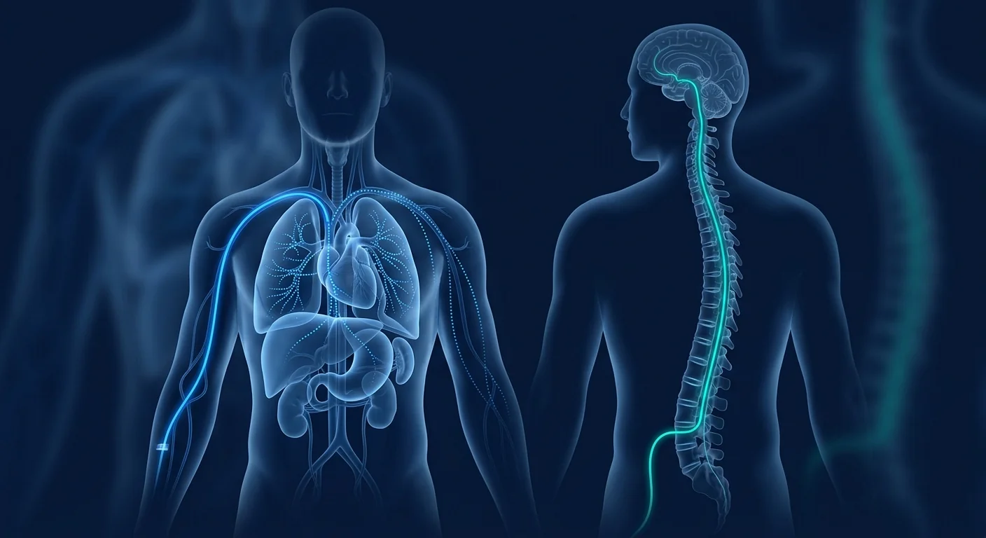
When mesenchymal stem cells (MSCs) are administered systemically, the delivery route is not a trivial choice. It determines biodistribution, cell survival, target-tissue engraftment, and ultimately clinical efficacy. Two routes dominate regenerative medicine practice — intravenous (IV) infusion and intrathecal (IT) injection — and they produce profoundly different pharmacokinetic profiles. This article compares them head-to-head, drawing on published preclinical and clinical data, so that clinicians and informed patients can understand the rationale behind route selection.[1][2]
How Intravenous MSC Delivery Works — and Where the Cells Actually Go
Intravenous infusion delivers MSCs directly into the peripheral circulation. From a peripheral vein, the cells travel through the right heart and into the pulmonary capillary bed — and this is where the single most important pharmacokinetic fact about IV MSCs reveals itself. Within minutes of infusion, the vast majority of cells are trapped in the lungs, a phenomenon known as first-pass pulmonary entrapment. Studies using labeled MSCs consistently show that 60–90% of infused cells are retained in the pulmonary microvasculature within the first hour.[3][4]
This entrapment is both a limitation and an advantage. It severely limits the number of cells that reach distant organs — the brain, spinal cord, joints, and kidneys each receive only a small fraction of the infused dose. But pulmonary entrapment also creates a sustained paracrine signaling depot: the trapped MSCs continue secreting anti-inflammatory cytokines, growth factors, and extracellular vesicles for 24–72 hours before clearance. This makes IV delivery well-suited for systemic inflammatory conditions — sepsis, ARDS, graft-versus-host disease, and autoimmune disorders where a broad immunomodulatory signal is the therapeutic goal.[5][6]
Key pharmacokinetic numbers for IV MSCs: ~80% pulmonary first-pass retention within 15 minutes; circulating half-life of 10–30 minutes; detectable paracrine activity for 48–72 hours post-infusion; less than 2% of the injected dose reaches the brain parenchyma.[7]
How Intrathecal MSC Delivery Works — Bypassing the Blood-Brain Barrier
Intrathecal injection places MSCs directly into the cerebrospinal fluid (CSF) via lumbar puncture. This route bypasses the blood-brain barrier (BBB) entirely, delivering cells into the subarachnoid space where they distribute through CSF flow along the spinal cord and into the cerebral ventricles. Because the BBB is the single greatest obstacle to delivering biologics into the central nervous system, IT administration represents a fundamentally different pharmacokinetic strategy than IV infusion.[8]
CSF distribution follows a pressure-gradient flow pattern. MSCs injected into the lumbar cistern travel rostrally — upward — propelled by CSF bulk flow toward the basal cisterns and, over 4–24 hours, into the ventricular system and perivascular spaces. Labeled MSCs have been detected in the brain parenchyma, spinal cord gray matter, and dorsal root ganglia within 6–24 hours of lumbar IT injection in animal models.[9] Cell survival in the CSF compartment is limited — typically 7–14 days — but the paracrine signals they release (BDNF, GDNF, NGF, anti-inflammatory cytokines) persist and exert trophic effects on neurons and glia.
Key pharmacokinetic numbers for IT MSCs: CSF distribution throughout the neuraxis within 4–6 hours; detectable paracrine activity for 7–14 days; direct access to the CNS compartment without BBB limitation; localized delivery means minimal systemic exposure — an advantage for neurological targeting but a limitation for multi-organ conditions.[10]
Head-to-Head: IV vs IT — What the Preclinical Data Shows
Animal studies comparing the two routes consistently show that IT delivery achieves far higher CNS cell concentrations than IV delivery. In rodent models of spinal cord injury, IT-injected MSCs achieved 20–50× higher cell counts in the spinal cord parenchyma compared to IV-injected cells.[11] In models of ischemic stroke, IT delivery produced significantly greater neurological recovery scores than IV delivery at equivalent cell doses, despite the IV group receiving cells earlier post-injury.
However, the picture is not one-sided. IV delivery, despite its low CNS penetration, has shown meaningful efficacy in CNS conditions — not through direct engraftment, but through systemic immunomodulation. In multiple sclerosis models, IV MSCs reduced peripheral T-cell activation and inflammatory cytokine levels, effects that were actually diminished with IT delivery because the cells were sequestered within the CNS compartment and could not interact with peripheral immune organs.[12]
The critical insight: IT delivery excels when the therapeutic target is neural tissue itself — neuroprotection, remyelination, axonal regeneration. IV delivery excels when the goal is systemic immunomodulation — even for neurological conditions with a strong autoimmune component. This is why several clinical programs now use both routes in combination: IV for systemic immune reset, IT for direct neurotrophic support.[13]
IV vs IT: a decision framework. IV is the default for systemic, multi-organ, and inflammatory conditions. IT is indicated when the BBB is the critical barrier and the therapeutic target is neural. Combined IV+IT protocols are emerging for complex neuroinflammatory conditions like MS, ALS, and cerebral palsy, where both systemic and compartmental pathology are present.
Clinical Evidence by Indication
Spinal Cord Injury
IT delivery is the dominant route in SCI clinical trials, and for good reason. Direct CSF administration places cells adjacent to the injury site, and early-phase trials have shown signals of motor and sensory improvement — particularly when MSCs are delivered within weeks to months of injury. A 2023 meta-analysis of 19 clinical studies found that IT MSC delivery in SCI was associated with statistically significant improvements in ASIA motor scores, with a favorable safety profile (no serious adverse events directly attributable to the injection procedure).[14] IV delivery has been explored but shows less consistent neurological benefit.
Neurodegenerative Disease (ALS, MS, Parkinson's)
The evidence is more nuanced here. In ALS, both IV and IT MSC delivery have been tested — IV trials emphasize systemic anti-inflammatory effects (reduced plasma TNF-α, IL-6), while IT trials report modest slowing of functional decline measured by ALSFRS-R.[15] In MS, IV MSCs have the stronger clinical evidence base, with Phase II trials demonstrating reduced gadolinium-enhancing lesions and stabilized EDSS scores — an effect likely mediated by peripheral Treg expansion rather than CNS engraftment. In Parkinson's disease, IT delivery (and the even more targeted intra-striatal route) is favored due to the need for local dopaminergic support.[16]
Orthopedic and Musculoskeletal Conditions
IV delivery is the standard for systemic musculoskeletal conditions. For osteoarthritis, joint pain, and sports injuries, IV MSCs distribute broadly and exert anti-inflammatory effects across multiple joints — a practical advantage over localized intra-articular injections when multiple sites are affected. IT delivery has no role in orthopedic indications; the cells would be sequestered in the CNS with no access to joints, tendons, or bones.
Systemic Inflammatory and Autoimmune Conditions
IV is the unquestioned route for systemic conditions. In graft-versus-host disease, lupus, rheumatoid arthritis, and Crohn's disease, the therapeutic mechanism — broad immunomodulation via pulmonary-trapped MSC secretome — is exactly what IV delivery provides. IT delivery would be counterproductive: it confines cells to the CNS, away from the peripheral immune organs where they need to act.
Safety Considerations — Both Routes, Different Profiles
IV MSC infusion has an established safety record across thousands of patients. The most common adverse events are mild infusion reactions (fever, headache, transient chills) occurring in 3–8% of infusions, typically self-limiting within hours.[17] Pulmonary embolism from cell clumping is a theoretical concern that has not materialized in clinical practice when appropriate cell preparation protocols (filtration, single-cell suspension, controlled infusion rate) are followed.
IT injection carries procedure-specific risks that IV does not. Lumbar puncture itself has a ~5–15% incidence of post-dural puncture headache, which is usually self-limiting but can be severe. More serious complications — spinal hematoma, infection (meningitis), nerve root irritation — are rare (<1 in 1,000) when performed by experienced clinicians under sterile technique.[18] The volume of cell suspension injected intrathecally must be carefully controlled; excessive volume or rapid injection can elevate intracranial pressure.
Safety summary: IV MSCs have the larger safety database and a lower procedure-risk profile. IT MSCs add the risk of lumbar puncture itself — small but real — and require a clinician experienced in intrathecal technique. Both routes have demonstrated acceptable safety in clinical trials when proper protocols are followed.
Practical Considerations — Cost, Convenience, and Patient Experience
IV infusion is logistically simpler. It requires peripheral venous access — a standard nursing procedure — and can be performed in an outpatient infusion suite. Sessions typically last 60–90 minutes including observation. IT injection requires lumbar puncture, which demands a trained clinician, sterile field preparation, and post-procedure observation (typically 1–2 hours lying flat to reduce PDPH risk). The procedure itself takes 15–30 minutes but the total clinic time is longer.
Patient experience differs markedly between the two routes. IV infusion is minimally invasive — a single venipuncture, similar to any IV drip. IT injection involves needle insertion between lumbar vertebrae, which most patients find more anxiety-provoking, though local anesthetic makes it tolerable. Some clinics offer light sedation for IT procedures. Recovery time is also different: IV patients can resume normal activities immediately; IT patients are advised to avoid strenuous activity for 24 hours.
Combination Protocols — When IV + IT Makes Sense
A growing body of clinical practice uses IV and IT MSCs together for complex neurological conditions. The rationale is pharmacokinetically sound: IV MSCs provide systemic immunomodulation (damping peripheral inflammation that drives neuroinflammation), while IT MSCs deliver direct neurotrophic and neuroprotective signals to the CNS compartment. This dual approach has been explored in cerebral palsy, ALS, and traumatic brain injury, with early reports suggesting additive benefit.[13][19]
Combination protocols are more resource-intensive and carry both sets of procedural risks. They should be reserved for conditions where there is a clear dual-compartment rationale — not used as a default. The strongest evidence for combination IV+IT exists in neuroinflammatory and neurodegenerative conditions where both peripheral immune dysregulation and CNS tissue damage are well-documented drivers of pathology.
How VELAR Approaches Route Selection
At VELAR Center, route selection is made individually for each patient based on three factors: the condition being treated, the therapeutic mechanism that matters most, and the patient's clinical history. For systemic autoimmune and inflammatory conditions, IV delivery is our standard. For spinal cord pathology — SCI, compressive myelopathy, spinal muscular atrophy — IT delivery is typically recommended. For complex neurological conditions with both systemic and compartmental components, we discuss combined IV+IT protocols where the evidence supports it.
Every IT procedure at VELAR is performed by a physician experienced in lumbar puncture technique, under sterile conditions, with ultrasound guidance where indicated. Every IV infusion uses freshly prepared, viability-verified MSCs in single-cell suspension and infused over 30–45 minutes — the protocol that has demonstrated the lowest incidence of infusion reactions in published series.[17]
Frequently Asked Questions
Which route is better — IV or intrathecal?
Neither is universally better; each is optimal for different clinical scenarios. IV is preferred for systemic inflammatory and multi-organ conditions. Intrathecal is preferred when the therapeutic target is the central nervous system and the blood-brain barrier must be bypassed. The right choice depends entirely on what is being treated.
Can I have both IV and intrathecal MSCs in the same treatment session?
Yes, combined IV+IT protocols are used for complex neurological conditions where both systemic immunomodulation and direct CNS support are desired. This approach is more resource-intensive and carries both sets of procedural risks but may offer additive benefit for select indications like ALS and cerebral palsy.
How painful is an intrathecal injection?
Local anesthetic is used at the injection site, and most patients describe the procedure as pressure rather than pain. Some discomfort during needle placement is normal. The most common post-procedure issue is a positional headache (post-dural puncture headache), which resolves with rest and hydration in most cases.
How long do the cells stay active after IV vs IT delivery?
After IV infusion, MSCs are largely cleared from circulation within hours but continue secreting paracrine factors for 48–72 hours from the pulmonary depot. After IT injection, paracrine activity is detectable in the CSF for 7–14 days. The therapeutic window — the period during which biologically meaningful signals are being sent — is longer with IT delivery for CNS targets.
Is IT delivery safe for children?
Lumbar puncture in children requires pediatric-specific expertise, but the procedure itself has been performed safely for decades (it is the standard route for intrathecal chemotherapy in pediatric oncology). MSC IT delivery in children has been studied most extensively in cerebral palsy trials, with acceptable safety profiles reported.
Does insurance cover intrathecal MSC therapy?
MSC therapy — regardless of delivery route — is generally not covered by standard health insurance plans as it remains investigational for most indications. Patients should confirm coverage directly with their insurer. VELAR provides transparent cost breakdowns during consultation.
Limitations and Realistic Expectations
MSC therapy by any route remains an investigational treatment for most indications. The pharmacokinetic data reviewed here comes predominantly from preclinical models and early-phase clinical trials; large-scale randomized controlled trials comparing IV and IT delivery head-to-head for specific conditions are limited.[20] Individual patient responses vary significantly, and no route of administration can guarantee a specific clinical outcome. This article is intended for educational purposes and does not constitute medical advice. Any decision about MSC therapy — including route selection — should be made in consultation with a qualified physician who has reviewed your full medical history.
References
- Pittenger MF, Mackay AM, Beck SC, et al. Multilineage potential of adult human mesenchymal stem cells. Science. 1999;284(5411):143-147. doi:10.1126/science.284.5411.143 ↩
- Caplan AI, Correa D. The MSC: an injury drugstore. Cell Stem Cell. 2011;9(1):11-15. doi:10.1016/j.stem.2011.06.008 ↩
- Fischer UM, Harting MT, Jimenez F, et al. Pulmonary passage is a major obstacle for intravenous stem cell delivery: the pulmonary first-pass effect. Stem Cells and Development. 2009;18(5):683-692. doi:10.1089/scd.2008.0253 ↩
- Kurtz A. Mesenchymal stem cell delivery routes and fate. International Journal of Stem Cells. 2008;1(1):1-7. doi:10.15283/ijsc.2008.1.1.1 ↩
- Lee RH, Pulin AA, Seo MJ, et al. Intravenous hMSCs improve myocardial infarction in mice because cells embolized in lung are activated to secrete the anti-inflammatory protein TSG-6. Cell Stem Cell. 2009;5(1):54-63. doi:10.1016/j.stem.2009.05.003 ↩
- Németh K, Leelahavanichkul A, Yuen PS, et al. Bone marrow stromal cells attenuate sepsis via prostaglandin E2-dependent reprogramming of host macrophages to increase their interleukin-10 production. Nature Medicine. 2009;15(1):42-49. doi:10.1038/nm.1905 ↩
- Schrepfer S, Deuse T, Reichenspurner H, et al. Stem cell transplantation: the lung barrier. Transplantation Proceedings. 2007;39(2):573-576. doi:10.1016/j.transproceed.2006.12.019 ↩
- Bakshi A, Hunter C, Swanger S, et al. Minimally invasive delivery of stem cells for spinal cord injury: advantages of the lumbar puncture technique. Journal of Neurosurgery: Spine. 2004;1(3):330-337. doi:10.3171/spi.2004.1.3.0330 ↩
- Paul C, Samdani AF, Betz RR, et al. Grafting of human bone marrow stromal cells into spinal cord injury: a comparison of delivery methods. Spine. 2009;34(4):328-334. doi:10.1097/BRS.0b013e31819403ce ↩
- Shin DA, Kim JM, Kim HI, et al. Comparison of intrathecal and intravenous administration of human mesenchymal stem cells in a rat model of spinal cord injury. Spine. 2013;38(15):E915-E922. doi:10.1097/BRS.0b013e318296fc1e ↩
- Syková E, Jendelová P, Urdzíková L, et al. Bone marrow stem cells and polymer hydrogels — two strategies for spinal cord injury repair. Cellular and Molecular Neurobiology. 2006;26(7-8):1113-1129. doi:10.1007/s10571-006-9007-2 ↩
- Zappia E, Casazza S, Pedemonte E, et al. Mesenchymal stem cells ameliorate experimental autoimmune encephalomyelitis inducing T-cell anergy. Blood. 2005;106(5):1755-1761. doi:10.1182/blood-2005-04-1496 ↩
- Karussis D, Karageorgiou C, Vaknin-Dembinsky A, et al. Safety and immunological effects of mesenchymal stem cell transplantation in patients with multiple sclerosis and amyotrophic lateral sclerosis. Archives of Neurology. 2010;67(10):1187-1194. doi:10.1001/archneurol.2010.248 ↩
- Zhao T, Yan W, Sun P, et al. Intrathecal transplantation of mesenchymal stem cells for spinal cord injury: a systematic review and meta-analysis. Stem Cell Research & Therapy. 2023;14:88. doi:10.1186/s13287-023-03309-4 ↩
- Petrou P, Gothelf Y, Argov Z, et al. Safety and clinical effects of mesenchymal stem cells secreting neurotrophic factor transplantation in patients with amyotrophic lateral sclerosis. JAMA Neurology. 2016;73(3):337-344. doi:10.1001/jamaneurol.2015.4321 ↩
- Venkataramana NK, Kumar SK, Balaraju S, et al. Open-labeled study of unilateral autologous bone-marrow-derived mesenchymal stem cell transplantation in Parkinson's disease. Translational Research. 2010;155(2):62-70. doi:10.1016/j.trsl.2009.07.006 ↩
- Lalu MM, McIntyre L, Pugliese C, et al. Safety of cell therapy with mesenchymal stromal cells (SafeCell): a systematic review and meta-analysis of clinical trials. PLoS One. 2012;7(10):e47559. doi:10.1371/journal.pone.0047559 ↩
- Evans RW, Armon C, Frohman EM, Goodin DS. Assessment: prevention of post-lumbar puncture headaches. Neurology. 2000;55(7):909-914. doi:10.1212/WNL.55.7.909 ↩
- Rah WJ, Lee YH, Moon JH, et al. Neuroregenerative potential of intravenous and intrathecal mesenchymal stem cells in neonatal hypoxic-ischemic brain injury. Stem Cells Translational Medicine. 2017;6(11):1951-1962. doi:10.1002/sctm.17-0034 ↩
- Galipeau J, Sensébé L. Mesenchymal stromal cells: clinical challenges and therapeutic opportunities. Cell Stem Cell. 2018;22(6):824-833. doi:10.1016/j.stem.2018.05.004 ↩
当间充质干细胞(MSCs)以系统性方式给药时,递送途径并非无关紧要的选择。它决定了生物分布、细胞存活、靶组织植入以及最终的临床疗效。在再生医学实践中,两种途径占据主导地位——静脉(IV)输注和鞘内(IT)注射——它们产生截然不同的药代动力学特征。本文基于已发表的临床前和临床数据进行头对头比较,使临床医生和知情患者能够理解途径选择背后的原理。[1][2]
静脉MSC递送如何工作——以及细胞实际上去了哪里
静脉输注将MSCs直接送入外周循环。从外周静脉出发,细胞通过右心进入肺毛细血管床——这就是关于IV MSCs最重要的药代动力学事实显现的地方。输注后几分钟内,绝大多数细胞被困在肺部,这一现象被称为首过肺截留。使用标记MSCs的研究一致表明,60–90%的输注细胞在最初一小时内滞留在肺微血管系统中。[3][4]
这种截留既是限制也是优势。它严重限制了到达远处器官的细胞数量——大脑、脊髓、关节和肾脏各自仅接受输注剂量的一小部分。但肺截留也创造了一个持续的旁分泌信号库:被困的MSCs在清除前持续分泌抗炎细胞因子、生长因子和细胞外囊泡24–72小时。这使得IV递送非常适合系统性炎症疾病——败血症、ARDS、移植物抗宿主病和自身免疫性疾病,其中广泛的免疫调节信号是治疗目标。[5][6]
IV MSCs的关键药代动力学数据:15分钟内约80%的肺首过滞留;循环半衰期10–30分钟;输注后可检测的旁分泌活性持续48–72小时;不到2%的注射剂量到达脑实质。[7]
鞘内MSC递送如何工作——绕过血脑屏障
鞘内注射通过腰椎穿刺将MSCs直接送入脑脊液(CSF)。该途径完全绕过血脑屏障(BBB),将细胞送入蛛网膜下腔,在那里它们通过CSF流动沿脊髓分布并进入脑室。由于BBB是将生物制剂送入中枢神经系统的最大障碍,IT给药代表了一种与IV输注根本不同的药代动力学策略。[8]
CSF分布遵循压力梯度流动模式。注入腰椎池的MSCs向上——头侧——移动,由CSF整体流推动朝向基底池,并在4–24小时内进入脑室系统和血管周围间隙。在动物模型中,标记的MSCs已在腰IT注射后6–24小时内在脑实质、脊髓灰质和后根神经节中被检测到。[9]CSF区室中的细胞存活有限——通常7–14天——但它们释放的旁分泌信号(BDNF、GDNF、NGF、抗炎细胞因子)持续存在并对神经元和胶质细胞发挥神经营养效应。
IT MSCs的关键药代动力学数据:4–6小时内CSF分布至整个神经轴;可检测的旁分泌活性持续7–14天;直接进入CNS区室,不受BBB限制;局部递送意味着最小的全身暴露——对神经靶向是优势,但对多器官疾病是限制。[10]
头对头:IV vs IT——临床前数据所示
比较两种途径的动物研究一致表明,IT递送在CNS中达到的细胞浓度远高于IV递送。在脊髓损伤的啮齿动物模型中,与IV注射的细胞相比,IT注射的MSCs在脊髓实质中达到的细胞计数高20–50倍。[11]在缺血性卒中模型中,在等效细胞剂量下,IT递送产生的神经功能恢复评分显著优于IV递送,尽管IV组在损伤后更早接受了细胞。
然而,情况并非一边倒。IV递送尽管CNS渗透率低,但在CNS疾病中已显示出有意义的疗效——不是通过直接植入,而是通过系统性免疫调节。在多发性硬化模型中,IV MSCs减少了外周T细胞活化和炎性细胞因子水平,这些效果在IT递送时实际上减弱了,因为细胞被隔离在CNS区室内,无法与外周免疫器官相互作用。[12]
关键见解:IT递送在治疗靶点是神经组织本身时表现优异——神经保护、髓鞘再生、轴突再生。IV递送在目标是系统性免疫调节时表现优异——即使是具有强自身免疫成分的神经系统疾病也是如此。这就是为什么几个临床项目现在联合使用两种途径:IV用于系统性免疫重置,IT用于直接神经营养支持。[13]
IV vs IT:决策框架。IV是系统性、多器官和炎性疾病的默认选择。当BBB是关键障碍且治疗靶点是神经时,IT是指征。联合IV+IT方案正在出现用于MS、ALS和脑瘫等复杂神经炎症疾病,其中全身性和局部病变同时存在。
按适应症划分的临床证据
脊髓损伤
IT递送是SCI临床试验中的主导途径,这是有充分理由的。直接CSF给药将细胞置于损伤部位附近,早期试验已显示出运动和感觉改善的信号——特别是在损伤后数周至数月内给予MSCs时。2023年一项包含19项临床研究的荟萃分析发现,SCI中的IT MSC递送与ASIA运动评分的统计学显著改善相关,且安全性良好(无直接归因于注射操作的严重不良事件)。[14]IV递送已被探索,但显示的神经功能获益不太一致。
神经退行性疾病(ALS、MS、帕金森病)
此处的证据更为微妙。在ALS中,IV和IT MSC递送均已测试——IV试验强调系统性抗炎效应(降低血浆TNF-α、IL-6),而IT试验报告通过ALSFRS-R测量的功能衰退略有减缓。[15]在MS中,IV MSCs具有更强的临床证据基础,II期试验显示钆增强病灶减少和EDSS评分稳定——这种效果很可能由外周Treg扩增而非CNS植入介导。在帕金森病中,由于需要局部多巴胺能支持,IT递送(以及更具靶向性的纹状体内途径)更受青睐。[16]
骨科和肌肉骨骼疾病
IV递送是系统性肌肉骨骼疾病的标准。对于骨关节炎、关节疼痛和运动损伤,IV MSCs广泛分布并在多个关节发挥抗炎作用——当多个部位受累时,这比局部关节内注射具有实际优势。IT递送在骨科适应症中无作用;细胞将被隔离在CNS中,无法进入关节、肌腱或骨骼。
系统性炎症和自身免疫性疾病
IV是系统性疾病的明确途径。在移植物抗宿主病、狼疮、类风湿关节炎和克罗恩病中,治疗机制——通过肺截留MSC分泌组进行广泛免疫调节——正是IV递送所提供的。IT递送将适得其反:它将细胞限制在CNS内,远离需要它们发挥作用的外周免疫器官。
安全考虑——两种途径,不同的安全性特征
IV MSC输注在数千名患者中已建立安全性记录。最常见的不良事件是轻度输注反应(发热、头痛、暂时性寒战),发生在3–8%的输注中,数小时内通常自限。[17]当遵循适当的细胞制备方案(过滤、单细胞悬液、控制输注速度)时,细胞聚集引起的肺栓塞是一个理论上的担忧,在临床实践中尚未出现。
IT注射带有IV没有的操作特异性风险。腰椎穿刺本身有约5–15%的硬膜穿刺后头痛发生率,通常自限但可能严重。更严重的并发症——脊髓血肿、感染(脑膜炎)、神经根刺激——在有经验的临床医生在无菌技术下操作时罕见(<1/1000)。[18]鞘内注射的细胞悬液体积必须谨慎控制;过量体积或快速注射可升高颅内压。
安全性总结:IV MSCs具有更大的安全性数据库和更低的操作风险特征。IT MSCs增加了腰椎穿刺本身的风险——虽小但真实——并且需要具备鞘内技术经验的临床医生。在遵循适当方案时,两种途径在临床试验中均显示出可接受的安全性。
实际考虑——费用、便利性和患者体验
IV输注在后勤上更简单。它需要外周静脉通路——一种标准护理操作——可在门诊输注室进行。疗程通常持续60–90分钟,包括观察。IT注射需要腰椎穿刺,这要求一名训练有素的临床医生、无菌区域准备和术后观察(通常平卧1–2小时以降低PDPH风险)。操作本身需要15–30分钟,但总门诊时间更长。
两种途径的患者体验差异显著。IV输注是微创的——单次静脉穿刺,类似于任何IV滴注。IT注射涉及在腰椎之间插入针头,大多数患者对此更为焦虑,尽管局部麻醉使其可耐受。一些诊所为IT操作提供轻度镇静。恢复时间也不同:IV患者可立即恢复正常活动;IT患者建议24小时内避免剧烈活动。
联合方案——IV + IT何时有意义
越来越多的临床实践对复杂神经系统疾病联合使用IV和IT MSCs。其原理在药代动力学上是合理的:IV MSCs提供系统性免疫调节(抑制驱动神经炎症的外周炎症),而IT MSCs向CNS区室提供直接神经营养和神经保护信号。这种双重方法已在脑瘫、ALS和创伤性脑损伤中进行了探索,早期报告提示具有附加效益。[13][19]
联合方案资源消耗更大,并承载两组操作风险。应保留用于有明确双区室原理的疾病——不用作默认选择。联合IV+IT的最强证据存在于神经炎症和神经退行性疾病中,其中外周免疫失调和CNS组织损伤都是已充分记录的病理驱动因素。
VELAR如何选择递送途径
在VELAR中心,途径选择基于三个因素为每位患者个体化制定:正在治疗的疾病、最重要的治疗机制以及患者的临床病史。对于系统性自身免疫和炎症性疾病,IV递送是我们的标准。对于脊髓病理——SCI、压迫性脊髓病、脊髓性肌萎缩症——通常推荐IT递送。对于具有全身性和局部成分的复杂神经系统疾病,当证据支持时,我们讨论联合IV+IT方案。
VELAR的每次IT操作均由具有腰椎穿刺技术经验的医生在无菌条件下进行,并在需要时使用超声引导。每次IV输注使用新鲜制备、活力验证的MSCs,以单细胞悬液形式在30–45分钟内输注——该方案在已发表的系列中显示出最低的输注反应发生率。[17]
常见问题
哪种途径更好——IV还是鞘内?
没有普遍更好的途径;每种途径在不同临床场景下最优。IV是系统性炎症和多器官疾病的首选。当治疗靶点是中枢神经系统且必须绕过血脑屏障时,鞘内是首选。正确的选择完全取决于正在治疗的是什么。
我可以在同一次治疗中同时接受IV和鞘内MSCs吗?
是的,联合IV+IT方案用于同时需要系统性免疫调节和直接CNS支持的复杂神经系统疾病。这种方法资源消耗更大,携带两组操作风险,但可能为ALS和脑瘫等特定适应症提供附加效益。
鞘内注射有多痛?
在注射部位使用局部麻醉,大多数患者将操作描述为压力而非疼痛。针头置入期间的一些不适是正常的。最常见的术后问题是体位性头痛(硬膜穿刺后头痛),大多数情况下通过休息和补液可缓解。
IV vs IT递送后细胞保持活性多久?
IV输注后,MSCs在数小时内大部分从循环中清除,但继续从肺部储存库分泌旁分泌因子48–72小时。IT注射后,在CSF中可检测到旁分泌活性持续7–14天。对于CNS靶点,IT递送的治疗窗口——即发出具有生物学意义信号的时间段——更长。
IT递送对儿童安全吗?
儿童腰椎穿刺需要儿科专业知识,但该操作本身已安全执行数十年(它是儿科肿瘤学中鞘内化疗的标准途径)。儿童中的MSC IT递送在脑瘫试验中研究最为广泛,报告的安全性特征可接受。
保险是否覆盖鞘内MSC治疗?
MSC治疗——无论递送途径如何——通常不在标准健康保险计划覆盖范围内,因为对大多数适应症而言仍处于研究阶段。患者应直接与保险公司确认覆盖范围。VELAR在咨询期间提供透明的费用明细。
局限性和现实期望
通过任何途径的MSC治疗对大多数适应症仍是一种研究性治疗。本文回顾的药代动力学数据主要来自临床前模型和早期临床试验;针对特定疾病头对头比较IV和IT递送的大规模随机对照试验有限。[20]个体患者反应差异显著,没有任何给药途径能保证特定的临床结果。本文仅供教育目的,不构成医疗建议。任何关于MSC治疗的决定——包括途径选择——都应在咨询已审查您完整病史的合格医生后做出。
参考文献
- Pittenger MF, Mackay AM, Beck SC, et al. Multilineage potential of adult human mesenchymal stem cells. Science. 1999;284(5411):143-147. doi:10.1126/science.284.5411.143 ↩
- Caplan AI, Correa D. The MSC: an injury drugstore. Cell Stem Cell. 2011;9(1):11-15. doi:10.1016/j.stem.2011.06.008 ↩
- Fischer UM, Harting MT, Jimenez F, et al. Pulmonary passage is a major obstacle for intravenous stem cell delivery: the pulmonary first-pass effect. Stem Cells and Development. 2009;18(5):683-692. doi:10.1089/scd.2008.0253 ↩
- Kurtz A. Mesenchymal stem cell delivery routes and fate. International Journal of Stem Cells. 2008;1(1):1-7. doi:10.15283/ijsc.2008.1.1.1 ↩
- Lee RH, Pulin AA, Seo MJ, et al. Intravenous hMSCs improve myocardial infarction in mice because cells embolized in lung are activated to secrete the anti-inflammatory protein TSG-6. Cell Stem Cell. 2009;5(1):54-63. doi:10.1016/j.stem.2009.05.003 ↩
- Németh K, Leelahavanichkul A, Yuen PS, et al. Bone marrow stromal cells attenuate sepsis via prostaglandin E2-dependent reprogramming of host macrophages to increase their interleukin-10 production. Nature Medicine. 2009;15(1):42-49. doi:10.1038/nm.1905 ↩
- Schrepfer S, Deuse T, Reichenspurner H, et al. Stem cell transplantation: the lung barrier. Transplantation Proceedings. 2007;39(2):573-576. doi:10.1016/j.transproceed.2006.12.019 ↩
- Bakshi A, Hunter C, Swanger S, et al. Minimally invasive delivery of stem cells for spinal cord injury: advantages of the lumbar puncture technique. Journal of Neurosurgery: Spine. 2004;1(3):330-337. doi:10.3171/spi.2004.1.3.0330 ↩
- Paul C, Samdani AF, Betz RR, et al. Grafting of human bone marrow stromal cells into spinal cord injury: a comparison of delivery methods. Spine. 2009;34(4):328-334. doi:10.1097/BRS.0b013e31819403ce ↩
- Shin DA, Kim JM, Kim HI, et al. Comparison of intrathecal and intravenous administration of human mesenchymal stem cells in a rat model of spinal cord injury. Spine. 2013;38(15):E915-E922. doi:10.1097/BRS.0b013e318296fc1e ↩
- Syková E, Jendelová P, Urdzíková L, et al. Bone marrow stem cells and polymer hydrogels — two strategies for spinal cord injury repair. Cellular and Molecular Neurobiology. 2006;26(7-8):1113-1129. doi:10.1007/s10571-006-9007-2 ↩
- Zappia E, Casazza S, Pedemonte E, et al. Mesenchymal stem cells ameliorate experimental autoimmune encephalomyelitis inducing T-cell anergy. Blood. 2005;106(5):1755-1761. doi:10.1182/blood-2005-04-1496 ↩
- Karussis D, Karageorgiou C, Vaknin-Dembinsky A, et al. Safety and immunological effects of mesenchymal stem cell transplantation in patients with multiple sclerosis and amyotrophic lateral sclerosis. Archives of Neurology. 2010;67(10):1187-1194. doi:10.1001/archneurol.2010.248 ↩
- Zhao T, Yan W, Sun P, et al. Intrathecal transplantation of mesenchymal stem cells for spinal cord injury: a systematic review and meta-analysis. Stem Cell Research & Therapy. 2023;14:88. doi:10.1186/s13287-023-03309-4 ↩
- Petrou P, Gothelf Y, Argov Z, et al. Safety and clinical effects of mesenchymal stem cells secreting neurotrophic factor transplantation in patients with amyotrophic lateral sclerosis. JAMA Neurology. 2016;73(3):337-344. doi:10.1001/jamaneurol.2015.4321 ↩
- Venkataramana NK, Kumar SK, Balaraju S, et al. Open-labeled study of unilateral autologous bone-marrow-derived mesenchymal stem cell transplantation in Parkinson's disease. Translational Research. 2010;155(2):62-70. doi:10.1016/j.trsl.2009.07.006 ↩
- Lalu MM, McIntyre L, Pugliese C, et al. Safety of cell therapy with mesenchymal stromal cells (SafeCell): a systematic review and meta-analysis of clinical trials. PLoS One. 2012;7(10):e47559. doi:10.1371/journal.pone.0047559 ↩
- Evans RW, Armon C, Frohman EM, Goodin DS. Assessment: prevention of post-lumbar puncture headaches. Neurology. 2000;55(7):909-914. doi:10.1212/WNL.55.7.909 ↩
- Rah WJ, Lee YH, Moon JH, et al. Neuroregenerative potential of intravenous and intrathecal mesenchymal stem cells in neonatal hypoxic-ischemic brain injury. Stem Cells Translational Medicine. 2017;6(11):1951-1962. doi:10.1002/sctm.17-0034 ↩
- Galipeau J, Sensébé L. Mesenchymal stromal cells: clinical challenges and therapeutic opportunities. Cell Stem Cell. 2018;22(6):824-833. doi:10.1016/j.stem.2018.05.004 ↩
عندما تُعطى الخلايا الجذعية الوسيطة (MSCs) جهازياً، فإن طريق الإعطاء ليس خياراً تافهاً. إنه يحدد التوزع الحيوي، وبقاء الخلايا، والانغراس في النسيج المستهدف، وفي النهاية الفعالية السريرية. يهيمن طريقان على ممارسة الطب التجديدي — التسريب الوريدي (IV) والحقن داخل القراب (IT) — وينتجان ملفات حركية دوائية مختلفة تماماً. تقارن هذه المقالة بينهما وجهاً لوجه، مستندة إلى البيانات المنشورة قبل السريرية والسريرية، ليتمكن الأطباء والمرضى المطلعون من فهم الأساس المنطقي وراء اختيار الطريق.[1][2]
كيف يعمل التوصيل الوريدي للخلايا الجذعية الوسيطة — وأين تذهب الخلايا فعلياً
يوصل التسريب الوريدي الخلايا الجذعية الوسيطة مباشرة إلى الدورة الدموية المحيطية. من الوريد المحيطي، تنتقل الخلايا عبر القلب الأيمن إلى السرير الشعري الرئوي — وهنا تظهر أهم حقيقة حركية دوائية حول الخلايا الجذعية الوسيطة الوريدية. في غضون دقائق من التسريب، تحتجز الغالبية العظمى من الخلايا في الرئتين، وهي ظاهرة تعرف باسم الاحتجاز الرئوي الأولي. تُظهر الدراسات التي تستخدم خلايا جذعية موسومة باستمرار أن 60–90% من الخلايا المعطاة تحتجز في الأوعية الدقيقة الرئوية خلال الساعة الأولى.[3][4]
هذا الاحتجاز هو قيد وميزة في آن واحد. إنه يحد بشدة من عدد الخلايا التي تصل إلى الأعضاء البعيدة — يتلقى الدماغ والحبل الشوكي والمفاصل والكلى كل منها جزءاً صغيراً فقط من الجرعة المعطاة. لكن الاحتجاز الرئوي يخلق أيضاً مستودعاً مستداماً للإشارات نظيرة الصماوية: تستمر الخلايا الجذعية الوسيطة المحتجزة في إفراز السيتوكينات المضادة للالتهابات وعوامل النمو والحويصلات خارج الخلية لمدة 24–72 ساعة قبل التصفية. هذا يجعل التوصيل الوريدي مناسباً تماماً للحالات الالتهابية الجهازية — الإنتان، متلازمة الضائقة التنفسية الحادة، داء الطعم حيال الثوي، واضطرابات المناعة الذاتية حيث تكون الإشارة المناعية الواسعة هي الهدف العلاجي.[5][6]
الأرقام الحركية الدوائية الرئيسية للخلايا الجذعية الوسيطة الوريدية: ~80% احتجاز رئوي أولي خلال 15 دقيقة؛ عمر نصف في الدوران 10–30 دقيقة؛ نشاط نظير صماوي قابل للكشف لمدة 48–72 ساعة بعد التسريب؛ أقل من 2% من الجرعة المحقونة تصل إلى النسيج الدماغي.[7]
كيف يعمل التوصيل داخل القراب للخلايا الجذعية الوسيطة — تجاوز الحاجز الدموي الدماغي
يضع الحقن داخل القراب الخلايا الجذعية الوسيطة مباشرة في السائل الدماغي الشوكي (CSF) عبر البزل القطني. يتجاوز هذا الطريق الحاجز الدموي الدماغي (BBB) تماماً، موصلاً الخلايا إلى الحيز تحت العنكبوتي حيث تتوزع من خلال تدفق السائل الدماغي الشوكي على طول الحبل الشوكي وإلى البطينات الدماغية. نظراً لأن الحاجز الدموي الدماغي هو أكبر عقبة أمام توصيل المواد البيولوجية إلى الجهاز العصبي المركزي، فإن الإعطاء داخل القراب يمثل استراتيجية حركية دوائية مختلفة جوهرياً عن التسريب الوريدي.[8]
يتبع توزع السائل الدماغي الشوكي نمط تدفق بتدرج ضغطي. تنتقل الخلايا الجذعية الوسيطة المحقونة في الصهريج القطني نحو الأعلى — باتجاه الرأس — مدفوعة بالتدفق الكتلي للسائل الدماغي الشوكي نحو الصهاريج القاعدية، وعلى مدى 4–24 ساعة، إلى الجهاز البطيني والمساحات حول الوعائية. تم الكشف عن خلايا جذعية موسومة في النسيج الدماغي والمادة الرمادية للحبل الشوكي والعقد الجذرية الظهرية في غضون 6–24 ساعة من الحقن القطني داخل القراب في النماذج الحيوانية.[9] بقاء الخلايا في حيز السائل الدماغي الشوكي محدود — عادة 7–14 يوماً — لكن الإشارات نظيرة الصماوية التي تطلقها (BDNF، GDNF، NGF، السيتوكينات المضادة للالتهابات) تستمر وتمارس تأثيرات تغذوية عصبية على العصبونات والدبق.
الأرقام الحركية الدوائية الرئيسية للخلايا الجذعية الوسيطة داخل القراب: توزع السائل الدماغي الشوكي في جميع أنحاء المحور العصبي خلال 4–6 ساعات؛ نشاط نظير صماوي قابل للكشف لمدة 7–14 يوماً؛ وصول مباشر إلى حيز الجهاز العصبي المركزي دون قيود الحاجز الدموي الدماغي؛ التوصيل الموضعي يعني تعرضاً جهازياً أدنى — ميزة للاستهداف العصبي لكنه قيد للحالات متعددة الأعضاء.[10]
وجهاً لوجه: وريدي مقابل داخل القراب — ما تظهره البيانات قبل السريرية
تُظهر الدراسات الحيوانية التي تقارن الطريقين باستمرار أن التوصيل داخل القراب يحقق تركيزات خلوية في الجهاز العصبي المركزي أعلى بكثير من التوصيل الوريدي. في نماذج القوارض لإصابة الحبل الشوكي، حققت الخلايا الجذعية الوسيطة المحقونة داخل القراب تعدادات خلوية أعلى بـ 20–50 ضعفاً في نسيج الحبل الشوكي مقارنة بالخلايا المحقونة وريدياً.[11] في نماذج السكتة الدماغية الإقفارية، أنتج التوصيل داخل القراب درجات تعافي عصبي أفضل بشكل ملحوظ من التوصيل الوريدي عند جرعات خلوية متكافئة، على الرغم من أن المجموعة الوريدية تلقت الخلايا في وقت أبكر بعد الإصابة.
ومع ذلك، الصورة ليست من جانب واحد. التوصيل الوريدي، على الرغم من انخفاض اختراقه للجهاز العصبي المركزي، أظهر فعالية ذات معنى في حالات الجهاز العصبي المركزي — ليس من خلال الانغراس المباشر، ولكن من خلال التعديل المناعي الجهازي. في نماذج التصلب المتعدد، قللت الخلايا الجذعية الوسيطة الوريدية من تنشيط الخلايا التائية المحيطية ومستويات السيتوكينات الالتهابية، وهي تأثيرات تضاءلت فعلياً مع التوصيل داخل القراب لأن الخلايا كانت محتجزة داخل حيز الجهاز العصبي المركزي ولم تستطع التفاعل مع الأعضاء المناعية المحيطية.[12]
الرؤية الحرجة: يتفوق التوصيل داخل القراب عندما يكون الهدف العلاجي هو النسيج العصبي نفسه — الحماية العصبية، إعادة التغطية بالنخاعين، التجدد المحواري. يتفوق التوصيل الوريدي عندما يكون الهدف هو التعديل المناعي الجهازي — حتى للحالات العصبية ذات المكون المناعي الذاتي القوي. لهذا السبب تستخدم عدة برامج سريرية الآن كلا الطريقين معاً: وريدي لإعادة الضبط المناعي الجهازي، وداخل القراب للدعم التغذوي العصبي المباشر.[13]
وريدي مقابل داخل القراب: إطار قرار. الوريدي هو الخيار الافتراضي للحالات الجهازية ومتعددة الأعضاء والالتهابية. يُشار إلى داخل القراب عندما يكون الحاجز الدموي الدماغي هو الحاجز الحرج والهدف العلاجي عصبي. تظهر بروتوكولات وريدي + داخل القراب المشتركة للحالات الالتهابية العصبية المعقدة مثل التصلب المتعدد والتصلب الجانبي الضموري والشلل الدماغي، حيث يوجد كل من الأمراض الجهازية والحيزية.
الأدلة السريرية حسب الاستطباب
إصابة الحبل الشوكي
التوصيل داخل القراب هو الطريق السائد في تجارب إصابة الحبل الشوكي السريرية، ولسبب وجيه. يضع الإعطاء المباشر في السائل الدماغي الشوكي الخلايا بجوار موقع الإصابة، وقد أظهرت تجارب المرحلة المبكرة إشارات تحسن حركي وحسي — خاصة عندما تُعطى الخلايا الجذعية الوسيطة في غضون أسابيع إلى أشهر من الإصابة. وجد تحليل تلوي لعام 2023 شمل 19 دراسة سريرية أن التوصيل داخل القراب للخلايا الجذعية الوسيطة في إصابة الحبل الشوكي ارتبط بتحسنات ذات دلالة إحصائية في درجات ASIA الحركية، مع ملف أمان مناسب (لا أحداث ضائرة خطيرة تُعزى مباشرة إلى إجراء الحقن).[14] تم استكشاف التوصيل الوريدي لكنه أظهر فائدة عصبية أقل اتساقاً.
الأمراض التنكسية العصبية (ALS، MS، باركنسون)
الأدلة هنا أكثر دقة. في ALS، تم اختبار كل من التوصيل الوريدي وداخل القراب — تؤكد التجارب الوريدية على التأثيرات المضادة للالتهابات الجهازية (انخفاض TNF-α، IL-6 في البلازما)، بينما تبلغ تجارب داخل القراب عن تباطؤ متواضع في التدهور الوظيفي مقاساً بـ ALSFRS-R.[15] في التصلب المتعدد، تمتلك الخلايا الجذعية الوسيطة الوريدية قاعدة أدلة سريرية أقوى، مع تجارب المرحلة الثانية التي تظهر انخفاضاً في آفات تعزيز الغادولينيوم واستقرار درجات EDSS — وهو تأثير يتوسطه على الأرجح توسع Treg المحيطي وليس الانغراس في الجهاز العصبي المركزي. في مرض باركنسون، يُفضل التوصيل داخل القراب (والطريق داخل الجسم المخطط الأكثر استهدافاً) بسبب الحاجة إلى دعم دوباميني موضعي.[16]
الحالات العظمية والعضلية الهيكلية
التوصيل الوريدي هو المعيار للحالات العضلية الهيكلية الجهازية. للفصال العظمي وآلام المفاصل والإصابات الرياضية، تتوزع الخلايا الجذعية الوسيطة الوريدية على نطاق واسع وتمارس تأثيرات مضادة للالتهابات عبر مفاصل متعددة — ميزة عملية على الحقن الموضعي داخل المفصل عندما تتأثر مواقع متعددة. ليس للتوصيل داخل القراب دور في الاستطبابات العظمية؛ ستُحتجز الخلايا في الجهاز العصبي المركزي دون وصول إلى المفاصل أو الأوتار أو العظام.
الحالات الالتهابية الجهازية والمناعة الذاتية
الوريدي هو الطريق غير القابل للجدل للحالات الجهازية. في داء الطعم حيال الثوي، والذئبة، والتهاب المفاصل الروماتويدي، وداء كرون، فإن الآلية العلاجية — التعديل المناعي الواسع عبر الإفراز نظير الصماوي للخلايا الجذعية الوسيطة المحتجزة رئوياً — هي بالضبط ما يوفره التوصيل الوريدي. سيكون التوصيل داخل القراب بنتيجة عكسية: إنه يحصر الخلايا في الجهاز العصبي المركزي، بعيداً عن الأعضاء المناعية المحيطية حيث تحتاج إلى العمل.
اعتبارات السلامة — كلا الطريقين، ملفات مختلفة
للتسريب الوريدي للخلايا الجذعية الوسيطة سجل أمان راسخ عبر آلاف المرضى. الأحداث الضائرة الأكثر شيوعاً هي تفاعلات تسريب خفيفة (حمى، صداع، قشعريرة عابرة) تحدث في 3–8% من عمليات التسريب، وعادة ما تكون محددة ذاتياً خلال ساعات.[17] الانصمام الرئوي من تكتل الخلايا هو مصدر قلق نظري لم يتحقق في الممارسة السريرية عند اتباع بروتوكولات تحضير الخلايا المناسبة (الترشيح، المعلق أحادي الخلية، معدل التسريب المضبوط).
يحمل الحقن داخل القراب مخاطر خاصة بالإجراء لا توجد في الوريدي. البزل القطني نفسه لديه حدوث ~5–15% من صداع ما بعد البزل الجافوي، والذي عادة ما يكون محدداً ذاتياً لكنه قد يكون شديداً. المضاعفات الأكثر خطورة — الورم الدموي الشوكي، العدوى (التهاب السحايا)، تهيج جذر العصب — نادرة (<1 من 1000) عندما يُجرى من قبل أطباء ذوي خبرة تحت تقنية معقمة.[18] يجب التحكم بعناية في حجم معلق الخلايا المحقون داخل القراب؛ يمكن للحجم المفرط أو الحقن السريع أن يرفع الضغط داخل القحف.
ملخص السلامة: تمتلك الخلايا الجذعية الوسيطة الوريدية قاعدة بيانات أمان أكبر وملف مخاطر إجرائية أقل. تضيف الخلايا الجذعية الوسيطة داخل القراب خطر البزل القطني نفسه — صغير لكنه حقيقي — وتتطلب طبيباً ذا خبرة في تقنية داخل القراب. أظهر كلا الطريقين أماناً مقبولاً في التجارب السريرية عند اتباع البروتوكولات المناسبة.
اعتبارات عملية — التكلفة والملاءمة وتجربة المريض
التسريب الوريدي أبسط لوجستياً. يتطلب وصولاً وريدياً محيطياً — إجراء تمريضي قياسي — ويمكن إجراؤه في جناح التسريب الخارجي. تستمر الجلسات عادة 60–90 دقيقة بما في ذلك المراقبة. يتطلب الحقن داخل القراب البزل القطني، الذي يستدعي طبيباً مدرباً وتحضير حقل معقم ومراقبة ما بعد الإجراء (عادة 1–2 ساعة استلقاء لتقليل خطر صداع ما بعد البزل). يستغرق الإجراء نفسه 15–30 دقيقة لكن وقت العيادة الإجمالي أطول.
تختلف تجربة المريض بشكل ملحوظ بين الطريقين. التسريب الوريدي طفيف التوغل — بزل وريدي واحد، مشابه لأي تنقيط وريدي. يتضمن الحقن داخل القراب إدخال إبرة بين الفقرات القطنية، وهو ما يجده معظم المرضى أكثر إثارة للقلق، على الرغم من أن المخدر الموضعي يجعله محتملاً. تقدم بعض العيادات تخديراً خفيفاً لإجراءات داخل القراب. يختلف وقت التعافي أيضاً: يمكن لمرضى الوريدي استئناف الأنشطة الطبيعية فوراً؛ يُنصح مرضى داخل القراب بتجنب النشاط الشاق لمدة 24 ساعة.
البروتوكولات المركبة — متى يكون الوريدي + داخل القراب منطقياً
تستخدم مجموعة متنامية من الممارسات السريرية الخلايا الجذعية الوسيطة الوريدية وداخل القراب معاً للحالات العصبية المعقدة. الأساس المنطقي سليم حركياً دوائياً: توفر الخلايا الجذعية الوسيطة الوريدية تعديلاً مناعياً جهازياً (تثبيط الالتهاب المحيطي الذي يدفع الالتهاب العصبي)، بينما توفر الخلايا الجذعية الوسيطة داخل القراب إشارات تغذوية عصبية ووقائية عصبية مباشرة إلى حيز الجهاز العصبي المركزي. تم استكشاف هذا النهج المزدوج في الشلل الدماغي وALS وإصابة الدماغ الرضية، مع تقارير مبكرة تشير إلى فائدة إضافية.[13][19]
البروتوكولات المركبة أكثر استهلاكاً للموارد وتحمل كلا مجموعتي المخاطر الإجرائية. يجب حجزها للحالات التي يوجد فيها أساس منطقي واضح للحيزين — لا تستخدم كخيار افتراضي. توجد أقوى الأدلة للدمج الوريدي + داخل القراب في الحالات الالتهابية العصبية والتنكسية العصبية حيث يكون كل من خلل التنظيم المناعي المحيطي وتلف نسيج الجهاز العصبي المركزي محركين موثقين جيداً للمرض.
كيف تختار VELAR طريق الإعطاء
في مركز VELAR، يتم اختيار الطريق بشكل فردي لكل مريض بناءً على ثلاثة عوامل: الحالة التي تُعالج، والآلية العلاجية الأكثر أهمية، والتاريخ السريري للمريض. للحالات المناعية الذاتية والالتهابية الجهازية، التوصيل الوريدي هو معيارنا. لأمراض الحبل الشوكي — إصابة الحبل الشوكي، الاعتلال النخاعي الانضغاطي، الضمور العضلي الشوكي — يُوصى عادة بالتوصيل داخل القراب. للحالات العصبية المعقدة ذات المكونات الجهازية والحيزية، نناقش بروتوكولات وريدي + داخل القراب المركبة عندما تدعمها الأدلة.
كل إجراء داخل القراب في VELAR يُجرى من قبل طبيب ذي خبرة في تقنية البزل القطني، تحت ظروف معقمة، مع توجيه بالأمواج فوق الصوتية عند الحاجة. كل تسريب وريدي يستخدم خلايا جذعية وسيطة محضّرة طازجة ومتحقق من حيويتها كمعلق أحادي الخلية وتُعطى على مدى 30–45 دقيقة — البروتوكول الذي أظهر أقل حدوث لتفاعلات التسريب في السلاسل المنشورة.[17]
الأسئلة الشائعة
أي طريق أفضل — وريدي أم داخل القراب؟
لا يوجد طريق أفضل عالمياً؛ كل منهما مثالي لسيناريوهات سريرية مختلفة. الوريدي مفضل للحالات الالتهابية الجهازية ومتعددة الأعضاء. داخل القراب مفضل عندما يكون الهدف العلاجي هو الجهاز العصبي المركزي ويجب تجاوز الحاجز الدموي الدماغي. يعتمد الاختيار الصحيح كلياً على ما يُعالج.
هل يمكنني الحصول على الخلايا الجذعية الوسيطة الوريدية وداخل القراب في نفس جلسة العلاج؟
نعم، تُستخدم بروتوكولات وريدي + داخل القراب المركبة للحالات العصبية المعقدة حيث يكون كل من التعديل المناعي الجهازي والدعم المباشر للجهاز العصبي المركزي مرغوباً. هذا النهج أكثر استهلاكاً للموارد ويحمل كلا مجموعتي المخاطر الإجرائية لكنه قد يقدم فائدة إضافية لاستطبابات مختارة مثل ALS والشلل الدماغي.
ما مدى ألم الحقن داخل القراب؟
يُستخدم المخدر الموضعي في موقع الحقن، ويصف معظم المرضى الإجراء بأنه ضغط وليس ألماً. بعض الانزعاج أثناء وضع الإبرة طبيعي. المشكلة الأكثر شيوعاً بعد الإجراء هي صداع وضعي (صداع ما بعد البزل الجافوي)، والذي يزول بالراحة والترطيب في معظم الحالات.
كم من الوقت تبقى الخلايا نشطة بعد التوصيل الوريدي مقابل داخل القراب؟
بعد التسريب الوريدي، تُصفى الخلايا الجذعية الوسيطة إلى حد كبير من الدوران في غضون ساعات لكنها تستمر في إفراز العوامل نظيرة الصماوية لمدة 48–72 ساعة من المستودع الرئوي. بعد الحقن داخل القراب، يمكن الكشف عن النشاط نظير الصماوي في السائل الدماغي الشوكي لمدة 7–14 يوماً. النافذة العلاجية — الفترة التي تُرسل خلالها إشارات ذات معنى بيولوجي — أطول مع التوصيل داخل القراب لأهداف الجهاز العصبي المركزي.
هل التوصيل داخل القراب آمن للأطفال؟
يتطلب البزل القطني عند الأطفال خبرة خاصة بالأطفال، لكن الإجراء نفسه تم إجراؤه بأمان لعقود (إنه الطريق القياسي للعلاج الكيميائي داخل القراب في أورام الأطفال). تمت دراسة التوصيل داخل القراب للخلايا الجذعية الوسيطة عند الأطفال على نطاق واسع في تجارب الشلل الدماغي، مع ملفات أمان مقبولة تم الإبلاغ عنها.
هل يغطي التأمين العلاج بالخلايا الجذعية الوسيطة داخل القراب؟
العلاج بالخلايا الجذعية الوسيطة — بغض النظر عن طريق الإعطاء — لا يغطيه عموماً خطط التأمين الصحي القياسية لأنه لا يزال قيد البحث لمعظم الاستطبابات. يجب على المرضى تأكيد التغطية مباشرة مع شركة التأمين الخاصة بهم. تقدم VELAR تفاصيل تكلفة شفافة أثناء الاستشارة.
القيود والتوقعات الواقعية
يبقى العلاج بالخلايا الجذعية الوسيطة بأي طريق علاجاً قيد البحث لمعظم الاستطبابات. تأتي البيانات الحركية الدوائية المستعرضة هنا في الغالب من النماذج قبل السريرية والتجارب السريرية المبكرة؛ التجارب المعشاة ذات الشواهد واسعة النطاق التي تقارن التوصيل الوريدي وداخل القراب وجهاً لوجه لحالات محددة محدودة.[20] تختلف استجابات المرضى الأفراد بشكل كبير، ولا يمكن لأي طريق إعطاء أن يضمن نتيجة سريرية محددة. هذه المقالة مخصصة للأغراض التعليمية ولا تشكل نصيحة طبية. أي قرار بشأن العلاج بالخلايا الجذعية الوسيطة — بما في ذلك اختيار الطريق — يجب أن يُتخذ بالتشاور مع طبيب مؤهل راجع تاريخك الطبي الكامل.
المراجع
- Pittenger MF, Mackay AM, Beck SC, et al. Multilineage potential of adult human mesenchymal stem cells. Science. 1999;284(5411):143-147. doi:10.1126/science.284.5411.143 ↩
- Caplan AI, Correa D. The MSC: an injury drugstore. Cell Stem Cell. 2011;9(1):11-15. doi:10.1016/j.stem.2011.06.008 ↩
- Fischer UM, Harting MT, Jimenez F, et al. Pulmonary passage is a major obstacle for intravenous stem cell delivery: the pulmonary first-pass effect. Stem Cells and Development. 2009;18(5):683-692. doi:10.1089/scd.2008.0253 ↩
- Kurtz A. Mesenchymal stem cell delivery routes and fate. International Journal of Stem Cells. 2008;1(1):1-7. doi:10.15283/ijsc.2008.1.1.1 ↩
- Lee RH, Pulin AA, Seo MJ, et al. Intravenous hMSCs improve myocardial infarction in mice because cells embolized in lung are activated to secrete the anti-inflammatory protein TSG-6. Cell Stem Cell. 2009;5(1):54-63. doi:10.1016/j.stem.2009.05.003 ↩
- Németh K, Leelahavanichkul A, Yuen PS, et al. Bone marrow stromal cells attenuate sepsis via prostaglandin E2-dependent reprogramming of host macrophages to increase their interleukin-10 production. Nature Medicine. 2009;15(1):42-49. doi:10.1038/nm.1905 ↩
- Schrepfer S, Deuse T, Reichenspurner H, et al. Stem cell transplantation: the lung barrier. Transplantation Proceedings. 2007;39(2):573-576. doi:10.1016/j.transproceed.2006.12.019 ↩
- Bakshi A, Hunter C, Swanger S, et al. Minimally invasive delivery of stem cells for spinal cord injury: advantages of the lumbar puncture technique. Journal of Neurosurgery: Spine. 2004;1(3):330-337. doi:10.3171/spi.2004.1.3.0330 ↩
- Paul C, Samdani AF, Betz RR, et al. Grafting of human bone marrow stromal cells into spinal cord injury: a comparison of delivery methods. Spine. 2009;34(4):328-334. doi:10.1097/BRS.0b013e31819403ce ↩
- Shin DA, Kim JM, Kim HI, et al. Comparison of intrathecal and intravenous administration of human mesenchymal stem cells in a rat model of spinal cord injury. Spine. 2013;38(15):E915-E922. doi:10.1097/BRS.0b013e318296fc1e ↩
- Syková E, Jendelová P, Urdzíková L, et al. Bone marrow stem cells and polymer hydrogels — two strategies for spinal cord injury repair. Cellular and Molecular Neurobiology. 2006;26(7-8):1113-1129. doi:10.1007/s10571-006-9007-2 ↩
- Zappia E, Casazza S, Pedemonte E, et al. Mesenchymal stem cells ameliorate experimental autoimmune encephalomyelitis inducing T-cell anergy. Blood. 2005;106(5):1755-1761. doi:10.1182/blood-2005-04-1496 ↩
- Karussis D, Karageorgiou C, Vaknin-Dembinsky A, et al. Safety and immunological effects of mesenchymal stem cell transplantation in patients with multiple sclerosis and amyotrophic lateral sclerosis. Archives of Neurology. 2010;67(10):1187-1194. doi:10.1001/archneurol.2010.248 ↩
- Zhao T, Yan W, Sun P, et al. Intrathecal transplantation of mesenchymal stem cells for spinal cord injury: a systematic review and meta-analysis. Stem Cell Research & Therapy. 2023;14:88. doi:10.1186/s13287-023-03309-4 ↩
- Petrou P, Gothelf Y, Argov Z, et al. Safety and clinical effects of mesenchymal stem cells secreting neurotrophic factor transplantation in patients with amyotrophic lateral sclerosis. JAMA Neurology. 2016;73(3):337-344. doi:10.1001/jamaneurol.2015.4321 ↩
- Venkataramana NK, Kumar SK, Balaraju S, et al. Open-labeled study of unilateral autologous bone-marrow-derived mesenchymal stem cell transplantation in Parkinson's disease. Translational Research. 2010;155(2):62-70. doi:10.1016/j.trsl.2009.07.006 ↩
- Lalu MM, McIntyre L, Pugliese C, et al. Safety of cell therapy with mesenchymal stromal cells (SafeCell): a systematic review and meta-analysis of clinical trials. PLoS One. 2012;7(10):e47559. doi:10.1371/journal.pone.0047559 ↩
- Evans RW, Armon C, Frohman EM, Goodin DS. Assessment: prevention of post-lumbar puncture headaches. Neurology. 2000;55(7):909-914. doi:10.1212/WNL.55.7.909 ↩
- Rah WJ, Lee YH, Moon JH, et al. Neuroregenerative potential of intravenous and intrathecal mesenchymal stem cells in neonatal hypoxic-ischemic brain injury. Stem Cells Translational Medicine. 2017;6(11):1951-1962. doi:10.1002/sctm.17-0034 ↩
- Galipeau J, Sensébé L. Mesenchymal stromal cells: clinical challenges and therapeutic opportunities. Cell Stem Cell. 2018;22(6):824-833. doi:10.1016/j.stem.2018.05.004 ↩