Mesenchymal stem cell therapy has earned its place in regenerative medicine through a growing body of evidence — but the most compelling outcomes rarely come from MSCs alone. The patients who report the most substantial, sustained improvements are often those whose treatment integrates complementary modalities: PRP to amplify the local healing signal, exosomes to extend paracrine activity, peptide protocols to support tissue remodelling, and structured rehabilitation to translate cellular repair into functional gain. This article examines the evidence behind each combination and the biological rationale that makes synergy more than a marketing claim. [1]
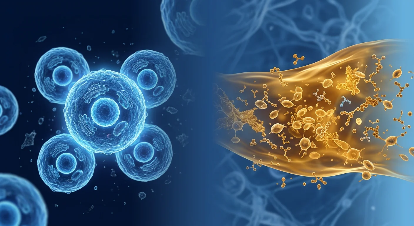
The central insight: MSCs create the environment — other therapies sustain it. When MSCs are infused intravenously or injected locally, they release a burst of bioactive molecules — cytokines, growth factors, extracellular vesicles, and microRNAs — over a period of days to weeks. This secretome initiates a cascade: inflammation is modulated, apoptotic cells are cleared, angiogenic signals are released, and resident progenitor cells are recruited. But the secretome is transient. Once the MSCs are cleared — typically within one to four weeks — the regenerative window begins to close unless it is actively maintained. [2] [3]
This is the gap that combination therapy fills. Each complementary modality has a distinct temporal profile and mechanism of action. Deployed in the right sequence, they extend and deepen the regeneration that MSCs initiate — turning a weeks-long therapeutic pulse into a months-long remodelling process.
Why combination, not competition
None of the therapies discussed in this article competes with or replaces MSC therapy. Each addresses a different layer of the regenerative process — cellular signalling, structural scaffolding, metabolic support, neuromuscular retraining — that MSCs alone cannot fully cover. The goal is not to find a single "best" therapy but to design a protocol where each component amplifies the others.
MSC + PRP: the foundational combination
Platelet-rich plasma is the most extensively studied MSC combination partner, and for good reason. PRP provides a concentrated dose of growth factors — PDGF, TGF-β, VEGF, EGF, IGF-1, and others — that creates a chemotactic gradient, drawing MSCs (both endogenous and exogenous) toward the injury site while simultaneously providing proliferative and anti-apoptotic signals that enhance their survival and function. [4]
The evidence for synergy. Preclinical models consistently show that MSCs co-delivered with PRP exhibit higher viability, enhanced proliferation, and greater differentiation capacity than MSCs delivered alone. PRP's fibrin matrix also serves as a provisional scaffold, improving cell retention at the injection site — a meaningful advantage given that MSCs injected without a carrier can disperse rapidly. [5]
Clinically, the MSC+PRP combination has shown the strongest results in orthopaedic applications: knee osteoarthritis, partial tendon tears, and cartilage defects. In one randomized trial, intra-articular MSC+PRP produced significantly greater improvements in WOMAC scores and MRI cartilage thickness at 12 months compared to MSC or PRP alone. [6]
Protocol considerations. Timing matters. The current consensus favours delivering PRP either simultaneously with MSCs (using PRP as the carrier medium) or 48–72 hours after MSC delivery — after the initial inflammatory modulation phase but while the MSC secretome is still active. Delivering PRP too early may compete with the MSC's own signalling; delivering it too late misses the synergistic window.
MSC + Exosomes: extending the paracrine reach
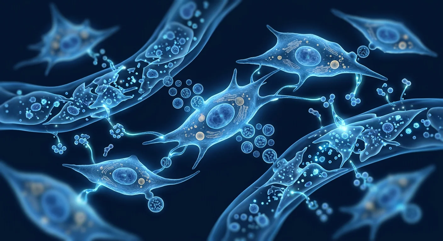
Exosomes — nano-scale extracellular vesicles shed by MSCs — are increasingly understood to carry a significant portion of MSC therapeutic activity. They package and deliver proteins, lipids, mRNA, and microRNA across biological barriers, effectively extending the reach of MSC signalling beyond what direct cell-cell contact can achieve. [7]
Why add exogenous exosomes to MSC therapy? Cultured MSCs produce exosomes as part of their natural secretome, but the quantity and composition vary with cell source, passage number, and culture conditions. Supplementing with concentrated, well-characterized exosome preparations — often derived from the same MSC source — amplifies the paracrine signal, particularly for conditions where the blood-brain barrier or other tissue barriers limit direct MSC access. [8]
The strongest evidence for MSC-exosome combination therapy currently exists in neurology — stroke, traumatic brain injury, and spinal cord injury models — where exosomes cross the blood-brain barrier far more efficiently than whole MSCs. Early clinical applications in aesthetics (skin rejuvenation, hair restoration) and orthopaedics are also emerging, though high-quality trials remain limited. [9]
Exosomes are not a replacement for MSCs
While exosomes carry a substantial fraction of MSC paracrine activity, they cannot replicate the full therapeutic repertoire of live cells — particularly sustained cytokine production, dynamic environmental sensing, and direct cell-cell immunomodulation. Exosomes complement MSCs; they do not substitute for them in most indications.
MSC + Peptide Protocols: biochemical support for tissue remodelling
Bioactive peptides — short chains of amino acids that act as signalling molecules — have gained attention as MSC adjuncts because they target specific pathways that complement MSC activity without overlapping mechanisms or competing for receptor occupancy. Several peptides have emerging evidence for use in combination with MSC therapy. [10]
BPC-157 (Body Protection Compound). A synthetic pentadecapeptide derived from gastric juice protein, BPC-157 promotes angiogenesis, accelerates fibroblast migration, and upregulates growth factor receptor expression. In musculoskeletal applications, BPC-157 combined with MSCs has shown enhanced tendon-to-bone healing in preclinical rotator cuff and Achilles models — likely by improving the local vascular environment that MSCs depend on for survival and function. [11]
Thymosin Beta-4 (Tβ4). A naturally occurring 43-amino-acid peptide, Tβ4 promotes cell migration, angiogenesis, and hair follicle stem cell activation while inhibiting apoptosis and inflammation. When administered alongside MSCs in cardiac and dermal wound models, Tβ4 enhances MSC engraftment, survival, and functional integration into host tissue. [12]
Clinical reality check. Both BPC-157 and Tβ4 remain investigative for most regenerative applications. Human data are sparse, and neither peptide is regulated as a pharmaceutical in most jurisdictions. Their use in combination protocols should be understood as an experimental adjunct — not an evidence-based standard of care. Patients considering peptide co-therapy should discuss the regulatory status, sourcing quality, and evidence limitations with their clinical team.
MSC + NAD+ and Metabolic Optimisation
Nicotinamide adenine dinucleotide (NAD+) is a coenzyme central to cellular energy metabolism, DNA repair, and sirtuin-mediated longevity pathways. NAD+ levels decline with age and chronic disease — the same contexts in which MSC therapy is most commonly sought. The hypothesis behind NAD+ supplementation as an MSC adjunct is straightforward: improving the metabolic health of the host environment may improve the conditions in which MSCs must function. [13]
Preclinical support. In vitro studies demonstrate that NAD+ precursors (nicotinamide riboside, NMN) reduce MSC senescence, enhance proliferation capacity, and preserve differentiation potential through SIRT1-dependent pathways. Aged MSCs cultured in NAD+-supplemented media show restored mitochondrial function, reduced oxidative stress markers, and improved secretome profiles — effects that are particularly relevant when using autologous (patient-derived) MSCs, which may be functionally compromised by age and disease. [14]
Practical protocol. NAD+ co-therapy is typically administered as intravenous NAD+ infusion or oral precursor supplementation (NR or NMN) beginning one to two weeks before MSC delivery and continuing for four to eight weeks after — covering the period of peak MSC activity and early tissue remodelling.
MSC + Physiotherapy & Structured Rehabilitation
Perhaps the most overlooked MSC combination partner is the one with the strongest evidence: structured rehabilitation. Cellular repair creates the biological substrate for improvement, but functional recovery requires neuromuscular retraining, progressive loading, and movement pattern correction — all of which are the domain of physiotherapy. [15]
The evidence is clear. Across orthopaedic MSC trials, the protocols that pair cell therapy with structured rehabilitation consistently outperform those that do not. In knee osteoarthritis, MSC injection plus a 12-week supervised exercise programme produced significantly greater functional gains than MSC injection alone at 6- and 12-month follow-ups. Similar patterns appear in spinal cord injury, rotator cuff repair, and cartilage restoration studies — rehabilitation acts as a force multiplier on the biological repair that MSCs provide. [16]
Phase 1: Weeks 1–2
Protection. Gentle range-of-motion, pain-free isometric activation, inflammation management. Protect the MSC secretome window while preventing deconditioning.
Phase 2: Weeks 3–6
Rebuilding. Progressive loading, neuromuscular control, movement pattern correction. Tissue remodelling begins — load drives structural adaptation.
Phase 3: Weeks 7–12+
Integration. Sport-specific or activity-specific training, full kinetic chain retraining, return-to-activity progression. Functional gains consolidate.
MSC + Nutritional Protocols: the substrate layer
Regeneration is metabolically demanding. MSCs require a rich supply of amino acids, fatty acids, vitamins, and trace minerals to sustain their synthetic activity. The host tissue — undergoing active remodelling — has similarly elevated nutritional requirements. A well-designed nutritional protocol ensures that neither the cells nor the tissue they are repairing is limited by substrate availability. [17]
Key nutritional components with evidence:
- Omega-3 fatty acids (EPA/DHA). Modulate the resolution of inflammation through specialized pro-resolving mediators (SPMs) — supporting the transition from inflammatory to regenerative phases without suppressing the initial healing signal. [18]
- Vitamin D. Modulates MSC differentiation, with adequate levels favouring osteogenic and chondrogenic lineages. Vitamin D deficiency is associated with impaired MSC function and has been linked to poorer orthopaedic regenerative outcomes.
- Collagen peptides and amino acid support. Provide the glycine, proline, and hydroxyproline that type II collagen synthesis requires — directly relevant to cartilage and connective tissue repair.
- Antioxidant support (vitamin C, N-acetylcysteine, alpha-lipoic acid). MSC culture studies show that oxidative stress impairs proliferation and promotes senescence. Systemic antioxidant status may influence the functional lifespan and secretome quality of infused MSCs.
Emerging Combinations: ozone, hyperbaric oxygen, and beyond
Several additional combination approaches have emerging preclinical or early clinical support, though definitive evidence remains limited:
Ozone therapy. Systemic ozone delivery (major autohemotherapy) has been studied as an MSC pre-treatment, with the hypothesis that controlled oxidative stress upregulates antioxidant defences and improves MSC resilience. Limited clinical data exist, but small studies in musculoskeletal and autoimmune contexts report subjective benefit when ozone is delivered before and after MSC infusion.
Hyperbaric oxygen therapy (HBOT). HBOT increases tissue oxygen tension and has been shown in preclinical models to enhance MSC proliferation, migration, and angiogenic factor secretion. In combination protocols, HBOT is typically delivered as a post-MSC adjunct — the elevated oxygen environment may improve MSC survival and function in hypoxic target tissues. Clinical evidence remains largely case-series level. [19]
Pulsed electromagnetic field (PEMF) therapy. PEMF has been shown to upregulate growth factor receptors and enhance MSC osteogenic differentiation in vitro. Its non-invasive nature makes it an attractive adjunct — though, like ozone and HBOT, the clinical evidence base for MSC-specific synergy is early-stage.
Building a coherent combination protocol: practical principles
Designing a multi-modal regenerative protocol is not about stacking every available therapy. It is about matching modalities to the specific biological needs of the condition, respecting temporal sequence, and avoiding antagonistic interactions. The principles below reflect current clinical consensus at centres that routinely combine MSC therapy with complementary modalities:
- Start with the biology of the condition. Is the primary deficit immune dysregulation, structural degeneration, vascular insufficiency, or neuromuscular dysfunction? Each points toward different combination partners.
- Respect the temporal sequence. MSC secretome peaks at 24–72 hours post-infusion. Modalities that support this phase (PRP, NAD+) should be timed to overlap with it. Modalities that translate repair into function (physiotherapy, progressive loading) should follow.
- Don't combine everything at once. Overly aggressive multi-modal protocols risk overwhelming the body's regulatory capacity, producing unpredictable inflammatory responses and making it impossible to determine which component is responsible for benefit or adverse effect.
- Source quality matters for every component. The same quality standards that apply to MSCs — cGMP manufacturing, third-party testing, sterility assurance — should guide the selection of every complementary product. The weakest link in the chain defines the overall safety profile.
- Monitor and adjust. The best protocols are iterative. Biomarker tracking, functional assessments, and patient-reported outcomes should inform adjustments to the combination over subsequent treatment cycles.
Limitations and honest disclosure
Most combination protocols discussed in this article are supported by preclinical evidence and early clinical experience — not large-scale randomized controlled trials. Direct head-to-head comparisons of different combination strategies are essentially non-existent. The recommendations here represent a synthesis of mechanistic reasoning, preclinical data, and expert clinical practice; they are not formal treatment guidelines. Patients should approach combination protocols as an evolving science, not a settled standard, and make decisions in close consultation with a qualified regenerative medicine physician.
References
- Pittenger MF, Discher DE, Péault BM, Phinney DG, Hare JM, Caplan AI. Mesenchymal stem cell perspective: cell biology to clinical progress. npj Regenerative Medicine. 2019;4:22. doi:10.1038/s41536-019-0083-6 ↩
- Caplan AI, Correa D. The MSC: an injury drugstore. Cell Stem Cell. 2011;9(1):11-15. doi:10.1016/j.stem.2011.06.008 ↩
- Galipeau J, Sensébé L. Mesenchymal stromal cells: clinical challenges and therapeutic opportunities. Cell Stem Cell. 2018;22(6):824-833. doi:10.1016/j.stem.2018.05.004 ↩
- Alsousou J, Thompson M, Hulley P, Noble A, Willett K. The biology of platelet-rich plasma and its application in trauma and orthopaedic surgery. Journal of Bone and Joint Surgery (Br). 2009;91(8):987-996. doi:10.1302/0301-620X.91B8.22546 ↩
- Qian Y, Han Q, Chen W, et al. Platelet-rich plasma derived growth factors contribute to stem cell differentiation in musculoskeletal regeneration. Frontiers in Chemistry. 2017;5:89. doi:10.3389/fchem.2017.00089 ↩
- Bastos R, Mathias M, Andrade R, et al. Intra-articular injection of culture-expanded mesenchymal stem cells with or without addition of platelet-rich plasma is effective in decreasing pain and symptoms in knee osteoarthritis. Knee Surgery, Sports Traumatology, Arthroscopy. 2020;28(6):1989-1999. doi:10.1007/s00167-019-05732-8 ↩
- Phinney DG, Pittenger MF. Concise review: MSC-derived exosomes for cell-free therapy. Stem Cells. 2017;35(4):851-858. doi:10.1002/stem.2575 ↩
- Keshtkar S, Azarpira N, Ghahremani MH. Mesenchymal stem cell-derived extracellular vesicles: novel frontiers in regenerative medicine. Stem Cell Research & Therapy. 2018;9(1):63. doi:10.1186/s13287-018-0791-7 ↩
- Xin H, Li Y, Chopp M. Exosomes/miRNAs as mediating cell-based therapy of stroke. Frontiers in Cellular Neuroscience. 2014;8:377. doi:10.3389/fncel.2014.00377 ↩
- Fosgerau K, Hoffmann T. Peptide therapeutics: current status and future directions. Drug Discovery Today. 2015;20(1):122-128. doi:10.1016/j.drudis.2014.10.003 ↩
- Chang CH, Chen CH, Su CI, et al. The effect of BPC 157 on Achilles tendon healing in a rat model. Journal of Orthopaedic Surgery and Research. 2022;17(1):315. doi:10.1186/s13018-022-03208-9 ↩
- Goldstein AL, Hannappel E, Sosne G, Kleinman HK. Thymosin β4: a multi-functional regenerative peptide. Expert Opinion on Biological Therapy. 2012;12(1):37-51. doi:10.1517/14712598.2012.634793 ↩
- Cantó C, Menzies KJ, Auwerx J. NAD+ metabolism and the control of energy homeostasis. Cell Metabolism. 2015;22(1):31-53. doi:10.1016/j.cmet.2015.05.023 ↩
- Yuan Y, Cruzat VF, Newsholme P, Cheng J, Chen Y, Lu Y. Regulation of SIRT1 in aging: roles in mitochondrial function and biogenesis. Mechanisms of Ageing and Development. 2016;155:10-21. doi:10.1016/j.mad.2016.02.003 ↩
- Kon E, Filardo G, Di Martino A, Marcacci M. Platelet-rich plasma (PRP) to treat sports injuries: evidence to support its use. Knee Surgery, Sports Traumatology, Arthroscopy. 2011;19(4):516-527. doi:10.1007/s00167-010-1306-y ↩
- Vega A, Martín-Ferrero MA, Del Canto F, et al. Treatment of knee osteoarthritis with allogeneic bone marrow mesenchymal stem cells: a randomized controlled trial. Transplantation. 2015;99(8):1681-1690. doi:10.1097/TP.0000000000000678 ↩
- Muschler GF, Nakamoto C, Griffith LG. Engineering principles of clinical cell-based tissue engineering. Journal of Bone and Joint Surgery (Am). 2004;86(7):1541-1558. doi:10.2106/00004623-200407000-00029 ↩
- Serhan CN, Chiang N, Dalli J. The resolution code of acute inflammation: novel pro-resolving lipid mediators in resolution. Seminars in Immunology. 2015;27(3):200-215. doi:10.1016/j.smim.2015.03.004 ↩
- Thom SR. Hyperbaric oxygen: its mechanisms and efficacy. Plastic and Reconstructive Surgery. 2011;127(Suppl 1):131S-141S. doi:10.1097/PRS.0b013e3181fbe2bf ↩
间充质干细胞疗法在再生医学领域已通过越来越多的证据确立了地位——但最显著的治疗结果很少来自MSC单独使用。那些报告最显著、最持久改善的患者,通常接受了整合互补疗法的治疗方案:PRP放大局部愈合信号,外泌体延长旁分泌活性,肽类方案支持组织重塑,结构化康复将细胞修复转化为功能提升。本文探讨每种组合背后的证据及使"协同"超越营销口号范畴的生物学原理。[1]
核心洞察:MSC创造环境——其他疗法维持环境。当MSC通过静脉输注或局部注射进入人体后,会在数天至数周内释放大量生物活性分子——细胞因子、生长因子、细胞外囊泡和microRNA。这种分泌组启动了一个级联反应:炎症得到调节,凋亡细胞被清除,血管生成信号被释放,常驻祖细胞被招募。但分泌组是暂时的。一旦MSC被清除——通常在一至四周内——再生窗口便开始关闭,除非得到积极维持。[2] [3]
这正是联合疗法填补的空白。每种互补疗法都有独特的时间特征和作用机制。按正确的时间序列部署,它们可以延长和深化MSC启动的再生过程——将数周的治疗脉冲转变为持续数月的组织重塑过程。
为何是组合而非竞争
本文讨论的任何疗法都不会与MSC疗法竞争或取代它。每种疗法针对再生过程的不同层面——细胞信号传导、结构支架、代谢支持、神经肌肉再训练——这些都是MSC单独无法完全覆盖的。目标不是找到单一的"最佳"疗法,而是设计一种协议,使每个组成部分放大其他部分的效果。
MSC + PRP:基础组合
富血小板血浆是研究最深入的MSC联合伙伴,这是有充分理由的。PRP提供浓缩的生长因子——PDGF、TGF-β、VEGF、EGF、IGF-1等——产生趋化梯度,将MSC(内源性和外源性)引导至损伤部位,同时提供增强其存活和功能的增殖和抗凋亡信号。[4]
协同作用的证据。临床前模型一致表明,与PRP共同递送的MSC比单独递送的MSC表现出更高的存活率、增强的增殖能力和更大的分化能力。PRP的纤维蛋白基质还可作为临时支架,改善注射部位的细胞保留——鉴于无载体注射的MSC可迅速分散,这是一个有意义的优势。[5]
临床上,MSC+PRP组合在骨科应用中显示出最强的效果:膝骨关节炎、部分肌腱撕裂和软骨缺损。在一项随机试验中,关节内MSC+PRP在12个月时的WOMAC评分和MRI软骨厚度方面比单独使用MSC或PRP产生了显著更大的改善。[6]
方案考虑。时机至关重要。目前的共识倾向于在MSC递送的同时(使用PRP作为载体介质)或在MSC递送后48-72小时——即初始炎症调节阶段之后、MSC分泌组仍然活跃之时——递送PRP。过早递送PRP可能与MSC自身的信号传导竞争;过晚递送则错过协同窗口。
MSC + 外泌体:扩展旁分泌范围
外泌体——MSC脱落的纳米级细胞外囊泡——越来越被认为承载了MSC治疗活性的重要部分。它们包装和递送蛋白质、脂质、mRNA和microRNA穿越生物屏障,有效扩展了MSC信号传导的范围,超越了直接细胞-细胞接触所能达到的程度。[7]
为什么要向MSC疗法添加外源外泌体?培养的MSC在其自然分泌组中产生外泌体,但数量和质量因细胞来源、传代次数和培养条件而异。补充浓缩的、经过良好表征的外泌体制剂——通常源自相同的MSC来源——可以放大旁分泌信号,特别适用于血脑屏障或其他组织屏障限制MSC直接进入的疾病。[8]
MSC-外泌体联合疗法的最有力证据目前存在于神经病学领域——卒中、创伤性脑损伤和脊髓损伤模型——在这些领域中,外泌体穿越血脑屏障的效率远高于完整MSC。美容(皮肤年轻化、毛发修复)和骨科领域的早期临床应用也在涌现,尽管高质量试验仍然有限。[9]
外泌体不是MSC的替代品
虽然外泌体承载了MSC旁分泌活性的重要部分,但它们无法复制活细胞的全部治疗功能——特别是持续的细胞因子产生、动态环境感知和直接细胞-细胞免疫调节。外泌体补充MSC;在大多数适应症中,它们不能替代MSC。
MSC + 肽类方案:组织重塑的生化支持
生物活性肽——作为信号分子的短链氨基酸——作为MSC辅助手段已获得关注,因为它们靶向特定的途径,补充MSC活性而不会机制重叠或竞争受体占用。几种肽在MSC联合治疗中具有新兴证据。[10]
BPC-157(身体保护化合物)。一种源自胃液蛋白的合成十五肽,BPC-157促进血管生成,加速成纤维细胞迁移,并上调生长因子受体表达。在肌肉骨骼应用中,BPC-157联合MSC在临床前肩袖和跟腱模型中显示出增强的肌腱-骨愈合——可能是通过改善MSC存活和功能所需的局部血管环境。[11]
胸腺素β4(Tβ4)。一种天然存在的43个氨基酸肽,Tβ4促进细胞迁移、血管生成和毛囊干细胞活化,同时抑制凋亡和炎症。在心脏和皮肤创伤模型中与MSC同时给药时,Tβ4增强MSC植入、存活和功能性整合入宿主组织。[12]
临床现实检查。BPC-157和Tβ4在大多数再生应用中仍处于研究阶段。人类数据稀少,这两种肽在大多数司法管辖区均未作为药品受到监管。它们在组合方案中的使用应被理解为实验性辅助手段——而非循证标准治疗。考虑肽联合治疗的患者应与临床团队讨论监管状态、来源质量和证据局限性。
MSC + NAD+与代谢优化
烟酰胺腺嘌呤二核苷酸(NAD+)是细胞能量代谢、DNA修复和sirtuin介导的长寿途径中核心的辅酶。NAD+水平随年龄和慢性疾病而下降——这正是MSC疗法最常见的应用背景。NAD+补充作为MSC辅助手段的假设很直接:改善宿主环境的代谢健康可能改善MSC必须发挥功能的条件。[13]
临床前支持。体外研究表明,NAD+前体(烟酰胺核苷、NMN)通过SIRT1依赖性途径减少MSC衰老、增强增殖能力并保持分化潜能。在NAD+补充培养基中培养的衰老MSC显示出恢复的线粒体功能、减少的氧化应激标志物和改善的分泌组谱——这些效果在使用自体(患者来源)MSC时尤为相关,因为这些细胞可能因年龄和疾病而功能受损。[14]
实用方案。NAD+联合疗法通常在MSC递送前一到两周开始以静脉NAD+输注或口服前体补充(NR或NMN)的形式给药,并持续四到八周——覆盖MSC活性峰值和早期组织重塑期。
MSC + 物理治疗与结构化康复
也许最被忽视的MSC联合伙伴是拥有最强证据的那一个:结构化康复。细胞修复创造了改善的生物基质,但功能恢复需要神经肌肉再训练、渐进负荷和运动模式纠正——所有这些都属于物理治疗的领域。[15]
证据是明确的。在骨科MSC试验中,将细胞疗法与结构化康复配对的方案始终优于不配对的方案。在膝骨关节炎中,MSC注射加12周监督运动计划在6个月和12个月随访时产生的功能提升显著大于单独MSC注射。类似的模式出现在脊髓损伤、肩袖修复和软骨修复研究中——康复作为MSC提供的生物修复的力量倍增器发挥作用。[16]
第1阶段:第1–2周
保护。温和的活动范围训练、无痛等长激活、炎症管理。保护MSC分泌组窗口同时防止功能退化。
第2阶段:第3–6周
重建。渐进负荷、神经肌肉控制、运动模式纠正。组织重塑开始——负荷驱动结构适应。
第3阶段:第7–12+周
整合。运动专项或活动专项训练、完整动力学链再训练、回归活动进程。功能提升巩固。
MSC + 营养方案:基质层
再生是代谢需求密集的过程。MSC需要丰富的氨基酸、脂肪酸、维生素和微量矿物质供应以维持其合成活性。宿主组织——处于主动重塑中——同样具有升高的营养需求。精心设计的营养方案确保无论是细胞还是它们正在修复的组织都不受基质可用性的限制。[17]
具有证据的关键营养成分:
- Omega-3脂肪酸(EPA/DHA)。通过专门的促消退介质(SPM)调节炎症消退——支持从炎症阶段向再生阶段的过渡而不抑制初始愈合信号。[18]
- 维生素D。调节MSC分化,充足水平有利于成骨和软骨形成谱系。维生素D缺乏与MSC功能受损相关,并与较差的骨科再生结果有关。
- 胶原蛋白肽和氨基酸支持。提供II型胶原合成所需的甘氨酸、脯氨酸和羟脯氨酸——与软骨和结缔组织修复直接相关。
- 抗氧化支持(维生素C、N-乙酰半胱氨酸、α-硫辛酸)。MSC培养研究表明氧化应激损害增殖并促进衰老。全身抗氧化状态可能影响输注MSC的功能寿命和分泌组质量。
新兴组合:臭氧、高压氧及其他
几种额外的联合方法拥有新兴的临床前或早期临床支持,尽管明确的证据仍然有限:
臭氧疗法。全身臭氧递送(主要自血疗法)已被研究作为MSC预处理,假设受控氧化应激上调抗氧化防御并改善MSC韧性。临床数据有限,但在肌肉骨骼和自身免疫背景下的小型研究报告了在MSC输注前后给予臭氧时的主观获益。
高压氧疗法(HBOT)。HBOT增加组织氧张力,临床前模型显示其可增强MSC增殖、迁移和血管生成因子分泌。在联合方案中,HBOT通常作为MSC后的辅助手段——升高的氧环境可能改善MSC在缺氧靶组织中的存活和功能。临床证据主要仍为病例系列级别。[19]
脉冲电磁场(PEMF)疗法。PEMF在体外已被证明可上调生长因子受体并增强MSC成骨分化。其无创性质使其成为有吸引力的辅助手段——尽管与臭氧和HBOT一样,MSC特异性协同作用的临床证据基础仍处于早期阶段。
构建协调的组合方案:实用原则
设计多模式再生方案不是堆叠所有可用疗法。它是将疗法模式与疾病的特定生物学需求相匹配,尊重时间序列,避免拮抗性相互作用。以下原则反映了常规将MSC疗法与互补疗法联合使用的中心的当前临床共识:
- 从疾病的生物学开始。主要缺陷是免疫失调、结构变性、血管功能不全还是神经肌肉功能障碍?每种指向不同的联合伙伴。
- 尊重时间序列。MSC分泌组在输注后24-72小时达到峰值。支持这一阶段的疗法(PRP、NAD+)应安排在此期间。将修复转化为功能的疗法(物理治疗、渐进负荷)应在之后进行。
- 不要一次组合所有东西。过于激进的多模式方案可能压倒身体的调节能力,产生不可预测的炎症反应,并使确定哪个组成部分负责获益或不良效应变得不可能。
- 每个组成部分的来源质量都很重要。适用于MSC的相同质量标准——cGMP制造、第三方检测、无菌保证——应指导每种互补产品的选择。链条中最薄弱的环节决定了整体安全性。
- 监测并调整。最好的方案是迭代的。生物标志物追踪、功能评估和患者报告的结果应为后续治疗周期中的组合调整提供信息。
局限性与诚实披露
本文讨论的大多数组合方案由临床前证据和早期临床经验支持——而非大规模随机对照试验。不同组合策略的直接头对头比较基本上不存在。本文的建议代表了机制推理、临床前数据和专家临床实践的综合;它们不是正式的治疗指南。患者应将组合方案视为一门不断发展的科学,而非已确定的标准,并在与合格的再生医学医师密切咨询下做出决定。
参考文献
- Pittenger MF, Discher DE, Péault BM, Phinney DG, Hare JM, Caplan AI. Mesenchymal stem cell perspective: cell biology to clinical progress. npj Regenerative Medicine. 2019;4:22. doi:10.1038/s41536-019-0083-6 ↩
- Caplan AI, Correa D. The MSC: an injury drugstore. Cell Stem Cell. 2011;9(1):11-15. doi:10.1016/j.stem.2011.06.008 ↩
- Galipeau J, Sensébé L. Mesenchymal stromal cells: clinical challenges and therapeutic opportunities. Cell Stem Cell. 2018;22(6):824-833. doi:10.1016/j.stem.2018.05.004 ↩
- Alsousou J, Thompson M, Hulley P, Noble A, Willett K. The biology of platelet-rich plasma and its application in trauma and orthopaedic surgery. Journal of Bone and Joint Surgery (Br). 2009;91(8):987-996. doi:10.1302/0301-620X.91B8.22546 ↩
- Qian Y, Han Q, Chen W, et al. Platelet-rich plasma derived growth factors contribute to stem cell differentiation in musculoskeletal regeneration. Frontiers in Chemistry. 2017;5:89. doi:10.3389/fchem.2017.00089 ↩
- Bastos R, Mathias M, Andrade R, et al. Intra-articular injection of culture-expanded mesenchymal stem cells with or without addition of platelet-rich plasma is effective in decreasing pain and symptoms in knee osteoarthritis. Knee Surgery, Sports Traumatology, Arthroscopy. 2020;28(6):1989-1999. doi:10.1007/s00167-019-05732-8 ↩
- Phinney DG, Pittenger MF. Concise review: MSC-derived exosomes for cell-free therapy. Stem Cells. 2017;35(4):851-858. doi:10.1002/stem.2575 ↩
- Keshtkar S, Azarpira N, Ghahremani MH. Mesenchymal stem cell-derived extracellular vesicles: novel frontiers in regenerative medicine. Stem Cell Research & Therapy. 2018;9(1):63. doi:10.1186/s13287-018-0791-7 ↩
- Xin H, Li Y, Chopp M. Exosomes/miRNAs as mediating cell-based therapy of stroke. Frontiers in Cellular Neuroscience. 2014;8:377. doi:10.3389/fncel.2014.00377 ↩
- Fosgerau K, Hoffmann T. Peptide therapeutics: current status and future directions. Drug Discovery Today. 2015;20(1):122-128. doi:10.1016/j.drudis.2014.10.003 ↩
- Chang CH, Chen CH, Su CI, et al. The effect of BPC 157 on Achilles tendon healing in a rat model. Journal of Orthopaedic Surgery and Research. 2022;17(1):315. doi:10.1186/s13018-022-03208-9 ↩
- Goldstein AL, Hannappel E, Sosne G, Kleinman HK. Thymosin β4: a multi-functional regenerative peptide. Expert Opinion on Biological Therapy. 2012;12(1):37-51. doi:10.1517/14712598.2012.634793 ↩
- Cantó C, Menzies KJ, Auwerx J. NAD+ metabolism and the control of energy homeostasis. Cell Metabolism. 2015;22(1):31-53. doi:10.1016/j.cmet.2015.05.023 ↩
- Yuan Y, Cruzat VF, Newsholme P, Cheng J, Chen Y, Lu Y. Regulation of SIRT1 in aging: roles in mitochondrial function and biogenesis. Mechanisms of Ageing and Development. 2016;155:10-21. doi:10.1016/j.mad.2016.02.003 ↩
- Kon E, Filardo G, Di Martino A, Marcacci M. Platelet-rich plasma (PRP) to treat sports injuries: evidence to support its use. Knee Surgery, Sports Traumatology, Arthroscopy. 2011;19(4):516-527. doi:10.1007/s00167-010-1306-y ↩
- Vega A, Martín-Ferrero MA, Del Canto F, et al. Treatment of knee osteoarthritis with allogeneic bone marrow mesenchymal stem cells: a randomized controlled trial. Transplantation. 2015;99(8):1681-1690. doi:10.1097/TP.0000000000000678 ↩
- Muschler GF, Nakamoto C, Griffith LG. Engineering principles of clinical cell-based tissue engineering. Journal of Bone and Joint Surgery (Am). 2004;86(7):1541-1558. doi:10.2106/00004623-200407000-00029 ↩
- Serhan CN, Chiang N, Dalli J. The resolution code of acute inflammation: novel pro-resolving lipid mediators in resolution. Seminars in Immunology. 2015;27(3):200-215. doi:10.1016/j.smim.2015.03.004 ↩
- Thom SR. Hyperbaric oxygen: its mechanisms and efficacy. Plastic and Reconstructive Surgery. 2011;127(Suppl 1):131S-141S. doi:10.1097/PRS.0b013e3181fbe2bf ↩
حصل العلاج بالخلايا الجذعية الوسيطة (MSC) على مكانته في الطب التجديدي من خلال مجموعة متنامية من الأدلة — لكن النتائج الأكثر إقناعاً نادراً ما تأتي من الخلايا الجذعية الوسيطة وحدها. المرضى الذين يبلغون عن التحسينات الأكثر جوهرية واستدامة هم غالباً أولئك الذين يدمج علاجهم طرائق تكميلية: PRP لتضخيم إشارة الشفاء المحلية، والإكسوسومات لتمديد النشاط الباراكريني، وبروتوكولات الببتيد لدعم إعادة تشكيل الأنسجة، وإعادة التأهيل المنظم لترجمة الإصلاح الخلوي إلى مكاسب وظيفية. تفحص هذه المقالة الأدلة وراء كل تركيبة والأساس البيولوجي الذي يجعل التآزر أكثر من مجرد ادعاء تسويقي. [1]
الرؤية المركزية: الخلايا الجذعية الوسيطة تخلق البيئة — والعلاجات الأخرى تحافظ عليها. عندما يتم حقن الخلايا الجذعية الوسيطة وريدياً أو موضعياً، فإنها تطلق دفعة من الجزيئات النشطة بيولوجياً — السيتوكينات وعوامل النمو والحويصلات خارج الخلية والـ microRNA — على مدى أيام إلى أسابيع. يبدأ هذا السيكريتوم سلسلة: يتم تعديل الالتهاب، وتصفية الخلايا المبرمجة للموت، وإطلاق إشارات تكوين الأوعية الدموية، وتجنيد الخلايا السلفية المقيمة. لكن السيكريتوم مؤقت. بمجرد إزالة الخلايا الجذعية الوسيطة — عادة في غضون أسبوع إلى أربعة أسابيع — تبدأ نافذة التجديد في الإغلاق ما لم يتم الحفاظ عليها بنشاط. [2] [3]
هذه هي الفجوة التي يملؤها العلاج المركب. كل طريقة تكميلية لها ملف زمني مميز وآلية عمل. عند نشرها بالتسلسل الصحيح، فإنها تمتد وتعمق التجديد الذي تبدأه الخلايا الجذعية الوسيطة — محولة النبضة العلاجية التي تدوم أسابيع إلى عملية إعادة تشكيل تستمر لأشهر.
لماذا التركيب وليس المنافسة
لا يتنافس أي من العلاجات التي تمت مناقشتها في هذه المقالة مع علاج الخلايا الجذعية الوسيطة أو يحل محله. كل منها يعالج طبقة مختلفة من عملية التجديد — الإشارات الخلوية، السقالات الهيكلية، الدعم الأيضي، إعادة التدريب العصبي العضلي — التي لا تستطيع الخلايا الجذعية الوسيطة وحدها تغطيتها بالكامل. الهدف ليس إيجاد علاج واحد "أفضل" بل تصميم بروتوكول يضخم فيه كل مكون الآخرين.
الخلايا الجذعية الوسيطة + PRP: التركيبة الأساسية
البلازما الغنية بالصفائح الدموية هي الشريك المركب الأكثر دراسة مع الخلايا الجذعية الوسيطة، وذلك لسبب وجيه. يوفر PRP جرعة مركزة من عوامل النمو — PDGF و TGF-β و VEGF و EGF و IGF-1 وغيرها — مما يخلق تدرجاً كيميائياً جاذباً، يسحب الخلايا الجذعية الوسيطة (الداخلية والخارجية) نحو موقع الإصابة مع توفير إشارات تكاثرية ومضادة للاستماتة تعزز بقائها ووظيفتها. [4]
أدلة التآزر. تظهر النماذج قبل السريرية باستمرار أن الخلايا الجذعية الوسيطة التي يتم توصيلها مع PRP تظهر قابلية أعلى للبقاء، وتكاثراً معززاً، وقدرة تمايز أكبر من الخلايا الجذعية الوسيطة التي يتم توصيلها وحدها. تعمل مصفوفة الفيبرين في PRP أيضاً كسقالة مؤقتة، محسنة احتفاظ الخلايا في موقع الحقن — وهي ميزة ذات معنى بالنظر إلى أن الخلايا الجذعية الوسيطة المحقونة بدون حامل يمكن أن تتشتت بسرعة. [5]
سريرياً، أظهرت تركيبة MSC+PRP أقوى النتائج في تطبيقات العظام: التهاب مفاصل الركبة، وتمزقات الأوتار الجزئية، وعيوب الغضاريف. في إحدى التجارب العشوائية، أنتج الحقن داخل المفصل بـ MSC+PRP تحسينات أكبر بكثير في درجات WOMAC وسماكة غضروف الرنين المغناطيسي عند 12 شهراً مقارنة بـ MSC أو PRP وحدهما. [6]
اعتبارات البروتوكول. التوقيت مهم. يفضل الإجماع الحالي توصيل PRP إما بالتزامن مع الخلايا الجذعية الوسيطة (باستخدام PRP كوسط ناقل) أو بعد 48-72 ساعة من توصيل الخلايا الجذعية الوسيطة — بعد مرحلة التعديل الالتهابي الأولية ولكن بينما لا يزال سيكريتوم الخلايا الجذعية الوسيطة نشطاً. توصيل PRP مبكراً جداً قد يتنافس مع إشارات الخلايا الجذعية الوسيطة نفسها؛ توصيله متأخراً جداً يفقد نافذة التآزر.
الخلايا الجذعية الوسيطة + الإكسوسومات: توسيع النطاق الباراكريني
الإكسوسومات — الحويصلات خارج الخلية بحجم النانو التي تفرزها الخلايا الجذعية الوسيطة — يُفهم بشكل متزايد أنها تحمل جزءاً كبيراً من النشاط العلاجي للخلايا الجذعية الوسيطة. إنها تغلف وتوصل البروتينات والدهون والـ mRNA والـ microRNA عبر الحواجز البيولوجية، موسعة بشكل فعال نطاق إشارات الخلايا الجذعية الوسيطة beyond ما يمكن أن يحققه الاتصال المباشر بين الخلايا. [7]
لماذا نضيف الإكسوسومات الخارجية إلى علاج الخلايا الجذعية الوسيطة؟ تنتج الخلايا الجذعية الوسيطة المستزرعة الإكسوسومات كجزء من سيكريتومها الطبيعي، لكن الكمية والتركيب يختلفان حسب مصدر الخلية ورقم المرور وظروف الاستزراع. المكملات بمستحضرات إكسوسوم مركزة ومحددة جيداً — غالباً مشتقة من نفس مصدر الخلايا الجذعية الوسيطة — تضخم الإشارة الباراكرينية، خاصة للحالات التي يحد فيها الحاجز الدموي الدماغي أو حواجز الأنسجة الأخرى من وصول الخلايا الجذعية الوسيطة المباشر. [8]
أقوى دليل على العلاج المركب بالخلايا الجذعية الوسيطة والإكسوسومات موجود حالياً في طب الأعصاب — نماذج السكتة الدماغية وإصابات الدماغ الرضحية وإصابات الحبل الشوكي — حيث تعبر الإكسوسومات الحاجز الدموي الدماغي بكفاءة أكبر بكثير من الخلايا الجذعية الوسيطة الكاملة. التطبيقات السريرية المبكرة في التجميل (تجديد البشرة، استعادة الشعر) وجراحة العظام تظهر أيضاً، رغم أن التجارب عالية الجودة لا تزال محدودة. [9]
الإكسوسومات ليست بديلاً عن الخلايا الجذعية الوسيطة
بينما تحمل الإكسوسومات جزءاً كبيراً من النشاط الباراكريني للخلايا الجذعية الوسيطة، إلا أنها لا تستطيع تكرار الذخيرة العلاجية الكاملة للخلايا الحية — خاصة إنتاج السيتوكين المستدام، والاستشعار البيئي الديناميكي، والتعديل المناعي المباشر بين الخلايا. الإكسوسومات تكمل الخلايا الجذعية الوسيطة؛ ولا تحل محلها في معظم المؤشرات.
الخلايا الجذعية الوسيطة + بروتوكولات الببتيد: الدعم البيوكيميائي لإعادة تشكيل الأنسجة
الببتيدات النشطة بيولوجياً — سلاسل قصيرة من الأحماض الأمينية تعمل كجزيئات إشارة — اكتسبت اهتماماً كمساعدات للخلايا الجذعية الوسيطة لأنها تستهدف مسارات محددة تكمل نشاط الخلايا الجذعية الوسيطة دون تداخل آليات أو تنافس على شغل المستقبلات. العديد من الببتيدات لديها أدلة ناشئة للاستخدام في العلاج المركب مع الخلايا الجذعية الوسيطة. [10]
BPC-157 (مركب حماية الجسم). بنتابيبتيد اصطناعي مشتق من بروتين العصارة المعدية، يعزز BPC-157 تكوين الأوعية الدموية، ويسرع هجرة الخلايا الليفية، ويرفع من تعبير مستقبلات عامل النمو. في تطبيقات العضلات والعظام، أظهر BPC-157 المدمج مع الخلايا الجذعية الوسيطة شفاءً معززاً للوتر-العظم في نماذج الكفة المدورة ووتر أخيل قبل السريرية — على الأرجح من خلال تحسين البيئة الوعائية المحلية التي تعتمد عليها الخلايا الجذعية الوسيطة للبقاء والوظيفة. [11]
ثيموسين بيتا-4 (Tβ4). ببتيد طبيعي مكون من 43 حمضاً أمينياً، يعزز Tβ4 هجرة الخلايا، وتكوين الأوعية الدموية، وتنشيط الخلايا الجذعية لبصيلات الشعر مع تثبيط الاستماتة والالتهاب. عند إعطائه جنباً إلى جنب مع الخلايا الجذعية الوسيطة في نماذج الجروح القلبية والجلدية، يعزز Tβ4 انغراس الخلايا الجذعية الوسيطة وبقائها واندماجها الوظيفي في الأنسجة المضيفة. [12]
فحص الواقع السريري. يبقى كل من BPC-157 و Tβ4 قيد البحث لمعظم تطبيقات التجديد. البيانات البشرية متناثرة، ولا يتم تنظيم أي من الببتيدين كدواء صيدلاني في معظم الولايات القضائية. يجب فهم استخدامها في بروتوكولات التركيب كمساعد تجريبي — وليس معيار رعاية قائم على الأدلة. يجب على المرضى الذين يفكرون في العلاج المشترك بالببتيد مناقشة الوضع التنظيمي، وجودة المصدر، ومحدودية الأدلة مع فريقهم السريري.
الخلايا الجذعية الوسيطة + NAD+ والتحسين الأيضي
نيكوتيناميد أدينين ثنائي النوكليوتيد (NAD+) هو أنزيم مساعد مركزي في استقلاب الطاقة الخلوية، وإصلاح الحمض النووي، ومسارات طول العمر بوساطة السيرتوين. تنخفض مستويات NAD+ مع التقدم في العمر والأمراض المزمنة — وهي نفس السياقات التي يُطلب فيها علاج الخلايا الجذعية الوسيطة بشكل شائع. الفرضية وراء مكملات NAD+ كمساعد للخلايا الجذعية الوسيطة مباشرة: تحسين الصحة الأيضية للبيئة المضيفة قد يحسن الظروف التي يجب أن تعمل فيها الخلايا الجذعية الوسيطة. [13]
الدعم قبل السريري. تظهر الدراسات المخبرية أن سلائف NAD+ (نيكوتيناميد ريبوسيد، NMN) تقلل من شيخوخة الخلايا الجذعية الوسيطة، وتعزز قدرة التكاثر، وتحافظ على إمكانية التمايز من خلال مسارات تعتمد على SIRT1. تظهر الخلايا الجذعية الوسيطة المسنة المستزرعة في وسط مكمل بـ NAD+ وظيفة ميتوكوندريا مستعادة، وعلامات إجهاد مؤكسد منخفضة، وملامح سيكريتوم محسنة — وهي تأثيرات ذات صلة خاصة عند استخدام الخلايا الجذعية الوسيطة الذاتية (المشتقة من المريض)، والتي قد تكون معطوبة وظيفياً بسبب العمر والمرض. [14]
البروتوكول العملي. يُعطى العلاج المشترك بـ NAD+ عادة كتسريب وريدي لـ NAD+ أو مكملات سلائف فموية (NR أو NMN) تبدأ قبل أسبوع إلى أسبوعين من توصيل الخلايا الجذعية الوسيطة وتستمر لأربعة إلى ثمانية أسابيع بعد ذلك — مغطية فترة ذروة نشاط الخلايا الجذعية الوسيطة وإعادة تشكيل الأنسجة المبكرة.
الخلايا الجذعية الوسيطة + العلاج الطبيعي وإعادة التأهيل المنظم
ربما يكون الشريك المركب الأكثر تجاهلاً مع الخلايا الجذعية الوسيطة هو الذي يمتلك أقوى الأدلة: إعادة التأهيل المنظم. الإصلاح الخلوي يخلق الركيزة البيولوجية للتحسين، لكن التعافي الوظيفي يتطلب إعادة تدريب عصبي عضلي، وتحميل تدريجي، وتصحيح أنماط الحركة — وكلها من اختصاص العلاج الطبيعي. [15]
الأدلة واضحة. عبر تجارب الخلايا الجذعية الوسيطة في جراحة العظام، تتفوق البروتوكولات التي تقرن العلاج الخلوي بإعادة التأهيل المنظم باستمرار على تلك التي لا تفعل ذلك. في التهاب مفاصل الركبة، أنتج حقن الخلايا الجذعية الوسيطة بالإضافة إلى برنامج تمارين مراقب لمدة 12 أسبوعاً مكاسب وظيفية أكبر بكثير من حقن الخلايا الجذعية الوسيطة وحدها عند متابعة 6 و 12 شهراً. تظهر أنماط مماثلة في دراسات إصابة الحبل الشوكي، وإصلاح الكفة المدورة، واستعادة الغضاريف — تعمل إعادة التأهيل كمضاعف قوة على الإصلاح البيولوجي الذي توفره الخلايا الجذعية الوسيطة. [16]
المرحلة 1: الأسابيع 1–2
الحماية. مدى حركة لطيف، تنشيط متساوي القياس غير مؤلم، إدارة الالتهاب. حماية نافذة سيكريتوم الخلايا الجذعية الوسيطة مع منع تدهور الحالة.
المرحلة 2: الأسابيع 3–6
إعادة البناء. تحميل تدريجي، تحكم عصبي عضلي، تصحيح أنماط الحركة. يبدأ إعادة تشكيل الأنسجة — التحميل يدفع التكيف الهيكلي.
المرحلة 3: الأسابيع 7–12+
الدمج. تدريب خاص بالرياضة أو النشاط، إعادة تدريب السلسلة الحركية الكاملة، تقدم العودة للنشاط. تترسخ المكاسب الوظيفية.
الخلايا الجذعية الوسيطة + البروتوكولات الغذائية: طبقة الركيزة
التجديد متطلب أيضياً. تحتاج الخلايا الجذعية الوسيطة إلى إمداد غني بالأحماض الأمينية والأحماض الدهنية والفيتامينات والمعادن النزرة للحفاظ على نشاطها التخليقي. الأنسجة المضيفة — التي تخضع لإعادة تشكيل نشطة — لديها متطلبات غذائية مرتفعة بالمثل. يضمن البروتوكول الغذائي المصمم جيداً ألا تكون الخلايا ولا الأنسجة التي تقوم بإصلاحها محدودة بتوفر الركيزة. [17]
المكونات الغذائية الرئيسية مع الأدلة:
- أحماض أوميغا-3 الدهنية (EPA/DHA). تعدل حل الالتهاب من خلال وسطاء الحل المتخصصين (SPMs) — داعمة الانتقال من المراحل الالتهابية إلى التجديدية دون تثبيط إشارة الشفاء الأولية. [18]
- فيتامين د. يعدل تمايز الخلايا الجذعية الوسيطة، مع تفضيل المستويات الكافية للنسل العظمي والغضروفي. يرتبط نقص فيتامين د بضعف وظيفة الخلايا الجذعية الوسيطة وتم ربطه بنتائج تجديدية أسوأ في جراحة العظام.
- ببتيدات الكولاجين ودعم الأحماض الأمينية. توفر الجلايسين والبرولين والهيدروكسي برولين التي يتطلبها تخليق الكولاجين من النوع الثاني — ذات صلة مباشرة بإصلاح الغضاريف والأنسجة الضامة.
- الدعم المضاد للأكسدة (فيتامين ج، N-أسيتيل سيستئين، حمض ألفا-ليبويك). تظهر دراسات استزراع الخلايا الجذعية الوسيطة أن الإجهاد التأكسدي يضعف التكاثر ويعزز الشيخوخة. قد تؤثر حالة مضادات الأكسدة الجهازية على العمر الوظيفي وجودة السيكريتوم للخلايا الجذعية الوسيطة المحقونة.
التركيبات الناشئة: الأوزون، والأكسجين عالي الضغط، وما بعده
العديد من النهج المركبة الإضافية لديها دعم سريري مبكر أو قبل سريري ناشئ، رغم أن الأدلة القاطعة لا تزال محدودة:
العلاج بالأوزون. تمت دراسة توصيل الأوزون الجهازي (العلاج الذاتي الرئيسي) كمعالجة مسبقة للخلايا الجذعية الوسيطة، بفرضية أن الإجهاد التأكسدي المسيطر عليه يرفع من الدفاعات المضادة للأكسدة ويحسن مرونة الخلايا الجذعية الوسيطة. توجد بيانات سريرية محدودة، لكن الدراسات الصغيرة في السياقات العضلية الهيكلية والمناعية الذاتية تبلغ عن فائدة ذاتية عند إعطاء الأوزون قبل وبعد تسريب الخلايا الجذعية الوسيطة.
العلاج بالأكسجين عالي الضغط (HBOT). يزيد HBOT من توتر الأكسجين في الأنسجة وقد ثبت في النماذج قبل السريرية أنه يعزز تكاثر الخلايا الجذعية الوسيطة وهجرتها وإفراز عامل تكوين الأوعية الدموية. في بروتوكولات التركيب، يتم توصيل HBOT عادة كمساعد بعد الخلايا الجذعية الوسيطة — قد تحسن بيئة الأكسجين المرتفعة بقاء ووظيفة الخلايا الجذعية الوسيطة في الأنسجة المستهدفة ناقصة الأكسجين. تبقى الأدلة السريرية بشكل رئيسي على مستوى سلاسل الحالات. [19]
العلاج بالمجال الكهرومغناطيسي النبضي (PEMF). ثبت أن PEMF يرفع من تعبير مستقبلات عامل النمو ويعزز التمايز العظمي للخلايا الجذعية الوسيطة في المختبر. طبيعته غير الغازية تجعله مساعداً جذاباً — رغم أن قاعدة الأدلة السريرية للتآزر الخاص بالخلايا الجذعية الوسيطة، كما هو الحال مع الأوزون و HBOT، لا تزال في مرحلة مبكرة.
بناء بروتوكول تركيبي متماسك: مبادئ عملية
تصميم بروتوكول تجديدي متعدد الوسائط لا يتعلق بتكديس كل علاج متاح. إنه يتعلق بمطابقة الطرائق مع الاحتياجات البيولوجية المحددة للحالة، واحترام التسلسل الزمني، وتجنب التفاعلات المتضادة. تعكس المبادئ أدناه الإجماع السريري الحالي في المراكز التي تجمع بشكل روتيني بين علاج الخلايا الجذعية الوسيطة والطرائق التكميلية:
- ابدأ ببيولوجيا الحالة. هل العجز الأساسي هو خلل في تنظيم المناعة، أم تنكس هيكلي، أم قصور وعائي، أم خلل عصبي عضلي؟ كل منها يشير إلى شركاء تركيبيين مختلفين.
- احترم التسلسل الزمني. يبلغ سيكريتوم الخلايا الجذعية الوسيطة ذروته عند 24-72 ساعة بعد التسريب. يجب توقيت الطرائق التي تدعم هذه المرحلة (PRP، NAD+) لتتداخل معها. الطرائق التي تترجم الإصلاح إلى وظيفة (العلاج الطبيعي، التحميل التدريجي) يجب أن تتبع.
- لا تجمع كل شيء دفعة واحدة. تخاطر البروتوكولات متعددة الوسائط شديدة العدوانية بإرباك قدرة الجسم التنظيمية، منتجة استجابات التهابية غير متوقعة وجعل من المستحيل تحديد أي مكون مسؤول عن الفائدة أو التأثير الضار.
- جودة المصدر مهمة لكل مكون. نفس معايير الجودة التي تنطبق على الخلايا الجذعية الوسيطة — التصنيع وفق cGMP، والاختبار من قبل طرف ثالث، وضمان التعقيم — يجب أن توجه اختيار كل منتج تكميلي. الحلقة الأضعف في السلسلة تحدد ملف السلامة العام.
- راقب واضبط. أفضل البروتوكولات هي تكرارية. يجب أن تفيد تتبع المؤشرات الحيوية، والتقييمات الوظيفية، والنتائج المبلغ عنها من قبل المريض التعديلات على التركيبة عبر دورات العلاج اللاحقة.
القيود والإفصاح الصادق
معظم بروتوكولات التركيب التي تمت مناقشتها في هذه المقالة مدعومة بأدلة قبل سريرية وخبرة سريرية مبكرة — وليس تجارب عشوائية محكومة واسعة النطاق. المقارنات المباشرة وجهاً لوجه بين استراتيجيات التركيب المختلفة غير موجودة أساساً. تمثل التوصيات هنا توليفة من التفكير الآلي، والبيانات قبل السريرية، والممارسة السريرية الخبيرة؛ وهي ليست إرشادات علاجية رسمية. يجب على المرضى التعامل مع بروتوكولات التركيب كعلم متطور، وليس معياراً مستقراً، واتخاذ القرارات بالتشاور الوثيق مع طبيب طب تجديدي مؤهل.
المراجع
- Pittenger MF, Discher DE, Péault BM, Phinney DG, Hare JM, Caplan AI. Mesenchymal stem cell perspective: cell biology to clinical progress. npj Regenerative Medicine. 2019;4:22. doi:10.1038/s41536-019-0083-6 ↩
- Caplan AI, Correa D. The MSC: an injury drugstore. Cell Stem Cell. 2011;9(1):11-15. doi:10.1016/j.stem.2011.06.008 ↩
- Galipeau J, Sensébé L. Mesenchymal stromal cells: clinical challenges and therapeutic opportunities. Cell Stem Cell. 2018;22(6):824-833. doi:10.1016/j.stem.2018.05.004 ↩
- Alsousou J, Thompson M, Hulley P, Noble A, Willett K. The biology of platelet-rich plasma and its application in trauma and orthopaedic surgery. Journal of Bone and Joint Surgery (Br). 2009;91(8):987-996. doi:10.1302/0301-620X.91B8.22546 ↩
- Qian Y, Han Q, Chen W, et al. Platelet-rich plasma derived growth factors contribute to stem cell differentiation in musculoskeletal regeneration. Frontiers in Chemistry. 2017;5:89. doi:10.3389/fchem.2017.00089 ↩
- Bastos R, Mathias M, Andrade R, et al. Intra-articular injection of culture-expanded mesenchymal stem cells with or without addition of platelet-rich plasma is effective in decreasing pain and symptoms in knee osteoarthritis. Knee Surgery, Sports Traumatology, Arthroscopy. 2020;28(6):1989-1999. doi:10.1007/s00167-019-05732-8 ↩
- Phinney DG, Pittenger MF. Concise review: MSC-derived exosomes for cell-free therapy. Stem Cells. 2017;35(4):851-858. doi:10.1002/stem.2575 ↩
- Keshtkar S, Azarpira N, Ghahremani MH. Mesenchymal stem cell-derived extracellular vesicles: novel frontiers in regenerative medicine. Stem Cell Research & Therapy. 2018;9(1):63. doi:10.1186/s13287-018-0791-7 ↩
- Xin H, Li Y, Chopp M. Exosomes/miRNAs as mediating cell-based therapy of stroke. Frontiers in Cellular Neuroscience. 2014;8:377. doi:10.3389/fncel.2014.00377 ↩
- Fosgerau K, Hoffmann T. Peptide therapeutics: current status and future directions. Drug Discovery Today. 2015;20(1):122-128. doi:10.1016/j.drudis.2014.10.003 ↩
- Chang CH, Chen CH, Su CI, et al. The effect of BPC 157 on Achilles tendon healing in a rat model. Journal of Orthopaedic Surgery and Research. 2022;17(1):315. doi:10.1186/s13018-022-03208-9 ↩
- Goldstein AL, Hannappel E, Sosne G, Kleinman HK. Thymosin β4: a multi-functional regenerative peptide. Expert Opinion on Biological Therapy. 2012;12(1):37-51. doi:10.1517/14712598.2012.634793 ↩
- Cantó C, Menzies KJ, Auwerx J. NAD+ metabolism and the control of energy homeostasis. Cell Metabolism. 2015;22(1):31-53. doi:10.1016/j.cmet.2015.05.023 ↩
- Yuan Y, Cruzat VF, Newsholme P, Cheng J, Chen Y, Lu Y. Regulation of SIRT1 in aging: roles in mitochondrial function and biogenesis. Mechanisms of Ageing and Development. 2016;155:10-21. doi:10.1016/j.mad.2016.02.003 ↩
- Kon E, Filardo G, Di Martino A, Marcacci M. Platelet-rich plasma (PRP) to treat sports injuries: evidence to support its use. Knee Surgery, Sports Traumatology, Arthroscopy. 2011;19(4):516-527. doi:10.1007/s00167-010-1306-y ↩
- Vega A, Martín-Ferrero MA, Del Canto F, et al. Treatment of knee osteoarthritis with allogeneic bone marrow mesenchymal stem cells: a randomized controlled trial. Transplantation. 2015;99(8):1681-1690. doi:10.1097/TP.0000000000000678 ↩
- Muschler GF, Nakamoto C, Griffith LG. Engineering principles of clinical cell-based tissue engineering. Journal of Bone and Joint Surgery (Am). 2004;86(7):1541-1558. doi:10.2106/00004623-200407000-00029 ↩
- Serhan CN, Chiang N, Dalli J. The resolution code of acute inflammation: novel pro-resolving lipid mediators in resolution. Seminars in Immunology. 2015;27(3):200-215. doi:10.1016/j.smim.2015.03.004 ↩
- Thom SR. Hyperbaric oxygen: its mechanisms and efficacy. Plastic and Reconstructive Surgery. 2011;127(Suppl 1):131S-141S. doi:10.1097/PRS.0b013e3181fbe2bf ↩