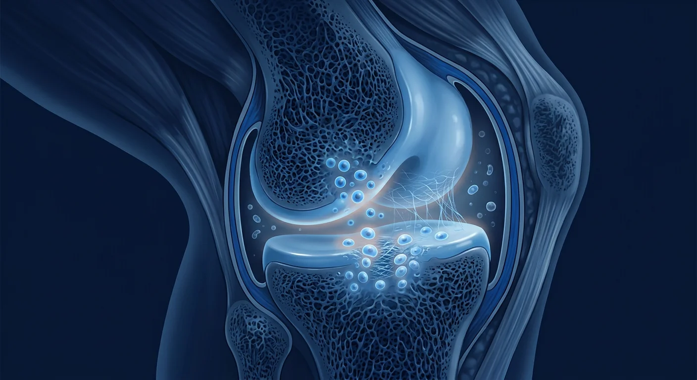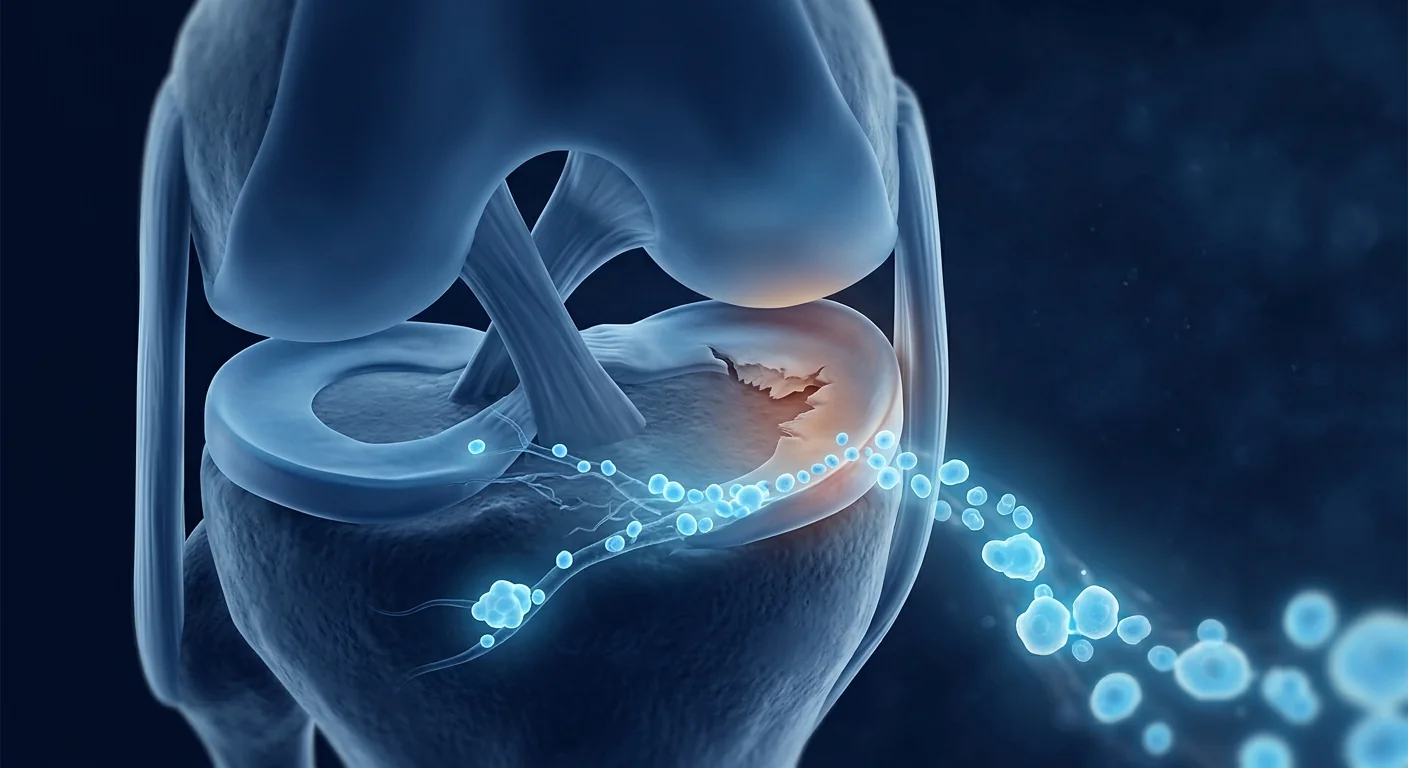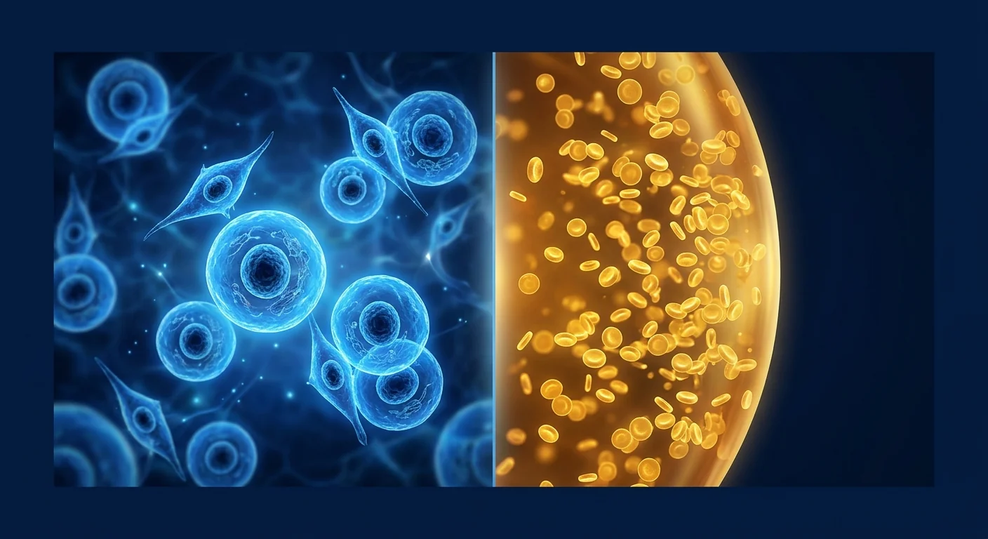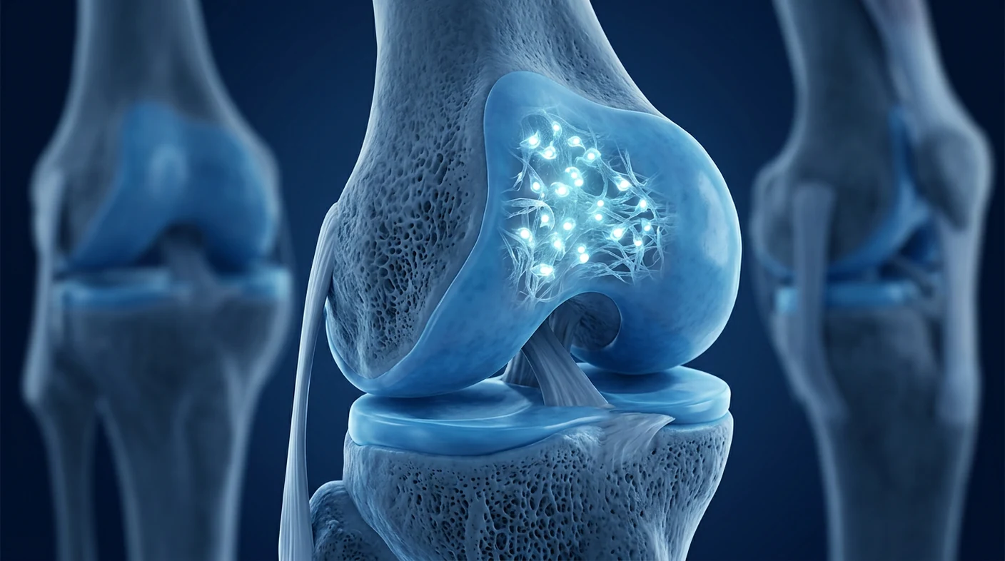
Cartilage regeneration means restoring a functional, load-bearing tissue surface rather than merely reducing joint pain. Mesenchymal stromal cell (MSC) therapy is being studied because MSCs can modify inflammation, support resident chondrocytes, and coordinate repair signals, but current human evidence does not show reliable regrowth of normal cartilage in every patient [1].
The biological challenge is unusually difficult. Articular cartilage has no blood vessels, few cells, and little access to circulating repair cells. Once a defect extends through the smooth surface, spontaneous healing is limited and the altered mechanics can accelerate degeneration.
The practical question is not simply whether stem cells “work.” It is whether a specific cell product, delivery method, joint environment, rehabilitation plan, and disease stage can produce durable tissue that behaves like cartilage. This review separates symptomatic improvement from structural regeneration and explains where MSC treatment in Bangkok may fit within an evidence-based joint-preservation plan.
What is articular cartilage regeneration?
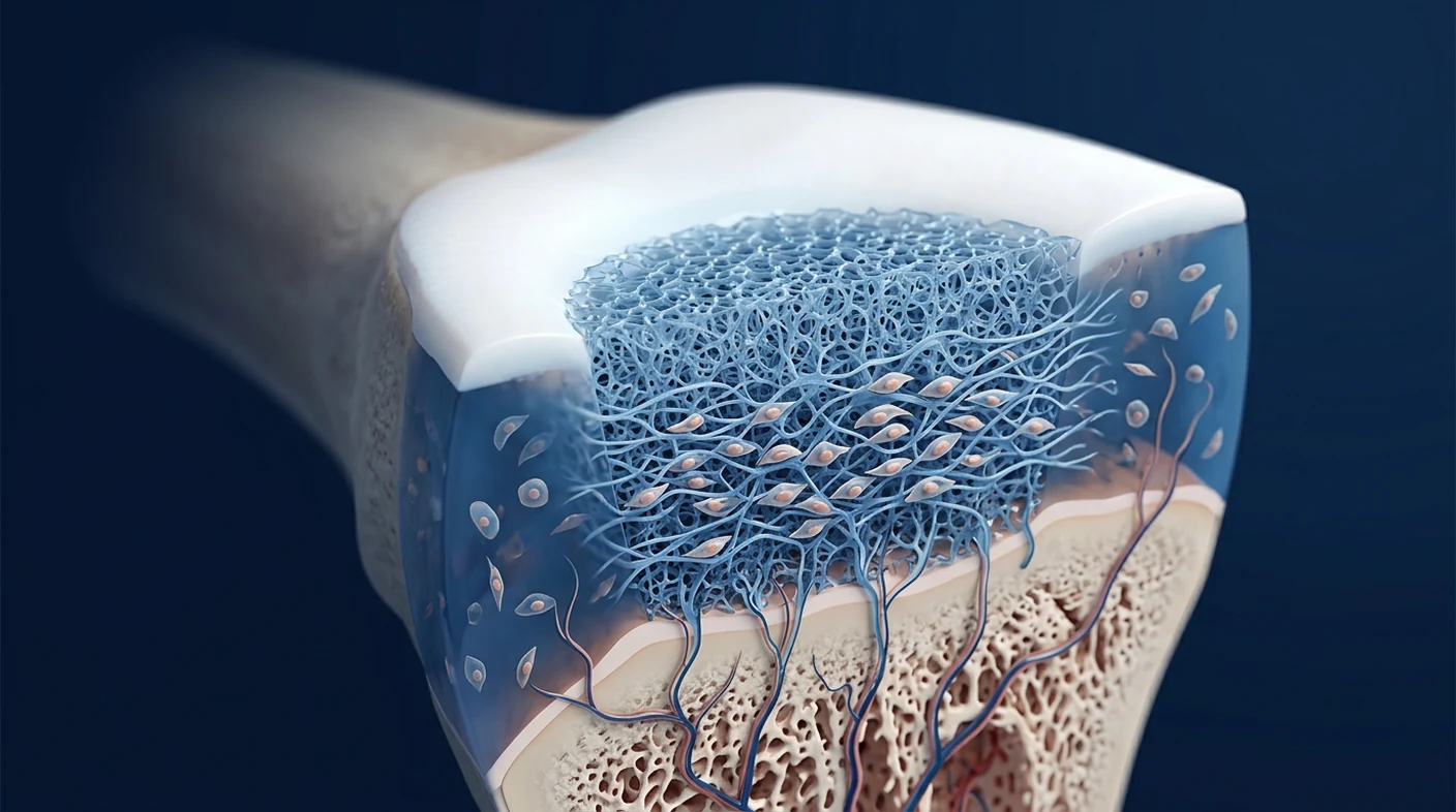
Articular cartilage regeneration is the restoration of organized cartilage that covers a joint, distributes load, and allows low-friction movement. Healthy hyaline cartilage is built from chondrocytes embedded in a dense extracellular matrix rich in type II collagen, aggrecan, and water. That architecture is different from scar-like fibrocartilage, which can fill a defect but usually has weaker mechanical durability [2].
A focal cartilage defect and osteoarthritis are related but not identical. A focal lesion is a defined area of damage, often after trauma or sport; osteoarthritis is a whole-joint process involving cartilage, synovium, subchondral bone, meniscus, ligaments, and mechanics. A treatment that helps a small contained lesion may not restore a diffusely worn, malaligned, bone-on-bone joint.
For patients, this distinction changes expectations. Pain relief may occur through reduced synovitis even when MRI shows little new tissue. Conversely, repair tissue visible on imaging does not automatically mean normal biomechanics or permanent protection from osteoarthritis. Good studies therefore measure symptoms, function, imaging, and—when surgery allows—arthroscopic or histological tissue quality.
Why does damaged cartilage heal so poorly?
Damaged cartilage heals poorly because it is avascular, has a sparse cell population, and contains a matrix that restricts cell migration. Superficial defects do not reach marrow blood supply, so they receive little fibrin clot formation or recruitment of reparative cells. Deep osteochondral defects can access marrow progenitors, but the tissue that forms is often fibrocartilage rather than durable hyaline cartilage [3].
Age and inflammation further suppress repair. Chondrocytes exposed to IL-1β, TNF-α, oxidative stress, and abnormal loading increase catabolic enzymes such as MMP-13 and ADAMTS-5. Senescent cells produce a secretory profile that reinforces inflammation and matrix loss. The result is a repair environment that actively works against regeneration.
Mechanical alignment matters as much as biology. Varus or valgus malalignment, instability, meniscal deficiency, obesity, and repetitive overload continue to concentrate force on the injured surface. Injecting cells without correcting an adverse mechanical environment is like repairing pavement while heavy traffic continues across the wet material.
How could MSC therapy support cartilage repair?
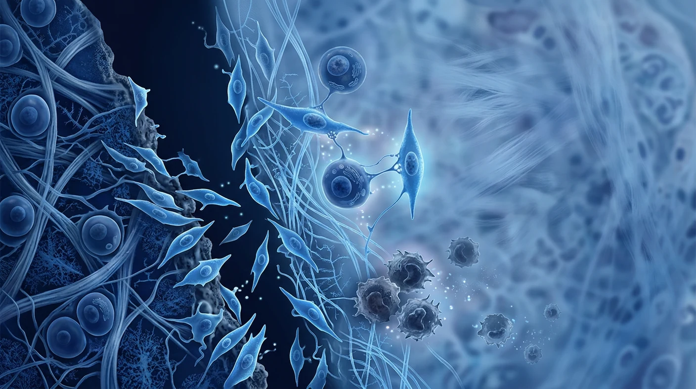
MSC therapy may support cartilage repair mainly through signaling, immune regulation, and trophic support—not by simply turning every injected cell into a chondrocyte. Laboratory differentiation is possible, but after clinical delivery many cells remain transient. Their secretome can still influence macrophages, synovial cells, chondrocytes, endothelial cells, and endogenous progenitors [4].
- Inflammation control: MSC-derived PGE2, TSG-6, IDO, and other mediators can reduce pro-inflammatory macrophage activity and support a repair-oriented immune state.
- Chondrocyte protection: trophic factors may reduce apoptosis, improve matrix synthesis, and help surviving chondrocytes maintain collagen II and aggrecan.
- Matrix remodeling: MSC signaling can influence the balance between matrix-building and matrix-degrading enzymes.
- Progenitor recruitment: chemokines and extracellular vesicles may attract endogenous reparative cells toward a defect.
- Chondrogenic contribution: under the right three-dimensional, mechanical, and biochemical conditions, MSCs can adopt cartilage-like phenotypes.
These mechanisms overlap, and their importance varies by treatment. A simple intra-articular injection is primarily a joint-environment intervention. Surgical implantation in a scaffold places cells inside a defined defect and is designed more directly for tissue formation.
Injection, scaffold, or surgery: which approach targets regeneration?
Intra-articular injection targets the biological environment of the joint, while scaffold-based or surgical approaches target a defined cartilage defect. These strategies should not be treated as interchangeable. The correct route depends on whether the main problem is diffuse osteoarthritis, a focal chondral lesion, an osteochondral defect, or a mechanical abnormality.
| Approach | Primary aim | Typical setting | Main limitation |
|---|---|---|---|
| Intra-articular MSC injection | Reduce inflammation and support cartilage homeostasis | Mild-to-moderate osteoarthritis | Cells are not fixed inside one defect |
| MSC plus hyaluronic-acid hydrogel | Retain cells and provide a three-dimensional matrix | Contained cartilage defects | Product and technique remain specialized |
| MSC implantation with microfracture | Augment marrow stimulation and defect filling | Focal full-thickness lesions | Requires arthroscopy and rehabilitation |
| Osteochondral graft or chondrocyte implantation | Replace or rebuild a structural surface | Larger focal defects | Surgical morbidity, cost, and eligibility |
Microfracture creates channels into subchondral bone so marrow cells enter the defect, but long-term results can deteriorate as fibrocartilage matures. Trials combining cultured MSCs with microfracture or cartilage-derived matrices aim to improve tissue organization; some report better MOCART imaging scores, but clinical superiority is not universal [5].
What do human trials show about cartilage regrowth?
Human trials show a clearer signal for pain and function than for consistent structural cartilage regrowth. Randomized studies of intra-articular MSCs often report improvements in WOMAC, KOOS, and pain scores, yet MRI findings range from no significant change to reduced defect size, improved cartilage quality, or slower volume loss [6].
A proof-of-concept adipose-derived MSC study reported dose-related clinical improvement and arthroscopic evidence of cartilage changes, but it was small and not sufficient to establish routine regeneration [7]. A randomized placebo-controlled phase IIb trial found symptom improvement but no significant MRI cartilage-volume change at six months, illustrating why symptom and structure must be reported separately [8].
One notable scaffold-based program combined allogeneic umbilical-cord-blood MSCs with hyaluronate hydrogel for cartilage defects. Extended follow-up supported safety and durable clinical improvement, with repair tissue assessed arthroscopically and histologically in early participants [9]. This is encouraging, but it represents a specialized implanted product for selected defects—not evidence that any IV or joint injection can rebuild cartilage.
Systematic reviews reach a cautious consensus: short-term patient-reported outcomes are often favorable, while heterogeneity in cell source, expansion, dose, co-interventions, imaging, and disease severity prevents a confident universal claim of regeneration [10].
The distinction that protects patients
Less pain is not proof of new cartilage. A responsible clinic should say whether its goal is inflammation control, joint preservation, focal-defect repair, or structural regeneration—and should use an outcome measure capable of testing that goal.
Who may be a candidate for MSC cartilage treatment in Bangkok?
The most plausible candidates have a clearly characterized cartilage problem, correctable mechanics, and enough remaining joint structure to preserve. Assessment should combine history, examination, weight-bearing radiographs, and MRI when the result will change the plan. Age alone is not decisive, but biological quality, activity, alignment, and lesion size influence the probability of benefit.
- Symptomatic focal chondral or osteochondral defects after appropriate conservative care
- Mild-to-moderate osteoarthritis with residual cartilage and no uncontrolled inflammatory flare
- Stable joints or instability that will be corrected as part of the plan
- Patients able to follow staged rehabilitation and load-management instructions
- Patients seeking joint preservation who understand that treatment remains investigational
Poor candidates may include people with active infection, untreated malignancy, uncontrolled coagulation problems, severe uncorrected malalignment, advanced bone-on-bone disease, or expectations of guaranteed regrowth. End-stage arthritis may be served more predictably by arthroplasty than by a biologic procedure.
How is a cartilage-regeneration plan built in Thailand?
An evidence-based cartilage plan begins with diagnosis and biomechanics, not with a preselected vial of cells. The clinician first identifies whether pain comes from cartilage, meniscus, tendon, synovium, subchondral bone, referred pain, or several structures. Imaging is interpreted alongside symptoms because many cartilage lesions are incidental.
- Define the lesion: location, surface area, depth, containment, subchondral bone status, and osteoarthritis grade.
- Map mechanical drivers: alignment, ligament stability, meniscal function, gait, body weight, and sport or work loading.
- Choose the therapeutic objective: symptom control, preservation, focal repair, or surgical restoration.
- Specify the product: tissue source, culture method, identity markers, sterility, potency rationale, dose, and fresh-delivery chain.
- Plan rehabilitation: protected loading when appropriate, range of motion, progressive strength, and return-to-activity criteria.
- Measure outcomes: baseline and follow-up KOOS/WOMAC, pain, function, and MRI only when structural assessment is justified.
For an injection, the procedure is usually performed under sterile conditions with ultrasound or fluoroscopic guidance when anatomy requires it. For an implanted scaffold, arthroscopy, defect preparation, and a longer protected-weight-bearing protocol may be necessary. A credible recommendation explains why the chosen route matches the lesion.
What quality controls matter for an MSC product?
Cell identity, sterility, viability, traceability, and manufacturing consistency matter more than a marketing label such as “stem cell.” Products described by the same broad name can differ substantially. The International Society for Cell & Gene Therapy minimum criteria help define cultured MSC identity, but they do not by themselves prove clinical potency for cartilage [11].
- Documented source and donor-screening process
- Phenotype consistent with MSC identity, including positive CD73/CD90/CD105 and exclusion markers
- Sterility, mycoplasma, endotoxin, and relevant pathogen testing
- Viability measured at delivery—not only at release from a distant laboratory
- Batch traceability and transport-temperature records
- A defensible dose and route linked to the treatment objective
Fresh, never-frozen delivery avoids a freeze-thaw step, but freshness alone does not establish efficacy. It must sit within a controlled manufacturing and clinical process. Similarly, very high cell counts are not automatically better; dose-response relationships can be nonlinear and remain incompletely standardized.
What are the risks and realistic recovery expectations?
The most common short-term risks of intra-articular MSC procedures are temporary pain, swelling, stiffness, or an inflammatory flare. Infection, bleeding, and injury to nearby structures are uncommon but important procedural risks. Surgical implantation adds anesthesia, thrombosis, wound, and rehabilitation risks.
Published knee studies have generally reported reassuring short- to medium-term safety, including long-term observational follow-up after autologous bone-marrow MSC transplantation [12]. However, safety evidence for one manufactured product cannot be transferred automatically to an uncharacterized product, and long-term surveillance remains important.
These are planning ranges, not promises. Recovery after an injection differs from recovery after microfracture or scaffold implantation. Rehabilitation adherence and correction of joint loading may influence durability as much as the cell intervention itself.
How should cartilage regeneration be measured?
Cartilage regeneration should be measured with separate clinical and structural endpoints because neither can substitute for the other. Patient-reported outcome measures such as KOOS and WOMAC capture pain, stiffness, sport, and daily function. Objective testing can include range of motion, strength, walking capacity, and return to activity.
Structural assessment may use MRI defect fill, cartilage thickness or volume, T2/T1rho compositional mapping, and the MOCART score after repair surgery. Arthroscopy can inspect surface integration, while biopsy can distinguish hyaline-like cartilage from fibrocartilage—but invasive assessment is rarely justified solely for research follow-up.
A before-and-after MRI image selected from different slices or sequences can be misleading. Meaningful comparison requires standardized acquisition, timing, blinded interpretation where possible, and awareness that hydration changes may alter cartilage signal without proving durable tissue formation.
What does the evidence not yet establish?
The evidence does not yet establish that MSC therapy routinely regenerates normal hyaline cartilage, reverses end-stage osteoarthritis, or prevents future joint replacement. Cell source and dose are not standardized, studies are often small, and many combine cells with surgery, hyaluronic acid, platelet products, or rehabilitation, making the active component difficult to isolate.
- No universal protocol has been proven superior across joints and lesion types.
- Most injection trials prioritize symptoms rather than cartilage histology.
- MRI improvement may not equal normal tissue composition or long-term durability.
- Evidence from focal defects cannot be generalized to diffuse bone-on-bone arthritis.
- Extracellular-vesicle and engineered-scaffold approaches remain largely preclinical or early-stage [13].
These limitations do not make the field irrelevant. They define the questions a responsible program must answer and protect patients from confusing plausible biology with guaranteed clinical regeneration.
Frequently Asked Questions
Can stem cells regrow knee cartilage?
Stem cells may support repair and some specialized implanted MSC products have produced cartilage-like tissue in selected focal defects. Routine injections have stronger evidence for pain and function than for consistent regrowth of normal hyaline cartilage.
Is cartilage regeneration treatment available in Bangkok?
Regenerative and orthopaedic clinics in Bangkok offer biologic joint procedures, but products, indications, and quality systems vary. Patients should seek imaging-based assessment, explicit cell-quality documentation, and conservative claims before proceeding.
Is an MSC injection the same as cartilage implantation?
No. An injection distributes cells in the joint and mainly targets inflammation and tissue support; implantation places cells or a cell-scaffold construct into a prepared focal defect, usually during surgery.
How long does cartilage regeneration take?
Biological remodeling is measured in months, not days. Symptom change can occur earlier than structural change, while scaffold or microfracture procedures often require months of protected loading and rehabilitation.
Does PRP regenerate cartilage as well as MSC therapy?
PRP provides concentrated platelet signals and can improve symptoms, but convincing evidence of durable hyaline cartilage regeneration is limited for both PRP and injectable MSC therapy. Their mechanisms, manufacturing, cost, and evidence should be compared for the specific diagnosis.
What is the best age for cartilage stem cell therapy?
There is no single age cutoff. Lesion type, remaining cartilage, alignment, metabolic health, activity goals, and ability to rehabilitate are usually more informative than chronological age alone.
Limitations and medical disclaimer
MSC-based cartilage regeneration remains investigational and should not be presented as guaranteed tissue regrowth. This review synthesizes published research across osteoarthritis and focal cartilage repair, but protocols are heterogeneous and results from one product cannot be assumed for another. Evidence is concentrated in the knee and cannot automatically be applied to the hip, ankle, shoulder, or spine.
This article is educational, not a diagnosis or treatment recommendation. A qualified clinician should review symptoms, examination findings, imaging, medical history, alternatives, costs, and uncertainties before any procedure. Standard care—including exercise therapy, weight management, medication, bracing, corrective surgery, or arthroplasty—may be more appropriate depending on disease stage.
References
- Barry F, Murphy M. Mesenchymal stem cells in joint disease and repair. Nature Reviews Rheumatology. 2013;9:584–594. doi:10.1038/nrrheum.2013.109 ↩
- Hunziker EB. Articular cartilage repair: basic science and clinical progress. Osteoarthritis and Cartilage. 2002;10(6):432–463. doi:10.1053/joca.2002.0801 ↩
- Makris EA, Gomoll AH, Malizos KN, Hu JC, Athanasiou KA. Repair and tissue engineering techniques for articular cartilage. Nature Reviews Rheumatology. 2015;11:21–34. doi:10.1038/nrrheum.2014.157 ↩
- Le H, Xu W, Zhuang X, et al. Mesenchymal stem cells for cartilage regeneration. Journal of Tissue Engineering. 2020;11:2041731420943839. doi:10.1177/2041731420943839 ↩
- Hashimoto Y, Nishida Y, Takahashi S, et al. Transplantation of autologous bone marrow-derived MSCs with microfracture versus microfracture alone. Regenerative Therapy. 2019;11:106–113. doi:10.1016/j.reth.2019.06.002 ↩
- Kim SH, Ha CW, Park YB, Nam E, Lee JE, Lee HJ. Intra-articular injection of MSCs for clinical outcomes and cartilage repair in knee osteoarthritis. Archives of Orthopaedic and Trauma Surgery. 2019;139:971–980. doi:10.1007/s00402-019-03140-8 ↩
- Jo CH, Lee YG, Shin WH, et al. Intra-articular injection of MSCs for knee osteoarthritis: a proof-of-concept clinical trial. Stem Cells. 2014;32(5):1254–1266. doi:10.1002/stem.1634 ↩
- Lee WS, Kim HJ, Kim KI, Kim GB, Jin W. Intra-articular autologous adipose-derived MSCs for knee osteoarthritis: a phase IIb randomized placebo-controlled trial. Stem Cells Translational Medicine. 2019;8(6):504–511. doi:10.1002/sctm.18-0122 ↩
- Park YB, Ha CW, Lee CH, Yoon YC, Park YG. Cartilage regeneration using allogeneic umbilical cord blood-derived MSCs and hyaluronate hydrogel. Stem Cells Translational Medicine. 2017;6(2):613–621. doi:10.5966/sctm.2016-0157 ↩
- Ha CW, Park YB, Kim SH, Lee HJ. Intra-articular MSCs in osteoarthritis of the knee: clinical outcomes and evidence of cartilage repair. Arthroscopy. 2019;35(1):277–288.e2. doi:10.1016/j.arthro.2018.07.028 ↩
- Dominici M, Le Blanc K, Mueller I, et al. Minimal criteria for defining multipotent mesenchymal stromal cells. Cytotherapy. 2006;8(4):315–317. doi:10.1080/14653240600855905 ↩
- Wakitani S, Okabe T, Horibe S, et al. Safety of autologous bone marrow-derived MSC transplantation for cartilage repair in 41 patients. Journal of Tissue Engineering and Regenerative Medicine. 2011;5(2):146–150. doi:10.1002/term.299 ↩
- Toh WS, Lai RC, Hui JHP, Lim SK. MSC exosome as a cell-free MSC therapy for cartilage regeneration. Seminars in Cell & Developmental Biology. 2017;67:56–64. doi:10.1016/j.semcdb.2016.11.008 ↩
- Matas J, Orrego M, Amenabar D, et al. Umbilical cord-derived MSCs for knee osteoarthritis: repeated dosing versus single dose and hyaluronic acid. Stem Cells Translational Medicine. 2019;8(3):215–224. doi:10.1002/sctm.18-0053 ↩
- Xiang XN, Zhu SY, He HC, Yu X, Xu Y, He CQ. Mesenchymal stromal cell-based therapy for cartilage regeneration in knee osteoarthritis. Stem Cell Research & Therapy. 2022;13:14. doi:10.1186/s13287-021-02689-9 ↩
软骨再生意味着恢复功能性、承重性组织表面,而不仅仅是减轻关节疼痛。间充质基质细胞 (MSC) 疗法正在研究中,因为 MSC 可以调节炎症、支持常驻软骨细胞并协调修复信号,但目前的临床证据并未显示每位患者都能可靠地再生正常软骨 [1]。
生物学挑战异常困难。关节软骨没有血管,细胞稀少,并且很少接触循环修复细胞。一旦缺陷穿透光滑表面,自发愈合就会受到限制,并且改变的力学特性会加速退化。
实际问题不仅仅是干细胞是否“有效”。它涉及特定的细胞产品、递送方法、关节环境、康复计划和疾病阶段是否能产生行为像软骨的持久组织。本综述将症状改善与结构再生区分开来,并解释了曼谷的 MSC 治疗如何融入循证关节保护计划。
什么是关节软骨再生?
关节软骨再生是恢复覆盖关节、分散负荷并允许低摩擦运动的有序软骨。健康的透明软骨由嵌入富含 II 型胶原蛋白、聚集蛋白聚糖和水的致密细胞外基质中的软骨细胞构成。这种结构与疤痕样纤维软骨不同,纤维软骨可以填充缺陷,但通常机械耐久性较弱 [2]。
局灶性软骨缺损和骨关节炎相关但不完全相同。局灶性病变是明确的损伤区域,通常发生在创伤或运动后;骨关节炎是一个涉及软骨、滑膜、软骨下骨、半月板、韧带和力学的全关节过程。对小型局限性病变有帮助的治疗可能无法恢复弥漫性磨损、对线不良、骨对骨的关节。
对于患者来说,这种区别改变了预期。即使 MRI 显示新组织很少,通过减少滑膜炎也可以缓解疼痛。相反,影像学上可见的修复组织并不自动意味着正常的生物力学或对骨关节炎的永久保护。因此,好的研究会测量症状、功能、影像学,以及——当手术允许时——关节镜或组织学组织质量。
为什么受损软骨愈合如此之差?
受损软骨愈合不良,因为它缺乏血管,细胞数量稀少,并且含有限制细胞迁移的基质。浅表缺损无法到达骨髓血液供应,因此很少形成纤维蛋白凝块或招募修复细胞。深层骨软骨缺损可以接触骨髓祖细胞,但形成的组织通常是纤维软骨而不是耐用的透明软骨 [3]。
年龄和炎症进一步抑制修复。暴露于 IL-1β、TNF-α、氧化应激和异常负荷的软骨细胞会增加分解酶,如 MMP-13 和 ADAMTS-5。衰老细胞产生的分泌谱会增强炎症和基质流失。结果是修复环境积极地对抗再生。
机械对线与生物学同样重要。内翻或外翻对线不良、不稳定、半月板缺失、肥胖和重复性过载会继续将力集中在受伤表面。在不纠正不利机械环境的情况下注射细胞,就像在重型交通持续通过湿材料时修复路面一样。
MSC 疗法如何支持软骨修复?
MSC 疗法主要通过信号传导、免疫调节和营养支持来支持软骨修复,而不是简单地将每个注射的细胞转化为软骨细胞。实验室分化是可能的,但在临床递送后,许多细胞仍然是瞬时的。它们的旁分泌组仍然可以影响巨噬细胞、滑膜细胞、软骨细胞、内皮细胞和内源性祖细胞 [4]。
- 炎症控制:MSC 衍生的 PGE2、TSG-6、IDO 和其他介质可以减少促炎巨噬细胞活性并支持以修复为导向的免疫状态。
- 软骨细胞保护:营养因子可以减少细胞凋亡,改善基质合成,并帮助存活的软骨细胞维持 II 型胶原蛋白和聚集蛋白聚糖。
- 基质重塑:MSC 信号传导可以影响基质构建酶和基质降解酶之间的平衡。
- 祖细胞招募:趋化因子和细胞外囊泡可以吸引内源性修复细胞向缺陷处移动。
- 软骨形成贡献:在正确的三维、机械和生化条件下,MSC 可以呈现软骨样表型。
这些机制相互重叠,其重要性因治疗而异。简单的关节内注射主要是关节环境干预。在支架中进行手术植入将细胞置于明确的缺陷内,并且更直接地用于组织形成。
注射、支架还是手术:哪种方法针对再生?
关节内注射针对关节的生物环境,而基于支架或手术方法针对明确的软骨缺损。这些策略不应被视为可互换的。正确的途径取决于主要问题是弥漫性骨关节炎、局灶性软骨病变、骨软骨缺损还是机械异常。
| 方法 | 主要目的 | 典型设置 | 主要限制 |
|---|---|---|---|
| 关节内 MSC 注射 | 减少炎症并支持软骨稳态 | 轻度至中度骨关节炎 | 细胞未固定在一个缺陷内 |
| MSC 加透明质酸水凝胶 | 保留细胞并提供三维基质 | 局限性软骨缺损 | 产品和技术仍属专业范畴 |
| MSC 植入结合微骨折 | 增强骨髓刺激和缺损填充 | 局灶性全层病变 | 需要关节镜检查和康复 |
| 骨软骨移植或软骨细胞植入 | 替换或重建结构表面 | 较大的局灶性缺损 | 手术并发症、成本和适用性 |
微骨折在软骨下骨中形成通道,使骨髓细胞进入缺损处,但随着纤维软骨的成熟,长期效果可能会恶化。将培养的 MSC 与微骨折或软骨衍生基质结合的试验旨在改善组织结构;一些报告显示 MOCART 影像学评分有所改善,但临床优势并非普遍存在 [5]。
人体试验显示软骨再生情况如何?
人体试验显示,疼痛和功能方面的信号比持续的结构性软骨再生更清晰。关节内 MSC 的随机研究通常报告 WOMAC、KOOS 和疼痛评分的改善,但 MRI 结果从无显著变化到缺陷尺寸减小、软骨质量改善或体积损失减慢不等 [6]。
一项概念验证的脂肪源性 MSC 研究报告了剂量相关的临床改善和关节镜下软骨变化的证据,但该研究规模较小,不足以建立常规再生 [7]。一项随机安慰剂对照 IIb 期试验发现症状改善,但在六个月时 MRI 软骨体积没有显著变化,这说明了为什么必须单独报告症状和结构 [8]。
一项值得注意的基于支架的方案将异体脐带血 MSC 与透明质酸水凝胶结合用于软骨缺损。长期随访支持安全性和持久的临床改善,早期参与者的修复组织通过关节镜和组织学评估 [9]。这令人鼓舞,但这代表了一种用于特定缺陷的专业植入产品,而不是任何静脉注射或关节注射都能重建软骨的证据。
系统评价达成谨慎共识:短期患者报告结果通常良好,但细胞来源、扩增、剂量、联合干预、影像学和疾病严重程度的异质性阻碍了对再生的普遍自信主张 [10]。
保护患者的区别
疼痛减轻并非新软骨的证据。负责任的诊所应说明其目标是炎症控制、关节保护、局灶性缺损修复还是结构再生——并应使用能够检验该目标的结局指标。
谁可能是曼谷 MSC 软骨治疗的候选人?
最有可能的候选人是具有明确特征的软骨问题、可纠正的力学问题以及足够多的剩余关节结构可供保留的患者。评估应结合病史、检查、负重 X 光片和 MRI,当结果会改变计划时。年龄本身并非决定性因素,但生物学质量、活动、对线和病变大小会影响获益的可能性。
- 经过适当保守治疗后出现症状的局灶性软骨或骨软骨缺损
- 轻度至中度骨关节炎,伴有残余软骨且无未受控制的炎症发作
- 稳定的关节或作为计划一部分将得到纠正的不稳定
- 能够遵循分阶段康复和负荷管理说明的患者
- 寻求关节保护并理解治疗仍在研究中的患者
不适合的候选人可能包括患有活动性感染、未经治疗的恶性肿瘤、未受控制的凝血问题、严重的未纠正对线不良、晚期骨对骨疾病或期望保证再生的患者。终末期关节炎通过关节置换术可能比通过生物学程序更可预测地得到治疗。
泰国如何制定软骨再生计划?
循证软骨计划始于诊断和生物力学,而不是预先选择的细胞瓶。临床医生首先确定疼痛是来自软骨、半月板、肌腱、滑膜、软骨下骨、牵涉痛还是多种结构。影像学与症状一起解读,因为许多软骨病变是偶然发现的。
- 定义病变:位置、表面积、深度、包容性、软骨下骨状况和骨关节炎分级。
- 绘制机械驱动因素:对线、韧带稳定性、半月板功能、步态、体重以及运动或工作负荷。
- 选择治疗目标:症状控制、保护、局灶性修复或手术重建。
- 指定产品:组织来源、培养方法、身份标志物、无菌性、效力原理、剂量和新鲜递送链。
- 计划康复:适当的保护性负荷、运动范围、渐进性力量训练和恢复活动标准。
- 测量结果:基线和随访 KOOS/WOMAC、疼痛、功能,以及仅在结构评估合理时进行 MRI。
对于注射,通常在无菌条件下进行,当解剖结构需要时,可采用超声或荧光透视引导。对于植入支架,可能需要关节镜检查、缺损准备和更长的保护性负重方案。可靠的建议会解释为什么选择的途径与病变相符。
MSC 产品有哪些质量控制很重要?
细胞身份、无菌性、活力、可追溯性和生产一致性比“干细胞”等营销标签更重要。用相同宽泛名称描述的产品可能存在显著差异。国际细胞与基因治疗学会的最低标准有助于定义培养的 MSC 身份,但它们本身并不能证明软骨的临床效力 [11]。
- 有记录的来源和供体筛选过程
- 与 MSC 身份一致的表型,包括阳性 CD73/CD90/CD105 和排除标志物
- 无菌性、支原体、内毒素和相关病原体检测
- 在递送时测量的活力——而不仅仅是在远离实验室时释放
- 批次可追溯性和运输温度记录
- 与治疗目标相关的合理剂量和途径
新鲜、从未冷冻的递送避免了冻融步骤,但新鲜本身并不能确立疗效。它必须在受控的生产和临床过程中。同样,非常高的细胞计数并非自动更好;剂量-反应关系可能是非线性的,并且尚未完全标准化。
风险和实际恢复预期是什么?
关节内 MSC 手术最常见的短期风险是暂时性疼痛、肿胀、僵硬或炎症发作。感染、出血和邻近结构损伤是不常见但重要的手术风险。手术植入增加了麻醉、血栓形成、伤口和康复风险。
已发表的膝关节研究通常报告了令人放心的短期至中期安全性,包括自体骨髓 MSC 移植后的长期观察性随访 [12]。然而,一种制造产品的安全证据不能自动转移到未表征的产品,长期监测仍然很重要。
这些是计划范围,而非承诺。注射后的恢复与微骨折或支架植入后的恢复不同。康复依从性和关节负荷的纠正可能与细胞干预本身一样影响耐久性。
如何衡量软骨再生?
软骨再生应通过独立的临床和结构终点来衡量,因为两者都不能替代对方。患者报告结局指标,如 KOOS 和 WOMAC,可捕捉疼痛、僵硬、运动和日常功能。客观测试可包括运动范围、力量、步行能力和恢复活动。
结构评估可使用 MRI 缺损填充、软骨厚度或体积、T2/T1rho 成分图谱以及修复手术后的 MOCART 评分。关节镜可检查表面整合,而活检可区分透明软骨样软骨和纤维软骨——但侵入性评估很少仅为研究随访而合理。
从不同切片或序列中选择的术前术后 MRI 图像可能具有误导性。有意义的比较需要标准化采集、时间、尽可能盲法解读,并意识到水合变化可能会改变软骨信号,而不能证明持久组织形成。
证据尚未确立什么?
证据尚未确立 MSC 疗法能常规再生正常透明软骨、逆转终末期骨关节炎或预防未来的关节置换。细胞来源和剂量尚未标准化,研究通常规模较小,许多研究将细胞与手术、透明质酸、血小板产品或康复相结合,使得活性成分难以分离。
- 尚未证明任何通用方案在关节和病变类型方面优越。
- 大多数注射试验优先考虑症状而非软骨组织学。
- MRI 改善可能不等于正常的组织成分或长期耐久性。
- 局灶性缺损的证据不能推广到弥漫性骨对骨关节炎。
- 细胞外囊泡和工程支架方法仍主要处于临床前或早期阶段 [13]。
这些限制并未使该领域变得无关紧要。它们定义了负责任的计划必须回答的问题,并保护患者免于将看似合理的生物学与有保证的临床再生混淆。
常见问题
干细胞能再生膝关节软骨吗?
干细胞可能支持修复,一些专门的植入式 MSC 产品已在选定的局灶性缺损中产生软骨样组织。常规注射在疼痛和功能方面有更强的证据,而不是持续再生正常透明软骨。
曼谷有软骨再生治疗吗?
曼谷的再生和骨科诊所提供生物关节手术,但产品、适应症和质量体系各不相同。患者在进行治疗前应寻求基于影像学的评估、明确的细胞质量文件和保守的声明。
MSC 注射与软骨植入相同吗?
不。注射将细胞分布在关节中,主要针对炎症和组织支持;植入将细胞或细胞支架结构置于准备好的局灶性缺损中,通常在手术期间进行。
软骨再生需要多长时间?
生物重塑以月为单位衡量,而不是天。症状变化可能早于结构变化,而支架或微骨折手术通常需要数月的保护性负重和康复。
PRP 能像 MSC 疗法一样再生软骨吗?
PRP 提供浓缩的血小板信号并能改善症状,但对于 PRP 和可注射 MSC 疗法,持久透明软骨再生的令人信服的证据有限。应针对具体诊断比较它们的机制、制造、成本和证据。
软骨干细胞治疗的最佳年龄是多少?
没有单一的年龄限制。病变类型、剩余软骨、对线、代谢健康、活动目标和康复能力通常比单独的实际年龄更能提供信息。
局限性和医疗免责声明
基于 MSC 的软骨再生仍在研究中,不应被视为有保证的组织再生。本综述综合了骨关节炎和局灶性软骨修复的已发表研究,但方案具有异质性,一种产品的结果不能假定适用于另一种产品。证据集中在膝关节,不能自动应用于髋关节、踝关节、肩关节或脊柱。
本文旨在教育,而非诊断或治疗建议。在进行任何手术之前,合格的临床医生应审查症状、检查结果、影像学、病史、替代方案、费用和不确定性。根据疾病阶段,标准护理——包括运动疗法、体重管理、药物、支具、矫正手术或关节置换术——可能更合适。
参考文献
- Barry F, Murphy M. Mesenchymal stem cells in joint disease and repair. Nature Reviews Rheumatology. 2013;9:584–594. doi:10.1038/nrrheum.2013.109 ↩
- Hunziker EB. Articular cartilage repair: basic science and clinical progress. Osteoarthritis and Cartilage. 2002;10(6):432–463. doi:10.1053/joca.2002.0801 ↩
- Makris EA, Gomoll AH, Malizos KN, Hu JC, Athanasiou KA. Repair and tissue engineering techniques for articular cartilage. Nature Reviews Rheumatology. 2015;11:21–34. doi:10.1038/nrrheum.2014.157 ↩
- Le H, Xu W, Zhuang X, et al. Mesenchymal stem cells for cartilage regeneration. Journal of Tissue Engineering. 2020;11:2041731420943839. doi:10.1177/2041731420943839 ↩
- Hashimoto Y, Nishida Y, Takahashi S, et al. Transplantation of autologous bone marrow-derived MSCs with microfracture versus microfracture alone. Regenerative Therapy. 2019;11:106–113. doi:10.1016/j.reth.2019.06.002 ↩
- Kim SH, Ha CW, Park YB, Nam E, Lee JE, Lee HJ. Intra-articular injection of MSCs for clinical outcomes and cartilage repair in knee osteoarthritis. Archives of Orthopaedic and Trauma Surgery. 2019;139:971–980. doi:10.1007/s00402-019-03140-8 ↩
- Jo CH, Lee YG, Shin WH, et al. Intra-articular injection of MSCs for knee osteoarthritis: a proof-of-concept clinical trial. Stem Cells. 2014;32(5):1254–1266. doi:10.1002/stem.1634 ↩
- Lee WS, Kim HJ, Kim KI, Kim GB, Jin W. Intra-articular autologous adipose-derived MSCs for knee osteoarthritis: a phase IIb randomized placebo-controlled trial. Stem Cells Translational Medicine. 2019;8(6):504–511. doi:10.1002/sctm.18-0122 ↩
- Park YB, Ha CW, Lee CH, Yoon YC, Park YG. Cartilage regeneration using allogeneic umbilical cord blood-derived MSCs and hyaluronate hydrogel. Stem Cells Translational Medicine. 2017;6(2):613–621. doi:10.5966/sctm.2016-0157 ↩
- Ha CW, Park YB, Kim SH, Lee HJ. Intra-articular MSCs in osteoarthritis of the knee: clinical outcomes and evidence of cartilage repair. Arthroscopy. 2019;35(1):277–288.e2. doi:10.1016/j.arthro.2018.07.028 ↩
- Dominici M, Le Blanc K, Mueller I, et al. Minimal criteria for defining multipotent mesenchymal stromal cells. Cytotherapy. 2006;8(4):315–317. doi:10.1080/14653240600855905 ↩
- Wakitani S, Okabe T, Horibe S, et al. Safety of autologous bone marrow-derived MSC transplantation for cartilage repair in 41 patients. Journal of Tissue Engineering and Regenerative Medicine. 2011;5(2):146–150. doi:10.1002/term.299 ↩
- Toh WS, Lai RC, Hui JHP, Lim SK. MSC exosome as a cell-free MSC therapy for cartilage regeneration. Seminars in Cell & Developmental Biology. 2017;67:56–64. doi:10.1016/j.semcdb.2016.11.008 ↩
- Matas J, Orrego M, Amenabar D, et al. Umbilical cord-derived MSCs for knee osteoarthritis: repeated dosing versus single dose and hyaluronic acid. Stem Cells Translational Medicine. 2019;8(3):215–224. doi:10.1002/sctm.18-0053 ↩
- Xiang XN, Zhu SY, He HC, Yu X, Xu Y, He CQ. Mesenchymal stromal cell-based therapy for cartilage regeneration in knee osteoarthritis. Stem Cell Research & Therapy. 2022;13:14. doi:10.1186/s13287-021-02689-9 ↩
تجديد الغضروف يعني استعادة سطح نسيجي وظيفي يتحمل الأحمال بدلاً من مجرد تقليل آلام المفاصل. تُدرس علاجات الخلايا الجذعية الوسيطة (MSC) لأنها تستطيع تعديل الالتهاب، ودعم الخلايا الغضروفية المقيمة، وتنسيق إشارات الإصلاح، لكن الأدلة البشرية الحالية لا تظهر إعادة نمو موثوقة للغضروف الطبيعي في كل مريض [1].
التحدي البيولوجي صعب بشكل غير عادي. الغضروف المفصلي لا يحتوي على أوعية دموية، وعدد قليل من الخلايا، وقليل من الوصول إلى خلايا الإصلاح المتداولة. بمجرد أن يمتد العيب عبر السطح الأملس، يكون الشفاء التلقائي محدودًا ويمكن أن تسرع الميكانيكا المتغيرة من التدهور.
السؤال العملي ليس ببساطة ما إذا كانت الخلايا الجذعية "تعمل". بل هو ما إذا كان منتج خلوي معين، وطريقة توصيل، وبيئة مفصلية، وخطة إعادة تأهيل، ومرحلة مرضية يمكن أن تنتج نسيجًا دائمًا يتصرف مثل الغضروف. تفصل هذه المراجعة التحسن في الأعراض عن التجديد الهيكلي وتشرح أين يمكن أن يتناسب علاج MSC في بانكوك ضمن خطة للحفاظ على المفاصل قائمة على الأدلة.
ما هو تجديد الغضروف المفصلي؟
تجديد الغضروف المفصلي هو استعادة الغضروف المنظم الذي يغطي المفصل، ويوزع الحمل، ويسمح بحركة منخفضة الاحتكاك. يتكون الغضروف الزجاجي الصحي من خلايا غضروفية مغمورة في مصفوفة خارج الخلية كثيفة غنية بالكولاجين من النوع الثاني، والأجريكان، والماء. يختلف هذا التركيب عن الغضروف الليفي الشبيه بالندوب، والذي يمكن أن يملأ العيب ولكنه عادة ما يكون أقل متانة ميكانيكية [2].
عيب الغضروف البؤري والتهاب المفاصل هما أمران مرتبطان ولكنهما ليسا متطابقين. الآفة البؤرية هي منطقة محددة من التلف، غالبًا بعد صدمة أو رياضة؛ التهاب المفاصل هو عملية مفصلية كاملة تشمل الغضروف، والغشاء الزليلي، والعظم تحت الغضروفي، والغضروف الهلالي، والأربطة، والميكانيكا. قد لا يعيد العلاج الذي يساعد آفة صغيرة محتواة مفصلاً متآكلاً بشكل منتشر، أو غير متوازن، أو عظمًا على عظم.
بالنسبة للمرضى، يغير هذا التمييز التوقعات. قد يحدث تخفيف الألم من خلال تقليل التهاب الغشاء الزليلي حتى عندما يظهر التصوير بالرنين المغناطيسي القليل من الأنسجة الجديدة. على العكس من ذلك، فإن نسيج الإصلاح المرئي في التصوير لا يعني تلقائيًا ميكانيكا حيوية طبيعية أو حماية دائمة من التهاب المفاصل. لذلك، تقيس الدراسات الجيدة الأعراض، والوظيفة، والتصوير، وعندما تسمح الجراحة، جودة الأنسجة بالمنظار أو النسيجية.
لماذا يلتئم الغضروف التالف بشكل سيء؟
يلتئم الغضروف التالف بشكل سيء لأنه لا يحتوي على أوعية دموية، ويحتوي على عدد قليل من الخلايا، ويحتوي على مصفوفة تحد من هجرة الخلايا. لا تصل العيوب السطحية إلى إمدادات الدم في نخاع العظم، لذلك تتلقى القليل من تكوين جلطة الفيبرين أو تجنيد الخلايا الإصلاحية. يمكن أن تصل العيوب العظمية الغضروفية العميقة إلى خلايا نخاع العظم الأولية، لكن النسيج الذي يتكون غالبًا ما يكون غضروفًا ليفيًا بدلاً من غضروف زجاجي دائم [3].
العمر والالتهاب يزيدان من تثبيط الإصلاح. تزيد الخلايا الغضروفية المعرضة لـ IL-1β و TNF-α والإجهاد التأكسدي والتحميل غير الطبيعي من الإنزيمات الهدمية مثل MMP-13 و ADAMTS-5. تنتج الخلايا الهرمة ملفًا إفرازيًا يعزز الالتهاب وفقدان المصفوفة. والنتيجة هي بيئة إصلاح تعمل بنشاط ضد التجديد.
المحاذاة الميكانيكية مهمة بقدر البيولوجيا. يستمر سوء المحاذاة التقوسية أو الروحية، وعدم الاستقرار، ونقص الغضروف الهلالي، والسمنة، والحمل الزائد المتكرر في تركيز القوة على السطح المصاب. حقن الخلايا دون تصحيح بيئة ميكانيكية معاكسة يشبه إصلاح الرصيف بينما تستمر حركة المرور الكثيفة عبر المادة الرطبة.
كيف يمكن أن يدعم علاج MSC إصلاح الغضروف؟
قد يدعم علاج MSC إصلاح الغضروف بشكل أساسي من خلال الإشارات، وتنظيم المناعة، والدعم الغذائي - وليس ببساطة تحويل كل خلية محقونة إلى خلية غضروفية. التمايز المخبري ممكن، ولكن بعد التوصيل السريري، تظل العديد من الخلايا عابرة. لا يزال إفرازها يمكن أن يؤثر على البلاعم، والخلايا الزليلية، والخلايا الغضروفية، والخلايا البطانية، والخلايا الأولية الذاتية [4].
- التحكم في الالتهاب: يمكن لـ PGE2 و TSG-6 و IDO وغيرها من الوسائط المشتقة من MSC أن تقلل من نشاط البلاعم المؤيدة للالتهابات وتدعم حالة مناعية موجهة نحو الإصلاح.
- حماية الخلايا الغضروفية: قد تقلل العوامل الغذائية من موت الخلايا المبرمج، وتحسن تخليق المصفوفة، وتساعد الخلايا الغضروفية الباقية على الحفاظ على الكولاجين II والأجريكان.
- إعادة تشكيل المصفوفة: يمكن أن تؤثر إشارات MSC على التوازن بين الإنزيمات البانية للمصفوفة والإنزيمات المحللة للمصفوفة.
- تجنيد الخلايا الأولية: قد تجذب الكيموكينات والحويصلات خارج الخلوية الخلايا الإصلاحية الذاتية نحو العيب.
- المساهمة الغضروفية: في ظل الظروف ثلاثية الأبعاد والميكانيكية والكيميائية الحيوية الصحيحة، يمكن للخلايا الجذعية الوسيطة أن تتبنى أنماطًا ظاهرية شبيهة بالغضروف.
تتداخل هذه الآليات، وتختلف أهميتها حسب العلاج. الحقن داخل المفصل البسيط هو في المقام الأول تدخل في بيئة المفصل. يضع الزرع الجراحي في سقالة الخلايا داخل عيب محدد وهو مصمم بشكل مباشر لتكوين الأنسجة.
الحقن، السقالة، أو الجراحة: أي نهج يستهدف التجديد؟
يستهدف الحقن داخل المفصل البيئة البيولوجية للمفصل، بينما تستهدف الأساليب القائمة على السقالة أو الجراحية عيبًا غضروفيًا محددًا. لا ينبغي التعامل مع هذه الاستراتيجيات على أنها قابلة للتبادل. يعتمد المسار الصحيح على ما إذا كانت المشكلة الرئيسية هي التهاب المفاصل المنتشر، أو آفة غضروفية بؤرية، أو عيب عظمي غضروفي، أو شذوذ ميكانيكي.
| النهج | الهدف الأساسي | الإعداد النموذجي | القيود الرئيسية |
|---|---|---|---|
| حقن MSC داخل المفصل | تقليل الالتهاب ودعم استتباب الغضروف | التهاب المفاصل الخفيف إلى المتوسط | الخلايا ليست ثابتة داخل عيب واحد |
| MSC بالإضافة إلى هيدروجيل حمض الهيالورونيك | احتجاز الخلايا وتوفير مصفوفة ثلاثية الأبعاد | عيوب الغضروف المحتواة | المنتج والتقنية لا يزالان متخصصين |
| زرع MSC مع التفتيت الدقيق | تعزيز تحفيز نخاع العظم وملء العيب | الآفات البؤرية كاملة السماكة | يتطلب تنظير المفصل وإعادة التأهيل |
| زرع طعم عظمي غضروفي أو زرع خلايا غضروفية | استبدال أو إعادة بناء سطح هيكلي | عيوب بؤرية أكبر | مضاعفات جراحية، تكلفة، وأهلية |
يخلق التفتيت الدقيق قنوات في العظم تحت الغضروفي بحيث تدخل خلايا نخاع العظم إلى العيب، لكن النتائج طويلة الأمد يمكن أن تتدهور مع نضوج الغضروف الليفي. تهدف التجارب التي تجمع بين الخلايا الجذعية الوسيطة المزروعة مع التفتيت الدقيق أو المصفوفات المشتقة من الغضروف إلى تحسين تنظيم الأنسجة؛ تشير بعض التقارير إلى درجات تصوير MOCART أفضل، لكن التفوق السريري ليس عالميًا [5].
ماذا تظهر التجارب البشرية حول إعادة نمو الغضروف؟
تظهر التجارب البشرية إشارة أوضح للألم والوظيفة أكثر من إعادة نمو الغضروف الهيكلي المتسق. غالبًا ما تشير الدراسات العشوائية للخلايا الجذعية الوسيطة داخل المفصل إلى تحسينات في درجات WOMAC و KOOS والألم، ومع ذلك تتراوح نتائج التصوير بالرنين المغناطيسي من عدم وجود تغيير كبير إلى تقليل حجم العيب، أو تحسين جودة الغضروف، أو تباطؤ فقدان الحجم [6].
أفادت دراسة إثبات المفهوم للخلايا الجذعية الوسيطة المشتقة من الأنسجة الدهنية بتحسن سريري مرتبط بالجرعة وأدلة بالمنظار على تغيرات الغضروف، لكنها كانت صغيرة وغير كافية لتأسيس تجديد روتيني [7]. وجدت تجربة عشوائية مضبوطة بالغفل من المرحلة الثانية ب تحسنًا في الأعراض ولكن لم يكن هناك تغيير كبير في حجم الغضروف بالرنين المغناطيسي بعد ستة أشهر، مما يوضح سبب ضرورة الإبلاغ عن الأعراض والهيكل بشكل منفصل [8].
جمع برنامج بارز قائم على السقالة بين الخلايا الجذعية الوسيطة الخيفية المشتقة من دم الحبل السري مع هيدروجيل الهيالورونات لعيوب الغضروف. دعم المتابعة الممتدة السلامة والتحسن السريري الدائم، مع تقييم نسيج الإصلاح بالمنظار والنسيجي في المشاركين الأوائل [9]. هذا مشجع، لكنه يمثل منتجًا مزروعًا متخصصًا لعيوب مختارة - وليس دليلًا على أن أي حقن وريدي أو مفصلي يمكن أن يعيد بناء الغضروف.
تصل المراجعات المنهجية إلى توافق حذر: غالبًا ما تكون النتائج المبلغ عنها من قبل المرضى على المدى القصير مواتية، بينما يمنع عدم التجانس في مصدر الخلايا، والتوسع، والجرعة، والتدخلات المشتركة، والتصوير، وشدة المرض، ادعاءً عالميًا واثقًا بالتجديد [10].
التمييز الذي يحمي المرضى
تقليل الألم ليس دليلاً على وجود غضروف جديد. يجب على العيادة المسؤولة أن توضح ما إذا كان هدفها هو التحكم في الالتهاب، أو الحفاظ على المفصل، أو إصلاح العيوب البؤرية، أو التجديد الهيكلي - ويجب أن تستخدم مقياس نتائج قادر على اختبار هذا الهدف.
من قد يكون مرشحًا لعلاج الغضروف بالخلايا الجذعية الوسيطة في بانكوك؟
المرشحون الأكثر ترجيحًا لديهم مشكلة غضروفية موصوفة بوضوح، وميكانيكا قابلة للتصحيح، وهيكل مفصلي متبقي كافٍ للحفاظ عليه. يجب أن يجمع التقييم بين التاريخ، والفحص، والأشعة السينية الحاملة للوزن، والتصوير بالرنين المغناطيسي عندما ستغير النتيجة الخطة. العمر وحده ليس حاسمًا، لكن الجودة البيولوجية، والنشاط، والمحاذاة، وحجم الآفة تؤثر على احتمالية الاستفادة.
- عيوب غضروفية أو عظمية غضروفية بؤرية مصحوبة بأعراض بعد الرعاية التحفظية المناسبة
- التهاب المفاصل الخفيف إلى المتوسط مع غضروف متبقي وعدم وجود تفاقم التهابي غير متحكم فيه
- مفاصل مستقرة أو عدم استقرار سيتم تصحيحه كجزء من الخطة
- المرضى القادرون على اتباع تعليمات إعادة التأهيل المرحلية وإدارة الحمل
- المرضى الذين يسعون للحفاظ على المفاصل والذين يدركون أن العلاج لا يزال قيد البحث
قد يشمل المرشحون غير المناسبين الأشخاص الذين يعانون من عدوى نشطة، أو ورم خبيث غير معالج، أو مشاكل تخثر غير متحكم فيها، أو سوء محاذاة شديد غير مصحح، أو مرض متقدم عظم على عظم، أو توقعات إعادة نمو مضمونة. قد يكون استبدال المفصل أكثر قابلية للتنبؤ به في حالات التهاب المفاصل في المرحلة النهائية من الإجراء البيولوجي.
كيف يتم بناء خطة تجديد الغضروف في تايلاند؟
تبدأ خطة الغضروف القائمة على الأدلة بالتشخيص والميكانيكا الحيوية، وليس بزجاجة خلايا مختارة مسبقًا. يحدد الطبيب أولاً ما إذا كان الألم يأتي من الغضروف، أو الغضروف الهلالي، أو الوتر، أو الغشاء الزليلي، أو العظم تحت الغضروفي، أو الألم المرجعي، أو عدة هياكل. يتم تفسير التصوير جنبًا إلى جنب مع الأعراض لأن العديد من آفات الغضروف عرضية.
- تحديد الآفة: الموقع، مساحة السطح، العمق، الاحتواء، حالة العظم تحت الغضروفي، ودرجة التهاب المفاصل.
- تحديد المحركات الميكانيكية: المحاذاة، استقرار الأربطة، وظيفة الغضروف الهلالي، المشي، وزن الجسم، وحمل الرياضة أو العمل.
- اختيار الهدف العلاجي: التحكم في الأعراض، الحفاظ، الإصلاح البؤري، أو الاستعادة الجراحية.
- تحديد المنتج: مصدر الأنسجة، طريقة الزراعة، علامات الهوية، التعقيم، أساس الفعالية، الجرعة، وسلسلة التوصيل الطازج.
- خطة إعادة التأهيل: التحميل المحمي عند الاقتضاء، نطاق الحركة، القوة التدريجية، ومعايير العودة إلى النشاط.
- قياس النتائج: خط الأساس والمتابعة KOOS/WOMAC، الألم، الوظيفة، والتصوير بالرنين المغناطيسي فقط عندما يكون التقييم الهيكلي مبررًا.
للحقن، يتم الإجراء عادة تحت ظروف معقمة مع توجيه بالموجات فوق الصوتية أو التنظير الفلوري عندما تتطلب التشريح ذلك. لزرع السقالة، قد يكون تنظير المفصل، وتحضير العيب، وبروتوكول تحميل الوزن المحمي الأطول ضروريًا. يشرح التوصية الموثوقة سبب تطابق المسار المختار مع الآفة.
ما هي ضوابط الجودة المهمة لمنتج MSC؟
هوية الخلية، والتعقيم، والحيوية، والتتبع، واتساق التصنيع أهم من علامة تسويقية مثل "الخلايا الجذعية". يمكن أن تختلف المنتجات الموصوفة بنفس الاسم الواسع بشكل كبير. تساعد المعايير الدنيا للجمعية الدولية للعلاج الخلوي والجيني في تحديد هوية الخلايا الجذعية الوسيطة المزروعة، لكنها لا تثبت بحد ذاتها الفعالية السريرية للغضروف [11].
- مصدر موثق وعملية فحص المتبرعين
- نمط ظاهري متوافق مع هوية MSC، بما في ذلك علامات CD73/CD90/CD105 الإيجابية وعلامات الاستبعاد
- اختبار التعقيم، الميكوبلازما، السموم الداخلية، ومسببات الأمراض ذات الصلة
- الحيوية المقاسة عند التسليم - وليس فقط عند الإفراج من مختبر بعيد
- تتبع الدفعة وسجلات درجة حرارة النقل
- جرعة وطريق يمكن الدفاع عنهما مرتبطان بالهدف العلاجي
التسليم الطازج، الذي لم يتم تجميده أبدًا، يتجنب خطوة التجميد والذوبان، لكن النضارة وحدها لا تثبت الفعالية. يجب أن تكون ضمن عملية تصنيع وسريرية خاضعة للرقابة. وبالمثل، فإن أعداد الخلايا العالية جدًا ليست أفضل تلقائيًا؛ يمكن أن تكون علاقات الجرعة والاستجابة غير خطية وتظل غير موحدة بشكل كامل.
ما هي المخاطر والتوقعات الواقعية للتعافي؟
أكثر المخاطر قصيرة المدى شيوعًا لإجراءات MSC داخل المفصل هي الألم المؤقت، والتورم، والتصلب، أو التفاقم الالتهابي. العدوى، والنزيف، وإصابة الهياكل القريبة هي مخاطر إجرائية غير شائعة ولكنها مهمة. يضيف الزرع الجراحي مخاطر التخدير، والتخثر، والجروح، وإعادة التأهيل.
أفادت الدراسات المنشورة على الركبة بشكل عام عن سلامة مطمئنة على المدى القصير إلى المتوسط، بما في ذلك المتابعة الملاحظة طويلة الأمد بعد زرع MSC ذاتي المنشأ من نخاع العظم [12]. ومع ذلك، لا يمكن نقل أدلة السلامة لمنتج مصنع واحد تلقائيًا إلى منتج غير موصوف، وتظل المراقبة طويلة الأمد مهمة.
هذه نطاقات تخطيط، وليست وعودًا. يختلف التعافي بعد الحقن عن التعافي بعد التفتيت الدقيق أو زرع السقالة. قد يؤثر الالتزام بإعادة التأهيل وتصحيح تحميل المفصل على المتانة بقدر ما يؤثر التدخل الخلوي نفسه.
كيف يجب قياس تجديد الغضروف؟
يجب قياس تجديد الغضروف بنقاط نهاية سريرية وهيكلية منفصلة لأن أحدهما لا يمكن أن يحل محل الآخر. تقيس مقاييس النتائج المبلغ عنها من قبل المرضى مثل KOOS و WOMAC الألم، والتصلب، والرياضة، والوظيفة اليومية. يمكن أن يشمل الاختبار الموضوعي نطاق الحركة، والقوة، والقدرة على المشي، والعودة إلى النشاط.
قد يستخدم التقييم الهيكلي ملء عيب التصوير بالرنين المغناطيسي، أو سمك أو حجم الغضروف، أو رسم خرائط التركيب T2/T1rho، ودرجة MOCART بعد جراحة الإصلاح. يمكن لتنظير المفصل فحص تكامل السطح، بينما يمكن للخزعة التمييز بين الغضروف الشبيه بالزجاجي والغضروف الليفي - لكن التقييم الغازي نادرًا ما يكون مبررًا فقط للمتابعة البحثية.
يمكن أن تكون صورة التصوير بالرنين المغناطيسي قبل وبعد مختارة من شرائح أو تسلسلات مختلفة مضللة. تتطلب المقارنة الهادفة الحصول الموحد، والتوقيت، والتفسير الأعمى حيثما أمكن، والوعي بأن تغيرات الترطيب قد تغير إشارة الغضروف دون إثبات تكوين نسيج دائم.
ما الذي لم تثبته الأدلة بعد؟
لم تثبت الأدلة بعد أن علاج MSC يجدد بشكل روتيني الغضروف الزجاجي الطبيعي، أو يعكس التهاب المفاصل في المرحلة النهائية، أو يمنع استبدال المفصل في المستقبل. مصدر الخلايا والجرعة غير موحدين، والدراسات غالبًا ما تكون صغيرة، ويجمع العديد منها الخلايا مع الجراحة، وحمض الهيالورونيك، ومنتجات الصفائح الدموية، أو إعادة التأهيل، مما يجعل المكون النشط صعب العزل.
- لم يثبت أي بروتوكول عالمي أنه متفوق عبر المفاصل وأنواع الآفات.
- تعطي معظم تجارب الحقن الأولوية للأعراض بدلاً من نسيج الغضروف.
- قد لا يساوي تحسن التصوير بالرنين المغناطيسي التركيب النسيجي الطبيعي أو المتانة طويلة الأمد.
- لا يمكن تعميم الأدلة من العيوب البؤرية على التهاب المفاصل المنتشر عظم على عظم.
- تظل أساليب الحويصلات خارج الخلوية والسقالات الهندسية في الغالب قبل السريرية أو في المراحل المبكرة [13].
هذه القيود لا تجعل المجال غير ذي صلة. إنها تحدد الأسئلة التي يجب على البرنامج المسؤول الإجابة عليها وتحمي المرضى من الخلط بين البيولوجيا المعقولة والتجديد السريري المضمون.
الأسئلة المتكررة
هل يمكن للخلايا الجذعية إعادة نمو غضروف الركبة؟
قد تدعم الخلايا الجذعية الإصلاح وقد أنتجت بعض منتجات MSC المزروعة المتخصصة نسيجًا شبيهًا بالغضروف في عيوب بؤرية مختارة. الحقن الروتينية لديها أدلة أقوى للألم والوظيفة أكثر من إعادة النمو المتسقة للغضروف الزجاجي الطبيعي.
هل علاج تجديد الغضروف متاح في بانكوك؟
تقدم عيادات الطب التجديدي والعظام في بانكوك إجراءات مفصلية بيولوجية، لكن المنتجات، والمؤشرات، وأنظمة الجودة تختلف. يجب على المرضى البحث عن تقييم قائم على التصوير، ووثائق جودة الخلايا الصريحة، والمطالبات المحافظة قبل المضي قدمًا.
هل حقن MSC هو نفسه زرع الغضروف؟
لا. الحقن يوزع الخلايا في المفصل ويستهدف بشكل أساسي الالتهاب ودعم الأنسجة؛ الزرع يضع الخلايا أو بناء خلوي-سقالة في عيب بؤري مُجهز، عادة أثناء الجراحة.
كم يستغرق تجديد الغضروف؟
يُقاس إعادة التشكيل البيولوجي بالأشهر، وليس الأيام. يمكن أن يحدث تغير الأعراض في وقت أبكر من التغيير الهيكلي، بينما تتطلب إجراءات السقالة أو التفتيت الدقيق غالبًا أشهرًا من التحميل المحمي وإعادة التأهيل.
هل PRP يجدد الغضروف بنفس فعالية علاج MSC؟
يوفر PRP إشارات صفائح دموية مركزة ويمكن أن يحسن الأعراض، لكن الأدلة المقنعة على تجديد الغضروف الزجاجي الدائم محدودة لكل من PRP وعلاج MSC القابل للحقن. يجب مقارنة آلياتهما، وتصنيعهما، وتكلفتهما، وأدلتهما للتشخيص المحدد.
ما هو أفضل عمر لعلاج الخلايا الجذعية للغضروف؟
لا يوجد حد عمري واحد. عادة ما تكون نوع الآفة، والغضروف المتبقي، والمحاذاة، والصحة الأيضية، وأهداف النشاط، والقدرة على إعادة التأهيل أكثر إفادة من العمر الزمني وحده.
القيود وإخلاء المسؤولية الطبية
يظل تجديد الغضروف القائم على MSC قيد البحث ولا ينبغي تقديمه على أنه إعادة نمو مضمونة للأنسجة. تستخلص هذه المراجعة الأبحاث المنشورة عبر التهاب المفاصل وإصلاح الغضروف البؤري، لكن البروتوكولات غير متجانسة ولا يمكن افتراض نتائج منتج واحد لمنتج آخر. تتركز الأدلة في الركبة ولا يمكن تطبيقها تلقائيًا على الورك، أو الكاحل، أو الكتف، أو العمود الفقري.
هذه المقالة تعليمية، وليست تشخيصًا أو توصية علاجية. يجب على طبيب مؤهل مراجعة الأعراض، ونتائج الفحص، والتصوير، والتاريخ الطبي، والبدائل، والتكاليف، والشكوك قبل أي إجراء. قد تكون الرعاية القياسية - بما في ذلك العلاج بالتمارين الرياضية، وإدارة الوزن، والأدوية، والدعامات، والجراحة التصحيحية، أو استبدال المفصل - أكثر ملاءمة اعتمادًا على مرحلة المرض.
المراجع
- Barry F, Murphy M. Mesenchymal stem cells in joint disease and repair. Nature Reviews Rheumatology. 2013;9:584–594. doi:10.1038/nrrheum.2013.109 ↩
- Hunziker EB. Articular cartilage repair: basic science and clinical progress. Osteoarthritis and Cartilage. 2002;10(6):432–463. doi:10.1053/joca.2002.0801 ↩
- Makris EA, Gomoll AH, Malizos KN, Hu JC, Athanasiou KA. Repair and tissue engineering techniques for articular cartilage. Nature Reviews Rheumatology. 2015;11:21–34. doi:10.1038/nrrheum.2014.157 ↩
- Le H, Xu W, Zhuang X, et al. Mesenchymal stem cells for cartilage regeneration. Journal of Tissue Engineering. 2020;11:2041731420943839. doi:10.1177/2041731420943839 ↩
- Hashimoto Y, Nishida Y, Takahashi S, et al. Transplantation of autologous bone marrow-derived MSCs with microfracture versus microfracture alone. Regenerative Therapy. 2019;11:106–113. doi:10.1016/j.reth.2019.06.002 ↩
- Kim SH, Ha CW, Park YB, Nam E, Lee JE, Lee HJ. Intra-articular injection of MSCs for clinical outcomes and cartilage repair in knee osteoarthritis. Archives of Orthopaedic and Trauma Surgery. 2019;139:971–980. doi:10.1007/s00402-019-03140-8 ↩
- Jo CH, Lee YG, Shin WH, et al. Intra-articular injection of MSCs for knee osteoarthritis: a proof-of-concept clinical trial. Stem Cells. 2014;32(5):1254–1266. doi:10.1002/stem.1634 ↩
- Lee WS, Kim HJ, Kim KI, Kim GB, Jin W. Intra-articular autologous adipose-derived MSCs for knee osteoarthritis: a phase IIb randomized placebo-controlled trial. Stem Cells Translational Medicine. 2019;8(6):504–511. doi:10.1002/sctm.18-0122 ↩
- Park YB, Ha CW, Lee CH, Yoon YC, Park YG. Cartilage regeneration using allogeneic umbilical cord blood-derived MSCs and hyaluronate hydrogel. Stem Cells Translational Medicine. 2017;6(2):613–621. doi:10.5966/sctm.2016-0157 ↩
- Ha CW, Park YB, Kim SH, Lee HJ. Intra-articular MSCs in osteoarthritis of the knee: clinical outcomes and evidence of cartilage repair. Arthroscopy. 2019;35(1):277–288.e2. doi:10.1016/j.arthro.2018.07.028 ↩
- Dominici M, Le Blanc K, Mueller I, et al. Minimal criteria for defining multipotent mesenchymal stromal cells. Cytotherapy. 2006;8(4):315–317. doi:10.1080/14653240600855905 ↩
- Wakitani S, Okabe T, Horibe S, et al. Safety of autologous bone marrow-derived MSC transplantation for cartilage repair in 41 patients. Journal of Tissue Engineering and Regenerative Medicine. 2011;5(2):146–150. doi:10.1002/term.299 ↩
- Toh WS, Lai RC, Hui JHP, Lim SK. MSC exosome as a cell-free MSC therapy for cartilage regeneration. Seminars in Cell & Developmental Biology. 2017;67:56–64. doi:10.1016/j.semcdb.2016.11.008 ↩
- Matas J, Orrego M, Amenabar D, et al. Umbilical cord-derived MSCs for knee osteoarthritis: repeated dosing versus single dose and hyaluronic acid. Stem Cells Translational Medicine. 2019;8(3):215–224. doi:10.1002/sctm.18-0053 ↩
- Xiang XN, Zhu SY, He HC, Yu X, Xu Y, He CQ. Mesenchymal stromal cell-based therapy for cartilage regeneration in knee osteoarthritis. Stem Cell Research & Therapy. 2022;13:14. doi:10.1186/s13287-021-02689-9 ↩