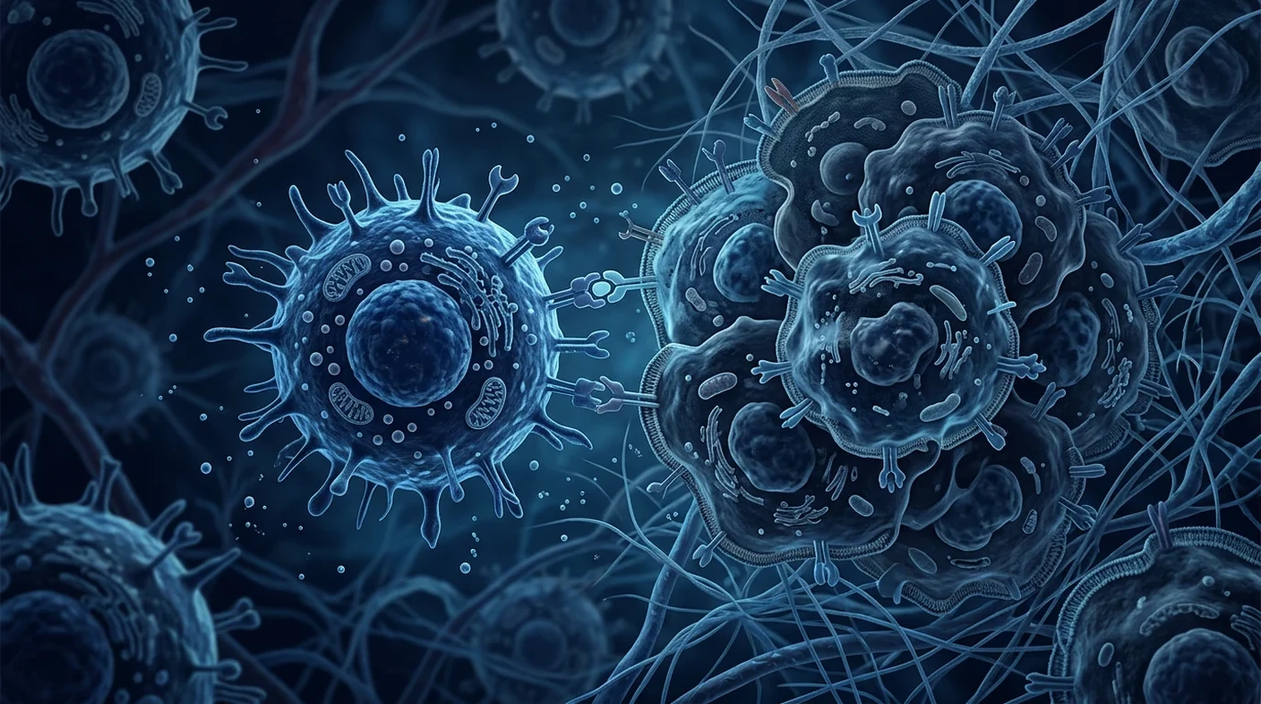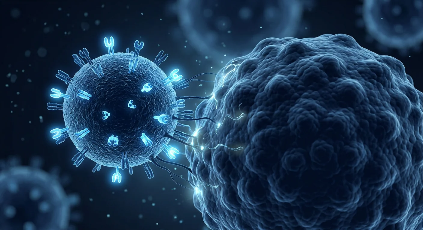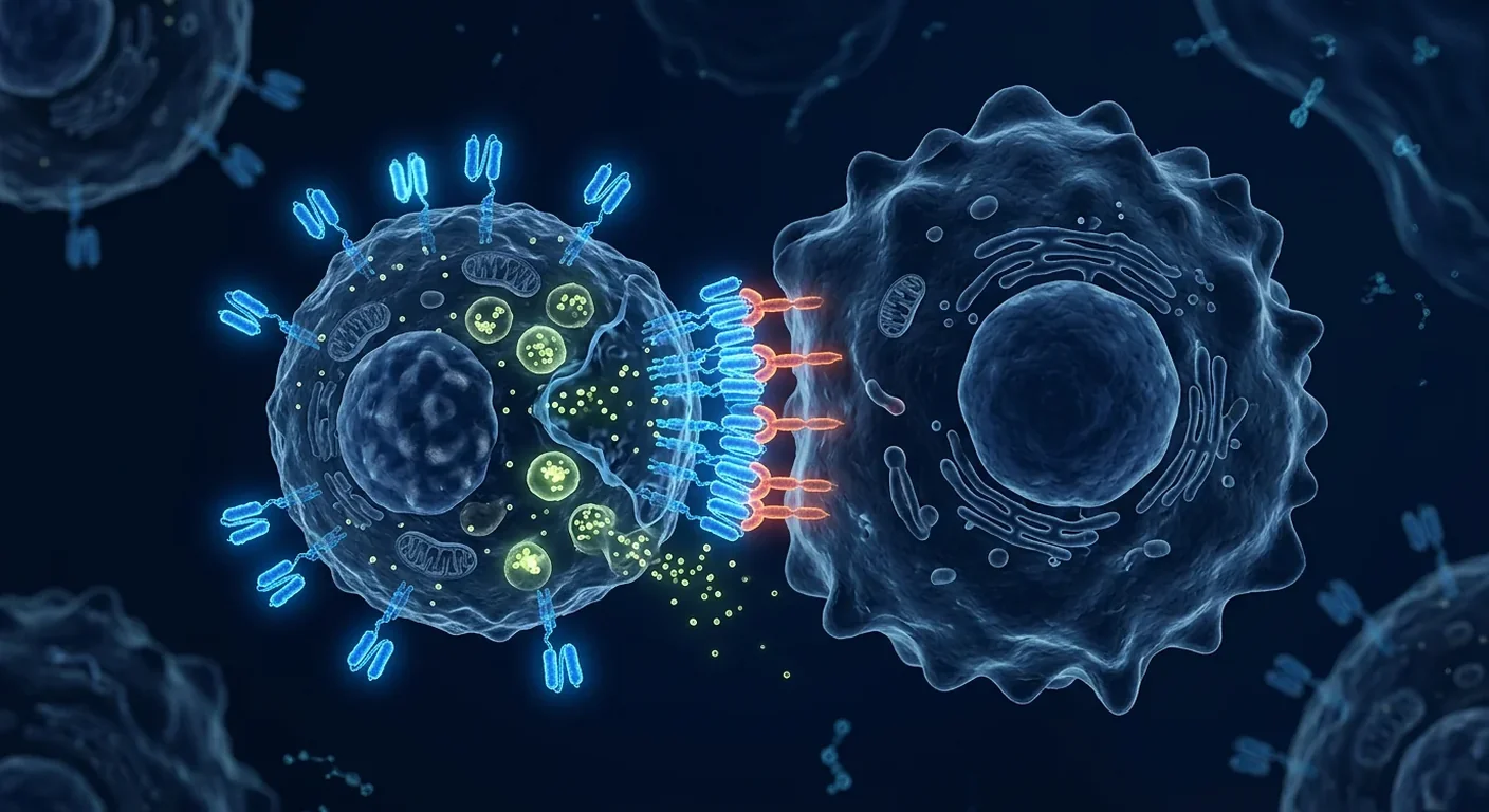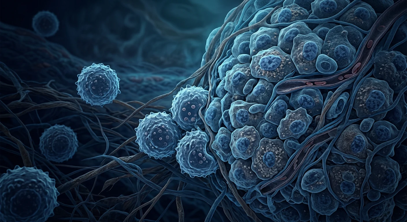
NK-cell exhaustion in the tumor microenvironment is a research description of reduced or altered NK-cell function, not a standalone diagnosis. Tumor tissue can limit trafficking, persistence, cytotoxicity, cytokine production and metabolic fitness at the same time. The result may look like exhaustion, suppression, tissue adaptation, anergy or senescence, and these states are not interchangeable.
The direct answer is cautious. A tumor’s surrounding cells and chemistry can make an otherwise competent NK cell less effective, but no universal test shows that a person will benefit from an NK-cell product or a checkpoint inhibitor. Most proposed ways to reverse exhaustion remain mechanistic, preclinical or early clinical research.
What this review does. It separates laboratory observations, animal models, observational human tissue studies and interventional clinical evidence. It does not diagnose exhaustion, promise tumor control, or suggest replacing standard oncology care. The strongest clinical example discussed here is CD19 CAR-NK therapy in lymphoid cancers; that result does not establish efficacy in solid tumors.
What do healthy NK cells normally do?
Healthy NK cells decide whether to kill by integrating activating and inhibitory signals rather than reading one universal tumor marker. Inhibitory receptors survey HLA class I, while activating receptors detect stress-associated ligands. When the balance favours activation, NK cells release perforin and granzymes, produce interferon-gamma and tumor-necrosis-factor, and can kill antibody-coated targets through CD16-mediated antibody-dependent cellular cytotoxicity.
That flexibility is useful but makes NK biology context-dependent. Cells circulating in blood are not identical to cells resident in liver, uterus, lung or a tumor. A laboratory cytotoxicity assay may describe one function under one stimulus; it cannot by itself predict trafficking, persistence, safety or response after infusion.
A report of a high or low NK-cell count is therefore not a treatment-selection result. Functional assays differ in target cells, effector-to-target ratios, cytokines and time points. Even a sophisticated receptor panel is a snapshot, not proof that an intervention will work in a particular tumor.
Is exhaustion the same as anergy, senescence or suppression?
No; “exhaustion” is an overlapping research phenotype, not a single biological switch. Chronic stimulation may produce sustained inhibitory-receptor expression, altered transcriptional programs and weaker cytokine or killing responses. Anergy can mean poor function despite receptor engagement; senescence implies durable changes associated with cellular aging; suppression can be imposed by neighboring cells or soluble mediators; tissue adaptation can be physiological.
Reviews describe recurring features such as reduced degranulation, impaired interferon-gamma output, changed NKG2D or natural-cytotoxicity-receptor expression, and metabolic rewiring [1][2]. None is a universal diagnostic threshold. The same checkpoint receptor can mark recent activation, adaptation or dysfunction depending on tissue, disease stage and assay.
For patients, this distinction prevents a common overclaim: finding an “exhausted” signature does not prove that an exhaustion-reversal drug will restore durable tumor control. It identifies a hypothesis to test alongside pathology, imaging, disease biology and clinical endpoints.
How does the tumor microenvironment suppress NK cells?
The tumor microenvironment is a multi-hit system in which cancer cells, stromal cells, myeloid cells, vessels and local chemistry constrain NK cells together. Fibroblasts, endothelial cells, tumor-associated macrophages, myeloid-derived suppressor cells, regulatory T cells and platelets can alter recruitment, receptor ligation, nutrient access and exposure to suppressive mediators [3][4].
These influences are not uniform across cancers or even across lesions in one patient. A well-perfused tumor edge may contain activated NK cells while a hypoxic, acidic core contains few cells with weaker function. Detecting NK cells in a biopsy therefore does not mean that they are reaching the right compartment or making productive contacts.
The microenvironment also changes over time. Treatment, necrosis, inflammation, fibrosis and metastatic spread can alter chemokines and oxygen gradients. Any proposed therapy must therefore be evaluated in the actual disease context rather than inferred from a single marker.
Why do trafficking and retention fail?
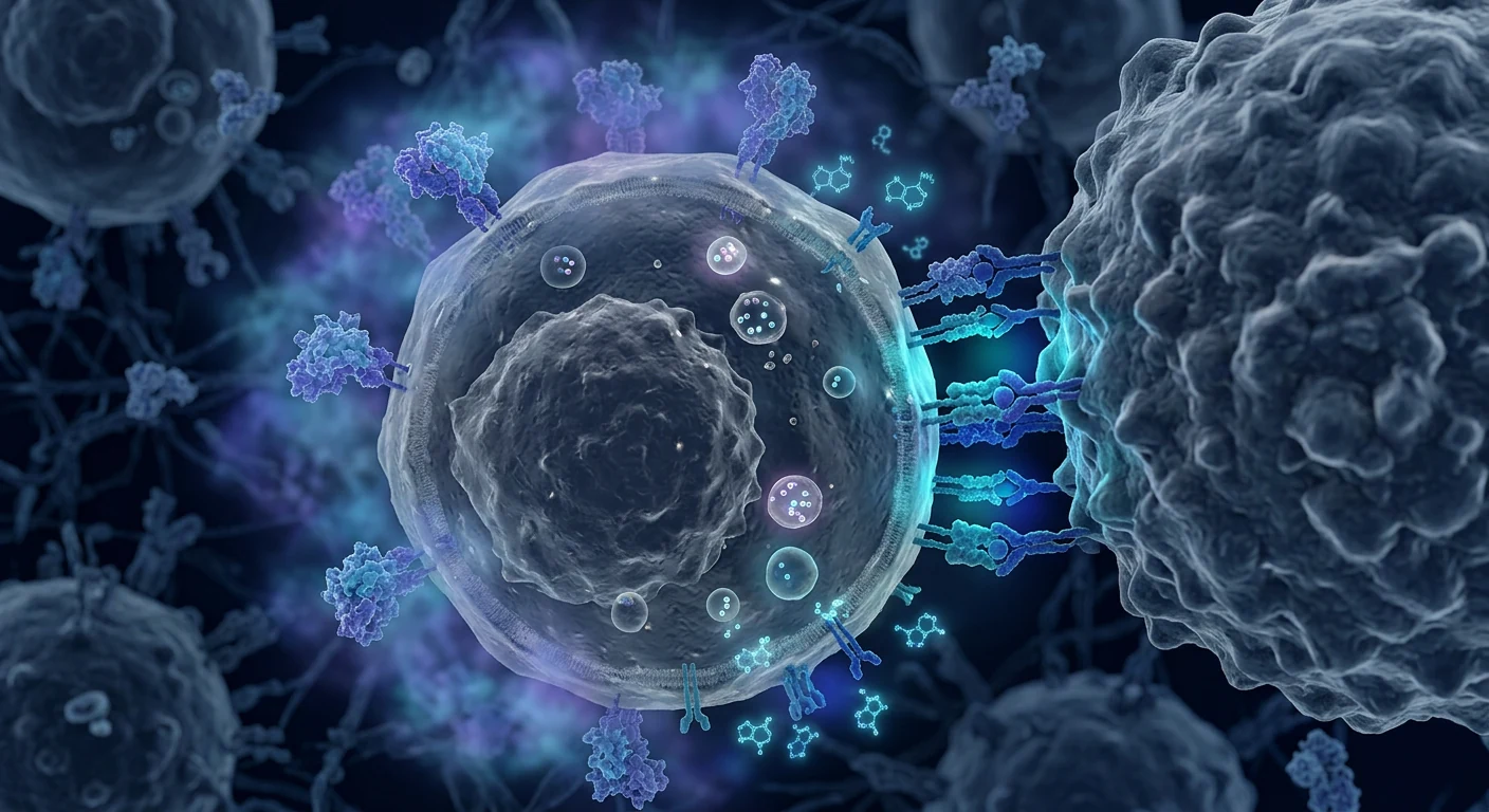
NK cells cannot kill a tumor cell they cannot reach, and infiltration is not equivalent to effective immune surveillance. Tumors may produce chemokines that do not match the receptors on available NK cells, develop abnormal vessels, compress tissue with extracellular matrix, or retain suppressive cells at the invasive margin.
Human tissue work in endometrial tumors found altered recruitment and resident NK-cell phenotype and function, illustrating that a tissue can reshape the cells that enter it [15]. Similar principles are being studied in other solid tumors, but one organ’s resident NK population cannot be treated as a universal model.
Engineering a chemokine receptor or changing local delivery could improve access in a model, yet better entry does not solve antigen heterogeneity, inhibitory ligands, nutrient scarcity or the risk of placing activated cells near healthy tissue. Trafficking is one gate in a sequence of gates.
How do TGF-beta, PGE2 and IDO reduce NK function?
TGF-beta can reduce NK activation and cytotoxic function partly by suppressing mTOR-linked cellular programs and weakening activating-receptor signals. Experimental work shows TGF-beta can impair NKG2D-mediated tumor immunity and reshape NK differentiation [6][7]. These are strong mechanistic reasons to investigate the pathway, not proof that systemic TGF-beta blockade is safe or effective for a patient.
Cancer-associated fibroblasts can add other brakes. In hepatocellular-carcinoma models and co-culture systems, PGE2 and IDO were linked with NK dysfunction [11]. Such findings help explain why an infused product may lose activity after entering tumor tissue, but cell culture concentrations and animal dosing do not define a clinically acceptable regimen.
Suppressive cytokines may also coexist with inflammation. A tumor can be inflamed enough to attract immune cells while still preventing stable cytotoxic synapses. Combination strategies targeting more than one signal are consequently being studied, but the benefit-risk balance remains disease- and product-specific.
What are the metabolic and oxygen barriers?
Low oxygen, glucose competition, acidity, lactate and adenosine can leave NK cells without the energy or signaling conditions needed for sustained killing. NK activation requires metabolic adaptation. When tumor cells consume nutrients and release lactate, acidification can blunt immune surveillance; LDHA-associated lactic-acid production has been linked to impaired T- and NK-cell activity [8].
Hypoxia adds a second constraint. In a multiple-myeloma model, low oxygen impaired NK cytotoxicity and IL-2 activation improved function in that experimental setting [9]. This does not mean high-dose IL-2 is a general solution: cytokines can cause fever, capillary effects, inflammation and other clinically important toxicities, and a model response may not persist in a heterogeneous tumor.
Adenosine is another local metabolite. Myeloid A2A-receptor signaling can suppress T- and NK-cell responses in solid-tumor models [10]. Targeting metabolism may therefore require coordinated changes to perfusion, stromal biology and immune-cell programming, not merely adding a cytokine to an infusion.
Which checkpoints and ligands are involved?
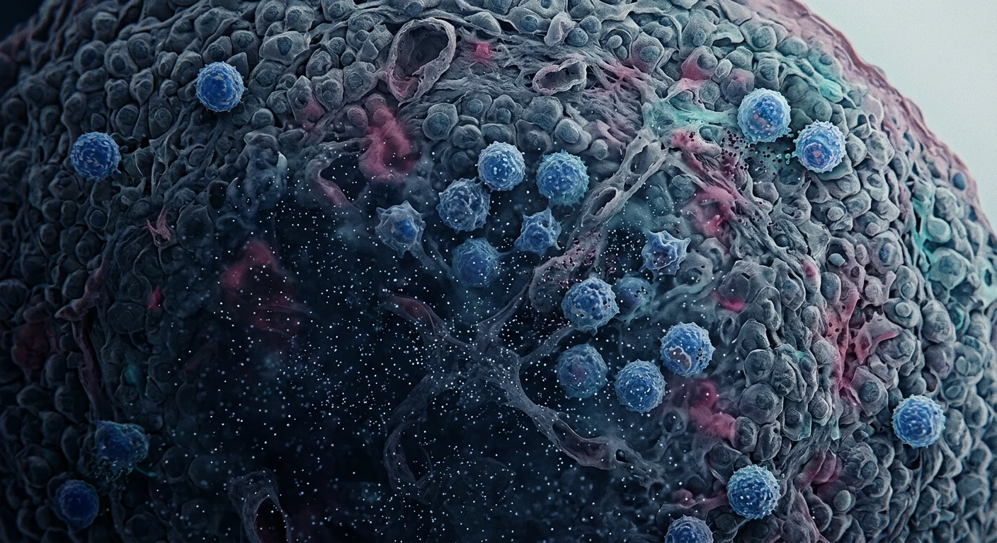
Checkpoint and ligand biology can reduce NK-cell responses, but receptor expression alone does not prove exhaustion. TIGIT can interact with CD155 or CD112 and has been implicated in NK dysfunction; blockade prevented exhaustion and enhanced antitumor immunity in experimental work [12]. Clinical translation requires evidence of pharmacology, safety and patient benefit, not just a plausible receptor.
Tumors can also alter the ligand side of the interaction. Platelets may shed NKG2D ligands, reducing immune surveillance [13]. In an oral-cancer study, N-cadherin was linked to KLRG1-mediated functional exhaustion during circulation [14]. These studies support several escape routes, not one universal checkpoint.
Checkpoint combinations can create immune toxicity and may activate cells in tissues where damage is unacceptable. A trial should therefore report dose, schedule, tissue distribution, immune-related adverse events and follow-up rather than presenting a receptor result as a clinical endpoint.
What has NK-cell therapy shown in patients?
Clinical NK-cell evidence is product-specific and strongest in selected blood cancers, while solid-tumor efficacy remains unproven as a general claim. Donor NK cells, cord-blood or iPSC-derived products, NK-92 cells, cytokine-induced memory-like cells and CAR-NK cells differ in source, expansion, phenotype, persistence and release testing.
The CD19 CAR-NK study in relapsed or refractory lymphoid tumors reported clinical activity with a comparatively limited early toxicity signal [16]. It is a bounded proof-of-concept in CD19-positive disease. It does not show that an NK cell can overcome a solid-tumor microenvironment, nor that every CAR-NK product has the same safety or durability.
Solid tumors add barriers that are often less prominent in circulating malignancies: access through abnormal stroma, antigen heterogeneity, regional hypoxia, suppressive myeloid cells and short persistence. Conditioning, cytokine support and local delivery can change outcomes and risks. The correct question is not “Do NK cells work?” but “What product, for which tumor, with which comparator and endpoint?”
Which exhaustion-reversal strategies are being investigated?
Researchers are testing combinations that improve entry, resist suppression, restore metabolism or increase target recognition, but none is a proven general solution. Investigational strategies include TGF-beta pathway edits or receptor designs, chemokine matching, checkpoint combinations, antibody-mediated ADCC, metabolic modulation, cytokine support, local administration and CAR engineering.
- TME-resistant products: genetic edits or signaling designs intended to reduce suppressive pathway effects.
- Better homing: matching chemokine receptors to the tumor’s recruitment signals.
- Combination therapy: pairing NK cells with antibodies, checkpoint agents or treatments that alter stromal and myeloid cells.
- Metabolic approaches: addressing lactate, adenosine, oxygen gradients or mitochondrial fitness.
- Local delivery: placing cells closer to a lesion, with site-specific procedural and off-target risks.
Laboratory rescue is not the same as a safe regimen. Gene editing may introduce manufacturing and genomic risks; cytokines may amplify inflammation; checkpoint inhibition may injure normal tissues; and local delivery may not reach every lesion. Each strategy needs controlled clinical testing.
How should a patient read an NK-cell therapy trial in Bangkok?
A credible trial identifies the product, protocol, endpoints, oversight and risks clearly before asking a patient to interpret a response claim. Ask the oncology team to review:
- Is the study registered, and is the site authorised for the intervention?
- What is the exact source, engineering, dose, schedule, viability and release testing?
- What conditioning or cytokine support is required, and who manages infection, cytopenia, cytokine-release symptoms or neurotoxicity?
- How are trafficking, persistence, target expression and response measured, and is there a comparator?
- What happens if the tumor progresses during manufacturing or follow-up?
- Which results are clinical endpoints, and which are exploratory biomarkers?
Marketing language deserves scrutiny when it uses “detox,” “immune reset,” or a single blood test as proof of tumor response. A regulated research pathway states uncertainty and records adverse events. It does not imply that an unapproved infusion is equivalent to an authorised cancer treatment.
What should patients and families take away?
Overcoming NK-cell dysfunction will probably require matching the cell product, tumor biology, delivery route and combination strategy, but no general exhaustion-reversal treatment has been proven. The microenvironment is not a single wall; it is a changing network of physical, metabolic and cellular barriers.
Discuss any trial with the clinician responsible for the cancer diagnosis. Do not delay, stop or replace standard treatment because a tumor is described as “NK exhausted,” and do not infer an individual prognosis from a research phenotype. A useful consultation focuses on the registered protocol, alternatives, expected uncertainties, monitoring and the patient’s goals.
Frequently asked questions
Is NK-cell exhaustion a diagnosis?
No. It is a research description that may combine reduced function, altered receptors, metabolic stress and tissue suppression. It does not by itself diagnose a patient or select a treatment.
Can the tumor microenvironment make NK-cell therapy fail?
It can create barriers to trafficking, persistence and killing, but failure or benefit depends on the product, tumor, dose, conditioning and trial design. Laboratory evidence cannot predict an individual outcome alone.
Does a checkpoint inhibitor reverse NK exhaustion?
Checkpoint pathways such as TIGIT are being studied, including in preclinical models. A mechanistic effect is not proof of a safe, durable benefit for every cancer or patient.
Are NK-cell therapies approved for solid tumors?
Most NK-cell interventions for solid tumors remain investigational. Verify the specific product, regulatory status and registered trial rather than relying on a clinic’s general description.
Is CAR-NK proven to overcome the tumor microenvironment?
No. CD19 CAR-NK clinical data in lymphoid tumors are an important proof-of-concept, but they do not establish solid-tumor efficacy or solve hypoxia, acidity, stromal suppression or antigen heterogeneity.
What should I ask before joining a trial?
Ask for the registry number, product identity, manufacturing and release criteria, conditioning, endpoints, comparator, follow-up and adverse-event plan. Keep the treating oncology team responsible for the overall cancer plan.
References
- Jia H, Yang H, Xiong H, Luo KQ. “NK cell exhaustion in the tumor microenvironment.” Frontiers in Immunology. 2023. doi:10.3389/fimmu.2023.1303605 ↩
- Bi J, Tian Z. “NK Cell Exhaustion.” Frontiers in Immunology. 2017. doi:10.3389/fimmu.2017.00760 ↩
- Stojanovic A, Correia MP, Cerwenka A. “Shaping of NK Cell Responses by the Tumor Microenvironment.” Cancer Microenvironment. 2013. doi:10.1007/s12307-012-0125-8 ↩
- Hu Z, Xu X, Wei H. “The Adverse Impact of Tumor Microenvironment on NK-Cell.” Frontiers in Immunology. 2021. doi:10.3389/fimmu.2021.633361 ↩
- Terren I, Orrantia A, Vitallé J, et al.. “NK Cell Metabolism and Tumor Microenvironment.” Frontiers in Immunology. 2019. doi:10.3389/fimmu.2019.02278 ↩
- Viel S, Marçais A, Souza-Fonseca Guimaraes F, et al.. “TGF-beta inhibits the activation and functions of NK cells by repressing the mTOR pathway.” Science Signaling. 2016. doi:10.1126/scisignal.aad1884 ↩
- Lazarova M, Steinle A. “Impairment of NKG2D-Mediated Tumor Immunity by TGF-beta.” Frontiers in Immunology. 2019. doi:10.3389/fimmu.2019.02689 ↩
- Brand A, Singer K, Koehl GE, et al.. “LDHA-Associated Lactic Acid Production Blunts Tumor Immunosurveillance by T and NK Cells.” Cell Metabolism. 2016. doi:10.1016/j.cmet.2016.08.011 ↩
- Sarkar S, Germeraad WTV, Rouschop KMA, et al.. “Hypoxia Induced Impairment of NK Cell Cytotoxicity against Multiple Myeloma Can Be Overcome by IL-2 Activation of the NK Cells.” PLoS ONE. 2013. doi:10.1371/journal.pone.0064835 ↩
- Cekic C, Day YJ, Sag D, Linden J. “Myeloid Expression of Adenosine A2A Receptor Suppresses T and NK Cell Responses in the Solid Tumor Microenvironment.” Cancer Research. 2014. doi:10.1158/0008-5472.CAN-13-3583 ↩
- Li T, Yang Y, Hua X, et al.. “Hepatocellular carcinoma-associated fibroblasts trigger NK cell dysfunction via PGE2 and IDO.” Cancer Letters. 2012. doi:10.1016/j.canlet.2011.12.020 ↩
- Zhang Q, Bi J, Zheng X, et al.. “Blockade of the checkpoint receptor TIGIT prevents NK cell exhaustion and elicits potent anti-tumor immunity.” Nature Immunology. 2018. doi:10.1038/s41590-018-0132-0 ↩
- Maurer S, Kropp KN, Klein G, et al.. “Platelet-mediated shedding of NKG2D ligands impairs NK cell immune-surveillance of tumor cells.” OncoImmunology. 2018. doi:10.1080/2162402X.2017.1364827 ↩
- Lou C, Wu K, Shi J, et al.. “N-cadherin protects oral cancer cells from NK cell killing in the circulation by inducing NK cell functional exhaustion via the KLRG1 receptor.” Journal for ImmunoTherapy of Cancer. 2022. doi:10.1136/jitc-2022-005061 ↩
- Degos C, Heinemann M, Barrou J, et al.. “Endometrial Tumor Microenvironment Alters Human NK Cell Recruitment, and Resident NK Cell Phenotype and Function.” Frontiers in Immunology. 2019. doi:10.3389/fimmu.2019.00877 ↩
- Liu E, Marin D, Banerjee P, et al.. “Use of CAR-Transduced Natural Killer Cells in CD19-Positive Lymphoid Tumors.” New England Journal of Medicine. 2020. doi:10.1056/NEJMoa1910607 ↩
肿瘤微环境中的 NK 细胞耗竭,是对 NK 细胞功能减弱或改变的研究性描述,并不是一个独立诊断。肿瘤组织可以同时限制细胞迁移、存续、细胞毒性、细胞因子产生和代谢适应。结果可能表现为耗竭、抑制、组织适应、无反应或衰老,而这些状态并不相同。
直接答案需要保持谨慎。肿瘤周围的细胞与化学环境确实可能使原本有功能的 NK 细胞难以杀伤,但没有一种通用检测能够证明某人一定会从 NK 细胞产品或检查点抑制剂中获益。多数逆转耗竭的方法仍属于机制、临床前或早期临床研究。
本文的范围。本文区分实验室观察、动物模型、人体组织观察和干预性临床证据,不诊断耗竭、不承诺肿瘤控制,也不建议替代标准肿瘤治疗。文中最明确的临床例子是淋巴系统肿瘤中的 CD19 CAR-NK;该结果不能证明实体瘤疗效。
健康 NK 细胞通常做什么?
健康 NK 细胞通过整合激活与抑制信号决定是否杀伤,而不是读取一个通用的肿瘤标志物。抑制性受体监测 HLA I 类分子,激活性受体识别应激配体;当激活信号占优势时,NK 细胞释放穿孔素和颗粒酶,产生干扰素-γ与肿瘤坏死因子,并可通过 CD16 介导抗体依赖性细胞毒作用。
血液中的 NK 细胞与肝脏、子宫、肺或肿瘤内的细胞并不相同。实验室细胞毒性试验只描述特定刺激下的一项功能,不能单独预测回输后的迁移、存续、安全性或疗效。NK 细胞数量高低也不是治疗选择结果;检测方法、靶细胞、细胞因子与时间点都会影响读数。
耗竭是否等同于无反应、衰老或抑制?
不是;“耗竭”是彼此重叠的研究表型,不是一个单一开关。持续刺激可带来抑制性受体表达、转录程序改变以及细胞因子或杀伤减弱。无反应、衰老、邻近细胞造成的抑制和正常组织适应,可能有相似表现却不应混为一谈。
综述常提到脱颗粒、干扰素-γ、NKG2D、自然细胞毒性受体和代谢程序的变化[1][2],但没有统一诊断阈值。同一检查点受体可能代表近期激活、适应或功能障碍,取决于组织、病程和检测。
肿瘤微环境如何抑制 NK 细胞?
肿瘤微环境是多重障碍系统,癌细胞、基质细胞、髓系细胞、血管和局部化学环境共同限制 NK 细胞。成纤维细胞、内皮细胞、肿瘤相关巨噬细胞、髓源抑制性细胞、调节性 T 细胞和血小板可改变募集、受体结合、营养供应与抑制性介质暴露[3][4]。
不同癌症、甚至同一患者的不同病灶,其微环境并不相同。活跃的肿瘤边缘可能有 NK 细胞,而缺氧、酸性的核心区域细胞很少且功能更弱。因此,活检发现 NK 细胞并不等于它们到达正确位置并完成有效杀伤。
为什么迁移和滞留会失败?
NK 细胞无法杀伤接触不到的肿瘤细胞,浸润也不等于有效免疫监视。肿瘤可能产生与 NK 细胞受体不匹配的趋化因子,形成异常血管,以细胞外基质压迫组织,或在侵袭边缘保留抑制性细胞。子宫内膜肿瘤组织研究发现,募集以及局部 NK 细胞表型和功能发生改变[15]。
改变趋化受体或局部给药也许能改善进入病灶,但不能解决抗原异质性、抑制配体、营养不足或健康组织的脱靶风险。迁移只是多个关卡中的一个。
TGF-β、PGE2 和 IDO 如何降低 NK 功能?
TGF-β 可通过抑制 mTOR 相关程序和削弱激活受体信号,降低 NK 的激活与细胞毒性。实验研究显示它能损害 NKG2D 介导的肿瘤免疫[6][7]。这支持继续研究该通路,但不能证明全身阻断 TGF-β 对患者安全有效。
癌相关成纤维细胞也会增加其他制动因素。肝细胞癌模型和共培养研究将 PGE2、IDO 与 NK 功能障碍联系起来[11]。细胞培养浓度和动物剂量不能直接规定临床方案。
代谢和缺氧障碍是什么?
低氧、葡萄糖竞争、酸化、乳酸和腺苷,可能让 NK 细胞缺少持续杀伤所需的能量与信号。肿瘤细胞消耗营养并释放乳酸,LDHA 相关乳酸生成与 T 细胞和 NK 细胞免疫监视减弱有关[8]。多发性骨髓瘤模型中,低氧降低 NK 细胞毒性,而 IL-2 在该实验环境中改善了功能[9];这不代表高剂量 IL-2 是普遍安全的解决方案。腺苷 A2A 受体信号也可抑制实体瘤模型中的 T、NK 反应[10]。
哪些检查点和配体参与其中?
检查点和配体会降低 NK 反应,但单独的受体表达不能证明耗竭。研究正在关注 TIGIT 与 CD155/CD112 的相互作用;实验中阻断 TIGIT 可阻止耗竭并增强抗肿瘤免疫[12]。血小板可脱落 NKG2D 配体而削弱免疫监视,N-钙黏蛋白也与 KLRG1 介导的功能耗竭有关[13][14]。这些结果说明存在多条逃逸路径,而不是一个通用检查点。
NK 细胞治疗在人类患者中显示了什么?
临床证据取决于产品;实体瘤疗效仍不能作为一般性结论。供者、脐带血、iPSC、NK-92、细胞因子诱导记忆样 NK 和 CAR-NK 在来源、扩增、表型、存续和放行检测方面均不同。
复发难治淋巴系统肿瘤中的 CD19 CAR-NK 研究报告了临床活性和较有限的早期毒性信号[16],但它只是 CD19 阳性疾病的受限概念验证。实体瘤还增加基质通行、抗原异质性、缺氧、髓系抑制和短暂存续等障碍。
哪些逆转耗竭策略正在研究?
研究者正在同时改善进入病灶、抵抗抑制、恢复代谢和增加靶向识别,但没有一种普遍方案已被证实。方向包括 TGF-β 通路改造、趋化受体匹配、检查点联合、抗体介导 ADCC、代谢调节、细胞因子支持、局部给药和 CAR 工程。
- 抗微环境产品:减少抑制信号影响的基因或受体设计。
- 改善归巢:让趋化受体匹配肿瘤信号。
- 联合治疗:与抗体、检查点药物或改变基质和髓系细胞的疗法联合。
- 代谢策略:研究乳酸、腺苷、氧梯度和线粒体适应。
实验室中的功能恢复不等于安全方案。基因编辑、细胞因子和检查点阻断都可能带来制造、炎症或正常组织损伤风险,需要受控临床测试。
在曼谷阅读 NK 细胞试验时应看什么?
可信试验会在患者解读疗效前清楚说明产品、方案、终点、监管和风险。请询问注册号、细胞来源与放行标准、预处理和细胞因子支持、感染和细胞因子释放管理、迁移与存续检测、比较组、随访,以及哪些是临床终点、哪些只是探索性生物标志物。不要把“排毒”“免疫重置”或一次血液检测当作肿瘤反应证明,也不要将未批准输注等同于获批治疗。
患者和家属应记住什么?
克服 NK 功能障碍可能需要匹配细胞产品、肿瘤生物学、给药方式和联合策略,但尚无普遍有效的耗竭逆转治疗。请与负责肿瘤诊断的医生讨论试验,不要因“NK 耗竭”描述而延迟、停止或替代标准治疗,也不要从研究表型推断个人预后。
常见问题
NK 细胞耗竭是诊断吗?
不是。它是可能包含功能下降、受体改变、代谢压力和组织抑制的研究描述,不能单独诊断或选择治疗。
肿瘤微环境会让 NK 治疗失败吗?
它可能限制迁移、存续和杀伤,但结果取决于产品、肿瘤、剂量、预处理和试验设计。
检查点抑制剂能逆转 NK 耗竭吗?
TIGIT 等通路正在研究,机制或动物结果不等于对每位患者安全且持久的获益。
实体瘤的 NK 治疗获批了吗?
大多数仍在研究阶段。应核实具体产品、监管状态和注册试验。
CAR-NK 已经解决肿瘤微环境了吗?
没有。淋巴瘤中的 CD19 数据不能证明实体瘤疗效,也不能解决缺氧、酸化、基质抑制和抗原异质性。
参加试验前应问什么?
询问注册号、产品、生产和放行标准、预处理、终点、比较组、随访和不良事件计划,并让肿瘤医生负责总体方案。
参考文献
- Jia H, Yang H, Xiong H, Luo KQ. “NK cell exhaustion in the tumor microenvironment.” Frontiers in Immunology. 2023. doi:10.3389/fimmu.2023.1303605 ↩
- Bi J, Tian Z. “NK Cell Exhaustion.” Frontiers in Immunology. 2017. doi:10.3389/fimmu.2017.00760 ↩
- Stojanovic A, Correia MP, Cerwenka A. “Shaping of NK Cell Responses by the Tumor Microenvironment.” Cancer Microenvironment. 2013. doi:10.1007/s12307-012-0125-8 ↩
- Hu Z, Xu X, Wei H. “The Adverse Impact of Tumor Microenvironment on NK-Cell.” Frontiers in Immunology. 2021. doi:10.3389/fimmu.2021.633361 ↩
- Terren I, Orrantia A, Vitallé J, et al.. “NK Cell Metabolism and Tumor Microenvironment.” Frontiers in Immunology. 2019. doi:10.3389/fimmu.2019.02278 ↩
- Viel S, Marçais A, Souza-Fonseca Guimaraes F, et al.. “TGF-beta inhibits the activation and functions of NK cells by repressing the mTOR pathway.” Science Signaling. 2016. doi:10.1126/scisignal.aad1884 ↩
- Lazarova M, Steinle A. “Impairment of NKG2D-Mediated Tumor Immunity by TGF-beta.” Frontiers in Immunology. 2019. doi:10.3389/fimmu.2019.02689 ↩
- Brand A, Singer K, Koehl GE, et al.. “LDHA-Associated Lactic Acid Production Blunts Tumor Immunosurveillance by T and NK Cells.” Cell Metabolism. 2016. doi:10.1016/j.cmet.2016.08.011 ↩
- Sarkar S, Germeraad WTV, Rouschop KMA, et al.. “Hypoxia Induced Impairment of NK Cell Cytotoxicity against Multiple Myeloma Can Be Overcome by IL-2 Activation of the NK Cells.” PLoS ONE. 2013. doi:10.1371/journal.pone.0064835 ↩
- Cekic C, Day YJ, Sag D, Linden J. “Myeloid Expression of Adenosine A2A Receptor Suppresses T and NK Cell Responses in the Solid Tumor Microenvironment.” Cancer Research. 2014. doi:10.1158/0008-5472.CAN-13-3583 ↩
- Li T, Yang Y, Hua X, et al.. “Hepatocellular carcinoma-associated fibroblasts trigger NK cell dysfunction via PGE2 and IDO.” Cancer Letters. 2012. doi:10.1016/j.canlet.2011.12.020 ↩
- Zhang Q, Bi J, Zheng X, et al.. “Blockade of the checkpoint receptor TIGIT prevents NK cell exhaustion and elicits potent anti-tumor immunity.” Nature Immunology. 2018. doi:10.1038/s41590-018-0132-0 ↩
- Maurer S, Kropp KN, Klein G, et al.. “Platelet-mediated shedding of NKG2D ligands impairs NK cell immune-surveillance of tumor cells.” OncoImmunology. 2018. doi:10.1080/2162402X.2017.1364827 ↩
- Lou C, Wu K, Shi J, et al.. “N-cadherin protects oral cancer cells from NK cell killing in the circulation by inducing NK cell functional exhaustion via the KLRG1 receptor.” Journal for ImmunoTherapy of Cancer. 2022. doi:10.1136/jitc-2022-005061 ↩
- Degos C, Heinemann M, Barrou J, et al.. “Endometrial Tumor Microenvironment Alters Human NK Cell Recruitment, and Resident NK Cell Phenotype and Function.” Frontiers in Immunology. 2019. doi:10.3389/fimmu.2019.00877 ↩
- Liu E, Marin D, Banerjee P, et al.. “Use of CAR-Transduced Natural Killer Cells in CD19-Positive Lymphoid Tumors.” New England Journal of Medicine. 2020. doi:10.1056/NEJMoa1910607 ↩
إنهاك خلايا NK في البيئة الدقيقة للورم هو وصف بحثي لانخفاض وظيفة الخلايا أو تبدلها، وليس تشخيصاً مستقلاً. يمكن لنسيج الورم أن يحد في الوقت نفسه من الهجرة والبقاء والسمية الخلوية وإنتاج السيتوكينات واللياقة الأيضية. وقد يبدو ذلك إنهاكاً أو تثبيطاً أو تكيّفاً نسيجياً أو لااستجابة أو شيخوخة، وهذه حالات ليست مترادفة.
الإجابة المباشرة تتطلب الحذر. قد تجعل الخلايا والمواد الكيميائية المحيطة بالورم خلية NK السليمة أقل قدرة على القتل، لكن لا يوجد اختبار عام يثبت أن شخصاً بعينه سيستفيد من منتج NK أو مثبط نقطة تفتيش. معظم طرق عكس الإنهاك ما زالت آلية أو قبل سريرية أو في مراحل سريرية مبكرة.
نطاق المقال. نفرق بين ملاحظات المختبر والنماذج الحيوانية ودراسات الأنسجة البشرية والأدلة السريرية التدخلية. لا يشخّص المقال الإنهاك ولا يعد بالسيطرة على الورم ولا يقترح استبدال الرعاية القياسية. المثال السريري الأوضح هو CAR-NK ضد CD19 في أورام لمفاوية، ولا يثبت ذلك فعالية في الأورام الصلبة.
ماذا تفعل خلايا NK السليمة عادةً؟
تقرر خلايا NK السليمة القتل بدمج إشارات التنشيط والتثبيط، لا بقراءة واسم ورمي عام واحد. تراقب المستقبلات المثبطة HLA-I، وتتعرف المستقبلات المنشطة على روابط الإجهاد؛ وعندما ترجح الإشارة المنشطة تطلق الخلايا البيرفورين والغرانزيمات وتنتج إنترفيرون-غاما وعامل نخر الورم، ويمكنها قتل الأهداف المغطاة بالأجسام المضادة عبر CD16.
ليست خلايا الدم مماثلة للخلايا المقيمة في الكبد أو الرحم أو الرئة أو الورم. يصف اختبار السمية في المختبر وظيفة واحدة تحت تحفيز محدد، ولا يتنبأ وحده بالهجرة أو البقاء أو الأمان أو الاستجابة بعد الحقن. كما أن عدد خلايا NK ليس نتيجة لاختيار العلاج؛ فطريقة القياس والهدف والسيتوكين والزمن تغير القراءة.
هل يساوي الإنهاك اللااستجابة أو الشيخوخة أو التثبيط؟
لا؛ الإنهاك نمط بحثي متداخل وليس مفتاحاً بيولوجياً واحداً. قد يسبب التحفيز المزمن مستقبلات مثبطة وبرامج نسخية متبدلة وضعف إنتاج السيتوكينات أو القتل. قد تتشابه اللااستجابة والشيخوخة والتثبيط المفروض من الخلايا المجاورة والتكيف الطبيعي، لكنها ليست شيئاً واحداً.
تذكر المراجعات تغير نزع الحبيبات وإنتاج إنترفيرون-غاما وNKG2D ومستقبلات السمية الطبيعية والبرمجة الأيضية[1][2]، ولا يوجد حد تشخيصي موحد. قد يدل المستقبل نفسه على تنشيط حديث أو تكيف أو خلل، بحسب النسيج والمرض والاختبار.
كيف تثبط البيئة الدقيقة للورم خلايا NK؟
البيئة الدقيقة منظومة عوائق متعددة، تتعاون فيها الخلايا السرطانية واللحمة والخلايا النخاعية والأوعية والكيمياء المحلية. يمكن للخلايا الليفية والبطانية والبلعميات المرتبطة بالورم والخلايا النخاعية الكابحة والخلايا التائية المنظمة والصفيحات تغيير التجنيد وارتباط المستقبلات والمواد الغذائية والتعرض للوسائط المثبطة[3][4].
لا تتشابه هذه التأثيرات بين السرطانات أو حتى بين آفات المريض نفسه. قد تحتوي حافة الورم على خلايا NK بينما يحوي القلب ناقص الأكسجين والحامضي خلايا أقل وأضعف. لذلك لا يعني وجودها في الخزعة وصولها إلى الموضع الصحيح أو قتلها المنتج.
لماذا تفشل الهجرة والاحتفاظ؟
لا تستطيع الخلية قتل هدف لا تصل إليه، كما أن الارتشاح لا يساوي مراقبة مناعية فعالة. قد تنتج الأورام كيموكينات لا تطابق مستقبلات NK، أو أوعية غير طبيعية، أو مصفوفة خارج خلوية ضاغطة، أو خلايا مثبطة عند الحافة الغازية. أظهرت دراسة في أورام بطانة الرحم تغير تجنيد خلايا NK المقيمة ونمطها ووظيفتها[15]، لكن لا يجوز تعميم عضو واحد.
قد تحسن هندسة مستقبل كيموكيني أو الإيصال الموضعي الوصول، لكنها لا تحل عدم تجانس المستضد أو الروابط المثبطة أو نقص الغذاء أو خطر الضرر خارج الهدف. الهجرة بوابة واحدة فقط.
كيف تخفض TGF-beta وPGE2 وIDO وظيفة NK؟
يمكن لـ TGF-beta خفض تنشيط NK والسمية الخلوية جزئياً عبر كبح برامج مرتبطة بـ mTOR وإضعاف إشارات المستقبلات المنشطة. وتظهر الدراسات التجريبية إضعاف مناعة الورم بوساطة NKG2D[6][7]، لكن ذلك لا يثبت أن الحجب الجهازي آمن أو فعال للمرضى.
وقد تضيف الخلايا الليفية المرتبطة بالسرطان مكابح أخرى؛ إذ ربطت نماذج سرطان الكبد وأنظمة المشاركة الخلوية بين PGE2 وIDO وخلل NK[11]. لا تحدد تراكيز الزراعة أو جرعات الحيوان نظاماً سريرياً مقبولاً.
ما عوائق الأيض ونقص الأكسجين؟
قد يترك نقص الأكسجين والتنافس على الغلوكوز والحموضة واللاكتات والأدينوزين خلايا NK دون الطاقة والإشارات اللازمة للقتل المستمر. تستهلك الخلايا الورمية المغذيات وتطلق اللاكتات، وارتبط إنتاج اللاكتات المرتبط بـ LDHA بضعف مراقبة الخلايا التائية وNK[8]. وفي نموذج الورم النقوي أضعف نقص الأكسجين السمية، وحسّن IL-2 الوظيفة في ذلك السياق التجريبي[9]، لكنه ليس حلاً عاماً آمناً. كما قد يكبح مستقبل A2A للأدينوزين استجابات T وNK في النماذج الصلبة[10].
ما نقاط التفتيش والروابط المعنية؟
قد تخفض نقاط التفتيش والروابط استجابة NK، لكن وجود المستقبل وحده لا يثبت الإنهاك. تجري دراسة TIGIT مع CD155 أو CD112، وقد منع حجبه الإنهاك وعزز المناعة في تجارب قبل سريرية[12]. ويمكن للصفيحات طرح روابط NKG2D، كما ارتبط N-cadherin بإنهاك وظيفي عبر KLRG1[13][14]. هذه مسارات هروب متعددة وليست نقطة واحدة.
ماذا أظهر علاج NK لدى البشر؟
الأدلة السريرية تعتمد على المنتج، بينما لا تزال فعالية الأورام الصلبة غير مثبتة كقاعدة عامة. تختلف خلايا المتبرعين ودم الحبل وiPSC وNK-92 وCIML-NK وCAR-NK في المصدر والتوسيع والنمط والبقاء واختبارات الإفراج.
أظهرت دراسة CAR-NK ضد CD19 في أورام لمفاوية ناكسة أو مقاومة نشاطاً سريرياً وإشارة سمية مبكرة محدودة نسبياً[16]؛ وهي برهان مفهوم محدود في مرض إيجابي CD19، لا برهان على تجاوز بيئة الورم الصلب. وتضيف الأورام الصلبة حواجز السدى ونقص الأكسجين والخلايا النخاعية المثبطة وقصر البقاء.
ما استراتيجيات عكس الإنهاك قيد البحث؟
يختبر الباحثون تحسين الدخول ومقاومة التثبيط واستعادة الأيض وزيادة التعرف، لكن لا توجد طريقة عامة مثبتة. تشمل الاتجاهات هندسة مسار TGF-beta، ومواءمة الكيموكينات، ودمج نقاط التفتيش، وADCC المعتمد على الأجسام المضادة، وتعديل الأيض، ودعم السيتوكينات، والإيصال الموضعي وهندسة CAR.
- منتجات مقاومة للبيئة: تعديلات جينية أو مستقبلية تقلل أثر المسارات المثبطة.
- توجيه أفضل: مطابقة المستقبلات لإشارات الورم.
- علاجات مشتركة: مع الأجسام المضادة أو الأدوية التي تغير السدى والخلايا النخاعية.
- مقاربات أيضية: اللاكتات والأدينوزين وتدرجات الأكسجين والميتوكوندريا.
إنقاذ الوظيفة في المختبر لا يساوي نظاماً آمناً؛ فالتعديل الجيني والسيتوكينات وحجب نقاط التفتيش قد تسبب مخاطر تصنيع أو التهاب أو أذى نسيجي، وتحتاج إلى اختبار سريري مضبوط.
كيف يقرأ المريض تجربة NK في بانكوك؟
تحدد التجربة الموثوقة المنتج والبروتوكول والنهايات والرقابة والمخاطر قبل مطالبة المريض بتفسير الاستجابة. اسأل عن رقم التسجيل، مصدر الخلية ومعايير الإفراج، التهيئة ودعم السيتوكينات، تدبير العدوى ونقص الدم وأعراض تحرر السيتوكينات، وقياس الهجرة والبقاء والمستضد والمقارنة والمتابعة، وما هو نقطة سريرية وما هو واسم استكشافي. لا تجعل عبارات «إزالة السموم» أو «إعادة ضبط المناعة» أو فحص دم واحد دليلاً على استجابة الورم، ولا تساوِ الحقن غير المعتمد بعلاج مرخص.
ما الخلاصة للمرضى والعائلات؟
قد يتطلب تجاوز خلل NK مطابقة المنتج وبيولوجيا الورم وطريق الإيصال والعلاج المشترك، لكن لم يثبت علاج عام لعكس الإنهاك. ناقش التجربة مع طبيب الأورام المسؤول، ولا تؤخر أو توقف أو تستبدل الرعاية القياسية بسبب وصف «إنهاك NK»، ولا تستنتج إنذاراً فردياً من نمط بحثي.
الأسئلة الشائعة
هل إنهاك NK تشخيص؟
لا. هو وصف بحثي قد يجمع انخفاض الوظيفة وتغير المستقبلات والضغط الأيضي والتثبيط النسيجي، ولا يختار العلاج وحده.
هل قد تجعل البيئة الورمية علاج NK يفشل؟
قد تعيق الهجرة والبقاء والقتل، لكن النتيجة تعتمد على المنتج والورم والجرعة والتهيئة وتصميم التجربة.
هل يعكس مثبط نقطة التفتيش إنهاك NK؟
تُدرس مسارات مثل TIGIT، لكن النتيجة الآلية أو الحيوانية لا تثبت فائدة آمنة ودائمة لكل مريض.
هل علاجات NK معتمدة للأورام الصلبة؟
معظمها بحثي. تحقق من المنتج المحدد ووضعه التنظيمي والتجربة المسجلة.
هل أثبت CAR-NK تجاوز البيئة الورمية؟
لا. بيانات CD19 في الأورام اللمفاوية لا تثبت فعالية الأورام الصلبة ولا تحل نقص الأكسجين أو الحموضة أو السدى أو عدم تجانس المستضد.
ماذا أسأل قبل التسجيل؟
اطلب رقم التسجيل وهوية المنتج ومعايير التصنيع والإفراج والتهيئة والنهايات والمقارنة والمتابعة وخطة الأحداث الضارة، ودع طبيب الأورام يدير الخطة العامة.
المراجع
- Jia H, Yang H, Xiong H, Luo KQ. “NK cell exhaustion in the tumor microenvironment.” Frontiers in Immunology. 2023. doi:10.3389/fimmu.2023.1303605 ↩
- Bi J, Tian Z. “NK Cell Exhaustion.” Frontiers in Immunology. 2017. doi:10.3389/fimmu.2017.00760 ↩
- Stojanovic A, Correia MP, Cerwenka A. “Shaping of NK Cell Responses by the Tumor Microenvironment.” Cancer Microenvironment. 2013. doi:10.1007/s12307-012-0125-8 ↩
- Hu Z, Xu X, Wei H. “The Adverse Impact of Tumor Microenvironment on NK-Cell.” Frontiers in Immunology. 2021. doi:10.3389/fimmu.2021.633361 ↩
- Terren I, Orrantia A, Vitallé J, et al.. “NK Cell Metabolism and Tumor Microenvironment.” Frontiers in Immunology. 2019. doi:10.3389/fimmu.2019.02278 ↩
- Viel S, Marçais A, Souza-Fonseca Guimaraes F, et al.. “TGF-beta inhibits the activation and functions of NK cells by repressing the mTOR pathway.” Science Signaling. 2016. doi:10.1126/scisignal.aad1884 ↩
- Lazarova M, Steinle A. “Impairment of NKG2D-Mediated Tumor Immunity by TGF-beta.” Frontiers in Immunology. 2019. doi:10.3389/fimmu.2019.02689 ↩
- Brand A, Singer K, Koehl GE, et al.. “LDHA-Associated Lactic Acid Production Blunts Tumor Immunosurveillance by T and NK Cells.” Cell Metabolism. 2016. doi:10.1016/j.cmet.2016.08.011 ↩
- Sarkar S, Germeraad WTV, Rouschop KMA, et al.. “Hypoxia Induced Impairment of NK Cell Cytotoxicity against Multiple Myeloma Can Be Overcome by IL-2 Activation of the NK Cells.” PLoS ONE. 2013. doi:10.1371/journal.pone.0064835 ↩
- Cekic C, Day YJ, Sag D, Linden J. “Myeloid Expression of Adenosine A2A Receptor Suppresses T and NK Cell Responses in the Solid Tumor Microenvironment.” Cancer Research. 2014. doi:10.1158/0008-5472.CAN-13-3583 ↩
- Li T, Yang Y, Hua X, et al.. “Hepatocellular carcinoma-associated fibroblasts trigger NK cell dysfunction via PGE2 and IDO.” Cancer Letters. 2012. doi:10.1016/j.canlet.2011.12.020 ↩
- Zhang Q, Bi J, Zheng X, et al.. “Blockade of the checkpoint receptor TIGIT prevents NK cell exhaustion and elicits potent anti-tumor immunity.” Nature Immunology. 2018. doi:10.1038/s41590-018-0132-0 ↩
- Maurer S, Kropp KN, Klein G, et al.. “Platelet-mediated shedding of NKG2D ligands impairs NK cell immune-surveillance of tumor cells.” OncoImmunology. 2018. doi:10.1080/2162402X.2017.1364827 ↩
- Lou C, Wu K, Shi J, et al.. “N-cadherin protects oral cancer cells from NK cell killing in the circulation by inducing NK cell functional exhaustion via the KLRG1 receptor.” Journal for ImmunoTherapy of Cancer. 2022. doi:10.1136/jitc-2022-005061 ↩
- Degos C, Heinemann M, Barrou J, et al.. “Endometrial Tumor Microenvironment Alters Human NK Cell Recruitment, and Resident NK Cell Phenotype and Function.” Frontiers in Immunology. 2019. doi:10.3389/fimmu.2019.00877 ↩
- Liu E, Marin D, Banerjee P, et al.. “Use of CAR-Transduced Natural Killer Cells in CD19-Positive Lymphoid Tumors.” New England Journal of Medicine. 2020. doi:10.1056/NEJMoa1910607 ↩