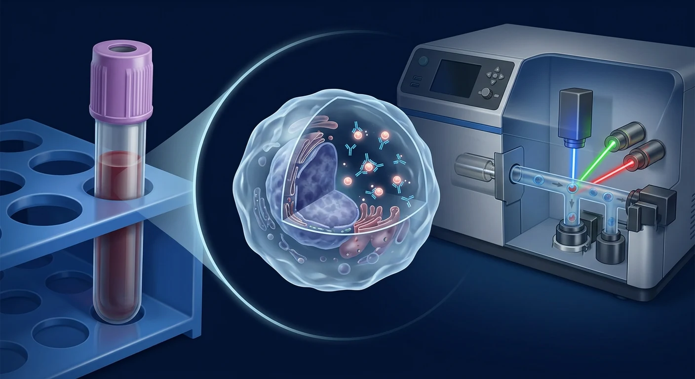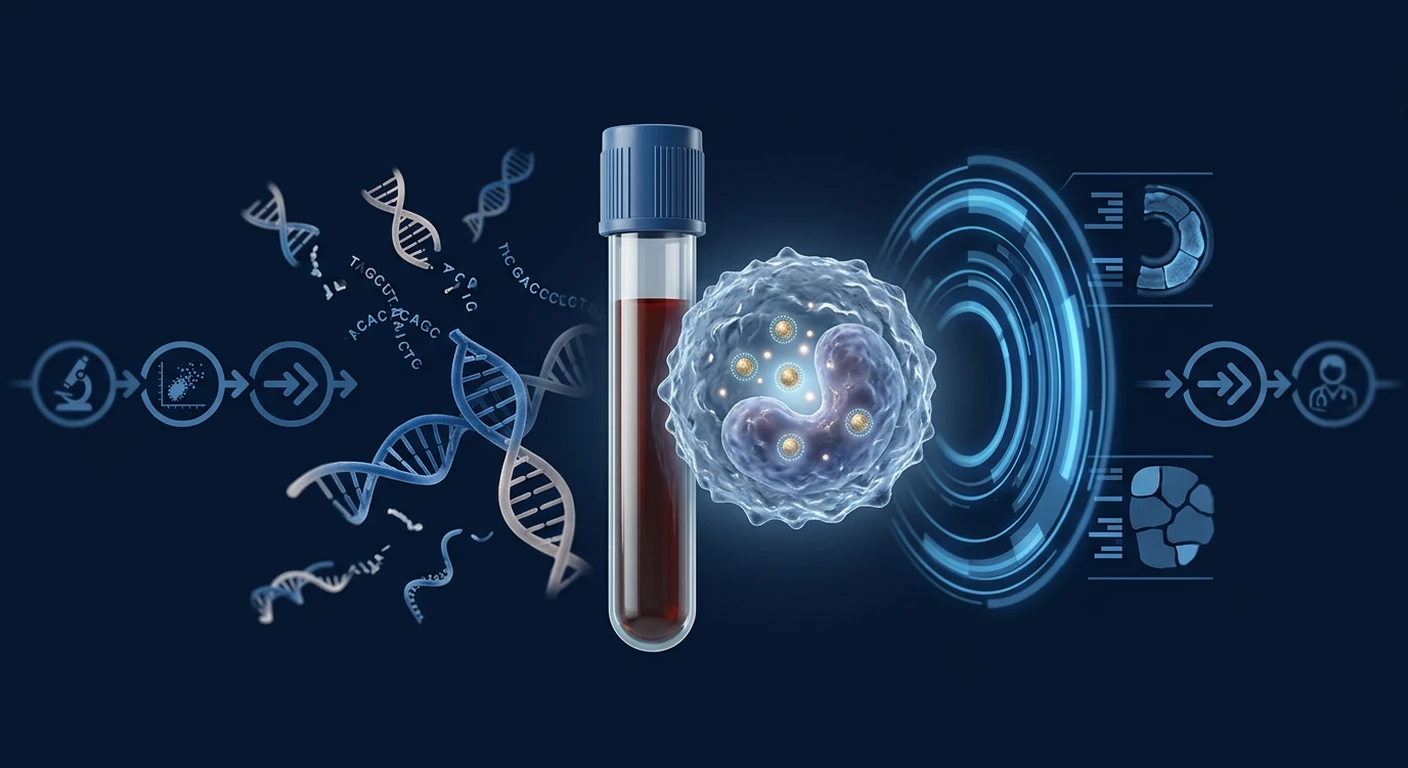
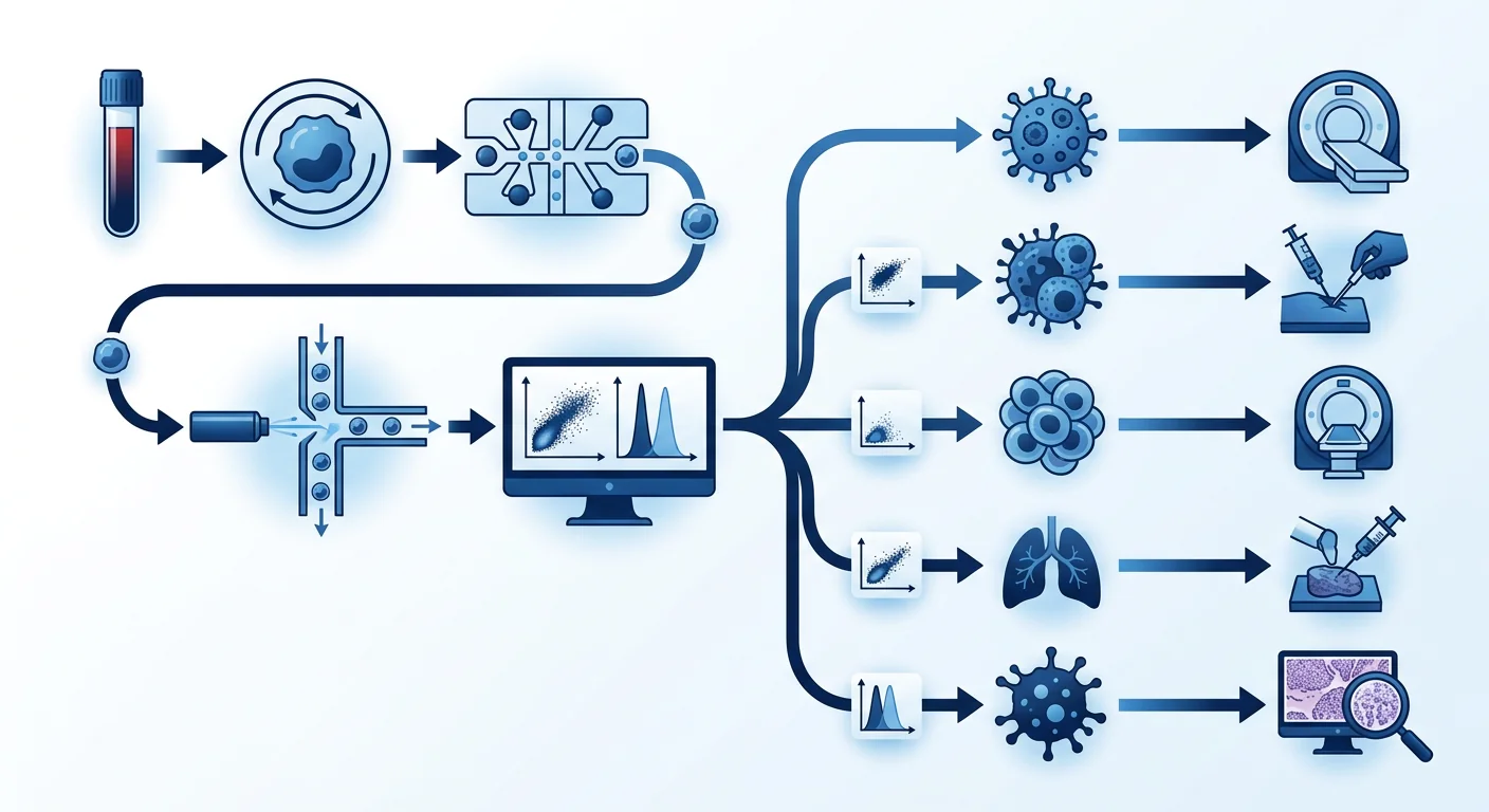
Epitope detection in monocytes\u2014EDIM\u2014is an investigational blood-test approach that uses flow cytometry to detect Apo10 and TKTL1 epitopes inside activated CD14+/CD16+ monocytes and macrophage-like cells. The biological premise is that phagocytic immune cells ingest material from abnormal tissue, return to the circulation, and carry detectable molecular traces. However, the performance of this approach is not uniform across every cancer type. Tumor biology, epitope expression levels, phagocytic activity, immune microenvironment, and disease stage all influence whether a given cancer produces a detectable monocyte signal.
This review examines the published evidence cancer type by cancer type\u2014breast, lung, colorectal, prostate, pancreatic, oral, pediatric, and less common tumors\u2014to clarify where the Apo10/TKTL1 signal is strongest, where uncertainty remains, and how patients and clinicians should interpret results in the context of established screening and diagnostic pathways.
Key message
EDIM test performance varies by cancer type, stage, and clinical context. No single sensitivity or specificity figure applies universally. A result from one cancer type population cannot be extrapolated to another. All positive results require confirmation through standard diagnostic pathways, and negative results cannot override symptoms, clinical findings, or guideline-recommended screening.
Breast cancer: moderate evidence in pilot cohorts
Breast cancer is one of the most studied cancer types in the EDIM literature, primarily through small case-control cohorts. A synthesis of EDIM studies reported 95.8% sensitivity and 97.3% specificity across selected cohorts that included breast, prostate, and oral cancer patients [1]. In a separate analysis, breast cancer cases showed statistically significantly higher Apo10 and TKTL1 scores than healthy controls, and combined-score thresholds achieved separation with reported area-under-the-curve values exceeding 0.90 [2].
The evidence has notable limitations. The published cohorts are small and predominantly contain patients with clinically apparent, biopsy-confirmed disease at the time of testing. Performance in subclinical, mammographically detected, or very early-stage disease has not been established in a prospective screening population. Additionally, the molecular heterogeneity of breast cancer subtypes\u2014estrogen-receptor-positive, HER2-positive, and triple-negative\u2014may influence Apo10 and TKTL1 expression; subtype-stratified analyses are absent from the available peer-reviewed data [1].
Practical implication. The EDIM signal appears detectable in established breast cancer, but data do not support its use as a standalone screening test for asymptomatic women. It should not replace mammography, clinical breast examination, or guideline-based screening intervals.
Lung cancer: promising signals with knowledge gaps
Lung cancer presents distinct challenges for EDIM because of tumor heterogeneity, variable epitope expression, and the high prevalence of benign pulmonary nodules and inflammatory lung disease in screening populations. Published EDIM studies have included lung cancer cases within broader mixed-cancer cohorts, showing elevated Apo10 and TKTL1 scores compared with healthy controls [3], [4]. However, no large prospective lung-cancer-specific screening cohort has been published.
The primary concern is specificity: chronic obstructive pulmonary disease, pulmonary fibrosis, active infections, and smoking-related inflammation can activate monocytes and macrophage-like cells, potentially producing false-positive EDIM signals [5]. This is particularly relevant for low-dose CT lung screening programs, where the baseline false-positive rate from benign nodules is already well documented [6].
Practical implication. EDIM may have a future adjunctive role in lung cancer risk stratification, but the current evidence does not support replacing or augmenting low-dose CT screening in high-risk populations without prospective validation in the intended screening cohort.
Colorectal cancer: surgical monitoring data available
Colorectal cancer has one of the richest EDIM evidence bases because of published surgical monitoring studies and a case report of early recurrence detection. In a mixed gastrointestinal study including colorectal cancer, 62 of 62 cancer patients had positive combined Apo10/TKTL1 scores, with no positives among 29 healthy controls and 1 positive among 13 participants with inflammatory conditions [7]. Surgical resection studies showed that EDIM scores declined after tumor removal, supporting the biological link between tumor presence and monocyte signal [8].
A reported case described an EDIM signal preceding radiological detection of colon cancer metastasis, suggesting potential for recurrence surveillance [9]. While intriguing, a single case cannot establish clinical utility for surveillance.
Practical implication. Colorectal cancer is one of the better evidence areas for EDIM, particularly for monitoring after curative treatment. However, EDIM cannot replace colonoscopy for screening, diagnosis, or polyp detection and removal.
Prostate cancer: prostatectomy data inform surveillance potential
Prostate cancer evidence includes an important contribution: a prospective monitoring study showing that EDIM scores fell after radical prostatectomy. Todenhöfer and colleagues reported that Apo10 and TKTL1 levels in circulating macrophages decreased significantly in the weeks following prostate removal, with the decline corresponding to prostate-specific antigen reduction [10]. This study provides mechanistic support for the monocyte signal originating at least in part from the prostate tumor.
However, the study population consisted of men with known, localized prostate cancer undergoing surgery\u2014not a screening cohort. Whether EDIM can distinguish clinically significant prostate cancer from indolent disease, or detect prostate cancer in asymptomatic men with elevated PSA, remains unstudied in a prospective screening context.
Practical implication. EDIM may have a future role in monitoring treated prostate cancer, but it is not validated for primary screening or for distinguishing aggressive from indolent prostate cancer. PSA, digital rectal examination, and MRI-guided biopsy remain the standard diagnostic pathway.
Pancreatic cancer: strong signal, small numbers
Pancreatic cancer is among the most lethal malignancies, and early detection is a critical unmet need. The published EDIM data for pancreatic cancer are striking but very small. In the mixed gastrointestinal study, pancreatic cancer cases within the cohort showed the same high detection rate, with combined Apo10/TKTL1 scores identifying all pancreatic cancer patients [7]. The urgent clinical need makes this an important area for further research.
The limitations are severe: fewer than 20 pancreatic cancer cases have been reported in the peer-reviewed EDIM literature, all with clinically apparent disease. Performance in early, resectable, asymptomatic pancreatic cancer\u2014the stage at which detection would meaningfully change outcomes\u2014is unknown. Additionally, benign pancreatic conditions such as chronic pancreatitis can produce inflammatory monocyte activation that may overlap with the malignant signal [11].
Practical implication. The preliminary signal in pancreatic cancer warrants larger prospective studies, but EDIM has no established role in pancreatic cancer screening or early diagnosis at present.
Oral and head and neck cancers: some of the best evidence
Oral squamous cell carcinoma has been studied across multiple publications, with results supporting both diagnostic and monitoring applications. EDIM scores correlated with tissue Apo10 and TKTL1 expression in oral cancer, and scores declined after surgical resection [12], [13]. The anatomic accessibility of oral lesions and the high rate of field cancerization make this an attractive application area.
The oral cancer literature benefits from both tissue correlation studies and surgical monitoring designs, providing mechanistic validation that the blood signal reflects the tumor. However, small sample sizes and lack of prospective screening cohorts limit generalizability.
Practical implication. Oral cancer is one of the stronger evidence areas for EDIM. Clinical examination and biopsy remain the diagnostic standard, and EDIM is not validated for population-based oral cancer screening in asymptomatic individuals.
Pediatric solid tumors: neuroblastoma and rhabdomyosarcoma
EDIM has been studied in pediatric oncology, specifically in neuroblastoma and rhabdomyosarcoma, representing a distinct application beyond adult epithelial cancers. A pilot study in neuroblastoma found that EDIM detected the ganglioside GD2 alongside Apo10 and TKTL1, achieving high discrimination between children with active neuroblastoma and those in remission or healthy controls [14]. A separate rhabdomyosarcoma pilot reported similar detection rates [15].
Pediatric solid tumors are rare, and building large screening cohorts is inherently difficult. The pilot studies provide biological plausibility but do not establish clinical utility for initial diagnosis, treatment monitoring, or relapse surveillance in routine practice.
Gynecological and less common cancers
Published evidence for EDIM in ovarian, cervical, endometrial, and other less common cancers is limited to case reports or inclusion within mixed-cancer studies. No dedicated prospective cohort has evaluated EDIM performance in ovarian or cervical cancer as a primary outcome. The available data are insufficient to draw meaningful conclusions about cancer-type-specific performance.
For cholangiocarcinoma (bile duct cancer), the mixed gastrointestinal study included cases that were detected alongside pancreatic and colorectal cases, suggesting the Apo10/TKTL1 signal is present but not organ-specific [7]. EDIM does not and cannot localize the tumor\u2014it provides a non-organ-specific signal that requires imaging and endoscopy for anatomical localization.
Cancer type summary: evidence table
| Cancer type | Published cohort size | Reported signal | Key limitation | Evidence level |
|---|---|---|---|---|
| Oral / Head & Neck | Small | Strong, tissue-validated | No screening cohort | Moderate |
| Colorectal | Small\u2013moderate | Strong, surgical decline | No screening cohort | Moderate |
| Prostate | Small | Moderate, surgical decline | Screening performance unknown | Low\u2013moderate |
| Breast | Small | Moderate | Subtype heterogeneity unknown | Low\u2013moderate |
| Pancreatic | Very small | Strong in advanced | Early-stage detection unknown | Low |
| Lung | Very small | Moderate | Inflammatory false positives | Low |
| Neuroblastoma (pediatric) | Small pilot | Moderate (including GD2) | Rare tumor, small pilot | Low |
| Rhabdomyosarcoma (pediatric) | Small pilot | Moderate | Rare tumor, small pilot | Low |
| Ovarian / Cervical | Minimal | Unknown | No dedicated study | Insufficient |
| Cholangiocarcinoma | Very small | Moderate | Included in mixed study only | Low |
Why cancer-type-specific evidence matters
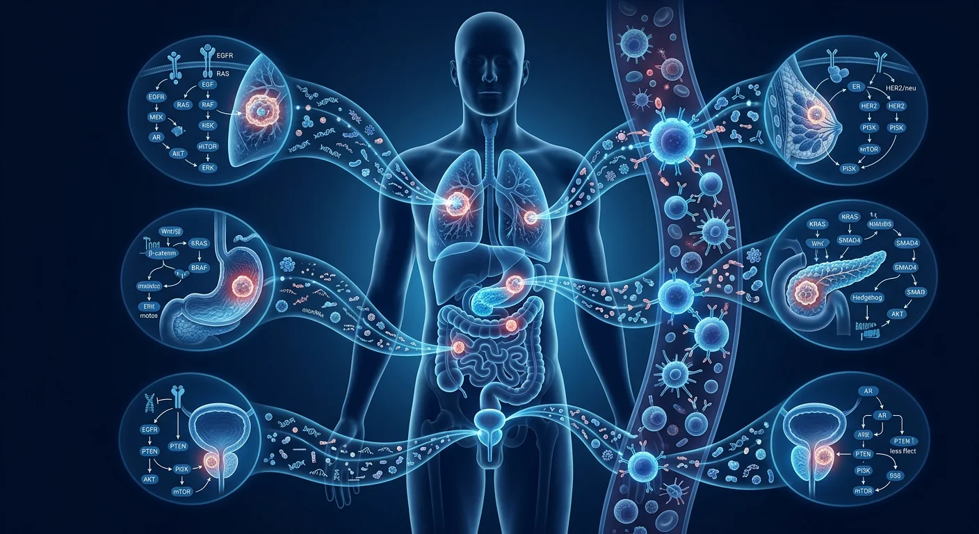
Cancer is not a single disease. Each type has a distinct tissue origin, genetic profile, immune microenvironment, and pattern of biomarker expression. A test that performs well in oral squamous cell carcinoma may not perform identically in pancreatic adenocarcinoma, and vice versa. Apo10 and TKTL1 expression levels, phagocytic activity of tumor-associated macrophages, and the degree of monocyte infiltration into the tumor microenvironment all vary by cancer type, grade, and stage [16].
This is why reported aggregate sensitivity and specificity figures across mixed-cancer cohorts can be misleading for individual patients. A woman concerned about breast cancer should not be reassured by a test validated primarily in oral and colorectal cancer populations. A negative result in a high-risk pancreatic cancer setting should not defer imaging.
Clinical pathway: integrating EDIM with standard cancer screening
Regardless of cancer type, EDIM should never replace guideline-recommended screening or diagnostic pathways. The appropriate clinical approach is to use EDIM, where offered, as an additional risk signal within a comprehensive evaluation framework:
- Breast cancer: mammography, ultrasound, and biopsy remain the standard. EDIM does not replace any of these.
- Lung cancer: low-dose CT screening in high-risk populations is guideline-based. EDIM has no validated add-on role.
- Colorectal cancer: colonoscopy with polypectomy is both diagnostic and preventive. FIT/fecal immunochemical testing is the non-invasive screening standard. EDIM does not replace either.
- Prostate cancer: PSA, digital rectal examination, and MRI followed by targeted biopsy are the diagnostic pathway. EDIM is not validated for this.
- Pancreatic cancer: no established screening exists for average-risk individuals. In high-risk populations (familial, genetic syndromes), endoscopic ultrasound and MRI are used. EDIM is investigational.
A positive or concerning EDIM score should trigger a clinician-led risk assessment, not self-directed imaging. The localizing investigation depends on the specific cancer type suspected based on the patient's risk factors, symptoms, and clinical context.
Limitations across cancer types
Across all cancer types studied, significant shared limitations apply. Cohorts are small and predominantly contain patients with known, clinically apparent disease. Case-control designs overestimate performance by excluding diagnostically challenging cases. Spectrum bias is inherent\u2014study populations do not represent screening populations [5]. Inter-laboratory standardization of the assay and threshold remains poorly documented in publicly available peer-reviewed sources. Clinical utility\u2014whether EDIM-guided care improves survival, quality of life, or treatment decisions\u2014has not been demonstrated for any cancer type through randomized or adequately controlled outcome studies.
Frequently asked questions
Does EDIM work equally well for all cancer types?
No. Published evidence is strongest for oral, colorectal, and prostate cancers. Evidence is moderate for breast and pancreatic cancers based on small cohorts. Evidence for lung, ovarian, cervical, and other cancers is limited or insufficient. Performance cannot be assumed to be uniform.
Can EDIM tell me which cancer type I have?
No. The Apo10/TKTL1 composite score is not organ-specific. It indicates a biological signal that could originate from any cancer type that expresses the target epitopes. Localization requires imaging, endoscopy, and tissue biopsy.
Should I stop my regular cancer screening if my EDIM score is normal?
No. A negative EDIM result cannot rule out any cancer type. False negatives occur, and standard age- and risk-appropriate screening must continue regardless of EDIM results. Symptoms and clinical findings always override a negative blood test.
Is EDIM more useful for certain cancers than others?
Based on current evidence, the strongest cases for EDIM are in oral squamous cell carcinoma (tissue-validation and monitoring data) and colorectal cancer (surgical decline and early recurrence case report). Pediatric neuroblastoma shows promise but is investigational. For all other cancer types, evidence is insufficient to recommend clinical use.
What confirmatory pathway follows a positive EDIM result?
The pathway is cancer-type-dependent. For a breast signal: mammography and possibly ultrasound and biopsy. For lung: chest CT. For colorectal: colonoscopy. For prostate: PSA and MRI. The choice of investigation should be guided by the patient's symptoms, risk factors, and clinical presentation\u2014not by the EDIM result alone. EDIM does not provide anatomical localization.
Medical disclaimer and limitations
This article reviews published EDIM evidence organized by cancer type for educational purposes. The evidence base is evolving, and the commercial assay versions and thresholds may vary between laboratories. Many studies involve investigators or entities connected with EDIM assay development; disclosure does not invalidate the findings but strengthens the case for independent replication and large-scale prospective validation. This article is not personal medical advice. Cancer screening and diagnostic decisions should be made in consultation with a qualified physician based on individual risk factors, symptoms, and guideline-recommended pathways.
References
- Coy JF. EDIM-TKTL1/Apo10 Blood Test: An Innate Immune System Based Liquid Biopsy for Early Detection, Prognosis and Therapy Monitoring of Malignancies. International Journal of Molecular Sciences. 2017;18(4):878. doi:10.3390/ijms18040878
- Grimm M, et al. A biomarker based detection and characterization of carcinomas exploiting two fundamental biophysical mechanisms in mammalian cells. BMC Cancer. 2013;13:569. doi:10.1186/1471-2407-13-569
- Grimm M, et al. Immunoreactivity of antibodies against DNaseX (Apo10) and TKTL1 in primary and metastatic lung cancer. Journal of Clinical Pathology. 2014;67:686\u2013691. doi:10.1136/jclinpath-2014-202127
- Grimm M, et al. Evaluation of a biomarker based blood test for monitoring surgical resection of oral squamous cell carcinomas. Clinical Oral Investigations. 2016;20:329\u2013338. doi:10.1007/s00784-015-1518-0
- Lachs MS, et al. Spectrum bias in the evaluation of diagnostic tests: lessons from the rapid dipstick for urinary tract infection. Annals of Internal Medicine. 1992;117:135\u2013140. doi:10.7326/0003-4819-117-2-135
- National Lung Screening Trial Research Team. Reduced lung-cancer mortality with low-dose CT screening. New England Journal of Medicine. 2011;365:395\u2013409. doi:10.1056/NEJMoa1102873
- Saman S, et al. Biomarkers Apo10 and TKTL1 as a new diagnostic approach for cholangiocellular, pancreatic and colorectal carcinoma. Cancer Biomarkers. 2020;27:129\u2013137. doi:10.3233/CBM-190414
- Todenhöfer T, et al. Effect of radical prostatectomy on levels of cancer related epitopes in circulating macrophages. Prostate. 2017;77:1251\u20131258. doi:10.1002/pros.23384
- Jansen N, Coy JF. Diagnostic use of epitope detection in monocytes (EDIM) blood test for early detection of colon cancer metastasis. Future Oncology. 2013;9:605\u2013609. doi:10.2217/fon.13.8
- Todenhöfer T, et al. Effect of radical prostatectomy on levels of cancer related epitopes in circulating macrophages. Prostate. 2017;77(12):1251\u20131258. doi:10.1002/pros.23384
- Zhang Y, et al. Macrophage polarization in pancreatic cancer: implications for tumor progression and immunotherapy. Journal of Experimental & Clinical Cancer Research. 2020;39:182. doi:10.1186/s13046-020-01689-4
- Grimm M, et al. Analysis of circulating CD14+/CD16+ monocyte-derived macrophages in patients with oral squamous cell carcinoma. Oral Surgery, Oral Medicine, Oral Pathology and Oral Radiology. 2016;121:301\u2013306. doi:10.1016/j.oooo.2015.10.024
- Grimm M, et al. Evaluation of a biomarker based blood test for monitoring surgical resection of oral squamous cell carcinomas. Clinical Oral Investigations. 2016;20:329\u2013338. doi:10.1007/s00784-015-1518-0
- Stagno MJ, et al. Epitope detection in monocytes (EDIM) for liquid biopsy including identification of GD2 in childhood neuroblastoma\u2014a pilot study. British Journal of Cancer. 2022;127:1324\u20131331. doi:10.1038/s41416-022-01855-x
- Urla C, et al. Epitope detection in monocytes (EDIM) as a new method of liquid biopsy in pediatric rhabdomyosarcoma. Biomedicines. 2022;10:1812. doi:10.3390/biomedicines10081812
- Ostuni R, et al. Macrophages and cancer: from mechanisms to therapeutic implications. Trends in Immunology. 2015;36:229\u2013239. doi:10.1016/j.it.2015.02.004
EDIM(单核细胞表位检测)是一种研究中的血液检测方法,利用流式细胞术检测活化 CD14+/CD16+ 单核细胞内的 Apo10 和 TKTL1 表位。其生物学原理是:吞噬性免疫细胞摄取异常组织物质后返回循环系统,携带可检测的分子痕迹。然而,EDIM 在不同癌症类型中的表现并不一致。肿瘤生物学特性、表位表达水平、吞噬活性、免疫微环境和疾病分期都会影响癌症能否产生可检测的单核细胞信号。
本文按癌种逐一审查已发表的证据——乳腺癌、肺癌、结直肠癌、前列腺癌、胰腺癌、口腔癌、儿童肿瘤及其他少见肿瘤——阐明 Apo10/TKTL1 信号在哪些癌种中最强、哪些仍存在不确定性,以及患者和临床医生应如何在标准筛查与诊断框架内解读结果。
核心信息
EDIM 检测性能因癌种、分期和临床情境而异。没有统一的敏感度或特异度适用于所有癌种。一个癌种人群的结果不能外推到另一种。所有阳性结果必须通过标准诊断路径确认,阴性结果也不能推翻症状、临床表现或指南推荐的筛查。
乳腺癌:小规模队列中的中等证据
乳腺癌是 EDIM 文献中研究较多的癌种之一,主要通过小型病例对照队列。一篇 EDIM 研究综合分析报告,在包含乳腺癌、前列腺癌和口腔癌患者的选定队列中,敏感度为 95.8%,特异度为 97.3% [1]。在另一项分析中,乳腺癌病例的 Apo10 和 TKTL1 评分显著高于健康对照,综合评分阈值报告的曲线下面积超过 0.90 [2]。
证据的局限性显著:已发表的队列规模小,且主要为检测时已具有临床表现、经活检确诊的患者。在亚临床、乳腺摄影检出或极早期疾病中的表现尚未在前瞻性筛查人群中确立。此外,乳腺癌分子亚型(雌激素受体阳性、HER2 阳性、三阴性)的异质性可能影响 Apo10 和 TKTL1 表达,可获得的同行评审数据中缺乏按亚型分层分析 [1]。
临床启示。EDIM 信号在已确诊乳腺癌中似乎可检测,但数据不支持将其作为无症状女性的独立筛查检测。它不应替代乳腺摄影、临床乳腺检查或指南推荐的筛查间隔。
肺癌:有希望的信号伴知识空白
肺癌因肿瘤异质性、表位表达变异以及筛查人群中肺良性结节和炎症性肺病的高患病率,对 EDIM 提出了独特挑战。已发表的 EDIM 研究将肺癌病例包含在更广泛的混合癌种队列中,显示与健康对照相比 Apo10 和 TKTL1 评分升高 [3]、[4]。然而,尚无大型前瞻性肺癌特异性筛查队列发表。
主要关注点是特异度:慢性阻塞性肺病、肺纤维化、活动性感染和吸烟相关炎症可激活单核细胞,可能产生 EDIM 假阳性信号 [5]。这与低剂量 CT 肺癌筛查项目尤其相关,良性结节的基线假阳性率已有充分文献记载 [6]。
临床启示。EDIM 未来可能在肺癌风险分层中起辅助作用,但当前证据不支持在无前瞻性验证的情况下替代或补充高危人群的低剂量 CT 筛查。
结直肠癌:手术监测数据可用
结直肠癌拥有最丰富的 EDIM 证据基础之一,得益于已发表的手术监测研究和早期复发检测个案报告。在一项混合胃肠道研究中,62 名结直肠癌患者中有 62 名综合 Apo10/TKTL1 评分阳性,29 名健康对照均阴性,13 名炎症参与者中 1 名阳性 [7]。手术切除研究显示 EDIM 评分在肿瘤切除后下降,支持肿瘤存在与单核细胞信号之间的生物学联系 [8]。
一例报告描述了 EDIM 信号在影像学发现结肠癌转移之前出现,提示复发监测的潜力 [9]。尽管令人关注,但单一个案不能确立临床效用。
临床启示。结直肠癌是 EDIM 证据较好的领域之一,特别是在治愈性治疗后的监测方面。然而,EDIM 不能替代结肠镜用于筛查、诊断或息肉检测与切除。
前列腺癌:前列腺切除术数据启示监测潜力
前列腺癌证据包含一项重要贡献:一项前瞻性监测研究显示 EDIM 评分在根治性前列腺切除术后下降。Todenhöfer 等人报告,循环巨噬细胞中的 Apo10 和 TKTL1 水平在前列腺切除后数周显著下降,变化幅度与前列腺特异性抗原降低相对应 [10]。该研究为单核细胞信号至少部分来自前列腺肿瘤提供了机制支持。
然而,研究对象为接受手术的已知局限性前列腺癌男性,而非筛查队列。EDIM 能否区分有临床意义的前列腺癌与惰性疾病,或在无症状男性中检测 PSA 升高背景下的前列腺癌,尚未在前瞻性筛查环境中得到研究。
临床启示。EDIM 未来可能在治疗后的前列腺癌监测中发挥作用,但未经验证用于初筛或区分侵袭性与惰性前列腺癌。PSA、直肠指检和 MRI 引导活检仍是标准诊断路径。
胰腺癌:信号强但样本数极小
胰腺癌是最致命的恶性肿瘤之一,早期检测是迫切未满足的需求。已发表的 EDIM 胰腺癌数据令人瞩目但样本量极小。在混合胃肠道研究中,队列内的胰腺癌病例显示出同样高的检出率,综合 Apo10/TKTL1 评分识别了所有胰腺癌患者 [7]。迫切的临床需求使这一领域成为未来研究的重要方向。
局限性严重:同行评审 EDIM 文献中报告的胰腺癌病例不足 20 例,且均为临床表现明显的疾病。在早期、可切除、无症状胰腺癌——即检测将显著改变预后的阶段——中的表现尚不清楚。此外,慢性胰腺炎等良性胰腺疾病可产生炎症性单核细胞活化,可能与恶性信号重叠 [11]。
临床启示。胰腺癌的初步信号值得更大规模的前瞻性研究,但 EDIM 目前对胰腺癌筛查或早期诊断尚无确定作用。
口腔及头颈部癌:部分最佳证据
口腔鳞状细胞癌已在多项出版物中得到研究,结果支持诊断和监测应用。EDIM 评分与口腔癌组织中 Apo10 和 TKTL1 表达相关,手术切除后评分下降 [12]、[13]。口腔病变的解剖可及性和高野癌化率使其成为有吸引力的应用领域。
口腔癌文献得益于组织相关性研究和手术监测设计,提供了血液信号反映肿瘤的机制验证。然而,小样本量和缺乏前瞻性筛查队列限制了普适性。
临床启示。口腔癌是 EDIM 证据较强的领域之一。临床检查和活检仍是诊断标准,EDIM 未经验证用于无症状人群的口腔癌人群筛查。
儿童实体肿瘤:神经母细胞瘤和横纹肌肉瘤
EDIM 已在儿童肿瘤学领域得到研究,特别是神经母细胞瘤和横纹肌肉瘤,代表了超越成人上皮性癌症的不同应用。一项神经母细胞瘤先导研究发现,EDIM 在检测 Apo10 和 TKTL1 的同时检测神经节苷脂 GD2,在活动性神经母细胞瘤患儿与缓解期或健康对照之间实现了高度区分 [14]。另一项横纹肌肉瘤先导研究报告了类似的检出率 [15]。
儿童实体肿瘤罕见,构建大型筛查队列固有困难。先导研究提供了生物学合理性,但未确立在常规实践中用于初始诊断、治疗监测或复发监测的临床效用。
妇科及较少见癌症
EDIM 在卵巢癌、宫颈癌、子宫内膜癌和其他较少见癌症中的已发表证据限于病例报告或混合癌种研究中的纳入数据。尚无专门的前瞻性队列以 EDIM 在卵巢癌或宫颈癌中的表现为主要结局进行评估。现有数据不足以对癌种特异性表现得出有意义的结论。
对于胆管癌,混合胃肠道研究将其与胰腺癌和结直肠癌病例一同检出,提示 Apo10/TKTL1 信号存在但非器官特异性 [7]。EDIM 不能也无法定位肿瘤——它提供的是非器官特异性信号,需影像学和内镜进行解剖定位。
癌种证据总结表
| 癌种 | 已发表队列规模 | 报告信号强度 | 主要局限 | 证据级别 |
|---|---|---|---|---|
| 口腔/头颈部 | 小 | 强,经组织验证 | 无筛查队列 | 中等 |
| 结直肠 | 小-中等 | 强,术后下降 | 无筛查队列 | 中等 |
| 前列腺 | 小 | 中等,术后下降 | 筛查表现未知 | 低-中等 |
| 乳腺 | 小 | 中等 | 亚型异质性未知 | 低-中等 |
| 胰腺 | 极小 | 晚期强信号 | 早期检测未知 | 低 |
| 肺 | 极小 | 中等 | 炎症假阳性 | 低 |
| 神经母细胞瘤(儿童) | 小型先导 | 中等(含 GD2) | 罕见肿瘤,小先导 | 低 |
| 横纹肌肉瘤(儿童) | 小型先导 | 中等 | 罕见肿瘤,小先导 | 低 |
| 卵巢/宫颈 | 极小 | 未知 | 无专项研究 | 不足 |
| 胆管 | 极小 | 中等 | 仅在混合研究中 | 低 |
癌种特异性证据为何重要
癌症不是单一疾病。每种类型都有不同的组织起源、遗传谱、免疫微环境和生物标志物表达模式。在口腔鳞状细胞癌中表现良好的检测在胰腺腺癌中可能表现不同。Apo10 和 TKTL1 的表达水平、肿瘤相关巨噬细胞的吞噬活性及单核细胞浸润肿瘤微环境的程度均因癌种、分级和分期而异 [16]。
这就是为什么在混合癌种队列中报告的汇总敏感度和特异度对个体患者可能具有误导性。关注乳腺癌的女性不应被主要在口腔和结直肠癌人群中验证的检测结果所安慰。胰腺癌高风险情境中的阴性结果不应推迟影像学检查。
临床路径:将 EDIM 整合到标准癌症筛查中
无论癌种如何,EDIM 绝不应替代指南推荐的筛查或诊断路径。适当的临床方法是将 EDIM(如提供的)作为综合评估框架内的额外风险信号:
- 乳腺癌:乳腺摄影、超声和活检仍是标准。EDIM 不替代其中任何一项。
- 肺癌:高危人群的低剂量 CT 筛查是指南推荐的。EDIM 无经验证的附加作用。
- 结直肠癌:结肠镜加息肉切除术兼具诊断和预防功能。FIT 是标准非侵入性筛查方法。EDIM 不替代任何一项。
- 前列腺癌:PSA、直肠指检和 MRI 后靶向活检是诊断路径。EDIM 未经验证用于此目的。
- 胰腺癌:普通风险人群无既定筛查。高风险人群中,内镜超声和 MRI 用于筛查。EDIM 为研究性质。
阳性或可疑的 EDIM 评分应触发医生主导的风险评估,而非自行安排的影像学检查。定位检查的选择取决于根据患者危险因素、症状和临床情境怀疑的特定癌种。
跨癌种局限性
在所有研究的癌种中,存在显著共享的局限性。队列规模小,且主要纳入已知临床明显疾病的患者。病例对照设计通过排除诊断困难的病例高估了性能。疾病谱偏倚固有——研究人群不代表筛查人群 [5]。检测和阈值的跨实验室标准化在公开可获得的同行评审来源中记录不充分。临床效用——EDIM 指导的照护是否能改善任何癌种的生存、生活质量或治疗决策——尚未通过随机或充分对照的结局研究得到证明。
常见问题
EDIM 对所有癌种效果相同吗?
不。已发表证据在口腔癌、结直肠癌和前列腺癌中最强。乳腺癌和胰腺癌基于小规模队列证据中等。肺癌、卵巢癌、宫颈癌和其他癌症的证据有限或不足。不能假设性能均匀。
EDIM 能告诉我患哪种癌吗?
不能。Apo10/TKTL1 综合评分非器官特异性。它指示可能来自任何表达靶表位的癌种的生物学信号。定位需要影像学、内镜和组织活检。
如果 EDIM 评分正常,应停止常规癌症筛查吗?
不。阴性 EDIM 结果不能排除任何癌种。假阴性存在,标准年龄和风险适宜筛查必须继续。症状和临床表现始终压倒阴性血液检测结果。
EDIM 对某些癌种更有用吗?
根据当前证据,EDIM 在口腔鳞状细胞癌(组织验证和监测数据)和结直肠癌(手术下降和早期复发个案报告)中的应用基础最强。儿童神经母细胞瘤有前景但为研究性质。所有其他癌种证据不足以推荐临床应用。
EDIM 阳性后应遵循什么确认路径?
路径取决于癌种。乳腺信号:乳腺摄影、超声和可能活检。肺部:胸部 CT。结直肠:结肠镜。前列腺:PSA 和 MRI。检查选择应基于患者症状、危险因素和临床表现,而非仅凭 EDIM 结果。EDIM 不提供解剖定位。
医疗声明与局限性
本文按癌种审阅已发表 EDIM 证据仅供教育目的。证据基础正在发展,商业检测版本和阈值可能因实验室而异。许多研究涉及与 EDIM 检测开发相关的研究者或机构;披露不自动否定研究结果,但加强了独立复制和大规模前瞻性验证的必要性。本文非个人医疗建议。癌症筛查和诊断决策应咨询合格医师,依据个体危险因素、症状和指南推荐路径作出。
参考文献
- Coy JF. EDIM-TKTL1/Apo10 Blood Test. Int J Mol Sci. 2017;18:878. doi:10.3390/ijms18040878
- Grimm M, et al. A biomarker based detection and characterization of carcinomas. BMC Cancer. 2013;13:569. doi:10.1186/1471-2407-13-569
- Grimm M, et al. DNaseX (Apo10) and TKTL1 immunoreactivity in lung cancer. J Clin Pathol. 2014;67:686-691. doi:10.1136/jclinpath-2014-202127
- Grimm M, et al. Blood test for monitoring surgical resection of oral SCC. Clin Oral Invest. 2016;20:329-338. doi:10.1007/s00784-015-1518-0
- Lachs MS, et al. Spectrum bias. Ann Intern Med. 1992;117:135-140. doi:10.7326/0003-4819-117-2-135
- National Lung Screening Trial. Reduced lung-cancer mortality with LDCT. NEJM. 2011;365:395-409. doi:10.1056/NEJMoa1102873
- Saman S, et al. Apo10 and TKTL1 for cholangiocellular, pancreatic and colorectal carcinoma. Cancer Biomark. 2020;27:129-137. doi:10.3233/CBM-190414
- Todenhöfer T, et al. Radical prostatectomy and EDIM macrophage epitopes. Prostate. 2017;77:1251-1258. doi:10.1002/pros.23384
- Jansen N, Coy JF. EDIM for colon cancer metastasis detection. Future Oncol. 2013;9:605-609. doi:10.2217/fon.13.8
- Todenhöfer T, et al. Effect of radical prostatectomy on macrophage epitope levels. Prostate. 2017;77(12):1251-1258. doi:10.1002/pros.23384
- Zhang Y, et al. Macrophage polarization in pancreatic cancer. J Exp Clin Cancer Res. 2020;39:182. doi:10.1186/s13046-020-01689-4
- Grimm M, et al. Circulating CD14+/CD16+ macrophages in oral SCC. Oral Surg Oral Med Oral Pathol Oral Radiol. 2016;121:301-306. doi:10.1016/j.oooo.2015.10.024
- Grimm M, et al. Blood test for monitoring surgical resection of oral SCC. Clin Oral Invest. 2016;20:329-338. doi:10.1007/s00784-015-1518-0
- Stagno MJ, et al. EDIM including GD2 in neuroblastoma. Br J Cancer. 2022;127:1324-1331. doi:10.1038/s41416-022-01855-x
- Urla C, et al. EDIM in pediatric rhabdomyosarcoma. Biomedicines. 2022;10:1812. doi:10.3390/biomedicines10081812
- Ostuni R, et al. Macrophages and cancer. Trends Immunol. 2015;36:229-239. doi:10.1016/j.it.2015.02.004
EDIM، أي كشف الحواتم داخل الوحيدات، هو نهج بحثي لفحص الدم يستخدم قياس التدفق الخلوي للكشف عن حواتم Apo10 وTKTL1 داخل خلايا CD14+/CD16+ المنشطة. الفكرة البيولوجية هي أن الخلايا البلعمية تبتلع مواد من أنسجة غير طبيعية وتعود إلى الدورة الدموية حاملة آثاراً جزيئية قابلة للكشف. لكن أداء هذا النهج ليس منتظماً عبر جميع أنواع السرطان. يؤثر بيولوجيا الورم، ومستويات التعبير عن الحواتم، والنشاط البلعمي، والبيئة المناعية للورم، ومرحلة المرض كلها على ما إذا كان سرطان معين ينتج إشارة وحيدات قابلة للكشف.
تستعرض هذه المراجعة الأدلة المنشورة نوعاً بنوع من السرطان\u2014الثدي والرئة والقولون والمستقيم والبروستات والبنكرياس والفم والأورام الطفولية وغيرها\u2014لتوضيح أين تكون إشارة Apo10/TKTL1 الأقوى وأين يبقى عدم اليقين وكيف ينبغي للمرضى والأطباء تفسير النتائج في سياق فحوص الكشف والتشخيص المعتمدة.
الرسالة الأساسية
أداء اختبار EDIM يختلف حسب نوع السرطان ومرحلته والسياق السريري. لا تنطبق قيمة واحدة للحساسية أو النوعية على جميع الأنواع. لا يمكن استقراء نتيجة من مجتمع سرطاني إلى آخر. كل النتائج الإيجابية تحتاج إلى تأكيد بالمسارات التشخيصية المعيارية، ولا يمكن للنتائج السلبية تجاوز الأعراض أو النتائج السريرية أو فحوص الكشف الموصى بها.
سرطان الثدي: أدلة معتدلة في أفواج استكشافية صغيرة
سرطان الثدي هو أحد أكثر الأنواع دراسةً في أدبيات EDIM، بشكل رئيسي عبر أفواج صغيرة من الحالات والشواهد. أبلغ توليف لدراسات EDIM عن حساسية 95.8% ونوعية 97.3% عبر أفواج مختارة شملت مرضى سرطان الثدي والبروستات والفم [1]. في تحليل منفصل، أظهرت حالات سرطان الثدي درجات Apo10 وTKTL1 أعلى إحصائياً من الأصحاء.
القيود واضحة: الأفواج المنشورة صغيرة وتحتوي غالباً على مرضى بمرض واضح سريرياً مؤكد بالخزعة. الأداء في المرض تحت السريري أو المبكر جداً لم يثبت في مجتمع فحص استباقي. كما أن عدم تجانس الأنواع الفرعية الجزيئية قد يؤثر على التعبير عن Apo10 وTKTL1 [1].
التطبيق العملي. تبدو إشارة EDIM قابلة للكشف في سرطان الثدي المُثبت، لكن البيانات لا تدعم استخدامه كفحص مستقل للنساء بلا أعراض. لا ينبغي أن يحل محل تصوير الثدي أو الفحص السريري أو فترات الكشف الموصى بها.
سرطان الرئة: إشارات واعدة مع فجوات معرفية
يشكل سرطان الرئة تحديات مميزة لـEDIM بسبب عدم تجانس الورم وتفاوت التعبير عن الحواتم وارتفاع انتشار العقيدات الرئوية الحميدة وأمراض الرئة الالتهابية في مجتمعات الفحص. أظهرت دراسات EDIM التي شملت سرطان الرئة ضمن أفواج مختلطة أوسع درجات Apo10 وTKTL1 مرتفعة مقارنة بالأصحاء [3]، [4]. لكن لم يُنشر أي فوج فحص استباقي كبير خاص بسرطان الرئة.
الاهتمام الأساسي هو النوعية: مرض الانسداد الرئوي المزمن والتليف الرئوي والعدوى النشطة والالتهاب المرتبط بالتدخين يمكن أن تنشط الوحيدات، مما قد ينتج نتائج EDIM إيجابية كاذبة [5]. هذا مهم بشكل خاص لبرامج فحص الرئة بالتصوير المقطعي منخفض الجرعة [6].
التطبيق العملي. قد يكون لـEDIM دور مساعد مستقبلي في تصنيف مخاطر سرطان الرئة، لكن الأدلة الحالية لا تدعم استبدال أو تعزيز فحص التصوير المقطعي منخفض الجرعة دون تحقق استباقي في مجتمع الفحص المستهدف.
سرطان القولون والمستقيم: بيانات المراقبة الجراحية متوفرة
يمتلك سرطان القولون والمستقيم أحد أغنى قواعد أدلة EDIM بفضل دراسات المراقبة الجراحية المنشورة وتقرير حالة للكشف المبكر عن النكس. في دراسة معدية معوية مختلطة شملت سرطان القولون، كانت درجات Apo10/TKTL1 المركبة إيجابية لدى 62 من 62 مريضاً، وسلبية لدى 29 سليماً، مع إيجابية واحدة بين 13 مصاباً بالالتهاب [7]. أظهرت دراسات الاستئصال الجراحي انخفاض درجات EDIM بعد إزالة الورم [8].
التطبيق العملي. سرطان القولون هو أحد مجالات أدلة EDIM الأفضل، خاصة للمراقبة بعد العلاج. لكن EDIM لا يمكن أن يحل محل تنظير القولون للكشف أو التشخيص.
سرطان البروستات: بيانات استئصال البروستات تفيد إمكانات المراقبة
تشمل أدلة سرطان البروستات مساهمة مهمة: دراسة مراقبة استباقية أظهرت انخفاض درجات EDIM بعد استئصال البروستات الجذري. أبلغ Todenhöfer وزملاؤه عن انخفاض كبير في مستويات Apo10 وTKTL1 في البلاعم المنتشرة بعد أسابيع من استئصال البروستات [10]. هذه الدراسة تقدم دعماً ميكانيزمياً لأصل إشارة الوحيدات من ورم البروستات على الأقل جزئياً.
التطبيق العملي. EDIM غير معتمد للفحص الأولي أو لتمييز سرطان البروستات العدواني من الخامل. تبقى PSA والفحص المستقيمي والتصوير بالرنين المغناطيسي والخزعة الموجهة هي المسار التشخيصي المعياري.
سرطان البنكرياس: إشارة قوية بأعداد صغيرة جداً
سرطان البنكرياس هو من أكثر الأورام الخبيثة فتكاً، والكشف المبكر حاجة ماسة غير ملباة. بيانات EDIM المنشورة لسرطان البنكرياس مذهلة لكن صغيرة جداً. في الدراسة المعدية المعوية المختلطة، أظهرت حالات سرطان البنكرياس نفس معدل الكشف المرتفع [7].
القيود شديدة: أقل من 20 حالة سرطان بنكرياس أُبلغ عنها في أدبيات EDIM المحكمة، كلها بمرض واضح سريرياً. الأداء في سرطان البنكرياس المبكر القابل للاستئصال بلا أعراض\u2014المرحلة التي يغير فيها الكشف النتائج\u2014غير معروف [11].
التطبيق العملي. الإشارة الأولية تستحق دراسات استباقية أكبر، لكن EDIM ليس له دور محدد في فحص أو تشخيص سرطان البنكرياس المبكر حالياً.
سرطان الفم والرأس والعنق: بعض من أفضل الأدلة
دُرس سرطان الخلايا الحرشفية الفموي عبر منشورات متعددة، بنتائج تدعم كلاً من التطبيقات التشخيصية والمراقبة. ارتبطت درجات EDIM بالتعبير النسيجي لـApo10 وTKTL1 في سرطان الفم، وانخفضت بعد الاستئصال الجراحي [12]، [13].
التطبيق العملي. سرطان الفم هو أحد مجالات أدلة EDIM الأقوى. يبقى الفحص السريري والخزعة المعيار التشخيصي، وEDIM غير معتمد لفحص سرطان الفم السكاني.
الأورام الصلبة الطفولية: الورم الأرومي العصبي والساركومة العضلية المخططة
دُرس EDIM في طب الأورام الطفولي، خاصة في الورم الأرومي العصبي والساركومة العضلية المخططة، ممثلاً تطبيقاً متميزاً يتجاوز سرطانات الخلايا الظهارية عند البالغين. وجدت دراسة استكشافية في الورم الأرومي العصبي أن EDIM كشف الغانغليوزيد GD2 إلى جانب Apo10 وTKTL1 [14]. دراسة منفصلة للساركومة العضلية المخططة أبلغت عن نتائج مماثلة [15].
السرطانات النسائية والأقل شيوعاً
الأدلة المنشورة لـEDIM في سرطان المبيض وعنق الرحم وبطانة الرحم محدودة لتقارير حالات أو ضمن دراسات مختلطة. لا توجد بيانات كافية لاستنتاجات ذات معنى عن الأداء النوعي.
القيود عبر أنواع السرطان
عبر جميع أنواع السرطان المدروسة، تنطبق قيود مشتركة مهمة. الأفواج صغيرة وتحتوي غالباً على مرضى بمرض معروف ظاهر سريرياً. التصاميم ذات الحالات والشواهد تبالغ في تقدير الأداء باستبعاد الحالات التشخيصية الصعبة. تحيز طيف المرض متأصل\u2014مجتمعات الدراسة لا تمثل مجتمعات الفحص [5]. توحيد المعايير المخبرية للفحص والعتبة غير موثق بشكل كافٍ. المنفعة السريرية لأي نوع سرطان لم تُثبت بدراسات نتائج عشوائية.
الخلاصة الطبية والقيود
هذه المقالة تستعرض أدلة EDIM المنشورة منظمة حسب نوع السرطان لأغراض تعليمية. قاعدة الأدلة تتطور وقد تختلف الإصدارات التجارية والعتبات بين المختبرات. العديد من الدراسات تشمل باحثين أو جهات مرتبطة بتطوير فحص EDIM؛ الإفصاح لا يبطل النتائج لكنه يعزز الحاجة للتكرار المستقل والتحقق الاستباقي واسع النطاق. هذه المقالة ليست نصيحة طبية شخصية. يجب اتخاذ قرارات فحص السرطان وتشخيصه بالتشاور مع طبيب مؤهل بناءً على عوامل الخطر الفردية والأعراض والمسارات الموصى بها في الإرشادات.
المراجع
- Coy JF. EDIM-TKTL1/Apo10 Blood Test. Int J Mol Sci. 2017;18:878. doi:10.3390/ijms18040878
- Grimm M, et al. A biomarker based detection. BMC Cancer. 2013;13:569. doi:10.1186/1471-2407-13-569
- Grimm M, et al. DNaseX and TKTL1 in lung cancer. J Clin Pathol. 2014;67:686-691. doi:10.1136/jclinpath-2014-202127
- Grimm M, et al. Blood test for monitoring surgical resection. Clin Oral Invest. 2016;20:329-338. doi:10.1007/s00784-015-1518-0
- Lachs MS, et al. Spectrum bias. Ann Intern Med. 1992;117:135-140. doi:10.7326/0003-4819-117-2-135
- National Lung Screening Trial. LDCT screening. NEJM. 2011;365:395-409. doi:10.1056/NEJMoa1102873
- Saman S, et al. Apo10 TKTL1 for GI cancers. Cancer Biomark. 2020;27:129-137. doi:10.3233/CBM-190414
- Todenhöfer T, et al. Prostatectomy and EDIM. Prostate. 2017;77:1251-1258. doi:10.1002/pros.23384
- Jansen N, Coy JF. EDIM colon metastasis. Future Oncol. 2013;9:605-609. doi:10.2217/fon.13.8
- Todenhöfer T, et al. Radical prostatectomy and macrophage epitopes. Prostate. 2017;77(12):1251-1258. doi:10.1002/pros.23384
- Zhang Y, et al. Macrophage polarization in pancreatic cancer. J Exp Clin Cancer Res. 2020;39:182. doi:10.1186/s13046-020-01689-4
- Grimm M, et al. CD14+/CD16+ macrophages in oral SCC. Oral Surg Oral Med Oral Pathol Oral Radiol. 2016;121:301-306. doi:10.1016/j.oooo.2015.10.024
- Grimm M, et al. Blood test oral SCC monitoring. Clin Oral Invest. 2016;20:329-338. doi:10.1007/s00784-015-1518-0
- Stagno MJ, et al. EDIM GD2 neuroblastoma. Br J Cancer. 2022;127:1324-1331. doi:10.1038/s41416-022-01855-x
- Urla C, et al. EDIM rhabdomyosarcoma. Biomedicines. 2022;10:1812. doi:10.3390/biomedicines10081812
- Ostuni R, et al. Macrophages and cancer. Trends Immunol. 2015;36:229-239. doi:10.1016/j.it.2015.02.004