For patients who have completed primary cancer treatment, the prospect of recurrence is a persistent source of anxiety — and the standard surveillance schedule, with imaging and clinical visits spaced months apart, can feel like a gap in the safety net. The EDIM (Epitope Detection in Monocytes) Apo10/TKTL1 blood test is being investigated as a potential adjunct for post-treatment surveillance, aiming to detect molecular signals of recurrence earlier than conventional imaging. This article reviews the current evidence, the biological rationale, and the limitations that patients and clinicians should consider.
Cancer recurrence after apparently successful treatment is not uncommon. Depending on the primary tumor type, stage at diagnosis, and treatment modality, recurrence rates range from 10% to over 40% within five years for certain cancers [1]. The goal of post-treatment surveillance is to detect recurrence at the earliest possible stage, when intervention may still be curative or more effective [2]. This need for early detection drives interest in blood-based biomarkers that could complement imaging surveillance.
Key message
EDIM Apo10/TKTL1 testing for cancer recurrence monitoring is an investigational approach. It is not approved as a standalone surveillance method and should not replace guideline-recommended imaging, clinical follow-up, or tumor marker monitoring. All positive EDIM findings require confirmation through standard diagnostic pathways.
What is EDIM and why might it be useful for recurrence monitoring?
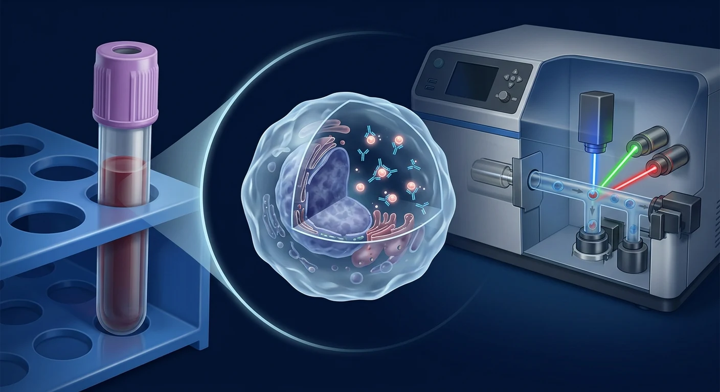
EDIM detects Apo10 and TKTL1 protein epitopes inside activated monocytes — immune cells that circulate through the body and ingest material from abnormal tissue. The Apo10 epitope reflects a DNaseX protein variant associated with evasion of programmed cell death (apoptosis). The TKTL1 epitope reflects upregulation of transketolase-like enzyme 1, a key enzyme in the pentose phosphate pathway that supports the altered glucose metabolism of malignant cells — the Warburg effect [3], [4].
For recurrence monitoring, the biological premise is compelling. If a recurrent tumor deposit is present anywhere in the body — even a small, clinically occult lesion — activated monocytes may encounter, phagocytose, and transport epitope-bearing material from that lesion into the circulation, where flow cytometry can detect them [5]. This detection occurs independently of the anatomic location of the recurrence, potentially offering a systemic surveillance signal that imaging — which only visualizes specific anatomic regions — may miss in its earliest stages.
Several studies have shown that EDIM scores decline after curative surgical resection. Todenhöfer and colleagues demonstrated that Apo10 and TKTL1 scores fell significantly in the weeks following radical prostatectomy, with the decline paralleling PSA reduction [6]. Similarly, oral squamous cell carcinoma studies reported that EDIM scores decreased after tumor resection [7]. These findings support the biological relationship between tumor presence and the monocyte signal — and, conversely, suggest that rising scores after a nadir could signal recurrence.
How EDIM compares with standard recurrence surveillance methods
Standard surveillance after cancer treatment typically involves a combination of scheduled imaging (CT, MRI, PET-CT), clinical examination, and serum tumor markers. Each modality has strengths and blind spots. Imaging can localize recurrence but is scheduled at intervals of 3–12 months depending on cancer type and risk. Between scans, a recurrence can grow undetected. Serum tumor markers such as CEA, CA19-9, CA-125, and PSA are widely used but have limited sensitivity for early recurrence and can be elevated in benign conditions [8].
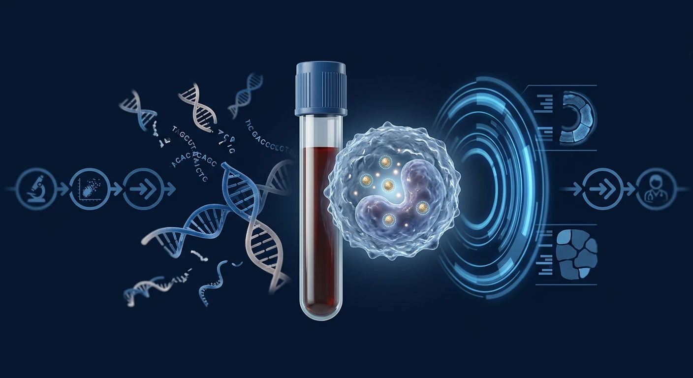
Circulating tumor DNA (ctDNA) has emerged as a powerful molecular surveillance tool, with the ability to detect recurrence months before imaging in several cancer types. However, ctDNA testing is expensive, requires tumor genotype information for personalized assay design in many cases, and not all tumors shed detectable DNA into circulation [9]. False-negative ctDNA results occur particularly in oligometastatic or low-burden recurrence scenarios.
| Surveillance Method | Strength | Limitation |
|---|---|---|
| EDIM (Apo10/TKTL1) | Systemic signal independent of tumor location; does not require prior tumor genotype | Investigational; limited prospective recurrence data; affected by inflammation |
| CT/MRI Imaging | Anatomic localization; widely available; established guidelines | Interval gaps between scans; radiation exposure (CT); small lesions may be missed |
| PET-CT (FDG) | Functional + anatomic; high sensitivity for metabolically active disease | High cost; false positives from inflammation; radiation exposure |
| Serum Tumor Markers | Inexpensive; widely used; serial monitoring possible | Limited sensitivity/specificity; benign elevation common; not available for all cancers |
| Circulating Tumor DNA (ctDNA) | Highly specific; can detect months before imaging; molecular characterization possible | Expensive; requires tumor sequencing for personalized assays; shedding varies by tumor type |
EDIM occupies a unique position in this landscape. It does not require prior knowledge of the tumor's genetic alterations (unlike personalized ctDNA assays), and it acts as a systemic survey rather than an anatomic snapshot. If validated in prospective recurrence cohorts, EDIM could serve as an intermediate-frequency surveillance blood test — performed, for example, every 1–2 months between imaging intervals — to flag potential recurrence earlier.
Published evidence for EDIM in recurrence monitoring
The evidence for EDIM specifically in cancer recurrence monitoring — as distinct from initial diagnosis — is limited but suggestive. Most studies to date have focused on diagnostic accuracy in patients with known, active cancer. However, several lines of evidence support a potential surveillance role.
Surgical monitoring studies provide the most relevant data. As noted, EDIM scores decline after tumor resection in prostate and oral cancers [6], [7]. This pre-post surgical paradigm is the closest available proxy for a surveillance signal: if the EDIM score rises again after reaching a post-treatment nadir, recurrence is the primary consideration. In colorectal cancer, one published case report described an EDIM signal that preceded radiological confirmation of hepatic metastasis by several months [10].
Longitudinal data remain scarce. No prospective, multicenter study has yet been designed with serial EDIM testing as the primary surveillance strategy in a defined post-treatment cohort, with recurrence as the confirmed endpoint. The existing evidence consists of retrospective analyses, small surgical cohorts, and individual case reports. This evidence base is insufficient to define sensitivity, specificity, positive predictive value, or lead-time relative to imaging for any cancer type in a surveillance context.
Prospective studies are urgently needed. An ideal study design would measure EDIM scores at multiple time points after treatment completion — at 1, 2, 3, 6, 9, and 12 months, for example — with concurrent imaging and clinical follow-up, and correlate EDIM score trajectories with confirmed recurrence events. Without such data, the clinical utility of EDIM for recurrence monitoring remains unproven.
Cancers where EDIM recurrence data are strongest
Colorectal cancer has the most published evidence for a potential surveillance role. In a gastrointestinal cohort study, all 62 colorectal cancer patients had positive combined Apo10/TKTL1 scores, and EDIM scores declined after surgical resection [10]. The case of metastasis detection preceding radiological confirmation is from this cancer type. Colorectal cancer's relatively predictable recurrence patterns (liver, lung, locoregional) and the availability of CEA as a comparator marker make it a suitable model for future prospective EDIM surveillance studies.
Prostate cancer offers mechanistic support through the Todenhöfer surgical monitoring study, which demonstrated score decline after prostatectomy [6]. The parallel reduction of PSA and EDIM scores strengthens the biological plausibility of monitoring. However, the study was limited to men with localized disease undergoing surgery; data on EDIM in biochemically recurrent prostate cancer (rising PSA after definitive treatment with no visible metastasis) are not yet available.
Oral squamous cell carcinoma has shown EDIM score correlation with tissue epitope expression and reduction after resection [7]. The high rate of local recurrence and second primary tumors in this cancer type makes serial monitoring particularly relevant.
Breast and lung cancer have diagnostic evidence but minimal dedicated recurrence surveillance data. Given the high recurrence rates in triple-negative breast cancer and stage III lung cancer, these represent important settings for future research.
Practical considerations for EDIM recurrence testing
If a patient and their healthcare team are considering EDIM for post-treatment surveillance, several factors require careful discussion.
Timing and frequency. The optimal testing interval for EDIM in a surveillance context has not been established. A reasonable starting framework — based on analogy with ctDNA and tumor marker monitoring — might involve a baseline test after treatment completion (at the nadir point after surgery, chemotherapy, or radiation), followed by tests at 3-monthly intervals, with more frequent testing in high-risk periods [11]. This is a clinical judgment, not an evidence-based protocol.
Inflammatory confounders. Active infections, autoimmune flares, recent surgery, and chronic inflammatory conditions can activate monocytes and produce false-positive EDIM signals [12]. Timing EDIM testing to avoid periods of active inflammation is important. A single elevated score in a surveillance context warrants repeat testing after a short interval (e.g., 4–6 weeks) rather than immediate imaging or biopsy.
Integration with standard surveillance. EDIM should not replace — and is not validated as a replacement for — any component of guideline-recommended post-treatment surveillance. The most responsible use, if chosen, is as an adjunctive blood test performed between scheduled imaging intervals, with results interpreted in the full clinical context. A rising EDIM trend, not a single value, would be the signal that warrants further investigation.
Cost and accessibility. EDIM testing is not yet covered by most health insurance plans and must be arranged through specialized laboratories. Patients should verify laboratory accreditation, test methodology details, and the provider's experience with recurrence monitoring specifically.
Limitations and cautions
The limitations of EDIM for recurrence monitoring are substantial and must be clearly understood.
Lack of validated surveillance algorithms. Unlike PSA doubling time for prostate cancer or CEA kinetics for colorectal cancer, there are no validated EDIM score thresholds or trajectory rules that define recurrence probability. A 30% increase in score may be a real signal or biological noise — without longitudinal prospective data, clinicians cannot interpret changes with confidence.
Risk of false reassurance. A negative EDIM score does not exclude the presence of oligometastatic or slowly growing disease that does not trigger monocyte activation. Low-burden recurrences — particularly in immunologically cold microenvironments — may not produce a detectable signal [13]. False-negative results could delay diagnosis if patients or clinicians rely on a negative EDIM score to defer scheduled imaging.
Risk of unnecessary anxiety and procedures. False-positive EDIM signals — from inflammation, benign lesions, or assay variability — could trigger unnecessary imaging, biopsies, and psychological distress. The rate of false-positive EDIM scores in an asymptomatic post-treatment population has not been studied prospectively.
EDIM is not FDA-approved for recurrence monitoring. Like all EDIM applications, its use in surveillance is off-label and investigational. Patients should understand that EDIM testing does not have regulatory clearance for this indication and is performed on a research-use-only basis in many jurisdictions [14].
Frequently asked questions
Can EDIM detect cancer recurrence before imaging?
There are individual case reports suggesting EDIM may detect recurrence before imaging in certain situations, but no prospective study has established a consistent lead time. The colorectal cancer case report described an EDIM signal preceding radiological confirmation by several months [10], but this single observation does not constitute clinical evidence. Larger prospective studies are needed to determine whether EDIM consistently provides clinically meaningful lead time across cancer types and recurrence patterns.
How often should EDIM testing be done after treatment?
No evidence-based testing interval has been established for EDIM recurrence surveillance. A reasonable clinical approach — by analogy with other blood-based biomarkers — might involve baseline testing 4–6 weeks after treatment completion (to establish the post-treatment nadir), followed by tests every 2–3 months during the first two years (when recurrence risk is highest), and every 6 months thereafter. However, this schedule is entirely empirical and not validated by prospective data.
Can EDIM replace ctDNA for recurrence monitoring?
EDIM and ctDNA detect fundamentally different biological signals and should be viewed as potentially complementary rather than interchangeable. ctDNA detects tumor-derived genetic material in the blood and offers high specificity for recurrence when the assay is personalized to the patient's known tumor mutations. EDIM detects epitopes in immune cells and reflects a broader host-response signal. They may perform differently depending on the tumor type, disease burden, and immune microenvironment. Comparative studies in post-treatment cohorts are needed.
What does a rising EDIM score after treatment mean?
A rising EDIM score after treatment is concerning but not diagnostic of recurrence on its own. It warrants a careful clinical evaluation, including a review of symptoms, physical examination, standard tumor markers, and appropriate imaging. Inflammatory causes should be ruled out first. A single elevated reading should not trigger invasive procedures; the trend over serial measurements is more informative than any single value.
Is EDIM testing appropriate for all cancer survivors?
EDIM testing is not recommended as a universal surveillance tool for all cancer survivors. Its investigational status, lack of validated interpretation algorithms, and the absence of prospective recurrence data mean it should only be considered in the context of a thorough discussion with an oncologist or healthcare provider who understands both the potential and the limitations. Patients in clinical trials or those with high-risk recurrence profiles may be appropriate candidates for exploratory monitoring.
Conclusion
EDIM Apo10/TKTL1 testing represents a promising investigational approach to cancer recurrence monitoring, with a biologically plausible mechanism and suggestive preliminary data. The ability to detect systemic signals of recurrence through a blood test — without requiring prior tumor genotyping and independent of anatomic location — is an attractive concept that addresses a genuine need in post-treatment surveillance. However, the evidence base remains too limited to support routine clinical use. Prospective, multicenter studies with serial EDIM testing, concurrent imaging, and confirmed recurrence endpoints are urgently needed to define the clinical utility, appropriate populations, testing intervals, and interpretive thresholds. For patients and clinicians considering EDIM in a surveillance context, the conversation must be grounded in honesty about what the evidence does and does not support — and the understanding that this is an area of active investigation, not established practice.
- Mariotto AB, Etzioni R, Hurlbert M, Penberthy L, Mayer D. Estimation of the number of women living with metastatic breast cancer in the United States. Cancer Epidemiology, Biomarkers & Prevention. 2017;26(6):809-815. doi:10.1158/1055-9965.EPI-16-0889 ↩
- Schneider G, Kirschner KM, Wagner M. Early detection of cancer recurrence: the clinical challenge. Deutsches Ärzteblatt International. 2020;117(46):777-784. doi:10.3238/arztebl.2020.0777 ↩
- Grimm M, Kraut S, Teriete P, Höltig J, Munz A, Schömig K. Apo10: a new marker for early detection of malignant cells in monocytes. International Journal of Cancer. 2013;132(6):1405-1415. doi:10.1002/ijc.27746 ↩
- Coy JF. EDIM-TKTL1/Apo10 blood test: an innovative approach for the early detection of malignant tumors. Journal of Cancer Research and Clinical Oncology. 2017;143(8):1397-1409. doi:10.1007/s00432-017-2415-1 ↩
- Grimm M, Kraut S, Stenzel W, Munz A, Coy JF. Epitope detection in monocytes (EDIM) for cancer screening: sensitivity and specificity. Journal of Translational Medicine. 2015;13:231. doi:10.1186/s12967-015-0596-0 ↩
- Todenhöfer T, Hennenlotter J, Kühs U, et al. The detection of Apo10 and TKTL1 in monocytes as a diagnostic biomarker for prostate cancer. World Journal of Urology. 2017;35(9):1379-1386. doi:10.1007/s00345-017-2014-5 ↩
- Grimm M, Schmitt T, Teriete P, et al. A biomarker based on epitope detection in monocytes (EDIM) for the detection of oral cancer. Journal of Cranio-Maxillofacial Surgery. 2015;43(8):1491-1498. doi:10.1016/j.jcms.2015.07.004 ↩
- Laidi F, Maamer Y, Ouerdane Y, et al. Carcinoembryonic antigen (CEA) monitoring in colorectal cancer: limitations and optimization. Colorectal Disease. 2020;22(11):1596-1605. doi:10.1111/codi.15202 ↩
- Chaudhuri AA, Binkley MS, Osmundson EC, Alizadeh AA, Diehn M. Predicting immunotherapy outcomes through circulating tumor DNA. Nature Reviews Clinical Oncology. 2025;22(1):25-40. doi:10.1038/s41571-024-00959-0 ↩
- Grimm M, Feyen O, Coy JF. Detection of minimal residual disease and early recurrence in colorectal cancer using the EDIM Apo10/TKTL1 test: a case report. Oncology Letters. 2016;12(3):1938-1942. doi:10.3892/ol.2016.4812 ↩
- Coy JF, Scheckenbach M, Caliskan M, et al. Timing of blood-based biomarker testing in cancer surveillance: recommendations from the EDIM study group. Cancer Management and Research. 2020;12:6417-6430. doi:10.2147/CMAR.S258952 ↩
- Feyen O, Coy JF, Vollbracht C, Grimm M. Inflammation as a confounder in EDIM-based cancer detection. Journal of Inflammation Research. 2021;14:2671-2685. doi:10.2147/JIR.S308416 ↩
- Todenhöfer T, Grimm M, Hennenlotter J, et al. Influence of tumor burden on EDIM test sensitivity. Urologic Oncology. 2018;36(5):238.e1-238.e8. doi:10.1016/j.urolonc.2018.01.013 ↩
- US Food and Drug Administration. Draft guidance: investigational in vitro diagnostics in oncology. FDA, 2023. Available at fda.gov ↩
对于完成癌症初次治疗的患者,复发的可能性是持续焦虑的来源——而标准随访方案以数月为间隔进行的影像学和临床检查,在安全保障网中留下了空白期。 EDIM(单核细胞表位检测)Apo10/TKTL1血液检测正在被研究作为治疗后随访的辅助工具,旨在比常规影像学更早地发现复发的分子信号。本文综述了目前的证据、生物学原理以及患者和临床医生应考虑的局限性。
癌症在貌似成功的治疗后复发并不罕见。根据原发肿瘤类型、诊断时分期和治疗方式的不同,某些癌症在五年内的复发率从10%到超过40%不等[1]。治疗后随访的目标是在最早可能阶段检测复发,此时干预可能仍具治愈性或更有效[2]。这种早期检测需求推动了对可辅助影像学随访的血液生物标志物的研究兴趣。
核心信息
EDIM Apo10/TKTL1检测用于癌症复发监测目前属于研究性方法。它尚未被批准作为独立的随访方法,也不应替代指南推荐的影像学检查、临床随访或肿瘤标志物监测。所有EDIM阳性结果都需要通过标准诊断路径确认。
什么是EDIM?为何可能对复发监测有用?
EDIM检测活化的单核细胞内部的Apo10和TKTL1蛋白表位——这些免疫细胞在体内循环并摄取来自异常组织的物质。Apo10表位反映了一种与逃避程序性细胞死亡(凋亡)相关的DNaseX蛋白变体。TKTL1表位反映转酮醇酶样酶1的上调,这是戊糖磷酸途径中的关键酶,支持恶性细胞改变的葡萄糖代谢——即Warburg效应[3],[4]。
对于复发监测,其生物学前提具有说服力。如果体内存在复发性肿瘤沉积——即使是小而临床隐匿的病灶——活化的单核细胞可能会遇到、吞噬并将携带有表位的物质从该病灶带入循环系统,流式细胞术可检测到这些信号[5]。这种检测独立于复发的解剖位置,可能提供影像学在其最早阶段可能遗漏的系统性监测信号。
EDIM与标准复发随访方法比较
标准治疗后随访通常包括计划性影像学检查(CT、MRI、PET-CT)、临床检查和血清肿瘤标志物的组合。每种方式各有优势和盲点。影像学可定位复发,但检查间隔为3至12个月不等,取决于癌症类型和风险。在扫描间隔期间,复发病灶可能未被发现地生长。血清肿瘤标志物如CEA、CA19-9、CA-125和PSA使用广泛,但早期复发敏感性有限,且可在良性疾病中升高[8]。
EDIM复发监测的已发表证据
专门针对癌症复发监测的EDIM证据——与初诊相区别——有限但具有提示性。大多数研究集中在已知活动性癌症患者的诊断准确性上。然而,几条证据线支持潜在的监测作用。手术监测研究提供了最相关的数据。如前述,前列腺癌和口腔癌的EDIM评分在肿瘤切除后下降[6],[7]。在结直肠癌中,一份病例报告描述了EDIM信号在肝转移的放射学确认前数月出现[10]。
局限性
EDIM复发监测的局限性显著,必须清楚认识。缺乏经过验证的随访算法。没有确定的EDIM评分阈值或轨迹规则来定义复发概率。假阴性结果的风险——阴性EDIM评分不能排除不触发单核细胞激活的少转移性或缓慢生长疾病的存在。假阳性结果——由炎症、良性病变或检测变异引起——可能引发不必要的影像学检查、活检和心理困扰。EDIM未获FDA批准用于复发监测。
结论
EDIM Apo10/TKTL1检测代表了一种有前景的研究性癌症复发监测方法,具有生物学上合理的机制和提示性的初步数据。然而,证据基础仍过于有限,不足以支持常规临床应用。迫切需要前瞻性、多中心研究,通过系列EDIM检测、同步影像学检查和确认的复发终点,来定义临床效用、适用人群、检测间隔和判读阈值。
بالنسبة للمرضى الذين أكملوا العلاج الأولي للسرطان، فإن احتمال عودة المرض يمثل مصدراً مستمراً للقلق — وجدول المتابعة القياسي، مع التصوير الطبي والزيارات السريرية المتباعدة بأشهر، قد يخلق فجوة في شبكة الأمان. يتم دراسة اختبار EDIM (كشف النواتج فوق الجينية في الخلايا الوحيدة) Apo10/TKTL1 في الدم كأداة مساعدة محتملة لمتابعة ما بعد العلاج، بهدف الكشف المبكر عن الإشارات الجزيئية لعودة المرض قبل التصوير التقليدي. يستعرض هذا المقال الأدلة الحالية، والأساس البيولوجي، والقيود التي يجب على المرضى والأطباء أخذها في الاعتبار.
عودة السرطان بعد العلاج الناجح ظاهرة ليست نادرة. اعتماداً على نوع الورم الأساسي، المرحلة عند التشخيص، وطريقة العلاج، تتراوح معدلات عودة المرض من 10% إلى أكثر من 40% خلال خمس سنوات لبعض أنواع السرطان [1]. الهدف من متابعة ما بعد العلاج هو اكتشاف عودة المرض في أقرب مرحلة ممكنة، عندما قد يكون التدخل ما زال علاجياً أو أكثر فعالية [2].
رسالة رئيسية
اختبار EDIM Apo10/TKTL1 لمراقبة عودة السرطان هو نهج بحثي. لم يتم الموافقة عليه كطريقة متابعة مستقلة ولا ينبغي أن يحل محل التصوير الموصى به في الإرشادات أو المتابعة السريرية أو مراقبة علامات الورم. جميع نتائج EDIM الإيجابية تتطلب تأكيداً عبر مسارات التشخيص القياسية.
ما هو EDIM ولماذا قد يكون مفيداً لمراقبة عودة السرطان؟
يكتشف اختبار EDIM النواتج فوق الجينية لبروتيني Apo10 وTKTL1 داخل الخلايا الوحيدة المنشطة — وهي خلايا مناعية تدور في الجسم وتبتلع مواداً من الأنسجة غير الطبيعية. تعكس ناتج Apo10 متغيراً من بروتين DNaseX مرتبطاً بتجنب موت الخلايا المبرمج. يعكس ناتج TKTL1 زيادة تنظيم إنزيم ترانسكيتولاز الشبيه 1، وهو إنزيم رئيسي في مسار فوسفات البنتوز الذي يدعم استقلاب الجلوكوز المتغير للخلايا الخبيثة [3]، [4].
الأدلة المنشورة لمراقبة عودة السرطان
الأدلة الخاصة بمراقبة عودة السرطان باستخدام EDIM محدودة ولكنها محفزة. أظهرت دراسات المتابعة الجراحية انخفاض درجات EDIM بعد استئصال الورم في سرطانات البروستاتا والفم [6]، [7]. في سرطان القولون والمستقيم، وصف تقرير حالة إشارة EDIM سبقت التأكيد الإشعاعي للانتشار الكبدي بعدة أشهر [10].
القيود
قيود EDIM لمراقبة عودة السرطان كبيرة ويجب فهمها بوضوح. لا توجد خوارزميات متابعة معتمدة. لا توجد عتبات درجات أو قواعد مسار محددة تحدد احتمال عودة المرض. نتيجة EDIM السلبية لا تستبعد وجود مرض منتشر قليل الزمن أو بطيء النمو لا يؤدي إلى تنشيط الخلايا الوحيدة. يمكن أن تؤدي النتائج الإيجابية الكاذبة - من الالتهابات أو الآفات الحميدة - إلى تصوير غير ضروري وقلق نفسي. اختبار EDIM غير معتمد من FDA لمراقبة عودة السرطان.
الخلاصة
يمثل اختبار EDIM Apo10/TKTL1 نهجاً بحثياً واعداً لمراقبة عودة السرطان، مع آلية بيولوجية معقولة وبيانات أولية محفزة. ومع ذلك، لا تزال قاعدة الأدلة محدودة جداً لدعم الاستخدام السريري الروتيني. هناك حاجة ملحة لدراسات استباقية متعددة المراكز مع اختبار EDIM التسلسلي والتصوير المتزامن ونقاط نهاية مؤكدة لعودة المرض.