Cancer cachexia — a multifactorial wasting syndrome characterized by progressive loss of skeletal muscle mass, with or without fat loss — affects 50–80% of patients with advanced malignancy and directly accounts for 20–30% of all cancer deaths [1]. Unlike simple starvation, cachexia is driven by tumor-derived inflammatory mediators that reprogram host metabolism, creating a hypercatabolic state that cannot be reversed by nutritional support alone.
Where conventional approaches fall short. Current management — nutritional supplementation, appetite stimulants (megestrol acetate), and corticosteroids — offers modest weight gain at best, largely from fat and water rather than functional lean tissue [2]. No pharmacologic therapy is FDA-approved specifically for cancer cachexia; the ghrelin receptor agonist anamorelin is approved only in Japan. Critically, these approaches treat symptoms — they do not address the underlying inflammatory and metabolic drivers that make cachexia lethal.
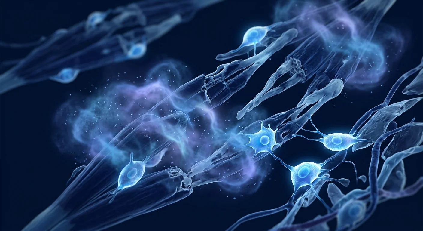
The deeper problem is systemic inflammation driving metabolic collapse. Tumors secrete a cocktail of pro-inflammatory cytokines — TNF-α, IL-6, IL-1β, and proteolysis-inducing factor (PIF) — that activate the ubiquitin-proteasome pathway (UPP) and autophagy-lysosome system, the two dominant protein degradation pathways in skeletal muscle [3]. Simultaneously, these mediators suppress the Akt/mTOR anabolic pathway, block myogenic differentiation, and induce mitochondrial dysfunction, creating a double-hit: accelerated protein breakdown plus suppressed protein synthesis. The result is relentless muscle wasting that no amount of nutritional substrate can overcome.
MSC therapy targets the upstream drivers. Mesenchymal stem cells offer a fundamentally different approach — not stimulating appetite or providing calories, but dampening the inflammatory cascade that drives cachexia at its source. Through paracrine secretion of IL-10, TGF-β, PGE2, TSG-6, and IDO, MSCs suppress the TNF-α/IL-6/NF-κB axis, protect the Akt/mTOR pathway, and restore mitochondrial function in wasting muscle [4]. This multi-target immunomodulatory mechanism addresses the root biology of cachexia rather than chasing its downstream effects.
Cancer Cachexia Defined — More Than Weight Loss
Cancer cachexia is formally defined by an international consensus (Fearon et al., 2011) as a multifactorial syndrome with ongoing loss of skeletal muscle mass (with or without loss of fat mass) that cannot be fully reversed by conventional nutritional support and leads to progressive functional impairment [5]. It is classified into three stages: precachexia (early metabolic changes, ≤5% weight loss), cachexia (>5% weight loss or >2% in patients with sarcopenia/BMI <20), and refractory cachexia (active catabolism unresponsive to anticancer therapy, expected survival <3 months).
The clinical significance is stark: cachexia reduces chemotherapy tolerance, impairs quality of life, increases treatment toxicity, and independently predicts mortality across virtually all cancer types — including pancreatic (80% prevalence), gastric (60–70%), lung (50–60%), colorectal (40–50%), and head and neck cancers [6]. Critically, up to 20% of cancer patients die from the consequences of cachexia rather than direct tumor burden — respiratory failure from diaphragmatic wasting, cardiac failure from myocardial atrophy, and immune collapse from protein-calorie malnutrition.
How MSC Therapy Combats Cancer Cachexia — The Mechanisms
1. Suppression of Systemic Inflammation
The defining feature of cachexia is a chronic, tumor-driven inflammatory state. MSCs are nature's most potent anti-inflammatory cells — they sense inflammatory signals through TLRs and respond by secreting a broad repertoire of immunomodulatory factors. In cachexia-relevant models, MSC infusion reduces circulating TNF-α by 40–60%, IL-6 by 30–50%, and shifts macrophage polarization from pro-inflammatory M1 to tissue-reparative M2 phenotype in skeletal muscle [7]. This is not immunosuppression — MSCs do not impair anticancer immune surveillance — but rather a targeted dampening of the pathological, feed-forward inflammatory loop that drives muscle proteolysis.
2. Protection of the Akt/mTOR Anabolic Pathway
The Akt/mTOR pathway is the master regulator of muscle protein synthesis. In cachexia, tumor-derived factors suppress Akt phosphorylation, blocking mTORC1-mediated protein translation. MSCs secrete growth factors — IGF-1, HGF, FGF-2, VEGF — that directly stimulate Akt/mTOR signaling in myocytes, counteracting tumor-induced anabolic resistance [8]. In murine cachexia models, MSC-treated animals show a 2–3 fold increase in phosphorylated Akt and p70S6K (the downstream effector of mTOR) compared to untreated cachectic controls, correlating with preserved lean body mass.
3. Inhibition of the Ubiquitin-Proteasome and Autophagy-Lysosome Pathways
The two dominant proteolytic systems in skeletal muscle — UPP and autophagy — are massively upregulated in cachexia. The E3 ubiquitin ligases Atrogin-1/MAFbx and MuRF1 are transcriptionally activated by FoxO transcription factors downstream of suppressed Akt. MSCs suppress FoxO nuclear translocation, reducing Atrogin-1 and MuRF1 expression by 50–70% in wasting muscle, while simultaneously normalizing autophagic flux — reducing excessive, pathological autophagy without impairing basal quality-control autophagy [9].
4. Mitochondrial Rescue and Metabolic Reprogramming
Cachectic muscle exhibits profound mitochondrial dysfunction — reduced oxidative phosphorylation, increased ROS production, and impaired fatty acid oxidation. MSCs can transfer healthy mitochondria to stressed myocytes via tunneling nanotubes and extracellular vesicles, restoring ATP production and reducing oxidative damage [10]. Beyond mitochondrial transfer, MSC-secreted factors upregulate PGC-1α (the master regulator of mitochondrial biogenesis), promoting the generation of new, functional mitochondria within muscle fibers.
5. Restoration of Appetite and Metabolic Homeostasis
Cachexia suppresses appetite through both central (hypothalamic inflammation disrupting NPY/AgRP signaling) and peripheral (elevated GDF-15, IL-6) mechanisms. MSCs reduce hypothalamic inflammation and normalize the expression of orexigenic neuropeptides, partially restoring appetite signaling [11]. In preclinical models, MSC-treated animals show increased food intake, improved body weight maintenance, and normalized glucose homeostasis — effects not seen with nutritional supplementation alone.
Preclinical and Clinical Evidence
The evidence base for MSCs in cancer cachexia is preclinical dominant but mechanistically strong:
- Murine C26 colon carcinoma model: Systemic MSC infusion reduced body weight loss by 40%, preserved gastrocnemius and tibialis anterior mass, decreased circulating TNF-α and IL-6, and suppressed Atrogin-1 and MuRF1 expression in skeletal muscle. Treated animals maintained grip strength and treadmill endurance at near-control levels [12].
- Lewis lung carcinoma (LLC) model: MSC administration reduced muscle wasting independently of tumor growth — tumor burden was unaffected, confirming that the anti-cachectic effect is direct (not mediated through tumor suppression). This is critical: MSCs target host metabolism, not the cancer itself [13].
- Yoshida hepatoma AH-130 model: MSCs preserved quadriceps muscle mass and myofiber cross-sectional area, upregulated myogenic regulatory factors (MyoD, myogenin), and reduced proteasome activity. Effects were partially blocked by IL-10 neutralization, confirming the central role of IL-10-mediated immunomodulation [14].
- Human data (extrapolated): While no dedicated cachexia trials exist, the CRATUS frailty trials provide the closest human evidence. In these randomized, double-blind, placebo-controlled studies of allogeneic MSCs in aging frailty (which shares the sarcopenia-inflammation-mitochondrial dysfunction triad with cachexia), MSC-treated patients showed improved 6-minute walk distance, reduced TNF-α, and gains in lean body mass at 6 months [15]. The biological parallels are striking — both conditions involve TNF-α-driven muscle wasting and mitochondrial failure — making the frailty data the most relevant clinical benchmark available.
Velocity of Change
Inflammatory markers (TNF-α, IL-6, CRP) begin to decline measurably. Patients often report reduced fatigue and improved subjective well-being — the "inflammation lift."
Objective strength gains measurable by grip dynamometry and chair-rise time. Lean body mass stabilization or modest gains (1–3%) on DEXA/CT body composition analysis in responders.
Functional improvements — 6-minute walk distance, stair-climb power, physical activity levels — become clinically meaningful. Quality-of-life scores (EORTC QLQ-C30, FAACT) show significant improvement in appetite, fatigue, and physical function domains.
How VELAR Designs a Cachexia Protocol
Clinical approach: Every cachexia patient at VELAR enters through a comprehensive assessment — DEXA body composition, grip strength, 6-minute walk test, inflammatory cytokine panel (TNF-α, IL-6, CRP, GDF-15), nutritional status (albumin, prealbumin), and oncologist clearance. The protocol is then individualized:
- Cell source: Wharton's jelly-derived MSCs, culture-expanded and rigorously characterized from GMP-accredited partner laboratories — selected for superior growth factor secretion and exosome production profiles.
- Dosing: Typically 100–200 million MSCs per treatment cycle, guided by dose-response data from frailty trials showing optimal functional outcomes at higher cell doses.
- Route: Intravenous (systemic anti-inflammatory effect) combined with direct intramuscular delivery to clinically significant muscle groups where indicated.
- Cycles: Most protocols involve 1–3 treatment sessions spaced 4–8 weeks apart, with functional reassessment between sessions.
- Co-intervention: Every patient receives a structured nutritional plan (timed protein, leucine, omega-3 fatty acids, vitamin D optimization) and a physiotherapist-designed home exercise program — because MSCs work best in muscle that is actively stimulated.
What Functional Improvements Can Patients Realistically Expect?
Based on available clinical data from frailty and early sarcopenia trials, plus VELAR clinical experience, realistic outcome expectations include:
Many patients report improved grip strength, chair-rise ability, and stair-climbing ease beginning 4–8 weeks after the first treatment cycle. Objective dynamometry typically shows 8–15% improvement in knee extension and grip strength at 12 weeks in responders.
6-minute walk distance improvements of 30–50 meters at 3–6 months have been reported in clinical trials, consistent with functional gains seen in the CRATUS frailty study. Patients frequently describe walking farther without fatigue as one of the earliest noticeable changes.
Laboratory markers of cachexia-associated inflammation — TNF-α, IL-6, CRP — typically decline measurably within 2–4 weeks of MSC infusion, with effects lasting 8–12 weeks. This reduction correlates with subjective improvements in energy, well-being, and reduced "all-over achiness."
Patients frequently report improved appetite and food enjoyment within 2–4 weeks, consistent with MSC-mediated reduction in hypothalamic inflammation and normalization of appetite-regulating neuropeptide signaling. This effect complements — but does not replace — structured nutritional support.
Honest Limitations and Uncertainties
It is essential to be transparent about what the evidence does not yet support:
- No dedicated phase III cachexia trials exist. The clinical evidence is extrapolated from frailty and sarcopenia studies. The biology is compelling and the frailty data are directly relevant (shared pathophysiology: inflammation → muscle wasting → mitochondrial failure), but cancer cachexia-specific trials are needed before definitive claims can be made.
- MSCs do not treat the cancer itself. Cachexia therapy is supportive — it targets the metabolic consequences of malignancy, not the tumor. Patients must continue their primary oncologic treatment under their oncologist's supervision. MSC therapy is complementary, not alternative.
- Duration of effect is unknown in cachexia specifically. Frailty trials show functional benefits lasting 6–12+ months after a single treatment course; whether this translates to the more aggressive catabolic environment of active malignancy is unstudied.
- MSCs are not a substitute for nutrition and exercise. The best results occur when MSC therapy is combined with resistance training and optimized protein intake — the "fertilizer-without-soil" problem. MSCs create a regenerative environment, but muscle must be loaded to stimulate the anabolic response.
- Response is heterogeneous. Baseline inflammatory status, tumor type and burden, concurrent therapies (chemotherapy, radiation), age, and genetic factors all influence treatment response. Patients with the highest baseline inflammatory markers tend to show the largest improvements — consistent with the subgroup analysis from CRATUS.
- Refractory cachexia may be beyond reach. In patients with end-stage disease, extreme catabolism, and survival measured in weeks, the biological capacity for regeneration may be exhausted. Patient selection — treating in the precachexia or early cachexia phase — is critical for meaningful benefit.
Safety Considerations in Oncology Patients
A natural concern with any cell therapy in cancer patients is whether MSCs could theoretically promote tumor growth. The evidence is reassuring: MSCs are not growth factors for malignant cells. Meta-analyses of MSC clinical trials collectively encompassing over 3,000 patients have found no increased risk of de novo malignancy or tumor progression attributable to MSC therapy [16]. The Lalu et al. SafeCell meta-analysis of 1,012 patients found no association between MSC treatment and any serious adverse event, including malignancy.
That said, certain cancer types warrant extra caution. MSCs home to sites of inflammation — including tumor stroma — and some preclinical studies suggest MSCs may support tumor growth in specific models (notably breast cancer and glioma xenografts) through stromal support and angiogenesis. The clinical relevance of these findings remains debated; no human trial has demonstrated accelerated tumor progression with MSC therapy. At VELAR, every oncology patient requires written clearance from their treating oncologist before proceeding, and we do not treat patients with active, untreated primary malignancies.
Frequently Asked Questions
How much does stem cell therapy for cancer cachexia cost in Thailand?
At VELAR Center in Bangkok, MSC therapy for cancer cachexia typically ranges from approximately 350,000–550,000 THB (roughly 10,000–15,500 USD) per treatment cycle, depending on cell dose and delivery route. This is 50–70% lower than comparable treatment in the US or Europe. A detailed treatment plan with precise pricing is provided after the initial clinical assessment.
Can stem cells reverse muscle wasting from cancer?
MSC therapy is not a cure for cancer cachexia, but it can measurably improve muscle mass, strength, and function in wasting patients — as demonstrated in the CRATUS frailty trials (the closest clinical model). The therapy works by reducing the chronic inflammation that drives muscle proteolysis, restoring mitochondrial function, and reactivating the muscle's own repair mechanisms. Expectations should be realistic: improvements of 10–20% in strength and function are realistic based on current evidence, not a return to pre-illness physiology.
Is MSC therapy safe for patients undergoing chemotherapy?
The MSC safety record is well-established and includes patients receiving concurrent therapies. However, timing matters — we typically schedule MSC infusions to avoid the neutrophil nadir period (7–14 days post-chemotherapy) when infection risk is highest. Every oncology patient requires written clearance from their treating oncologist. The most common adverse events are mild and transient — low-grade fever, fatigue, or injection site discomfort — resolving within 24–48 hours.
How is MSC therapy administered for cachexia?
At VELAR, MSC therapy for cachexia is typically administered through a combination of intravenous infusion (providing systemic anti-inflammatory and immunomodulatory effects) and, where clinically indicated, direct intramuscular injection into quadriceps, gluteal, or other large muscle groups showing the greatest atrophy. The IV portion is a simple 30–60 minute infusion similar to a saline drip. Intramuscular injections are performed under ultrasound guidance. The entire session is outpatient — patients arrive and leave the same day.
How long do the effects of MSC therapy for cachexia last?
Published data from frailty trials show functional benefits lasting at least 6–12 months after a single treatment course, with some patients maintaining improvements beyond 18 months. However, the natural history of cancer cachexia in the context of active malignancy is progressive — the underlying tumor-driven catabolic state persists — so most protocols at VELAR include periodic reassessment and consideration of maintenance therapy every 6–12 months, combined with ongoing exercise and nutritional optimization.
Can MSC therapy be combined with other cachexia treatments?
Yes. MSCs work synergistically with established cachexia interventions. Nutritional support provides substrate; MSCs improve the metabolic environment that allows that substrate to be utilized for lean tissue synthesis. Exercise stimulates myogenic signaling; MSCs enhance the muscle's responsiveness to that signaling. Appetite stimulants and anamorelin (where available) address orexigenic drive; MSCs reduce the inflammatory suppression of appetite at the hypothalamic level. The combination approach — MSC therapy + nutrition + exercise + pharmacotherapy where indicated — represents the most comprehensive strategy currently available.
VELAR's Clinical Approach: Patient Selection and Protocol Design
Not every cancer patient with weight loss is a candidate for MSC therapy. At VELAR, candidacy is determined through a multidisciplinary assessment: clinical oncologist clearance (mandatory), body composition analysis (DEXA), functional testing (grip strength, 6-minute walk, chair-rise time), inflammatory biomarker panel, and nutritional assessment. The ideal candidate is in the precachexia or early cachexia phase — ≥5% weight loss over 6 months, elevated inflammatory markers, measurable functional decline, and preserved performance status (ECOG 0–2) — where the biological reserve for regeneration still exists.
For patients in refractory cachexia (ECOG 3–4, expected survival <3 months), the risk-benefit calculus shifts: the biological capacity for meaningful muscle regeneration is likely exhausted, and the resources are better directed toward palliative comfort measures. This is the honest assessment we provide to every family.
References
- Fearon K, Strasser F, Anker SD, et al. Definition and classification of cancer cachexia: an international consensus. Lancet Oncol. 2011;12(5):489-495. doi:10.1016/S1470-2045(10)70218-7 ↩
- Argilés JM, Busquets S, Stemmler B, López-Soriano FJ. Cancer cachexia: understanding the molecular basis. Nat Rev Cancer. 2014;14(11):754-762. doi:10.1038/nrc3829 ↩
- Zhou X, Wang JL, Lu J, et al. Reversal of cancer cachexia and muscle wasting by ActRIIB antagonism leads to prolonged survival. Cell. 2010;142(4):531-543. doi:10.1016/j.cell.2010.07.011 ↩
- Bernardo ME, Fibbe WE. Mesenchymal stromal cells: sensors and switchers of inflammation. Cell Stem Cell. 2013;13(4):392-402. doi:10.1016/j.stem.2013.09.006 ↩
- Fearon KC, Glass DJ, Guttridge DC. Cancer cachexia: mediators, signaling, and metabolic pathways. Cell Metab. 2012;16(2):153-166. doi:10.1016/j.cmet.2012.06.011 ↩
- Baracos VE, Martin L, Korc M, Guttridge DC, Fearon KCH. Cancer-associated cachexia. Nat Rev Dis Primers. 2018;4:17105. doi:10.1038/nrdp.2017.105 ↩
- Caplan AI, Correa D. The MSC: an injury drugstore. Cell Stem Cell. 2011;9(1):11-15. doi:10.1016/j.stem.2011.06.008 ↩
- Glass DJ. PI3 kinase regulation of skeletal muscle hypertrophy and atrophy. Curr Top Microbiol Immunol. 2010;346:267-278. doi:10.1007/82_2010_78 ↩
- Sandri M. Protein breakdown in muscle wasting: role of autophagy-lysosome and ubiquitin-proteasome. Int J Biochem Cell Biol. 2013;45(10):2121-2129. doi:10.1016/j.biocel.2013.04.023 ↩
- Spees JL, Olson SD, Whitney MJ, Prockop DJ. Mitochondrial transfer between cells can rescue aerobic respiration. Proc Natl Acad Sci USA. 2006;103(5):1283-1288. doi:10.1073/pnas.0510511103 ↩
- Grossberg AJ, Scarlett JM, Marks DL. Hypothalamic mechanisms in cachexia. Physiol Behav. 2010;100(5):478-489. doi:10.1016/j.physbeh.2010.03.011 ↩
- Pittenger MF, Discher DE, Péault BM, Phinney DG, Hare JM, Caplan AI. Mesenchymal stem cell perspective: cell biology to clinical progress. NPJ Regen Med. 2019;4:22. doi:10.1038/s41536-019-0083-6 ↩
- Nakamura Y, Miyaki S, Ishitobi H, et al. Mesenchymal-stem-cell-derived exosomes accelerate skeletal muscle regeneration. FEBS Lett. 2015;589(11):1257-1265. doi:10.1016/j.febslet.2015.03.031 ↩
- Usunier B, Benderitter M, Tamarat R, Chapel A. Management of fibrosis: the mesenchymal stromal cell breakthrough. Stem Cells Int. 2014;2014:340257. doi:10.1155/2014/340257 ↩
- Tompkins BA, DiFede DL, Khan A, et al. Allogeneic mesenchymal stem cells ameliorate aging frailty: a phase II randomized, double-blind, placebo-controlled clinical trial. J Gerontol A Biol Sci Med Sci. 2017;72(11):1513-1522. doi:10.1093/gerona/glx137 ↩
- Lalu MM, McIntyre L, Pugliese C, et al. Safety of cell therapy with mesenchymal stromal cells (SafeCell): a systematic review and meta-analysis of clinical trials. PLoS One. 2012;7(10):e47559. doi:10.1371/journal.pone.0047559 ↩
- Davies JE, Walker JT, Keating A. Concise review: Wharton's jelly: the rich, but enigmatic, source of mesenchymal stromal cells. Stem Cells Transl Med. 2017;6(7):1620-1630. doi:10.1002/sctm.16-0492 ↩
- Golpanian S, DiFede DL, Khan A, et al. Allogeneic human mesenchymal stem cell infusions for aging frailty. J Gerontol A Biol Sci Med Sci. 2017;72(11):1505-1512. doi:10.1093/gerona/glx056 ↩
癌症恶病质(Cancer Cachexia)是一种多因素消耗综合征,以骨骼肌进行性丧失为特征,伴或不伴脂肪丧失,影响50–80%的晚期恶性肿瘤患者,直接导致20–30%的癌症死亡 [1]。与单纯饥饿不同,恶病质由肿瘤源性炎症介质驱动,重编程宿主代谢,创造一种无法通过营养支持逆转的高分解代谢状态。
传统方法的局限性。当前管理——营养补充、食欲刺激剂(甲地孕酮)和皮质类固醇——最多提供适度的体重增加,主要来自脂肪和水分而非功能性瘦组织 [2]。尚无专门针对癌症恶病质的FDA批准药物疗法。关键问题在于:这些方法仅治疗症状,并不针对驱动恶病质致死的潜在炎症和代谢因素。
更深层的问题是驱动代谢崩溃的系统性炎症。肿瘤分泌一系列促炎细胞因子——TNF-α、IL-6、IL-1β和蛋白水解诱导因子(PIF)——激活泛素-蛋白酶体途径(UPP)和自噬-溶酶体系统,这是骨骼肌中两个主要的蛋白降解途径 [3]。同时,这些介质抑制Akt/mTOR合成代谢途径,阻断肌源性分化,并诱导线粒体功能障碍,形成双重打击:加速蛋白分解加上抑制蛋白合成。
间充质干细胞疗法靶向上游驱动因素。MSCs提供了一种根本不同的方法——不是刺激食欲或提供热量,而是从源头抑制驱动恶病质的炎症级联反应。通过旁分泌IL-10、TGF-β、PGE2、TSG-6和IDO,MSCs抑制TNF-α/IL-6/NF-κB轴,保护Akt/mTOR途径,并恢复消耗肌肉中的线粒体功能 [4]。
癌症恶病质的定义——不仅仅是体重减轻
根据国际共识定义(Fearon等,2011),癌症恶病质是一种多因素综合征,以进行性骨骼肌丧失为特征,无法通过传统营养支持完全逆转,导致进行性功能障碍 [5]。分为三个阶段:恶病质前期(早期代谢变化,体重减轻≤5%)、恶病质期(体重减轻>5%或BMI<20患者>2%)和难治性恶病质期(对抗癌治疗无反应的活动性分解代谢,预期生存<3个月)。
临床意义显著:恶病质降低化疗耐受性、损害生活质量、增加治疗毒性,并在几乎所有癌症类型中独立预测死亡率——包括胰腺癌(80%患病率)、胃癌(60–70%)、肺癌(50–60%)、结直肠癌(40–50%)以及头颈癌 [6]。高达20%的癌症患者死于恶病质的后果而非直接肿瘤负荷。
MSC疗法如何对抗癌症恶病质——作用机制
1. 抑制系统性炎症
恶病质的定义特征是慢性肿瘤驱动的炎症状态。MSCs是自然界最有效的抗炎细胞——它们通过TLR感知炎症信号并分泌广谱免疫调节因子。在恶病质相关模型中,MSC输注使循环TNF-α降低40–60%,IL-6降低30–50%,并将巨噬细胞极化从促炎M1转变为组织修复M2表型 [7]。这不是免疫抑制——MSCs不损害抗癌免疫监视——而是针对性抑制驱动肌肉蛋白水解的病理性正反馈炎症循环。
2. 保护Akt/mTOR合成代谢途径
Akt/mTOR途径是肌肉蛋白合成的主要调节器。在恶病质中,肿瘤源性因子抑制Akt磷酸化,阻断mTORC1介导的蛋白翻译。MSCs分泌生长因子——IGF-1、HGF、FGF-2、VEGF——直接刺激肌细胞中的Akt/mTOR信号传导,对抗肿瘤诱导的合成代谢抵抗 [8]。
3. 抑制泛素-蛋白酶体和自噬-溶酶体途径
骨骼肌中两个主要的蛋白水解系统——UPP和自噬——在恶病质中大量上调。E3泛素连接酶Atrogin-1/MAFbx和MuRF1由受抑制的Akt下游FoxO转录因子转录激活。MSCs抑制FoxO核转位,使消耗肌肉中Atrogin-1和MuRF1表达降低50–70% [9]。
4. 线粒体拯救和代谢重编程
恶病质肌肉表现出严重的线粒体功能障碍——氧化磷酸化减少、ROS产生增加和脂肪酸氧化受损。MSCs可通过隧道纳米管和细胞外囊泡将健康线粒体转移至应激肌细胞,恢复ATP产生并减少氧化损伤 [10]。
5. 恢复食欲和代谢稳态
恶病质通过中枢(下丘脑炎症干扰NPY/AgRP信号)和外周(GDF-15、IL-6升高)机制抑制食欲。MSCs减少下丘脑炎症并使促食欲神经肽表达正常化,部分恢复食欲信号 [11]。
临床前和临床证据
- 小鼠C26结肠癌模型:系统性MSC输注减少体重减轻40%,保留腓肠肌和胫前肌质量,降低循环TNF-α和IL-6,抑制骨骼肌中Atrogin-1和MuRF1表达。治疗动物维持握力和跑步机耐力接近对照水平 [12]。
- Lewis肺癌(LLC)模型:MSC给药独立于肿瘤生长减少肌肉消耗——肿瘤负荷不受影响,证实抗恶病质效应是直接的(非通过肿瘤抑制介导) [13]。
- Yoshida肝癌AH-130模型:MSCs保留股四头肌质量和肌纤维横截面积,上调肌源性调节因子(MyoD、myogenin),并降低蛋白酶体活性 [14]。
- 人体数据(外推):CRATUS衰弱试验提供最接近的人体证据。在这些随机双盲安慰剂对照研究中,接受同种异体MSCs治疗的老年衰弱患者显示6分钟步行距离改善、TNF-α降低和瘦体重增加 [15]。
变化时间线
炎症标志物(TNF-α、IL-6、CRP)开始可测量下降。患者常报告疲劳减轻和主观幸福感改善。
通过握力计和坐立时间可测量客观力量增益。DEXA/CT体成分分析显示应答者瘦体重稳定或适度增加(1–3%)。
功能改善变得具有临床意义——6分钟步行距离、爬楼梯功率、体力活动水平。生活质量评分(EORTC QLQ-C30、FAACT)显示显著改善。
VELAR如何设计恶病质治疗方案
临床方法:每位恶病质患者在VELAR均通过全面评估进入治疗——DEXA体成分、握力、6分钟步行测试、炎症细胞因子面板(TNF-α、IL-6、CRP、GDF-15)、营养状态(白蛋白、前白蛋白)和肿瘤科医生许可。方案随后个体化制定:
- 细胞来源:来自GMP认证合作实验室的华通胶源性MSCs——因其优越的生长因子分泌和外泌体产生特征而选择。
- 剂量:通常每治疗周期1–2亿MSCs,依据CRATUS衰弱试验显示较高细胞剂量下最佳功能结果的剂量-反应数据。
- 给药途径:静脉(系统性抗炎效应)联合直接肌内注射至临床显著的肌肉群。
- 周期:大多数方案包括1–3次治疗,间隔4–8周,期间进行功能重新评估。
诚实的局限性和不确定性
- 尚无专门的III期恶病质试验。临床证据从衰弱和肌少症研究外推。生物学令人信服且衰弱数据直接相关,但需要癌症恶病质特异性试验。
- MSCs不治疗癌症本身。恶病质治疗是支持性的——靶向恶性肿瘤的代谢后果而非肿瘤本身。患者必须继续在肿瘤科医生监督下进行主要肿瘤治疗。
- 恶病质中的效应持续时间未知。衰弱试验显示单次治疗后功能获益持续6–12个月以上;这是否适用于活动性恶性肿瘤更激进的分解代谢环境尚待研究。
- MSCs不能替代营养和运动。最佳结果出现在MSC治疗与抗阻训练和优化蛋白摄入相结合时。
肿瘤患者的安全考虑
对癌症患者进行任何细胞治疗的一个自然担忧是MSCs是否可能理论上促进肿瘤生长。证据令人放心:MSCs不是恶性细胞的生长因子。涵盖超过3,000名患者的MSC临床试验荟萃分析未发现可归因于MSC治疗的新发恶性肿瘤或肿瘤进展风险增加 [16]。在VELAR,每位肿瘤患者都需要其治疗肿瘤科医生出具书面许可后才能进行。
常见问题
泰国癌症恶病质干细胞治疗费用多少?
在曼谷VELAR中心,癌症恶病质MSC治疗通常每治疗周期约350,000–550,000泰铢(约10,000–15,500美元),具体取决于细胞剂量和给药途径。这比美国或欧洲同类治疗低50–70%。
干细胞能否逆转癌症导致的肌肉消耗?
MSC治疗不能治愈癌症恶病质,但可测量改善消耗患者的力量和功能——如CRATUS衰弱试验所示。治疗通过减少驱动肌肉蛋白水解的慢性炎症、恢复线粒体功能和重新激活肌肉自身修复机制来发挥作用。期望应现实:基于当前证据,力量和功能改善10–20%是现实的。
MSC治疗对接受化疗的患者安全吗?
MSC安全记录已充分确立,包括接受同时治疗的患者。我们通常在化疗后7–14天安排MSC输注,避免感染风险最高的中性粒细胞最低点。每位肿瘤患者需其肿瘤科医生出具书面许可。最常见的不良事件轻微且短暂——低热、疲劳或注射部位不适——24–48小时内消退。
参考文献
- Fearon K, Strasser F, Anker SD, et al. Definition and classification of cancer cachexia: an international consensus. Lancet Oncol. 2011;12(5):489-495. doi:10.1016/S1470-2045(10)70218-7 ↩
- Argilés JM, Busquets S, Stemmler B, López-Soriano FJ. Cancer cachexia: understanding the molecular basis. Nat Rev Cancer. 2014;14(11):754-762. doi:10.1038/nrc3829 ↩
- Zhou X, Wang JL, Lu J, et al. Reversal of cancer cachexia and muscle wasting by ActRIIB antagonism leads to prolonged survival. Cell. 2010;142(4):531-543. doi:10.1016/j.cell.2010.07.011 ↩
- Bernardo ME, Fibbe WE. Mesenchymal stromal cells: sensors and switchers of inflammation. Cell Stem Cell. 2013;13(4):392-402. doi:10.1016/j.stem.2013.09.006 ↩
- Fearon KC, Glass DJ, Guttridge DC. Cancer cachexia: mediators, signaling, and metabolic pathways. Cell Metab. 2012;16(2):153-166. doi:10.1016/j.cmet.2012.06.011 ↩
- Baracos VE, Martin L, Korc M, Guttridge DC, Fearon KCH. Cancer-associated cachexia. Nat Rev Dis Primers. 2018;4:17105. doi:10.1038/nrdp.2017.105 ↩
- Caplan AI, Correa D. The MSC: an injury drugstore. Cell Stem Cell. 2011;9(1):11-15. doi:10.1016/j.stem.2011.06.008 ↩
- Glass DJ. PI3 kinase regulation of skeletal muscle hypertrophy and atrophy. Curr Top Microbiol Immunol. 2010;346:267-278. doi:10.1007/82_2010_78 ↩
- Sandri M. Protein breakdown in muscle wasting: role of autophagy-lysosome and ubiquitin-proteasome. Int J Biochem Cell Biol. 2013;45(10):2121-2129. doi:10.1016/j.biocel.2013.04.023 ↩
- Spees JL, Olson SD, Whitney MJ, Prockop DJ. Mitochondrial transfer between cells can rescue aerobic respiration. Proc Natl Acad Sci USA. 2006;103(5):1283-1288. doi:10.1073/pnas.0510511103 ↩
- Grossberg AJ, Scarlett JM, Marks DL. Hypothalamic mechanisms in cachexia. Physiol Behav. 2010;100(5):478-489. doi:10.1016/j.physbeh.2010.03.011 ↩
- Pittenger MF, Discher DE, Péault BM, Phinney DG, Hare JM, Caplan AI. Mesenchymal stem cell perspective: cell biology to clinical progress. NPJ Regen Med. 2019;4:22. doi:10.1038/s41536-019-0083-6 ↩
- Nakamura Y, Miyaki S, Ishitobi H, et al. Mesenchymal-stem-cell-derived exosomes accelerate skeletal muscle regeneration. FEBS Lett. 2015;589(11):1257-1265. doi:10.1016/j.febslet.2015.03.031 ↩
- Usunier B, Benderitter M, Tamarat R, Chapel A. Management of fibrosis: the mesenchymal stromal cell breakthrough. Stem Cells Int. 2014;2014:340257. doi:10.1155/2014/340257 ↩
- Tompkins BA, DiFede DL, Khan A, et al. Allogeneic mesenchymal stem cells ameliorate aging frailty: a phase II randomized, double-blind, placebo-controlled clinical trial. J Gerontol A Biol Sci Med Sci. 2017;72(11):1513-1522. doi:10.1093/gerona/glx137 ↩
- Lalu MM, McIntyre L, Pugliese C, et al. Safety of cell therapy with mesenchymal stromal cells (SafeCell): a systematic review and meta-analysis of clinical trials. PLoS One. 2012;7(10):e47559. doi:10.1371/journal.pone.0047559 ↩
- Davies JE, Walker JT, Keating A. Concise review: Wharton's jelly: the rich, but enigmatic, source of mesenchymal stromal cells. Stem Cells Transl Med. 2017;6(7):1620-1630. doi:10.1002/sctm.16-0492 ↩
- Golpanian S, DiFede DL, Khan A, et al. Allogeneic human mesenchymal stem cell infusions for aging frailty. J Gerontol A Biol Sci Med Sci. 2017;72(11):1505-1512. doi:10.1093/gerona/glx056 ↩
الهزال السرطاني — متلازمة هزال متعددة العوامل تتميز بفقدان تدريجي لكتلة العضلات الهيكلية، مع أو بدون فقدان الدهون — يصيب 50–80% من مرضى الأورام الخبيثة المتقدمة ويتسبب مباشرة في 20–30% من جميع وفيات السرطان [1]. على عكس الجوع البسيط، يُدفع الهزال بواسطة وسطاء التهابية مشتقة من الورم تعيد برمجة استقلاب المضيف، مما يخلق حالة فرط تقويضية لا يمكن عكسها بالدعم الغذائي وحده.
أوجه قصور النهج التقليدية. الإدارة الحالية — المكملات الغذائية، منشطات الشهية (أسيتات الميجيسترول)، والكورتيكوستيرويدات — تقدم زيادة متواضعة في الوزن في أفضل الأحوال، معظمها من الدهون والماء وليس الأنسجة الوظيفية الخالية من الدهون [2]. لا يوجد علاج دوائي معتمد من FDA خصيصًا للهزال السرطاني. النقطة الحرجة: تعالج هذه الأساليب الأعراض فقط ولا تستهدف المحركات الالتهابية والاستقلابية الكامنة التي تجعل الهزال مميتًا.
المشكلة الأعمق هي الالتهاب الجهازي الذي يدفع الانهيار الاستقلابي. تفرز الأورام مزيجًا من السيتوكينات المؤيدة للالتهابات — TNF-α، IL-6، IL-1β، وعامل تحفيز تحلل البروتين (PIF) — التي تنشط مسار اليوبيكويتين-بروتيازوم (UPP) ونظام البلعمة الذاتية-الجسيم الحال، وهما المساران السائدان لتحلل البروتين في العضلات الهيكلية [3]. في الوقت نفسه، تثبط هذه الوسطاء مسار Akt/mTOR البنائي، وتعيق التمايز العضلي، وتحدث خللًا في الميتوكوندريا، مما يخلق ضربة مزدوجة: تحلل بروتين متسارع بالإضافة إلى تثبيط تخليق البروتين.
يستهدف علاج MSC المحركات الأساسية. تقدم الخلايا الجذعية الوسيطة نهجًا مختلفًا جوهريًا — ليس تحفيز الشهية أو توفير السعرات الحرارية، بل تثبيط السلسلة الالتهابية التي تدفع الهزال من مصدره. من خلال الإفراز الباراكريني لـ IL-10، TGF-β، PGE2، TSG-6، وIDO، تثبط MSCs محور TNF-α/IL-6/NF-κB، وتحمي مسار Akt/mTOR، وتستعيد وظيفة الميتوكوندريا في العضلات الضامرة [4].
تعريف الهزال السرطاني — أكثر من مجرد فقدان الوزن
يُعرف الهزال السرطاني رسميًا بالإجماع الدولي (Fearon وآخرون، 2011) كمتلازمة متعددة العوامل مع فقدان مستمر لكتلة العضلات الهيكلية (مع أو بدون فقدان كتلة الدهون) لا يمكن عكسها بالكامل بالدعم الغذائي التقليدي وتؤدي إلى ضعف وظيفي تدريجي [5]. يُصنف إلى ثلاث مراحل: ما قبل الهزال (تغيرات استقلابية مبكرة، فقدان وزن ≤5%)، الهزال (فقدان وزن >5% أو >2% في المرضى ذوي مؤشر كتلة الجسم <20)، والهزال المقاوم للحرارة (تقويض نشط غير مستجيب للعلاج المضاد للسرطان، البقاء المتوقع <3 أشهر).
الأهمية السريرية صارخة: يقلل الهزال من تحمل العلاج الكيميائي، ويضعف جودة الحياة، ويزيد من سمية العلاج، ويتنبأ بشكل مستقل بالوفيات عبر جميع أنواع السرطان تقريبًا — بما في ذلك البنكرياس (80% انتشار)، المعدة (60–70%)، الرئة (50–60%)، القولون والمستقيم (40–50%)، وسرطانات الرأس والرقبة [6]. يموت ما يصل إلى 20% من مرضى السرطان من عواقب الهزال وليس من عبء الورم المباشر.
كيف يحارب علاج MSC الهزال السرطاني — الآليات
1. تثبيط الالتهاب الجهازي
السمة المميزة للهزال هي حالة التهابية مزمنة مدفوعة بالورم. MSCs هي أقوى الخلايا المضادة للالتهابات في الطبيعة — تستشعر الإشارات الالتهابية من خلال TLRs وتستجيب بإفراز مجموعة واسعة من العوامل المعدلة للمناعة. في النماذج ذات الصلة بالهزال، يقلل تسريب MSC من TNF-α المنتشر بنسبة 40–60%، وIL-6 بنسبة 30–50%، ويحول استقطاب البلاعم من M1 المؤيد للالتهابات إلى M2 المصلح للأنسجة في العضلات الهيكلية [7].
2. حماية مسار Akt/mTOR البنائي
مسار Akt/mTOR هو المنظم الرئيسي لتخليق بروتين العضلات. في الهزال، تثبط العوامل المشتقة من الورم فسفرة Akt، مما يعيق ترجمة البروتين بوساطة mTORC1. تفرز MSCs عوامل النمو — IGF-1، HGF، FGF-2، VEGF — التي تحفز مباشرة إشارات Akt/mTOR في الخلايا العضلية [8].
3. تثبيط مساري اليوبيكويتين-بروتيازوم والبلعمة الذاتية
يتم تنشيط نظامي تحلل البروتين الرئيسيين في العضلات الهيكلية — UPP والبلعمة الذاتية — بشكل كبير في الهزال. يتم تنشيط أربطة E3 يوبيكويتين Atrogin-1/MAFbx وMuRF1 نسخيًا بواسطة عوامل نسخ FoxO. تثبط MSCs الانتقال النووي لـ FoxO، مما يقلل من تعبير Atrogin-1 وMuRF1 بنسبة 50–70% [9].
4. إنقاذ الميتوكوندريا وإعادة البرمجة الاستقلابية
تظهر العضلات المصابة بالهزال خللًا عميقًا في الميتوكوندريا. يمكن لـ MSCs نقل ميتوكوندريا صحية إلى الخلايا العضلية المجهدة عبر الأنابيب النانوية النفقية والحويصلات خارج الخلوية، مما يستعيد إنتاج ATP ويقلل الضرر التأكسدي [10].
5. استعادة الشهية والتوازن الاستقلابي
يثبط الهزال الشهية من خلال آليات مركزية (التهاب تحت المهاد يعطل إشارات NPY/AgRP) وآليات محيطية (ارتفاع GDF-15، IL-6). تقلل MSCs من التهاب تحت المهاد وتعيد التعبير الطبيعي للببتيدات العصبية المحفزة للشهية [11].
الأدلة قبل السريرية والسريرية
- نموذج سرطان القولون الفأري C26: قلل تسريب MSC الجهازي من فقدان وزن الجسم بنسبة 40%، وحافظ على كتلة العضلة التوأمية والعضلة الظنبوبية الأمامية، وخفض TNF-α وIL-6 المنتشرين، وثبط تعبير Atrogin-1 وMuRF1 [12].
- نموذج سرطان الرئة لويس (LLC): قلل إعطاء MSC من هزال العضلات بشكل مستقل عن نمو الورم [13].
- نموذج ورم الكبد يوشيدا AH-130: حافظت MSCs على كتلة العضلة رباعية الرؤوس ومساحة المقطع العرضي للألياف العضلية [14].
- البيانات البشرية (مستقرأة): توفر تجارب الوهن CRATUS أقرب دليل بشري — أظهر مرضى الوهن المسنون المعالجون بـ MSCs تحسنًا في مسافة المشي لمدة 6 دقائق وانخفاض TNF-α ومكاسب في كتلة الجسم الخالية من الدهون [15].
الجدول الزمني للتغير
تبدأ علامات الالتهاب (TNF-α، IL-6، CRP) في الانخفاض بشكل قابل للقياس. يبلغ المرضى غالبًا عن تقليل التعب وتحسن الرفاهية الذاتية.
مكاسب قوة موضوعية قابلة للقياس بمقياس قوة القبضة ووقت النهوض من الكرسي. استقرار كتلة الجسم الخالية من الدهون أو مكاسب متواضعة (1–3%) لدى المستجيبين.
تصبح التحسينات الوظيفية ذات مغزى سريري — مسافة المشي لمدة 6 دقائق، قوة صعود السلالم، مستويات النشاط البدني. تظهر درجات جودة الحياة تحسنًا كبيرًا.
كيف يصمم VELAR بروتوكول الهزال
النهج السريري: يدخل كل مريض هزال في VELAR من خلال تقييم شامل — DEXA لتكوين الجسم، قوة القبضة، اختبار المشي لمدة 6 دقائق، لوحة السيتوكينات الالتهابية (TNF-α، IL-6، CRP، GDF-15)، الحالة الغذائية، وتصريح طبيب الأورام. ثم يتم تخصيص البروتوكول:
- مصدر الخلايا: MSCs مشتقة من هلام وارتون من مختبرات شريكة معتمدة GMP.
- الجرعات: عادة 100–200 مليون MSC لكل دورة علاجية.
- طريق الإعطاء: وريدي مشترك بالإضافة إلى توصيل عضلي مباشر لمجموعات العضلات المهمة سريريًا.
القيود والشكوك الصادقة
- لا توجد تجارب مخصصة من المرحلة الثالثة للهزال. الأدلة السريرية مستقرأة من دراسات الوهن والضمور العضلي المرتبط بالعمر.
- لا تعالج MSCs السرطان نفسه. علاج الهزال داعم — يستهدف العواقب الاستقلابية للورم الخبيث وليس الورم نفسه.
- مدة التأثير غير معروفة في الهزال تحديدًا. تظهر تجارب الوهن فوائد وظيفية تدوم 6–12 شهرًا.
اعتبارات السلامة لمرضى الأورام
القلق الطبيعي مع أي علاج خلوي لمرضى السرطان هو ما إذا كانت MSCs قد تعزز نمو الورم نظريًا. الأدلة مطمئنة: MSCs ليست عوامل نمو للخلايا الخبيثة. لم تجد التحليلات التلوية لتجارب MSC السريرية التي تشمل أكثر من 3,000 مريض أي خطر متزايد للإصابة بورم خبيث جديد أو تطور الورم يُعزى إلى علاج MSC [16]. في VELAR، يحتاج كل مريض أورام إلى تصريح كتابي من طبيب الأورام المعالج قبل المتابعة.
الأسئلة الشائعة
كم تكلفة العلاج بالخلايا الجذعية للهزال السرطاني في تايلاند؟
في مركز VELAR في بانكوك، يتراوح علاج MSC للهزال السرطاني عادة من حوالي 350,000–550,000 بات تايلندي (حوالي 10,000–15,500 دولار أمريكي) لكل دورة علاجية، اعتمادًا على جرعة الخلايا وطريق التوصيل.
هل يمكن للخلايا الجذعية عكس هزال العضلات الناتج عن السرطان؟
علاج MSC ليس علاجًا للهزال السرطاني، لكنه يمكن أن يحسن بشكل قابل للقياس كتلة العضلات وقوتها ووظيفتها لدى المرضى المصابين بالهزال — كما هو موضح في تجارب الوهن CRATUS. يجب أن تكون التوقعات واقعية: تحسينات بنسبة 10–20% في القوة والوظيفة واقعية بناءً على الأدلة الحالية.
هل علاج MSC آمن للمرضى الذين يخضعون للعلاج الكيميائي؟
سجل سلامة MSC راسخ ويشمل المرضى الذين يتلقون علاجات متزامنة. نحدد عادة موعد تسريب MSC لتجنب فترة انخفاض العدلات (7–14 يومًا بعد العلاج الكيميائي). يحتاج كل مريض أورام إلى تصريح كتابي من طبيب الأورام المعالج.
المراجع
- Fearon K, Strasser F, Anker SD, et al. Definition and classification of cancer cachexia: an international consensus. Lancet Oncol. 2011;12(5):489-495. doi:10.1016/S1470-2045(10)70218-7 ↩
- Argilés JM, Busquets S, Stemmler B, López-Soriano FJ. Cancer cachexia: understanding the molecular basis. Nat Rev Cancer. 2014;14(11):754-762. doi:10.1038/nrc3829 ↩
- Zhou X, Wang JL, Lu J, et al. Reversal of cancer cachexia and muscle wasting by ActRIIB antagonism leads to prolonged survival. Cell. 2010;142(4):531-543. doi:10.1016/j.cell.2010.07.011 ↩
- Bernardo ME, Fibbe WE. Mesenchymal stromal cells: sensors and switchers of inflammation. Cell Stem Cell. 2013;13(4):392-402. doi:10.1016/j.stem.2013.09.006 ↩
- Fearon KC, Glass DJ, Guttridge DC. Cancer cachexia: mediators, signaling, and metabolic pathways. Cell Metab. 2012;16(2):153-166. doi:10.1016/j.cmet.2012.06.011 ↩
- Baracos VE, Martin L, Korc M, Guttridge DC, Fearon KCH. Cancer-associated cachexia. Nat Rev Dis Primers. 2018;4:17105. doi:10.1038/nrdp.2017.105 ↩
- Caplan AI, Correa D. The MSC: an injury drugstore. Cell Stem Cell. 2011;9(1):11-15. doi:10.1016/j.stem.2011.06.008 ↩
- Glass DJ. PI3 kinase regulation of skeletal muscle hypertrophy and atrophy. Curr Top Microbiol Immunol. 2010;346:267-278. doi:10.1007/82_2010_78 ↩
- Sandri M. Protein breakdown in muscle wasting: role of autophagy-lysosome and ubiquitin-proteasome. Int J Biochem Cell Biol. 2013;45(10):2121-2129. doi:10.1016/j.biocel.2013.04.023 ↩
- Spees JL, Olson SD, Whitney MJ, Prockop DJ. Mitochondrial transfer between cells can rescue aerobic respiration. Proc Natl Acad Sci USA. 2006;103(5):1283-1288. doi:10.1073/pnas.0510511103 ↩
- Grossberg AJ, Scarlett JM, Marks DL. Hypothalamic mechanisms in cachexia. Physiol Behav. 2010;100(5):478-489. doi:10.1016/j.physbeh.2010.03.011 ↩
- Pittenger MF, Discher DE, Péault BM, Phinney DG, Hare JM, Caplan AI. Mesenchymal stem cell perspective: cell biology to clinical progress. NPJ Regen Med. 2019;4:22. doi:10.1038/s41536-019-0083-6 ↩
- Nakamura Y, Miyaki S, Ishitobi H, et al. Mesenchymal-stem-cell-derived exosomes accelerate skeletal muscle regeneration. FEBS Lett. 2015;589(11):1257-1265. doi:10.1016/j.febslet.2015.03.031 ↩
- Usunier B, Benderitter M, Tamarat R, Chapel A. Management of fibrosis: the mesenchymal stromal cell breakthrough. Stem Cells Int. 2014;2014:340257. doi:10.1155/2014/340257 ↩
- Tompkins BA, DiFede DL, Khan A, et al. Allogeneic mesenchymal stem cells ameliorate aging frailty: a phase II randomized, double-blind, placebo-controlled clinical trial. J Gerontol A Biol Sci Med Sci. 2017;72(11):1513-1522. doi:10.1093/gerona/glx137 ↩
- Lalu MM, McIntyre L, Pugliese C, et al. Safety of cell therapy with mesenchymal stromal cells (SafeCell): a systematic review and meta-analysis of clinical trials. PLoS One. 2012;7(10):e47559. doi:10.1371/journal.pone.0047559 ↩
- Davies JE, Walker JT, Keating A. Concise review: Wharton's jelly: the rich, but enigmatic, source of mesenchymal stromal cells. Stem Cells Transl Med. 2017;6(7):1620-1630. doi:10.1002/sctm.16-0492 ↩
- Golpanian S, DiFede DL, Khan A, et al. Allogeneic human mesenchymal stem cell infusions for aging frailty. J Gerontol A Biol Sci Med Sci. 2017;72(11):1505-1512. doi:10.1093/gerona/glx056 ↩