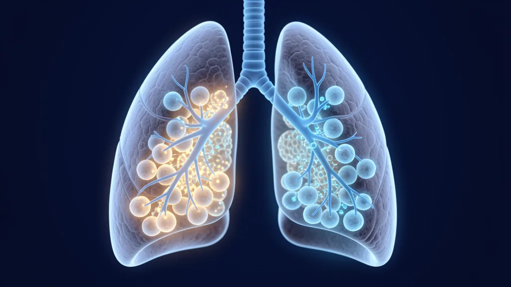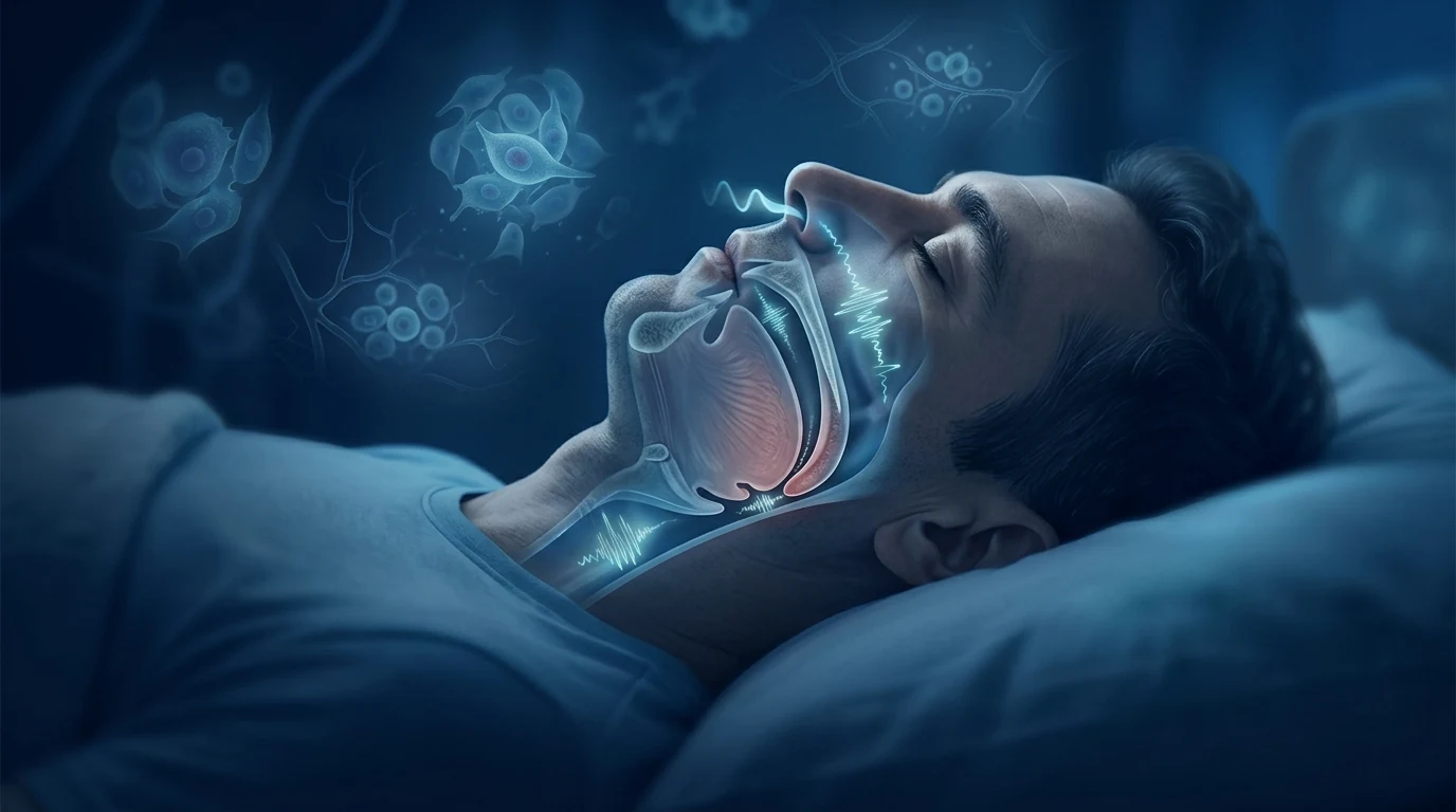
Obstructive sleep apnea is far more than a nightly nuisance. It is a chronic, progressive disease in which the upper airway repeatedly collapses during sleep, starving the body of oxygen dozens or even hundreds of times each night. The consequence is not just poor sleep — it is a systemic inflammatory and oxidative stress cascade that damages the heart, brain, blood vessels, and metabolism. That cascade is precisely what makes sleep apnea a plausible target for Mesenchymal Stem Cell therapy.
The scale of the problem is staggering. Nearly one billion adults worldwide have obstructive sleep apnea (OSA), with approximately 425 million classified as moderate to severe. [1] Despite this prevalence, the majority remain undiagnosed, and of those who are diagnosed, adherence to continuous positive airway pressure (CPAP) therapy — the gold-standard treatment — is poor, with roughly half of patients discontinuing use within the first year. [2]
Where standard treatment falls short. CPAP is highly effective when used consistently, but real-world adherence is a persistent clinical challenge. Oral appliances help a subset of patients but are less effective for moderate-to-severe disease. Surgical options exist (UPPP, hypoglossal nerve stimulation, maxillomandibular advancement) but carry complication risks and variable success rates. [3] What remains unaddressed by all these approaches is the underlying tissue-level damage — the oxidative stress, systemic inflammation, and endothelial injury — that OSA inflicts even when the airway is mechanically supported.
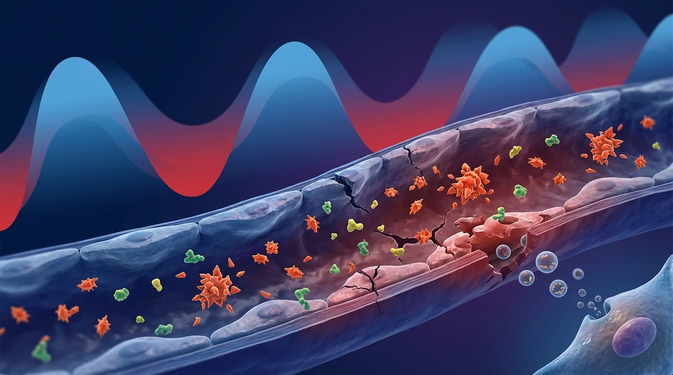
The tissue-level problem is intermittent hypoxia with reperfusion injury. Each apnoeic episode produces a drop in oxygen saturation followed by a sudden reoxygenation upon arousal. This pattern of intermittent hypoxia (IH) triggers a cascade identical to ischaemia-reperfusion injury: reactive oxygen species (ROS) surge, NF-κB is activated, pro-inflammatory cytokines (TNF-α, IL-6, IL-1β) rise, and the vascular endothelium becomes dysfunctional. [4] Over months and years, this IH-driven injury drives hypertension, insulin resistance, cognitive decline, atherosclerosis, and heart failure. [5]
MSC therapy targets the injury cascade itself. Rather than merely splinting the airway open, MSCs enter the inflamed, oxidatively stressed tissues and modulate the underlying pathology — dampening the NF-κB-driven inflammatory response, reducing oxidative injury, and supporting endothelial repair. The relevance to OSA is not about replacing CPAP; it is about repairing the damage that OSA causes and potentially improving the metabolic and cardiovascular outcomes that define the disease's long-term risk.
What is obstructive sleep apnea
Obstructive sleep apnea is a sleep-related breathing disorder characterised by repetitive episodes of complete (apnoea) or partial (hypopnoea) upper airway collapse during sleep. Each episode produces intermittent hypoxia, hypercapnia, sympathetic nervous system activation, and arousal from sleep. Severity is graded by the apnoea-hypopnoea index (AHI): mild (5–15 events/hour), moderate (15–30), and severe (≥30). [6]
OSA is strongly associated with obesity (approximately 60–70% of moderate-to-severe OSA occurs in people with obesity), but it also affects non-obese individuals with craniofacial anatomy predisposing to airway collapse. Men are 2–3 times more likely than premenopausal women to have OSA, though the gap narrows after menopause. [7]
Untreated OSA is an independent risk factor for systemic hypertension, pulmonary hypertension, atrial fibrillation, stroke, coronary artery disease, type 2 diabetes, cognitive impairment, and all-cause mortality. The unifying mechanism across these comorbidities is IH-driven systemic inflammation and oxidative stress. [5]
The inflammatory biology of OSA
OSA is not simply a mechanical airway problem — it is an inflammatory disease of the whole organism. Each night of untreated apnoeas increases circulating levels of TNF-α, IL-6, CRP, and adhesion molecules, while reducing endothelial repair capacity. Over time, this creates a state of chronic, low-grade systemic inflammation that accelerates cardiovascular, metabolic, and cognitive decline. [4]
What goes wrong at the tissue level in OSA
To understand where MSCs may intervene, it helps to map the key pathophysiological mechanisms of OSA at the tissue level:
- Intermittent hypoxia and oxidative stress — cycles of hypoxia-reoxygenation generate ROS that overwhelm antioxidant defences, damaging cellular lipids, proteins, and DNA. This oxidative injury is detectable in circulating leukocytes, vascular endothelium, and neural tissue of OSA patients. [8]
- NF-κB-driven systemic inflammation — IH activates the transcription factor NF-κB, which upregulates TNF-α, IL-6, IL-1β, and MCP-1, creating a self-sustaining inflammatory loop. These cytokines promote endothelial dysfunction, insulin resistance, and leukocyte adhesion. [9]
- Vascular endothelial injury — circulating endothelial cells and endothelial progenitor cells are reduced in number and function in OSA, reflecting ongoing vascular damage and impaired repair capacity. [5]
- Sympathetic overactivation — each apnoea triggers a surge of sympathetic activity that persists into daytime, driving hypertension, tachycardia, and metabolic dysregulation
- Neuroinflammation and cognitive injury — IH induces microglial activation, hippocampal oxidative stress, and blood-brain barrier disruption, contributing to the cognitive deficits — memory, attention, executive function — commonly seen in untreated OSA. [10]
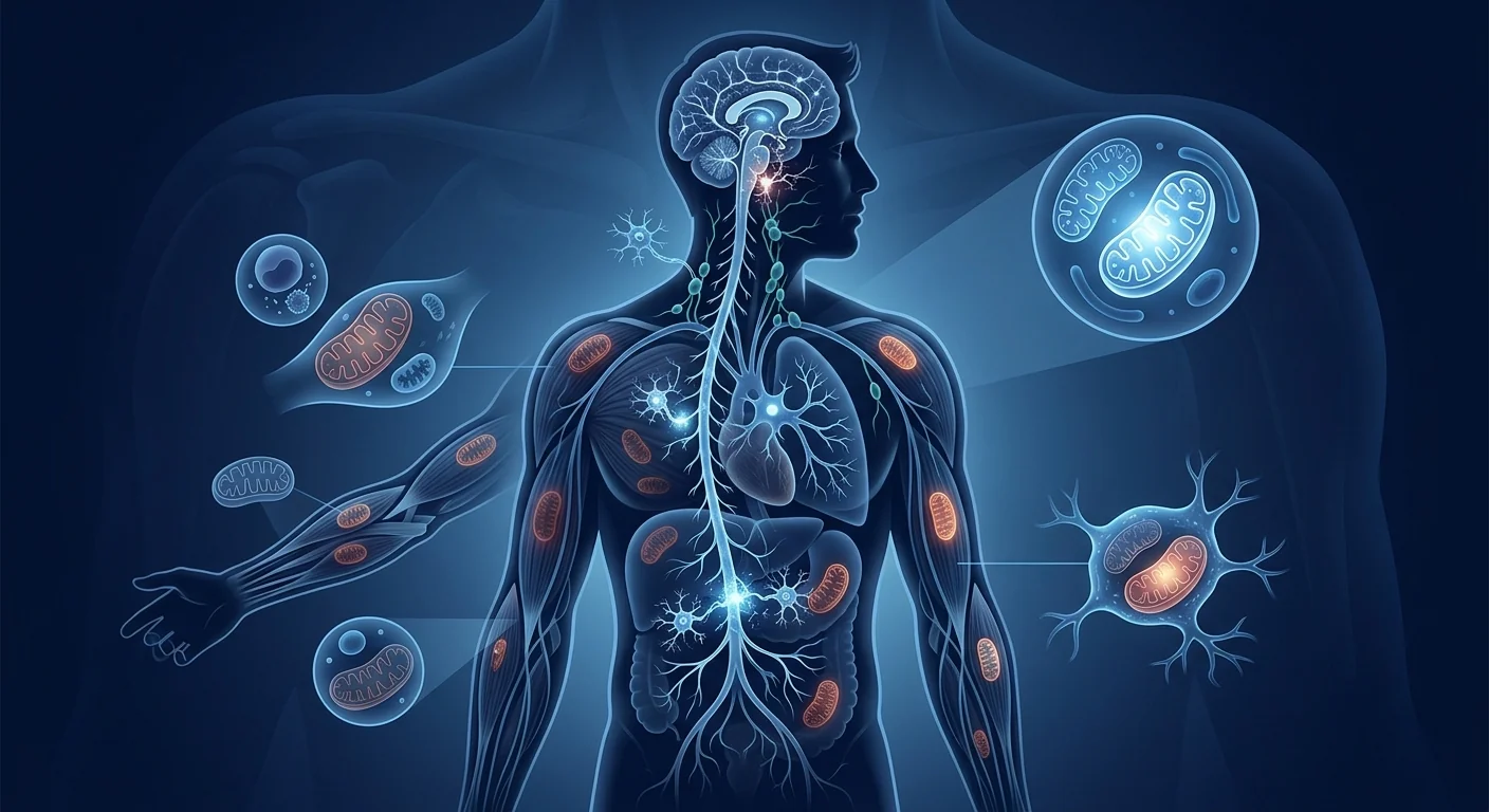
The cumulative effect is a multi-organ inflammatory state in which the capacity for endogenous repair is progressively overwhelmed by the nightly burden of IH-driven injury.
How MSCs may benefit obstructive sleep apnea
MSCs are not a treatment for the mechanical upper airway collapse of OSA. No evidence suggests that MSC therapy restores pharyngeal muscle tone or prevents the airway from closing during sleep. What MSCs may do is interrupt the downstream inflammatory and oxidative injury cascade that IH triggers — reducing the systemic damage and potentially improving the metabolic, cardiovascular, and cognitive outcomes that define OSA's health burden.
Anti-inflammatory modulation of IH-driven inflammation
MSCs sense the inflammatory milieu created by intermittent hypoxia and respond by secreting TSG-6, PGE2, IDO, IL-10, and other paracrine factors that suppress NF-κB signalling, reduce TNF-α and IL-6 output, and promote macrophage polarisation toward the anti-inflammatory M2 phenotype. [11] In a rat model of recurrent obstructive apnoeas, MSC infusion reduced serum IL-1β from 66.7 to 6.1 pg/mL — essentially normalising the IH-driven inflammatory spike. [12]
Oxidative stress attenuation
MSCs secrete antioxidants and upregulate the host's endogenous antioxidant systems, including Nrf2 and its target genes (HO-1, NQO1). This reduces the ROS burden that drives endothelial and neural injury in OSA. Preclinical data show that MSC treatment normalises markers of oxidative damage in IH-exposed tissues. [13]
Vascular endothelial repair
MSC-derived paracrine factors — VEGF, HGF, angiopoietin-1 — support endothelial integrity, stimulate angiogenesis, and mobilise endothelial progenitor cells. In a rat model of OSA-induced aortic remodelling, MSC infusion normalised vascular wall thickness, reduced elastin fragmentation, and restored endothelial function. [14]
Neuroprotection against IH-induced cognitive decline
Through their secretion of BDNF, GDNF, and NGF, MSCs support neuronal survival and synaptic plasticity in the hippocampus and prefrontal cortex — regions particularly vulnerable to IH-induced injury. MSC-derived exosomes also reduce microglial activation and restore blood-brain barrier integrity. [15]
Metabolic benefits relevant to OSA comorbidity
Given the strong bidirectional relationship between OSA and metabolic dysfunction, MSCs may offer ancillary improvements in insulin sensitivity, adipokine balance, and hepatic steatosis — potentially reducing the metabolic burden that both causes and is worsened by OSA. [16]
What the evidence supports — and what it does not
An honest assessment of where the published research stands on MSC therapy for OSA:
What is plausible: Reduction in systemic inflammatory markers (hsCRP, IL-6, TNF-α), attenuation of oxidative stress, improvement in endothelial function, and potential neuroprotection against IH-induced cognitive decline — if MSCs are administered in the context of ongoing airway management. Preclinical evidence (rodent models of recurrent obstructive apnoeas) is consistent and supportive. [12][14]
What is not supported by current evidence: Resolution of upper airway obstruction, reduction in AHI (apnoea-hypopnoea index), improvement in overnight oxygen saturation, replacement of CPAP or oral appliance therapy, or any claim that MSC therapy "cures" sleep apnea. No human clinical trial has yet tested MSCs specifically for OSA as a primary endpoint.
What remains under active investigation: The optimal dosing protocol for IH-related inflammatory injury, the best cell source (Wharton's jelly MSCs may be preferable to autologous adipose MSCs, which can be functionally impaired in the metabolic dysfunction that frequently accompanies OSA), the role of MSC-derived exosomes versus whole-cell therapy, and the interaction between MSC therapy and CPAP adherence.
The honest frame for sleep apnea and MSC therapy
MSC therapy for OSA is, at present, an adjunctive and investigational protocol — a possible addition to standard airway management (CPAP, oral appliance, or surgery), not a replacement for it. CPAP remains the first-line treatment because it directly addresses the mechanical airway collapse that MSCs cannot fix. The role of regenerative therapy is in repairing the downstream tissue damage that OSA causes and potentially improving the cardiovascular and metabolic outcomes that persist even with mechanical therapy.
Who is the strongest candidate
Within realistic boundaries, the patients most likely to see meaningful benefit from MSC therapy in the context of OSA are those who:
- Are already on CPAP or other airway management but continue to have elevated inflammatory markers or metabolic complications — indicating that airway management alone is insufficient to fully reverse the systemic injury
- Have OSA-related cardiovascular or metabolic comorbidities (hypertension, insulin resistance, systemic inflammation) that persist despite adequate CPAP adherence
- Are in the earlier stages of OSA-related end-organ damage, before irreversible structural cardiovascular or cognitive changes have occurred
- Are not candidates for or have declined surgical airway interventions and want to address the systemic inflammatory burden of OSA beyond what CPAP provides
- Are coordinating with their sleep physician — MSC therapy must be complementary to, not a substitute for, standard sleep medicine care
Patients with severe, long-standing OSA and established cardiovascular or cerebrovascular disease are less likely to see substantial benefit from MSC therapy alone, though it may still play a supportive anti-inflammatory role in carefully selected cases.
How MSC therapy for OSA is delivered
Given the systemic nature of OSA-related inflammatory injury, the most studied delivery route is intravenous infusion, which distributes MSCs broadly to tissues affected by IH-driven damage — the vasculature, heart, brain, and metabolically active organs. Intrathecal delivery has not been studied in the OSA context, though it may be relevant if cognitive or neuroinflammatory endpoints become more prominent trial targets.
A typical protocol at a regulated clinical centre:
- Evaluation: Polysomnography-confirmed OSA diagnosis, inflammatory marker assessment (hsCRP, IL-6), endothelial function evaluation, and cardiometabolic risk stratification
- Cell source: Allogeneic Wharton's jelly-derived MSCs (preferred for consistent potency; autologous adipose MSCs may be functionally compromised by the metabolic dysfunction that often accompanies OSA)
- Dose: Typically 100–200 million cells per infusion, administered intravenously over 30–60 minutes
- Schedule: One to three infusions over 2–4 weeks, with maintenance infusions every 6–12 months depending on clinical response
- Monitoring: Repeat inflammatory markers, endothelial function (flow-mediated dilation), cognitive assessment (MoCA), and sleep quality questionnaires at 3, 6, and 12 months
MSC infusion should never be scheduled on the same night as a sleep study or CPAP titration, as the sedation or sleep disruption may confound the evaluation of either intervention.
Safety considerations
MSC therapy has a well-established safety profile across hundreds of clinical trials. A 2020 systematic review and meta-analysis of intravascular MSC administration across 62 clinical trials found no association with acute infusion toxicity, organ system complications, infection, or thromboembolic events compared with controls. [17]
For OSA patients specifically, theoretical considerations include:
- Nocturnal oxygen desaturation: MSC infusion does not affect oxygen saturation or respiratory drive; patients should continue CPAP as prescribed during the treatment period
- Interaction with CPAP: No known interaction; MSCs and CPAP address different aspects of OSA pathology (mechanical vs. inflammatory)
- Interaction with medications: Anticoagulants and antiplatelet agents are relevant if intravenous access requires consideration; otherwise, no drug-specific contraindications
- Allogeneic MSCs: Fever or infusion reactions are rare and self-limited; pre-medication with antihistamines may be offered
Frequently asked questions
Can MSC therapy replace CPAP for sleep apnea?
No. MSC therapy does not address the mechanical upper airway collapse that defines OSA. CPAP, oral appliances, and surgical interventions are the only treatments that directly maintain airway patency during sleep. MSCs are being studied as an adjunctive therapy aimed at reducing the systemic inflammatory injury caused by untreated or partially treated OSA.
Will MSC therapy lower my AHI score?
There is no evidence that MSC therapy reduces the apnoea-hypopnoea index. The AHI reflects the frequency of airway collapse events, which is a mechanical — not inflammatory — parameter. Improvements in sleep quality, daytime fatigue, and cardiovascular markers may occur independently of changes in AHI.
How long do the effects of MSC therapy last in OSA?
The anti-inflammatory and tissue-repair effects of MSCs are transient, typically lasting 6–12 months after a treatment course. Maintenance infusions are likely needed to sustain the benefits. The duration of effect depends on the individual's ongoing burden of IH exposure, which is determined by CPAP adherence and OSA severity.
Who is not a candidate for MSC therapy for OSA?
Patients with severe, untreated OSA who are not using any form of airway management should focus on establishing effective CPAP or alternative therapy before considering regenerative interventions. MSC therapy is also not appropriate for patients with active malignancy, acute infection, or uncontrolled bleeding disorders.
How much does MSC therapy cost for sleep apnea?
MSC therapy is not currently approved for OSA by any regulatory authority, and costs vary widely between clinics. A single treatment course typically ranges from $15,000 to $25,000 USD depending on the cell source, dose, and number of infusions. Patients should verify that the clinic operates under registered medical supervision and uses independently tested, pathogen-screened cells. Our Thailand cost guide provides a detailed breakdown of pricing across Bangkok clinics. [18]
Is MSC therapy for sleep apnea covered by insurance?
No. Since MSC therapy for OSA is investigational and not FDA-approved (or equivalent regulatory approved) for this indication, it is not covered by health insurance. Patients must pay out-of-pocket.
Limitations and honest caveats
It is important to state plainly what the research does and does not support. All published studies on MSCs in OSA to date are preclinical rodent models. [12][14] There are no human clinical trials — not a single Phase I, II, or III study — investigating MSC therapy for obstructive sleep apnea as a primary or secondary endpoint. The mechanistic rationale is strong (MSCs address the inflammatory and oxidative injury that drives OSA's systemic complications), and the preclinical data are consistent, but clinical translation has not yet occurred.
Until human trials are conducted, the use of MSC therapy for OSA remains entirely off-label and investigational. Patients considering this approach should do so with the understanding that the evidence base is preclinical only, and that the primary value of MSC therapy in the OSA context is likely adjunctive — reducing the inflammatory and metabolic consequences of the disease, not treating the airway obstruction itself.
References
- Benjafield AV, Ayas NT, Eastwood PR, et al. Estimation of the global prevalence and burden of obstructive sleep apnoea: a literature-based analysis. The Lancet Respiratory Medicine. 2019;7(8):687-698. doi:10.1016/S2213-2600(19)30198-5 ↩
- Kapur VK, Auckley DH, Chowdhuri S, et al. Clinical Practice Guideline for Diagnostic Testing for Adult Obstructive Sleep Apnea. Journal of Clinical Sleep Medicine. 2017;13(3):479-504. doi:10.5664/jcsm.6506 ↩
- Ramar K, Dort LC, Katz SG, et al. Clinical Practice Guideline for the Treatment of Obstructive Sleep Apnea and Snoring with Oral Appliance Therapy: An Update for 2015. Journal of Clinical Sleep Medicine. 2015;11(7):773-827. doi:10.5664/jcsm.4858 ↩
- Lavie L. Oxidative stress in obstructive sleep apnea and intermittent hypoxia — Revisited — The bad ugly and good: Implications to the heart and brain. Sleep Medicine Reviews. 2015;20:27-45. doi:10.1016/j.smrv.2014.07.003 ↩
- Jelic S, Padeletti M, Kawut SM, et al. Inflammation, Oxidative Stress, and Repair Capacity of the Vascular Endothelium in Obstructive Sleep Apnea. Circulation. 2008;117(17):2270-2278. doi:10.1161/CIRCULATIONAHA.107.741512 ↩
- Ryan S, Taylor CT, McNicholas WT. Selective Activation of Inflammatory Pathways by Intermittent Hypoxia in Obstructive Sleep Apnea Syndrome. Circulation. 2005;112(17):2660-2667. doi:10.1161/CIRCULATIONAHA.105.556746 ↩
- Almendros I, Carreras A, Montserrat JM, Gozal D, Navajas D, Farre R. Potential Role of Adult Stem Cells in Obstructive Sleep Apnea. Frontiers in Neurology. 2012;3:112. doi:10.3389/fneur.2012.00112 ↩
- Lavie L. Oxidative stress in obstructive sleep apnea and intermittent hypoxia. Sleep Medicine Reviews. 2015;20:27-45. doi:10.1016/j.smrv.2014.07.003 ↩
- Ryan S, Taylor CT, McNicholas WT. Selective activation of inflammatory pathways by intermittent hypoxia in obstructive sleep apnea syndrome. Circulation. 2005;112(17):2660-2667. doi:10.1161/CIRCULATIONAHA.105.556746 ↩
- Zdravkovic M, Harrell CR, Jakovljevic V, Djonov V, Volarevic V. Molecular Mechanisms Responsible for Mesenchymal Stem Cell-Based Modulation of Obstructive Sleep Apnea. International Journal of Molecular Sciences. 2023;24(4):3708. doi:10.3390/ijms24043708 ↩
- Carreras A, Almendros I, Montserrat JM, Navajas D, Farre R. Potential Role of Bone Marrow Mesenchymal Stem Cells in Obstructive Sleep Apnea. International Journal of Stem Cells. 2011;4(1):43-49. doi:10.15283/ijsc.2011.4.1.43 ↩
- Carreras A, Almendros I, Montserrat JM, Navajas D, Farre R. Mesenchymal stem cells reduce inflammation in a rat model of obstructive sleep apnea. Respiratory Physiology and Neurobiology. 2010;172(3):210-212. doi:10.1016/j.resp.2010.05.009 ↩
- Zdravkovic M, Harrell CR, Jakovljevic V, Djonov V, Volarevic V. Molecular Mechanisms Responsible for Mesenchymal Stem Cell-Based Modulation of Obstructive Sleep Apnea. International Journal of Molecular Sciences. 2023;24(4):3708. doi:10.3390/ijms24043708 ↩
- Rubies C, Dantas AP, Batlle M, et al. Aortic remodelling induced by obstructive apneas is normalized with mesenchymal stem cells infusion. Scientific Reports. 2019;9(1):10977. doi:10.1038/s41598-019-47813-1 ↩
- Zdravkovic M, Harrell CR, Jakovljevic V, Djonov V, Volarevic V. Molecular mechanisms responsible for MSC-based modulation of OSA. International Journal of Molecular Sciences. 2023;24(4):3708. doi:10.3390/ijms24043708 ↩
- Bi H, He J, He X, et al. Bone marrow stem cells therapy alleviates vascular injury in a COPD-OSA overlap syndrome rat model. Molecular Medicine Reports. 2021;23(1):55. doi:10.3892/mmr.2020.11707 ↩
- Thompson M, Mei SHJ, Wolfe D, et al. Cell therapy with intravascular administration of mesenchymal stromal cells continues to appear safe: An updated systematic review and meta-analysis. EClinicalMedicine. 2020;19:100249. doi:10.1016/j.eclinm.2019.100249 ↩
- VELAR Editorial Team. Stem Cell Therapy in Thailand: Cost, Standards and How to Choose a Clinic. VELAR Insights. 2026. Read article ↩
阻塞性睡眠呼吸暂停远非夜间烦恼那么简单。它是一种慢性进行性疾病,上呼吸道在睡眠中反复塌陷,使身体每夜数十次甚至数百次缺氧。其后果不仅是睡眠质量差,还会引发全身性炎症和氧化应激级联反应,损害心脏、大脑、血管和新陈代谢。正是这种炎症级联反应使得睡眠呼吸暂停成为间充质干细胞疗法的合理靶点。
问题规模令人震惊。全球近十亿成年人患有阻塞性睡眠呼吸暂停(OSA),其中约4.25亿属于中重度。 [1]尽管患病率如此之高,但大多数人仍未得到诊断,而在已确诊的患者中,对金标准治疗——持续气道正压通气(CPAP)的依从性很差,约半数患者在第一年内停止使用。 [2]
标准治疗的局限。CPAP在持续使用时非常有效,但现实世界中的依从性始终是临床挑战。口腔矫治器对部分患者有帮助,但对中重度疾病效果较差。外科选择(UPPP、舌下神经刺激、颌前移术)存在并发症风险和不同的成功率。 [3]所有这些方法都未能解决潜在的组织层面损伤——即OSA即使在气道得到机械支持时仍然造成的氧化应激、全身炎症和内皮损伤。
组织层面问题是间歇性缺氧合并再灌注损伤。每次呼吸暂停事件都会导致血氧饱和度下降,随后在觉醒时突然再氧合。这种间歇性缺氧模式触发了与缺血再灌注损伤相同的级联反应:活性氧激增、NF-κB被激活、促炎细胞因子(TNF-α、IL-6、IL-1β)升高、血管内皮功能失调。 [4]经年累月,这种间歇性缺氧驱动的损伤导致高血压、胰岛素抵抗、认知衰退、动脉粥样硬化和心力衰竭。 [5]
MSC疗法靶向损伤级联反应本身。MSC并非简单撑开气道,而是进入发炎、氧化应激的组织中调节潜在病理——抑制NF-κB驱动的炎症反应、减少氧化损伤、支持内皮修复。MSC对OSA的相关性不在于替代CPAP,而在于修复OSA造成的损伤,并可能改善定义该疾病长期风险的心血管和代谢结局。
什么是阻塞性睡眠呼吸暂停
阻塞性睡眠呼吸暂停是一种睡眠相关呼吸障碍,其特征是睡眠期间上呼吸道反复完全(呼吸暂停)或部分(低通气)塌陷。每次发作都会导致间歇性缺氧、高碳酸血症、交感神经系统激活和睡眠觉醒。严重程度通过呼吸暂停低通气指数(AHI)分级:轻度(5-15次/小时)、中度(15-30)和重度(≥30)。 [6]
OSA与肥胖密切相关(约60-70%的中重度OSA发生于肥胖人群),但也影响因颅面解剖导致气道塌陷的非肥胖个体。男性患病率是绝经前女性的2-3倍,但绝经后差距缩小。 [7]
未经治疗的OSA是全身性高血压、肺动脉高压、心房颤动、中风、冠状动脉疾病、2型糖尿病、认知障碍和全因死亡的独立危险因素。这些合并症的共同机制是间歇性缺氧驱动的全身炎症和氧化应激。 [5]
OSA的炎症生物学
OSA不仅仅是机械性气道问题——它是一种全身性炎症疾病。每个未经治疗的呼吸暂停之夜都会增加循环中TNF-α、IL-6、CRP和粘附分子的水平,同时降低内皮修复能力。久而久之,这会造成慢性低度全身炎症状态,加速心血管、代谢和认知衰退。 [4]
OSA中组织层面出了什么问题
要理解MSC可能在何处介入,需要了解OSA在组织层面的关键病理生理机制:
- 间歇性缺氧与氧化应激——缺氧-再氧合循环产生大量活性氧,压倒抗氧化防御系统,损伤细胞脂质、蛋白质和DNA。OSA患者的循环白细胞、血管内皮和神经组织中均可检测到这种氧化损伤。 [8]
- NF-κB驱动的全身炎症——间歇性缺氧激活转录因子NF-κB,上调TNF-α、IL-6、IL-1β和MCP-1,形成自我维持的炎症循环。这些细胞因子促进内皮功能障碍、胰岛素抵抗和白细胞粘附。 [9]
- 血管内皮损伤——OSA患者中循环内皮细胞和内皮祖细胞的数量和功能均下降,反映持续的血管损伤和修复能力受损。 [5]
- 交感神经过度激活——每次呼吸暂停都会触发交感神经活动激增,并持续至白天,导致高血压、心动过速和代谢失调
- 神经炎症与认知损伤——间歇性缺氧诱导小胶质细胞活化、海马氧化应激和血脑屏障破坏,导致未经治疗OSA常见的认知缺陷——记忆、注意力、执行功能。 [10]
累积效应是一个多器官炎症状态,其中内源性修复能力逐渐被夜间间歇性缺氧损伤的负担所压倒。
MSC如何可能有益于阻塞性睡眠呼吸暂停
MSC并非治疗OSA机械性上呼吸道塌陷的方法。没有证据表明MSC疗法能恢复咽部肌肉张力或防止睡眠中气道关闭。MSC可能做到的是中断间歇性缺氧触发的下游炎症和氧化损伤级联反应——减少全身损伤,并可能改善定义OSA健康负担的代谢、心血管和认知结局。
对间歇性缺氧驱动炎症的抗炎调节
MSC感知间歇性缺氧产生的炎症环境,分泌TSG-6、PGE2、IDO、IL-10和其他旁分泌因子,抑制NF-κB信号传导,减少TNF-α和IL-6输出,促进巨噬细胞向抗炎M2表型极化。 [11]在反复阻塞性呼吸暂停的大鼠模型中,MSC输注将血清IL-1β从66.7 pg/mL降至6.1 pg/mL——基本上使间歇性缺氧驱动的炎症峰值正常化。 [12]
氧化应激减轻
MSC分泌抗氧化剂并上调宿主的內源性抗氧化系统,包括Nrf2及其靶基因(HO-1、NQO1)。这减少了驱动OSA内皮和神经损伤的活性氧负担。临床前数据显示,MSC治疗使间歇性缺氧暴露组织中的氧化损伤标志物正常化。 [13]
血管内皮修复
MSC衍生的旁分泌因子——VEGF、HGF、血管生成素-1——支持内皮完整性,刺激血管生成,并动员内皮祖细胞。在OSA诱导主动脉重塑的大鼠模型中,MSC输注使血管壁厚度正常化,减少弹性蛋白 fragmentation,并恢复内皮功能。 [14]
针对间歇性缺氧诱导认知衰退的神经保护
通过分泌BDNF、GDNF和NGF,MSC支持海马和前额叶皮层(尤其易受间歇性缺氧损伤的区域)的神经元存活和突触可塑性。MSC衍生的外泌体还减少小胶质细胞活化并恢复血脑屏障完整性。 [15]
与OSA合并症相关的代谢益处
鉴于OSA与代谢功能障碍之间的强双向关系,MSC可能在胰岛素敏感性、脂肪因子平衡和肝脂肪变性方面提供辅助改善——潜在地减轻既是OSA原因又因OSA恶化的代谢负担。 [16]
证据支持什么——不支持什么
对MSC治疗OSA已发表研究现状的诚实评估:
合理的预期:全身炎症标志物(hsCRP、IL-6、TNF-α)降低、氧化应激减轻、内皮功能改善,以及可能对间歇性缺氧诱导的认知衰退的神经保护——前提是在持续气道管理的情况下使用MSC。临床前证据(啮齿动物反复阻塞性呼吸暂停模型)一致且支持性良好。 [12][14]
当前证据不支持:上呼吸道阻塞的消除、AHI(呼吸暂停低通气指数)的降低、夜间血氧饱和度的改善、CPAP或口腔矫治器治疗的替代,或任何声称MSC疗法"治愈"睡眠呼吸暂停的说法。目前尚无人类临床试验将MSC专门用于OSA作为主要终点。
仍在积极研究中:针对间歇性缺氧相关炎症损伤的最佳给药方案、最佳细胞来源(华通氏胶MSC可能优于自体脂肪MSC,后者在OSA常伴随的代谢功能障碍中功能可能受损)、MSC衍生外泌体与全细胞治疗的作用比较,以及MSC治疗与CPAP依从性之间的相互作用。
睡眠呼吸暂停与MSC治疗的诚实框架
目前,MSC治疗OSA属于辅助性和研究性方案——可能作为标准气道管理(CPAP、口腔矫治器或手术)的补充,而非替代。CPAP仍然是一线治疗,因为它直接解决MSC无法修复的机械性气道塌陷。再生医学的作用在于修复OSA造成的下游组织损伤,并可能改善即使接受机械治疗后仍持续存在的心血管和代谢结局。
谁是最佳候选者
在合理范围内,最可能从OSA相关MSC治疗中获益的患者是:
- 已在使用CPAP或其他气道管理但炎症标志物或代谢并发症仍然升高的患者——表明仅气道管理不足以完全逆转全身损伤
- 存在OSA相关心血管或代谢合并症(高血压、胰岛素抵抗、全身炎症)的患者,即使在充分CPAP治疗后仍持续存在
- 处于OSA相关终末器官损伤早期阶段的患者,在发生不可逆的结构性心血管或认知改变之前
- 不适合或拒绝手术气道干预的患者,希望超越CPAP解决OSA的全身炎症负担
- 正在与睡眠医生协调治疗的患者——MSC治疗必须作为标准睡眠医学护理的补充,而非替代
安全考虑
MSC治疗在数百项临床试验中具有良好的安全性。2020年对62项临床试验中血管内MSC给药的系统综述和荟萃分析发现,与对照组相比,与急性输注毒性、器官系统并发症、感染或血栓栓塞事件无关联。 [17]对于OSA患者,理论上的考虑包括:MSC输注不影响血氧饱和度或呼吸驱动;与CPAP无已知相互作用;抗凝剂和抗血小板药物与静脉通路相关;同种异体MSC的发热或输注反应罕见且自限。
局限性与诚实警示
必须明确指出研究支持和不支持的内容。迄今为止所有关于OSA中MSC的已发表研究都是临床前啮齿动物模型。 [12][14]目前尚无人类临床试验——没有任何I、II或III期研究——将MSC治疗用于阻塞性睡眠呼吸暂停作为主要或次要终点。机制学依据是强有力的(MSC解决驱动OSA全身并发症的炎症和氧化损伤),临床前数据是一致的,但临床转化尚未实现。在人体试验完成之前,MSC治疗OSA仍然完全属于标签外和研究性用途。
参考文献
- Benjafield AV, Ayas NT, Eastwood PR, et al. Estimation of the global prevalence and burden of obstructive sleep apnoea. Lancet Respir Med. 2019;7(8):687-698. doi:10.1016/S2213-2600(19)30198-5 ↩
- Kapur VK, et al. Clinical Practice Guideline for Diagnostic Testing for Adult OSA. J Clin Sleep Med. 2017;13(3):479-504. doi:10.5664/jcsm.6506 ↩
- Ramar K, et al. Clinical Practice Guideline for Oral Appliance Therapy. J Clin Sleep Med. 2015;11(7):773-827. doi:10.5664/jcsm.4858 ↩
- Lavie L. Oxidative stress in OSA and intermittent hypoxia. Sleep Med Rev. 2015;20:27-45. doi:10.1016/j.smrv.2014.07.003 ↩
- Jelic S, et al. Inflammation, Oxidative Stress, and Repair Capacity of the Vascular Endothelium in OSA. Circulation. 2008;117(17):2270-2278. doi:10.1161/CIRCULATIONAHA.107.741512 ↩
- Ryan S, et al. Selective Activation of Inflammatory Pathways by Intermittent Hypoxia in OSA. Circulation. 2005;112(17):2660-2667. doi:10.1161/CIRCULATIONAHA.105.556746 ↩
- Almendros I, et al. Potential Role of Adult Stem Cells in OSA. Front Neurol. 2012;3:112. doi:10.3389/fneur.2012.00112 ↩
- Lavie L. Oxidative stress in OSA. Sleep Med Rev. 2015;20:27-45. doi:10.1016/j.smrv.2014.07.003 ↩
- Ryan S, et al. Selective activation of inflammatory pathways by IH in OSA. Circulation. 2005;112(17):2660-2667. doi:10.1161/CIRCULATIONAHA.105.556746 ↩
- Zdravkovic M, et al. Molecular Mechanisms Responsible for MSC-Based Modulation of OSA. Int J Mol Sci. 2023;24(4):3708. doi:10.3390/ijms24043708 ↩
- Carreras A, et al. Potential Role of Bone Marrow MSCs in OSA. Int J Stem Cells. 2011;4(1):43-49. doi:10.15283/ijsc.2011.4.1.43 ↩
- Carreras A, et al. MSCs reduce inflammation in a rat model of OSA. Respir Physiol Neurobiol. 2010;172(3):210-212. doi:10.1016/j.resp.2010.05.009 ↩
- Zdravkovic M, et al. MSC-based modulation of OSA. Int J Mol Sci. 2023;24(4):3708. doi:10.3390/ijms24043708 ↩
- Rubies C, et al. Aortic remodelling induced by obstructive apneas is normalized with MSC infusion. Sci Rep. 2019;9(1):10977. doi:10.1038/s41598-019-47813-1 ↩
- Zdravkovic M, et al. Molecular mechanisms responsible for MSC-based modulation of OSA. Int J Mol Sci. 2023;24(4):3708. doi:10.3390/ijms24043708 ↩
- Bi H, et al. BMSCs therapy alleviates vascular injury in COPD-OSA overlap syndrome rat model. Mol Med Rep. 2021;23(1):55. doi:10.3892/mmr.2020.11707 ↩
- Thompson M, et al. Cell therapy with intravascular administration of MSCs continues to appear safe. EClinicalMedicine. 2020;19:100249. doi:10.1016/j.eclinm.2019.100249 ↩
انقطاع النفس الانسدادي النومي هو أكثر بكثير من مجرد إزعاج ليلي. إنه مرض مزمن تقدمي ينهار فيه المجرى التنفسي العلوي بشكل متكرر أثناء النوم، مما يحرم الجسم من الأكسجين عشرات أو حتى مئات المرات كل ليلة. العواقب ليست مجرد نوم سيء — بل هي سلسلة التهابية جهازية وإجهاد تأكسدي تدمر القلب والدماغ والأوعية الدموية والتمثيل الغذائي. هذه السلسلة الالتهابية هي بالضبط ما يجعل انقطاع النفس النومي هدفًا معقولًا للعلاج بالخلايا الجذعية الميزنشيمية.
حجم المشكلة مذهل. ما يقرب من مليار بالغ حول العالم يعانون من انقطاع النفس الانسدادي النومي (OSA)، مع حوالي 425 مليون مصنفين ضمن الحالات المتوسطة إلى الشديدة. [1] على الرغم من هذا الانتشار، لا يزال الأغلبية غير مشخصين، ومن بين الذين تم تشخيصهم، فإن الالتزام بعلاج الضغط الهوائي الإيجابي المستمر (CPAP) — العلاج الذهبي — ضعيف، حيث يتوقف حوالي نصف المرضى عن الاستخدام خلال السنة الأولى. [2]
حيث يقصر العلاج القياسي. CPAP فعال للغاية عند الاستخدام المستمر، لكن الالتزام في العالم الحقيقي يشكل تحديًا سريريًا مستمرًا. تساعد الأجهزة الفموية مجموعة فرعية من المرضى ولكنها أقل فعالية في الحالات المتوسطة إلى الشديدة. الخيارات الجراحية موجودة (UPPP، تحفيز العصب تحت اللسان، تقديم الفك العلوي والسفلي) ولكنها تحمل مخاطر مضاعفات ومعدلات نجاح متغيرة. [3] ما يظل غير معالج من قبل جميع هذه الأساليب هو الضرر على مستوى الأنسجة — الإجهاد التأكسدي والالتهاب الجهازي وإصابة البطانة الوعائية — الذي يسببه OSA حتى عندما يكون المجرى التنفسي مدعومًا ميكانيكيًا.
المشكلة على مستوى الأنسجة هي نقص الأكسجين المتقطع مع إصابة إعادة التروية. كل نوبة انقطاع نفس تنتج انخفاضًا في تشبع الأكسجين يتبعه إعادة أكسجة مفاجئة عند الاستيقاظ. هذا النمط من نقص الأكسجين المتقطع (IH) يحفز سلسلة مطابقة لإصابة نقص التروية-إعادة التروية: اندفاع الأنواع التفاعلية من الأكسجين (ROS)، تفعيل NF-κB، ارتفاع السيتوكينات المؤيدة للالتهابات (TNF-α، IL-6، IL-1β)، وخلل وظيفي في البطانة الوعائية. [4] على مدى أشهر وسنوات، يؤدي هذا الضرر الناجم عن IH إلى ارتفاع ضغط الدم ومقاومة الأنسولين والتدهور المعرفي وتصلب الشرايين وفشل القلب. [5]
علاج MSC يستهدف سلسلة الضرر نفسها. بدلاً من مجرد تثبيت المجرى التنفسي ميكانيكيًا، تدخل الخلايا الجذعية الميزنشيمية إلى الأنسجة الملتهبة والمجهدة تأكسديًا وتعدل المرض الأساسي — مما يخفف الاستجابة الالتهابية المدفوعة بـ NF-κB، ويقلل الضرر التأكسدي، ويدعم إصلاح البطانة الوعائية. لا يتعلق ارتباط MSC بـ OSA باستبدال CPAP؛ بل يتعلق بإصلاح الضرر الذي يسببه OSA وتحسين النتائج الأيضية والقلبية الوعائية التي تحدد الخطر طويل المدى للمرض.
ما هو انقطاع النفس الانسدادي النومي
انقطاع النفس الانسدادي النومي هو اضطراب تنفسي مرتبط بالنوم يتميز بنوبات متكررة من الانهيار الكامل (انقطاع النفس) أو الجزئي (نقص التنفس) للمجرى التنفسي العلوي أثناء النوم. كل نوبة تنتج نقص أكسجين متقطع وفرط ثاني أكسيد الكربون وتنشيط الجهاز العصبي الودي والاستيقاظ من النوم. تصنف الشدة بمؤشر انقطاع النفس-نقص التنفس (AHI): خفيف (5-15 حدث/ساعة)، متوسط (15-30)، شديد (≥30). [6]
يرتبط OSA بقوة بالسمنة (حوالي 60-70% من حالات OSA المتوسطة إلى الشديدة تحدث لدى الأشخاص الذين يعانون من السمنة)، ولكنه يؤثر أيضًا على الأشخاص غير البدينين الذين لديهم تشريح قحفي وجهي مهيئ لانهيار المجرى التنفسي. الرجال أكثر عرضة 2-3 مرات من النساء قبل انقطاع الطمث للإصابة بـ OSA، على الرغم من أن الفجوة تضيق بعد انقطاع الطمث. [7]
OSA غير المعالج هو عامل خطر مستقل لارتفاع ضغط الدم الجهازي وارتفاع ضغط الدم الرئوي والرجفان الأذيني والسكتة الدماغية وأمراض الشرايين التاجية وداء السكري من النوع 2 والضعف الإدراكي والوفيات لجميع الأسباب. الآلية الموحدة عبر هذه الاعتلالات المشتركة هي الالتهاب الجهازي والإجهاد التأكسدي المدفوعين بـ IH. [5]
البيولوجيا الالتهابية لـ OSA
OSA ليس مجرد مشكلة ميكانيكية في المجرى التنفسي — إنه مرض التهابي للكائن الحي بأكمله. كل ليلة من انقطاع النفس غير المعالج تزيد مستويات TNF-α وIL-6 وCRP وجزيئات الالتصاق في الدورة الدموية، مع تقليل قدرة إصلاح البطانة الوعائية. بمرور الوقت، يخلق هذا حالة من الالتهاب الجهازي المزمن منخفض الدرجة الذي يسرع التدهور القلبي الوعائي والأيضي والمعرفي. [4]
الآليات الرئيسية لـ MSC في OSA
تستهدف الخلايا الجذعية الميزنشيمية الآلية المرضية لـ OSA من خلال عدة مسارات: التعديل المضاد للالتهابات عبر تثبيط NF-κB وتقليل TNF-α وIL-6 وتحويل البلاعم إلى النمط M2 المضاد للالتهابات؛ تقليل الإجهاد التأكسدي عبر إفراز مضادات الأكسدة وتنشيط مسار Nrf2/HO-1؛ إصلاح البطانة الوعائية عبر VEGF وHGF وعامل أنجيوبويتين-1؛ الحماية العصبية عبر BDNF وGDNF ودعم التشابك العصبي؛ والفوائد الأيضية من خلال تحسين حساسية الأنسولين وتوازن الأديبوكين. [10][11]
في نموذج الفئران لانقطاع النفس الانسدادي المتكرر، أدى تسريب MSC إلى خفض IL-1β في المصل من 66.7 إلى 6.1 pg/mL — وهو تطبيع شبه كامل للارتفاع الالتهابي الناجم عن IH. [12] كما ثبت أن تسريب MSC يطبيع إعادة تشكيل الأبهر الناجم عن OSA، مما يستعيد سمك جدار الوعاء الدموي ومرونة الألياف المرنة ووظيفة البطانة الوعائية. [14]
ما يدعمه الدليل — وما لا يدعمه
المعقول: انخفاض في علامات الالتهاب الجهازية (hsCRP، IL-6، TNF-α)، تخفيف الإجهاد التأكسدي، تحسن في وظيفة البطانة الوعائية، وحماية عصبية محتملة ضد التدهور المعرفي الناجم عن IH — إذا تم إعطاء MSC في سياق إدارة المجرى التنفسي المستمرة. الأدلة قبل السريرية (نماذج القوارض لانقطاع النفس الانسدادي المتكرر) متسقة وداعمة. [12][14]
ما لا يدعمه الدليل الحالي: إزالة انسداد المجرى التنفسي العلوي، انخفاض AHI (مؤشر انقطاع النفس-نقص التنفس)، تحسن في تشبع الأكسجين الليلي، استبدال CPAP أو العلاج بجهاز فموي، أو أي ادعاء بأن علاج MSC "يشفي" انقطاع النفس النومي. لا توجد تجربة سريرية بشرية اختبرت MSC خصيصًا لـ OSA كنقطة نهاية أولية.
الإطار الصادق لعلاج MSC في انقطاع النفس النومي
علاج MSC لـ OSA هو، في الوقت الحالي، بروتوكول مساعد وبحثي — إضافة محتملة لإدارة المجرى التنفسي القياسية (CPAP، جهاز فموي، أو جراحة)، وليس بديلاً عنها. يبقى CPAP العلاج الخط الأول لأنه يعالج بشكل مباشر الانهيار الميكانيكي للمجرى التنفسي الذي لا تستطيع MSC إصلاحه. دور الطب التجديدي هو في إصلاح الضرر النسيجي النهائي الذي يسببه OSA وتحسين النتائج القلبية الوعائية والأيضية التي تستمر حتى مع العلاج الميكانيكي.
من هو المرشح الأقوى
ضمن الحدود الواقعية، المرضى الأكثر احتمالاً للاستفادة من علاج MSC في سياق OSA هم:
- المستخدمون بالفعل لـ CPAP أو إدارة مجرى تنفسي أخرى ولكن لا تزال لديهم علامات التهابية مرتفعة أو مضاعفات أيضية
- الذين يعانون من اعتلالات مشتركة قلبية وعائية أو أيضية مرتبطة بـ OSA تستمر على الرغم من الالتزام الكافي بـ CPAP
- في المراحل المبكرة من ضرر الأعضاء النهائي المرتبط بـ OSA، قبل حدوث تغيرات قلبية وعائية أو معرفية هيكلية لا رجعة فيها
- غير المرشحين للتدخلات الجراحية للمجرى التنفسي أو من رفضوها ويريدون معالجة العبء الالتهابي الجهازي لـ OSA
- المنسقون مع طبيب النوم الخاص بهم — يجب أن يكون علاج MSC مكملاً لرعاية طب النوم القياسية، وليس بديلاً عنها
الاعتبارات السلامة
لعلاج MSC ملف سلامة راسخ عبر مئات التجارب السريرية. وجد تحليل منهجي ومراجعة تلوية لتجارب MSC عبر 62 تجربة عدم وجود ارتباط مع سمية التسريب الحادة أو مضاعفات أجهزة الأعضاء أو العدوى أو الأحداث الانصمامية الخثارية مقارنة بالضوابط. [17]
القيود والتحذيرات الصادقة
من المهم توضيح ما يدعمه البحث وما لا يدعمه. جميع الدراسات المنشورة حول MSC في OSA حتى الآن هي نماذج قبل سريرية للقوارض. [12][14] لا توجد تجارب سريرية بشرية — ولا دراسة واحدة من المرحلة الأولى أو الثانية أو الثالثة — تبحث في علاج MSC لانقطاع النفس الانسدادي النومي كنقطة نهاية أولية أو ثانوية. الأساس المنطقي الآلي قوي (MSC تعالج الضرر الالتهابي والتأكسدي الذي يدفع مضاعفات OSA الجهازية)، والبيانات قبل السريرية متسقة، ولكن الترجمة السريرية لم تحدث بعد. المرضى الذين يفكرون في هذا النهج يجب أن يفعلوا ذلك مع فهم أن قاعدة الأدلة هي قبل سريرية فقط.
المراجع
- Benjafield AV, et al. Estimation of the global prevalence and burden of OSA. Lancet Respir Med. 2019;7(8):687-698. doi:10.1016/S2213-2600(19)30198-5 ↩
- Kapur VK, et al. Clinical Practice Guideline for Diagnostic Testing for Adult OSA. J Clin Sleep Med. 2017;13(3):479-504. doi:10.5664/jcsm.6506 ↩
- Ramar K, et al. Clinical Practice Guideline for Oral Appliance Therapy. J Clin Sleep Med. 2015;11(7):773-827. doi:10.5664/jcsm.4858 ↩
- Lavie L. Oxidative stress in OSA and IH. Sleep Med Rev. 2015;20:27-45. doi:10.1016/j.smrv.2014.07.003 ↩
- Jelic S, et al. Inflammation, Oxidative Stress, and Endothelial Repair in OSA. Circulation. 2008;117(17):2270-2278. doi:10.1161/CIRCULATIONAHA.107.741512 ↩
- Ryan S, et al. Selective Activation of Inflammatory Pathways by IH in OSA. Circulation. 2005;112(17):2660-2667. doi:10.1161/CIRCULATIONAHA.105.556746 ↩
- Almendros I, et al. Potential Role of Adult Stem Cells in OSA. Front Neurol. 2012;3:112. doi:10.3389/fneur.2012.00112 ↩
- Lavie L. Oxidative stress in OSA. Sleep Med Rev. 2015;20:27-45. doi:10.1016/j.smrv.2014.07.003 ↩
- Ryan S, et al. Selective activation of inflammatory pathways by IH. Circulation. 2005;112(17):2660-2667. doi:10.1161/CIRCULATIONAHA.105.556746 ↩
- Zdravkovic M, et al. Molecular mechanisms responsible for MSC-based modulation of OSA. Int J Mol Sci. 2023;24(4):3708. doi:10.3390/ijms24043708 ↩
- Carreras A, et al. Potential Role of Bone Marrow MSCs in OSA. Int J Stem Cells. 2011;4(1):43-49. doi:10.15283/ijsc.2011.4.1.43 ↩
- Carreras A, et al. MSCs reduce inflammation in a rat model of OSA. Respir Physiol Neurobiol. 2010;172(3):210-212. doi:10.1016/j.resp.2010.05.009 ↩
- Zdravkovic M, et al. MSC-based modulation of OSA. Int J Mol Sci. 2023;24(4):3708. doi:10.3390/ijms24043708 ↩
- Rubies C, et al. Aortic remodelling induced by obstructive apneas normalized with MSC infusion. Sci Rep. 2019;9(1):10977. doi:10.1038/s41598-019-47813-1 ↩
- Zdravkovic M, et al. Molecular mechanisms for MSC-based modulation of OSA. Int J Mol Sci. 2023;24(4):3708. doi:10.3390/ijms24043708 ↩
- Bi H, et al. BMSCs therapy alleviates vascular injury in COPD-OSA overlap syndrome rat model. Mol Med Rep. 2021;23(1):55. doi:10.3892/mmr.2020.11707 ↩
- Thompson M, et al. Cell therapy with MSCs continues to appear safe. EClinicalMedicine. 2020;19:100249. doi:10.1016/j.eclinm.2019.100249 ↩