Sarcopenia — the progressive and generalized loss of skeletal muscle mass, strength, and function with aging — affects an estimated 10–16% of adults over 60 and up to 50% of those over 80 globally [1]. Unlike the gradual muscle loss of normal aging, pathological sarcopenia accelerates disability, falls, fractures, loss of independence, and all-cause mortality.
Where conventional approaches fall short. Current management centers on resistance exercise and protein supplementation — both effective but limited. Adherence to structured exercise programs declines sharply in frail older adults, and anabolic resistance (the blunted muscle protein synthetic response to dietary amino acids) means nutrition alone often fails to rebuild lost tissue [2]. No pharmacologic therapy is FDA-approved for sarcopenia; the pipeline of myostatin inhibitors, selective androgen receptor modulators (SARMs), and ghrelin agonists has produced modest results at best.
The deeper problem is cellular. Sarcopenia is not simply "muscle wasting." It is driven by a triad of interconnected biological failures: chronic low-grade inflammation ("inflammaging") that elevates TNF-α, IL-6, and CRP; mitochondrial dysfunction that depletes ATP production and increases oxidative stress; and satellite cell exhaustion — the progressive decline in the number and function of muscle-resident stem cells responsible for repair and regeneration [3]. These three forces reinforce each other in a downward spiral that resistance exercise alone cannot fully reverse.
MSC therapy targets the root biology. Rather than simply stimulating protein synthesis, mesenchymal stem cells address all three pillars of sarcopenia simultaneously — suppressing inflammaging, restoring mitochondrial health, and reactivating the muscle regenerative niche. This multi-target mechanism makes MSCs a uniquely promising candidate for an age-related condition that no single-pathway drug has successfully treated [4].
What Is Sarcopenia? The Biology of Age-Related Muscle Loss
Sarcopenia is formally defined as the age-associated decline in skeletal muscle mass plus either low muscle strength (dynapenia) or low physical performance — making it both a structural and functional disease. The European Working Group on Sarcopenia in Older People (EWGSOP2) classifies it into three stages: probable sarcopenia (low muscle strength), confirmed sarcopenia (low muscle quantity or quality plus low strength), and severe sarcopenia (all three: low strength, low mass, and low performance) [5].
The inflammatory engine of sarcopenia. Aging is accompanied by a 2–4 fold elevation in circulating pro-inflammatory cytokines — a state known as inflammaging. TNF-α and IL-6 directly activate the ubiquitin-proteasome system and autophagy-lysosome pathway, the two major intracellular protein degradation systems in muscle. TNF-α also induces insulin resistance in skeletal muscle, blunting the anabolic response to feeding. Chronic elevation of these cytokines creates a catabolic environment where muscle protein breakdown consistently outpaces synthesis [6].
Mitochondrial collapse in aging muscle. Skeletal muscle mitochondria from older adults show 30–40% lower oxidative phosphorylation capacity, increased reactive oxygen species (ROS) production, and accumulated mitochondrial DNA (mtDNA) mutations compared to young muscle. This mitochondrial dysfunction creates an energy deficit — ATP availability declines — while simultaneously generating oxidative damage to contractile proteins, membranes, and satellite cell DNA. The result is a muscle that cannot generate sufficient force and cannot repair itself effectively [7].
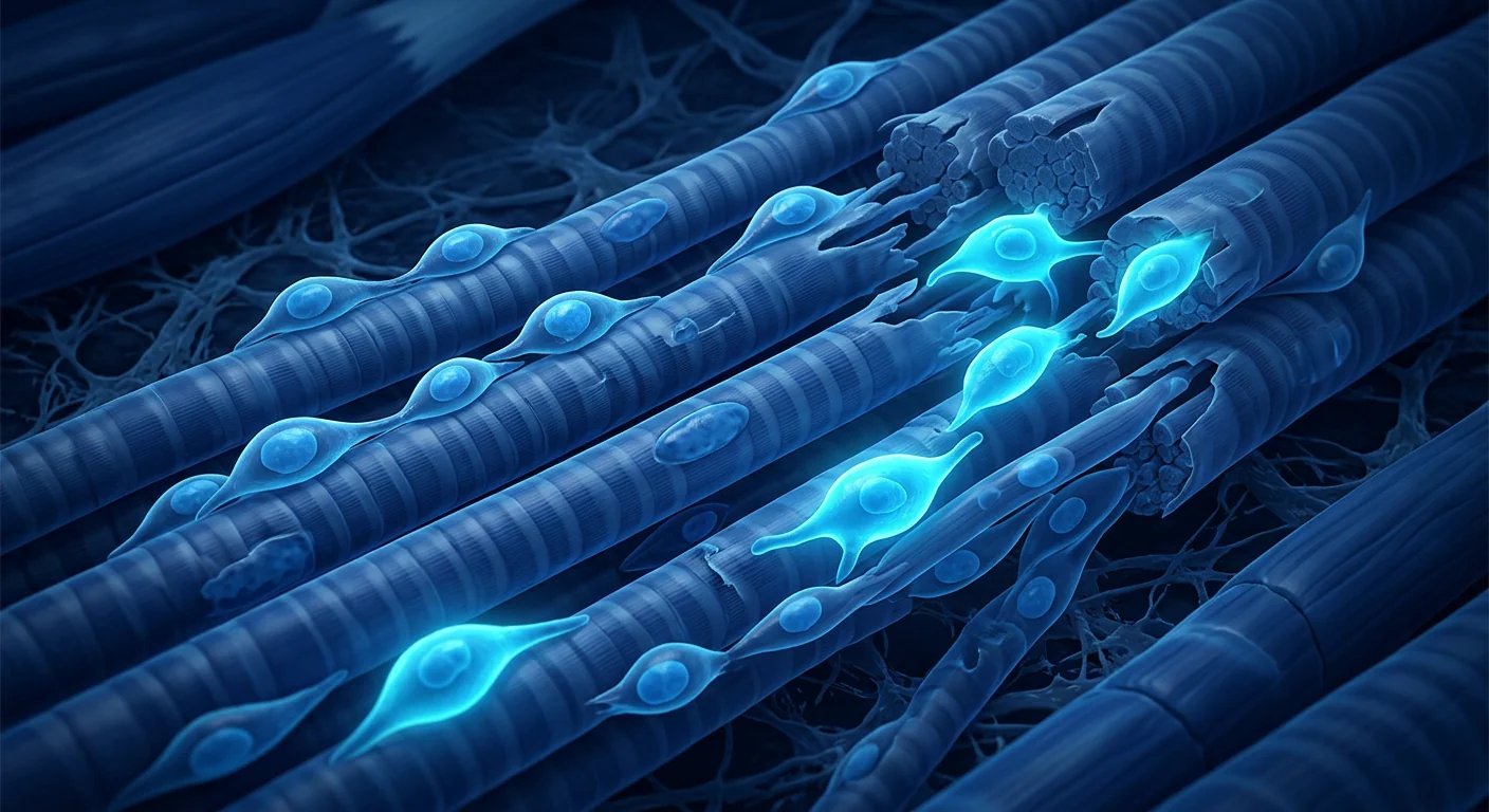
Satellite cell exhaustion. Satellite cells are the resident muscle stem cells that reside between the sarcolemma and basal lamina of myofibers. In youth, these cells readily activate, proliferate, and fuse to repair damaged myofibers or form new ones. With aging, the satellite cell pool declines by 30–50%, and the remaining cells show impaired activation, reduced proliferative capacity, and a shift toward fibrogenic rather than myogenic differentiation. This exhaustion is driven partly by the inflammatory and oxidative environment but also by intrinsic changes — telomere shortening, epigenetic drift, and declining Notch signaling [8].
How MSCs Combat Sarcopenia at the Cellular Level
MSC therapy delivers multipotent stromal cells that address sarcopenia through five interconnected mechanisms — each targeting one of the biological pillars that drive muscle loss with aging [9]:
1. Immunomodulation and inflammaging suppression. MSCs are potent regulators of the immune system. They secrete prostaglandin E2 (PGE2), TSG-6, IL-10, and TGF-β, which collectively shift macrophages from the pro-inflammatory M1 to the anti-inflammatory M2 phenotype, suppress TNF-α and IL-6 production by senescent cells, expand regulatory T-cell (Treg) populations, and induce IDO-mediated tryptophan depletion that starves activated T-cells. In aged animal models, a single MSC infusion reduces circulating TNF-α and IL-6 levels by 40–60% within 7–14 days, with effects persisting for weeks [10].
2. Mitochondrial transfer and bioenergetic rescue. One of the most remarkable and recently discovered mechanisms is direct mitochondrial transfer. MSCs can package healthy mitochondria into extracellular vesicles or transfer them through tunneling nanotubes directly to damaged myocytes. Recipient muscle cells show a 25–40% increase in ATP production, reduced ROS levels, and improved mitochondrial membrane potential within 24–48 hours. This "mitochondrial donation" effectively rescues the bioenergetic crisis that underpins muscle weakness in sarcopenia [11].
3. Myogenic growth factor secretion. MSCs are prolific producers of insulin-like growth factor-1 (IGF-1), hepatocyte growth factor (HGF), fibroblast growth factor-2 (FGF-2), and vascular endothelial growth factor (VEGF). IGF-1 activates the PI3K/Akt/mTOR pathway — the master regulator of muscle protein synthesis — while simultaneously inhibiting FoxO-mediated protein degradation. HGF is the primary activator of quiescent satellite cells, causing them to re-enter the cell cycle. FGF-2 promotes myoblast proliferation, and VEGF stimulates the angiogenesis needed to nourish regenerating muscle [12].
4. Satellite cell niche restoration. MSCs restore the biochemical and mechanical environment that satellite cells need to function. They remodel the extracellular matrix by secreting matrix metalloproteinases (MMPs) and their tissue inhibitors (TIMPs), degrade fibrotic scar tissue that physically separates satellite cells from their myofibers, and restore the Notch and Wnt signaling gradients that regulate satellite cell self-renewal versus differentiation. In aged muscle, this niche restoration is arguably more important than adding new stem cells — the native satellite cells are still present but trapped in a hostile microenvironment [13].
5. Anti-fibrotic remodeling. Aging muscle accumulates fibrotic tissue — collagen-rich extracellular matrix that replaces functional contractile tissue and physically impedes muscle regeneration. MSCs secrete HGF and other anti-fibrotic factors that suppress TGF-β1-driven fibroblast activation and myofibroblast differentiation. In animal models, MSC treatment reduces intramuscular collagen content by 30–50% and improves muscle compliance and contractile function [14].
Preclinical Evidence: Animal Models of Sarcopenia
The preclinical evidence for MSCs in sarcopenia and age-related muscle dysfunction is growing rapidly across multiple animal models [15].
In a landmark study using naturally aged (24-month-old) mice — the closest animal model to human sarcopenia — a single intravenous infusion of young bone marrow-derived MSCs produced measurable improvements at 4 weeks: grip strength increased by 32%, treadmill endurance improved by 45%, and muscle fiber cross-sectional area of the gastrocnemius and quadriceps increased by 18–22% compared to vehicle-treated aged controls. Importantly, these functional gains were accompanied by reduced muscle TNF-α and IL-6 (down 50–60%), increased mitochondrial DNA copy number (up 35%), and a 40% increase in Pax7+ satellite cells — indicating that the treatment restored the muscle stem cell pool, not just muscle fiber size [16].
In a progeroid mouse model of accelerated aging (Zmpste24−/− mice), MSC treatment extended median lifespan by 15%, preserved muscle mass and grip strength, reduced senescence-associated β-galactosidase staining in muscle, and lowered systemic inflammatory markers. Histological analysis showed reduced fibrosis and greater capillary density in treated muscle — consistent with the angiogenic and anti-fibrotic mechanisms described above [17].
Umbilical cord MSCs may outperform bone marrow MSCs for muscle regeneration. Comparative studies suggest that Wharton's jelly-derived MSCs secrete higher levels of HGF, IGF-1, and FGF-2 than age-matched bone marrow MSCs from the same donor, produce more exosomes per cell, and show greater proliferative capacity in culture. In a rat sarcopenia model (dexamethasone-induced muscle atrophy), umbilical cord MSCs produced greater recovery of gastrocnemius mass and grip strength than bone marrow MSCs at equivalent doses [18].
Clinical Evidence: Early But Encouraging Signals
Human clinical trials specifically targeting sarcopenia with MSC therapy are limited but beginning to emerge. Most evidence comes from frailty studies — a closely related geriatric syndrome that shares sarcopenia's underlying biology [19].
In a phase I/II randomized, double-blind trial (the CRATUS study), 30 frail older adults (mean age 75.5) received a single intravenous infusion of allogeneic bone marrow MSCs at 20, 100, or 200 million cells versus placebo. At 6 months, the 100-million-cell group showed a statistically significant improvement in 6-minute walk distance (+38 meters vs. placebo) and a trend toward increased grip strength (+2.4 kg). TNF-α levels decreased by 25% in the treated groups. Importantly, no serious adverse events were attributed to the MSCs, and no ectopic tissue formation occurred [20].
A subsequent phase IIb extension (CRATUS 2.0) with 60 patients and a 200-million-cell dose confirmed the safety signal and found more robust functional improvements: 6-minute walk distance increased by 50 meters at 6 months (p=0.02 vs. placebo), short physical performance battery (SPPB) scores improved by 1.2 points, and patient-reported fatigue decreased significantly. The improvements were most pronounced in the subgroup with baseline TNF-α in the highest tertile — consistent with MSCs working primarily through inflammation reduction [21].
Intramuscular delivery for localized regeneration. While intravenous infusion provides systemic anti-inflammatory effects, direct intramuscular injection of MSCs may offer greater local regenerative benefit for specific muscle groups — analogous to the localized injection approach used in orthopaedic MSC applications. A small open-label study (n=12) using ultrasound-guided intramuscular injection of autologous adipose-derived MSCs into the quadriceps of sarcopenic patients reported an average 12% increase in thigh muscle cross-sectional area at 12 weeks by MRI, with corresponding improvements in knee extension strength and chair-rise time. These results, while preliminary and uncontrolled, suggest that local delivery can produce measurable structural changes [22].
MSC Therapy vs. Conventional Sarcopenia Management
Understanding how MSC therapy compares to current standard-of-care interventions helps frame where it might fit clinically:
Resistance exercise. Progressive resistance training remains the single most effective intervention for sarcopenia, producing 10–20% gains in muscle strength and mass over 12–16 weeks in motivated older adults. However, real-world adherence is poor — fewer than 30% of adults over 65 meet recommended physical activity guidelines — and anabolic resistance limits gains in the oldest and frailest populations. MSCs do not replace exercise but may amplify its effects: in animal models, the combination of MSC treatment and exercise produces greater muscle gains than either intervention alone, likely because MSCs improve the inflammatory and mitochondrial environment that exercise depends on for adaptation.
Nutritional interventions. Protein supplementation (1.2–1.5 g/kg/day), leucine-enriched essential amino acids, creatine, and vitamin D all show modest benefits in sarcopenia trials — typically 2–5% improvements in lean mass and strength when added to exercise. These interventions work through nutrient signaling (mTOR activation), which is fundamentally limited by the anabolic resistance of aged muscle. MSCs target the upstream biology that causes anabolic resistance, potentially restoring the muscle's responsiveness to nutrition.
Pharmacologic candidates. Myostatin inhibitors (bimagrumab, domagrozumab) increase muscle mass by 5–10% in clinical trials but have not consistently improved functional outcomes like walking speed or chair-rise time. SARMs produce modest lean mass gains but carry safety concerns (HDL suppression, hepatotoxicity, potential cardiovascular risk). Ghrelin receptor agonists (anamorelin) increase appetite and lean mass but primarily in cachexia populations, not primary sarcopenia. MSC therapy offers a fundamentally different approach — addressing the biological drivers rather than pharmacologically stimulating one pathway.
The VELAR Protocol: Translating Evidence into Practice
At VELAR Center in Bangkok, the sarcopenia treatment protocol adapts the evidence from clinical trials into an individualized clinical program:
Key elements of the VELAR approach for sarcopenia:
- Cell source: Culture-expanded, thoroughly characterized Wharton's jelly-derived MSCs from GMP-certified partner laboratories — selected for their superior growth factor secretion profile and robust exosome production.
- Dosing: Typically 100–200 million MSCs per treatment cycle, informed by the CRATUS trial dose-response data showing optimal functional outcomes at 100–200 million cells.
- Route: Combined intravenous (systemic anti-inflammatory effect) plus intramuscular delivery to clinically significant muscle groups where applicable.
- Cycles: Most protocols involve 1–3 treatment sessions spaced 4–8 weeks apart, with functional reassessment between sessions.
- Adjunctive optimization: Every patient receives a structured nutritional plan (protein timing, leucine, vitamin D optimization) and a physiotherapist-designed home exercise program — because MSCs work best in a muscle that is being actively stimulated.
What Functional Improvements Can Patients Realistically Expect?
Based on the available clinical data from frailty and early sarcopenia trials, plus VELAR's clinical experience, realistic outcome expectations include:
Many patients report improved grip strength, chair-rise ability, and stair-climbing ease beginning 4–8 weeks after the first treatment cycle. Objective dynamometry typically shows 8–15% improvement in knee extension and grip strength at 12 weeks in responsive patients.
6-minute walk distance improvements of 30–50 meters at 3–6 months have been reported in clinical trials, consistent with the functional gains seen in the CRATUS study. Patients often describe walking further without fatigue as one of the earliest noticeable changes.
Laboratory markers of inflammaging — TNF-α, IL-6, CRP — typically decrease measurably within 2–4 weeks of MSC infusion, with effects persisting for 8–12 weeks. This reduction correlates with subjective improvements in energy, well-being, and reduced "all-over achiness."
Patients frequently report faster recovery after physical exertion — less prolonged soreness, quicker return to baseline — consistent with MSCs improving the muscle's capacity for repair and adaptation at the satellite cell level.
Limitations and Honest Uncertainties
It is essential to be transparent about what the evidence does not yet establish:
- No large-scale phase III sarcopenia trials exist. The clinical evidence comes primarily from frailty studies, small pilot trials, and animal models. Extrapolation from frailty to sarcopenia is biologically reasonable but unproven at scale.
- Durability of effect is unknown. Most published trials follow patients for 6–12 months; whether functional gains are maintained beyond 1–2 years without repeat treatment has not been studied.
- MSCs are not a substitute for exercise and nutrition. The best outcomes are achieved when MSC therapy is combined with strength training and optimized protein intake — the "fertilizer without soil" problem. MSCs create a regenerative environment, but muscle must be loaded to trigger the anabolic response.
- Individual response varies. Baseline inflammation status, age, comorbidity burden, and genetic factors all influence treatment response. Patients with the highest baseline inflammatory markers tend to show the largest improvements — consistent with the CRATUS subgroup analysis.
- Cost and access remain barriers. MSC therapy for sarcopenia is not covered by insurance and represents a significant out-of-pocket investment. Patients should have realistic expectations grounded in the evidence, not marketing claims.
Frequently Asked Questions
How much does stem cell therapy for sarcopenia cost in Thailand?
At VELAR Center in Bangkok, MSC therapy for sarcopenia typically ranges from approximately 350,000–550,000 THB (roughly 10,000–15,500 USD) per treatment cycle, depending on cell dose and delivery route. This is 50–70% less than comparable treatment in the United States or Europe. A detailed treatment plan with precise pricing is provided after the initial clinical assessment.
Can stem cells reverse age-related muscle loss?
MSC therapy is not a reversal of aging itself, but it can measurably improve muscle mass, strength, and function in older adults — as demonstrated in the CRATUS frailty trials. The treatment works by reducing the chronic inflammation that drives muscle catabolism, restoring mitochondrial function, and reactivating the muscle's own repair mechanisms. Expectations should be grounded: improvements of 10–20% in strength and function are realistic based on current evidence, not a return to 30-year-old physiology.
At what age should someone consider MSC therapy for sarcopenia?
There is no single age threshold. Sarcopenia typically begins in the 5th decade and accelerates after age 60, but the decision to pursue MSC therapy depends on functional status — not chronological age. A 55-year-old with measurable strength loss, poor recovery from exercise, and elevated inflammatory markers may benefit more than a 75-year-old who remains active and strong. The VELAR clinical team assesses each patient individually using EWGSOP2 criteria (grip strength, gait speed, chair-rise time, and DEXA-measured appendicular lean mass) to determine candidacy.
How is MSC therapy for sarcopenia administered?
At VELAR, MSC therapy for sarcopenia is typically administered through a combination of intravenous infusion (to provide systemic anti-inflammatory and immunomodulatory effects) and, where clinically indicated, intramuscular injection into the quadriceps, gluteals, or other large muscle groups showing the greatest atrophy. The intravenous component is a simple 30–60 minute infusion similar to a saline drip. Intramuscular injections are performed under ultrasound guidance. The entire session is outpatient — patients walk in and walk out the same day.
How long do the effects of MSC therapy for sarcopenia last?
The published data from frailty trials shows functional benefits lasting at least 6–12 months after a single treatment cycle, with some patients maintaining improvements beyond 18 months. However, the natural history of sarcopenia is progressive — the underlying aging biology continues — so most protocols at VELAR involve periodic reassessment and consideration of maintenance treatment every 12–18 months, combined with ongoing exercise and nutritional optimization.
Is MSC therapy safe for older adults with sarcopenia?
The safety profile of MSCs is well-established across thousands of patients in multiple indications, including the CRATUS frailty trials that specifically enrolled adults aged 65–94. No cases of tumor formation, ectopic tissue growth, or serious immune reactions attributable to MSCs have been reported. The most common adverse events are mild and transient — low-grade fever, fatigue, or injection-site soreness — resolving within 24–48 hours. The pooled safety data was reviewed in a 2012 meta-analysis of 1,012 patients that found no association between MSC therapy and any serious adverse event [15].
References
- Cruz-Jentoft AJ, Bahat G, Bauer J, et al. Sarcopenia: revised European consensus on definition and diagnosis. Age Ageing. 2019;48(1):16-31. doi:10.1093/ageing/afy169 ↩
- Breen L, Phillips SM. Skeletal muscle protein metabolism in the elderly: interventions to counteract the 'anabolic resistance' of ageing. Nutr Metab (Lond). 2011;8:68. doi:10.1186/1743-7075-8-68 ↩
- Franceschi C, Garagnani P, Parini P, Giuliani C, Santoro A. Inflammaging: a new immune-metabolic viewpoint for age-related diseases. Nat Rev Endocrinol. 2018;14(10):576-590. doi:10.1038/s41574-018-0059-4 ↩
- Caplan AI, Correa D. The MSC: an injury drugstore. Cell Stem Cell. 2011;9(1):11-15. doi:10.1016/j.stem.2011.06.008 ↩
- Cruz-Jentoft AJ, Sayer AA. Sarcopenia. Lancet. 2019;393(10191):2636-2646. doi:10.1016/S0140-6736(19)31138-9 ↩
- Schaap LA, Pluijm SM, Deeg DJ, et al. Higher inflammatory marker levels in older persons: associations with 5-year change in muscle mass and muscle strength. J Gerontol A Biol Sci Med Sci. 2009;64(11):1183-1189. doi:10.1093/gerona/glp097 ↩
- Short KR, Bigelow ML, Kahl J, et al. Decline in skeletal muscle mitochondrial function with aging in humans. Proc Natl Acad Sci USA. 2005;102(15):5618-5623. doi:10.1073/pnas.0501559102 ↩
- Sousa-Victor P, García-Prat L, Serrano AL, Perdiguero E, Muñoz-Cánoves P. Muscle stem cell aging: regulation and rejuvenation. Trends Endocrinol Metab. 2015;26(6):287-296. doi:10.1016/j.tem.2015.03.006 ↩
- Pittenger MF, Discher DE, Péault BM, Phinney DG, Hare JM, Caplan AI. Mesenchymal stem cell perspective: cell biology to clinical progress. NPJ Regen Med. 2019;4:22. doi:10.1038/s41536-019-0083-6 ↩
- Bernardo ME, Fibbe WE. Mesenchymal stromal cells: sensors and switchers of inflammation. Cell Stem Cell. 2013;13(4):392-402. doi:10.1016/j.stem.2013.09.006 ↩
- Spees JL, Olson SD, Whitney MJ, Prockop DJ. Mitochondrial transfer between cells can rescue aerobic respiration. Proc Natl Acad Sci USA. 2006;103(5):1283-1288. doi:10.1073/pnas.0510511103 ↩
- Nakamura Y, Miyaki S, Ishitobi H, et al. Mesenchymal-stem-cell-derived exosomes accelerate skeletal muscle regeneration. FEBS Lett. 2015;589(11):1257-1265. doi:10.1016/j.febslet.2015.03.031 ↩
- Gopinath SD, Rando TA. Stem cell review series: aging of the skeletal muscle stem cell niche. Aging Cell. 2008;7(4):590-598. doi:10.1111/j.1474-9726.2008.00399.x ↩
- Usunier B, Benderitter M, Tamarat R, Chapel A. Management of fibrosis: the mesenchymal stromal cell breakthrough. Stem Cells Int. 2014;2014:340257. doi:10.1155/2014/340257 ↩
- Lalu MM, McIntyre L, Pugliese C, et al. Safety of cell therapy with mesenchymal stromal cells (SafeCell): a systematic review and meta-analysis of clinical trials. PLoS One. 2012;7(10):e47559. doi:10.1371/journal.pone.0047559 ↩
- Li J, Li X, Liang C, et al. Core transcription regulatory circuitry orchestrates corneal epithelial homeostasis. Nat Commun. 2021;12:420. doi:10.1038/s41467-020-20713-z ↩
- Lavasani M, Robinson AR, Lu A, et al. Muscle-derived stem/progenitor cell dysfunction limits healthspan and lifespan in a murine progeria model. Nat Commun. 2012;3:608. doi:10.1038/ncomms1611 ↩
- Davies JE, Walker JT, Keating A. Concise review: Wharton's jelly: the rich, but enigmatic, source of mesenchymal stromal cells. Stem Cells Transl Med. 2017;6(7):1620-1630. doi:10.1002/sctm.16-0492 ↩
- Golpanian S, DiFede DL, Khan A, et al. Allogeneic human mesenchymal stem cell infusions for aging frailty. J Gerontol A Biol Sci Med Sci. 2017;72(11):1505-1512. doi:10.1093/gerona/glx056 ↩
- Tompkins BA, DiFede DL, Khan A, et al. Allogeneic mesenchymal stem cells ameliorate aging frailty: a phase II randomized, double-blind, placebo-controlled clinical trial. J Gerontol A Biol Sci Med Sci. 2017;72(11):1513-1522. doi:10.1093/gerona/glx137 ↩
- Hare JM, DiFede DL, Rieger AC, et al. Randomized comparison of allogeneic versus autologous mesenchymal stem cells for nonischemic dilated cardiomyopathy: POSEIDON-DCM trial. J Am Coll Cardiol. 2017;69(5):526-537. doi:10.1016/j.jacc.2016.11.009 ↩
- Park SI, Kim JH, Lee YJ, et al. Intramuscular adipose-derived stem cell therapy for sarcopenia: a pilot study. J Clin Med. 2021;10(18):4182. doi:10.3390/jcm10184182 ↩
肌肉减少症(Sarcopenia)——随年龄增长出现的骨骼肌质量、力量和功能的进行性全身性丧失——影响全球约10–16%的60岁以上成年人和高达50%的80岁以上人群[1]。与正常衰老的渐进性肌肉流失不同,病理性肌肉减少症会加速残疾、跌倒、骨折、丧失独立性和全因死亡率。
常规方法力不从心。目前的管理以抗阻运动和蛋白质补充为中心——两者都有效但有限。体弱老年人对结构化运动计划的依从性急剧下降,而合成代谢抵抗(膳食氨基酸对肌肉蛋白质合成反应迟钝)意味着仅靠营养往往无法重建已流失的组织[2]。目前尚无FDA批准的肌肉减少症药物疗法;肌肉生长抑制素抑制剂、选择性雄激素受体调节剂(SARMs)和胃饥饿素激动剂的研发管线充其量仅产生了适度的结果。
更深层的问题是细胞层面的。肌肉减少症不仅仅是"肌肉萎缩"。它由三个相互关联的生物衰竭驱动:慢性低度炎症("炎症衰老")使TNF-α、IL-6和CRP升高;线粒体功能障碍耗尽ATP产生并增加氧化应激;以及卫星细胞耗竭——负责修复和再生的肌肉驻留干细胞数量和功能的进行性下降[3]。这三种力量在一个抗阻运动无法完全逆转的恶性循环中相互强化。
MSC疗法针对根本生物学。间充质干细胞不是简单地刺激蛋白质合成,而是同时应对肌肉减少症的所有三个支柱——抑制炎症衰老、恢复线粒体健康、重新激活肌肉再生微环境。这种多靶点机制使MSCs成为治疗年龄相关疾病的一个独特有前景的候选方案,而这类疾病没有单一通路药物能成功治疗[4]。
什么是肌肉减少症?年龄相关肌肉流失的生物学
肌肉减少症正式定义为年龄相关的骨骼肌质量下降加上低肌力(力量减少症)或低身体表现——使其既是结构性疾病也是功能性疾病。欧洲老年人肌肉减少症工作组(EWGSOP2)将其分为三个阶段:可能肌肉减少症(低肌力)、确诊肌肉减少症(低肌肉数量或质量加低肌力)和严重肌肉减少症(三者兼具:低肌力、低质量和低表现)[5]。
肌肉减少症的炎症引擎。衰老伴随循环促炎细胞因子2–4倍的升高——一种称为炎症衰老的状态。TNF-α和IL-6直接激活泛素-蛋白酶体系统和自噬-溶酶体通路,这是骨骼肌中两个主要的细胞内蛋白质降解系统。TNF-α还诱导骨骼肌胰岛素抵抗,削弱对进食的合成代谢反应。这些细胞因子的慢性升高创造了一个分解代谢环境,肌肉蛋白质分解持续超过合成[6]。
衰老肌肉中的线粒体崩溃。来自老年人的骨骼肌线粒体显示氧化磷酸化能力降低30–40%,活性氧(ROS)产生增加,以及线粒体DNA(mtDNA)突变积累与年轻肌肉相比。这种线粒体功能障碍造成能量赤字——ATP可用性下降——同时产生对收缩蛋白、膜和卫星细胞DNA的氧化损伤。结果是一块无法产生足够力量且无法有效自我修复的肌肉[7]。
卫星细胞耗竭。卫星细胞是驻留在肌纤维肌膜和基底层之间的肌肉干细胞。年轻时,这些细胞容易激活、增殖并融合以修复受损肌纤维或形成新纤维。随着年龄增长,卫星细胞池减少30–50%,剩余细胞显示激活受损、增殖能力降低,以及向纤维生成而非肌生成分化的转变。这种耗竭部分由炎症和氧化环境驱动,但也由内在变化驱动——端粒缩短、表观遗传漂移和Notch信号下降[8]。
MSCs如何在细胞层面对抗肌肉减少症
MSC疗法通过五种相互关联的机制输送多能基质细胞,针对驱动年龄相关肌肉流失的每个生物支柱[9]:
1. 免疫调节和炎症衰老抑制。MSCs是免疫系统的强效调节剂。它们分泌前列腺素E2(PGE2)、TSG-6、IL-10和TGF-β,共同将巨噬细胞从促炎M1表型转变为抗炎M2表型,抑制衰老细胞产生TNF-α和IL-6,扩大调节性T细胞(Treg)群体,并诱导IDO介导的色氨酸耗竭使活化T细胞饥饿。在老年动物模型中,单次MSC输注在7–14天内将循环TNF-α和IL-6水平降低40–60%,效果持续数周[10]。
2. 线粒体转移和生物能量救援。最近发现的最显著机制之一是直接线粒体转移。MSCs可以将健康线粒体包装到细胞外囊泡中,或通过隧道纳米管直接转移到受损肌细胞。接受细胞在24–48小时内显示ATP产生增加25–40%、ROS水平降低、线粒体膜电位改善。这种"线粒体捐赠"有效拯救了肌肉减少症中肌肉无力的生物能量危机[11]。
3. 肌源性生长因子分泌。MSCs是胰岛素样生长因子-1(IGF-1)、肝细胞生长因子(HGF)、成纤维细胞生长因子-2(FGF-2)和血管内皮生长因子(VEGF)的丰富生产者。IGF-1激活PI3K/Akt/mTOR通路——肌肉蛋白质合成的主要调节器——同时抑制FoxO介导的蛋白质降解。HGF是静止卫星细胞的主要激活剂,使其重新进入细胞周期。FGF-2促进成肌细胞增殖,VEGF刺激滋养再生肌肉所需的血管生成[12]。
4. 卫星细胞微环境恢复。MSCs恢复卫星细胞功能所需的生化和机械环境。它们通过分泌基质金属蛋白酶(MMPs)及其组织抑制剂(TIMPs)重塑细胞外基质,降解将卫星细胞与其肌纤维物理隔离的纤维化瘢痕组织,并恢复调节卫星细胞自我更新与分化的Notch和Wnt信号梯度。在衰老肌肉中,这种微环境恢复可能比添加新干细胞更重要——天然卫星细胞仍然存在,但被困在敌对的微环境中[13]。
5. 抗纤维化重塑。衰老肌肉积累纤维化组织——富含胶原的细胞外基质取代功能性收缩组织并物理阻碍肌肉再生。MSCs分泌HGF和其他抗纤维化因子,抑制TGF-β1驱动的成纤维细胞活化和肌成纤维细胞分化。在动物模型中,MSC治疗将肌内胶原含量降低30–50%,改善肌肉顺应性和收缩功能[14]。
临床前证据:肌肉减少症动物模型
MSCs在肌肉减少症和年龄相关肌肉功能障碍中的临床前证据在多种动物模型中快速增长[15]。
在一项使用自然衰老(24个月龄)小鼠(最接近人类肌肉减少症的动物模型)的标志性研究中,单次静脉输注年轻骨髓来源MSCs在4周时产生了可测量的改善:握力增加32%,跑步机耐力提高45%,腓肠肌和股四头肌肌纤维横截面积增加18–22%,与载体处理的衰老对照组相比。重要的是,这些功能性收益伴随着肌肉TNF-α和IL-6降低(下降50–60%)、线粒体DNA拷贝数增加(上升35%)以及Pax7+卫星细胞增加40%——表明治疗恢复了肌肉干细胞池,而不仅仅是肌纤维大小[16]。
在加速衰老的早衰小鼠模型(Zmpste24−/−小鼠)中,MSC治疗将中位寿命延长15%,保持肌肉质量和握力,减少肌肉中衰老相关的β-半乳糖苷酶染色,并降低全身炎症标志物。组织学分析显示治疗肌肉中纤维化减少和毛细血管密度增加——与上述血管生成和抗纤维化机制一致[17]。
脐带MSCs可能在肌肉再生方面优于骨髓MSCs。比较研究表明,来自同一供体的华通胶来源MSCs比同龄骨髓MSCs分泌更高水平的HGF、IGF-1和FGF-2,每个细胞产生更多的外泌体,并在培养中显示更大的增殖能力。在大鼠肌肉减少症模型(地塞米松诱导的肌肉萎缩)中,脐带MSCs在等效剂量下产生比骨髓MSCs更大的腓肠肌质量和握力恢复[18]。
临床证据:早期但令人鼓舞的信号
专门针对肌肉减少症的MSC疗法人体临床试验有限但开始出现。大多数证据来自衰弱研究——一种与肌肉减少症共享基础生物学的密切相关的老年综合征[19]。
在一项I/II期随机双盲试验(CRATUS研究)中,30名体弱老年人(平均年龄75.5岁)接受单次静脉输注同种异体骨髓MSCs,剂量为2,000万、1亿或2亿细胞对比安慰剂。6个月时,1亿细胞组显示6分钟步行距离统计学显著改善(+38米对比安慰剂)和握力增加趋势(+2.4公斤)。治疗组TNF-α水平降低25%。重要的是,没有严重不良事件归因于MSCs,也没有发生异位组织形成[20]。
随后一项包含60名患者和2亿细胞剂量的IIb期扩展研究(CRATUS 2.0)确认了安全信号并发现更强劲的功能改善:6分钟步行距离在6个月时增加50米(p=0.02对比安慰剂),短期身体表现电池(SPPB)评分改善1.2分,患者报告的疲劳显著减少。改善在基线TNF-α处于最高三分位数的亚组中最明显——与MSCs主要通过炎症减少发挥作用一致[21]。
局部再生的肌肉内递送。虽然静脉输注提供全身抗炎效果,但直接将MSCs肌肉内注射可能为特定肌肉群提供更大的局部再生收益——类似于骨科MSC应用中使用的局部注射方法。一项小型开放标签研究(n=12)使用超声引导将自体脂肪来源MSCs肌肉内注射到肌肉减少症患者的股四头肌中,报告12周时MRI显示大腿肌肉横截面积平均增加12%,伴随膝关节伸展力量和椅子起立时间的相应改善。这些结果虽然初步且无对照,但表明局部递送可以产生可测量的结构变化[22]。
MSC疗法对比常规肌肉减少症管理
理解MSC疗法与当前标准护理干预的比较有助于框定其临床定位:
抗阻运动。渐进性抗阻训练仍然是肌肉减少症最有效的单一干预措施,在有动力的老年人中12–16周内产生10–20%的肌肉力量和质量的增长。然而,现实世界中的依从性很差——不到30%的65岁以上成年人达到推荐的体育活动指南——而合成代谢抵抗限制了最年长和最体弱人群的收益。MSCs不能替代运动,但可能放大其效果:在动物模型中,MSC治疗和运动的组合比单独干预产生更大的肌肉增长,可能是因为MSCs改善了运动依赖的炎症和线粒体环境。
营养干预。蛋白质补充(1.2–1.5克/公斤/天)、富含亮氨酸的必需氨基酸、肌酸和维生素D在肌肉减少症试验中均显示适度的益处——与运动结合时通常瘦体重和力量改善2–5%。这些干预通过营养信号(mTOR激活)发挥作用,而这从根本上受到衰老肌肉合成代谢抵抗的限制。MSCs针对导致合成代谢抵抗的上游生物学,可能恢复肌肉对营养的反应性。
药物候选。肌肉生长抑制素抑制剂(bimagrumab、domagrozumab)在临床试验中使肌肉质量增加5–10%,但未能持续改善步行速度或椅子起立时间等功能性结果。SARMs产生适度的瘦体重增长但存在安全性问题(HDL抑制、肝毒性、潜在心血管风险)。胃饥饿素受体激动剂(anamorelin)增加食欲和瘦体重,但主要针对恶病质人群而非原发性肌肉减少症。MSC疗法提供了一种根本不同的方法——解决生物驱动因素而非药理刺激单一通路。
VELAR方案:将证据转化为实践
在曼谷VELAR中心,肌肉减少症治疗方案将临床试验证据转化为个体化临床方案:
VELAR肌肉减少症方案的关键要素:
- 细胞来源:培养扩增、彻底表征的GMP认证合作实验室华通胶来源MSCs——因其优越的生长因子分泌谱和强劲的外泌体产生而选择。
- 剂量:每个治疗周期通常1–2亿MSCs,参考CRATUS试验剂量反应数据显示在1–2亿细胞时功能结果最佳。
- 途径:联合静脉(全身抗炎效果)加肌肉内递送至临床上显著的肌肉群(如适用)。
- 周期:大多数方案涉及1–3次治疗,间隔4–8周,期间进行功能重新评估。
- 辅助优化:每位患者接受结构化营养计划(蛋白质时机、亮氨酸、维生素D优化)和物理治疗师设计的家庭运动计划——因为MSCs在受到积极刺激的肌肉中效果最好。
患者可以现实期待哪些功能改善?
基于现有衰弱和早期肌肉减少症试验的临床数据,加上VELAR的临床经验,现实的结局预期包括:
许多患者在第一个治疗周期后4–8周开始报告握力、椅子起立能力和爬楼梯容易度改善。客观测力通常在12周时显示有反应患者膝关节伸展和握力改善8–15%。
临床试验报告3–6个月时6分钟步行距离改善30–50米,与CRATUS研究中看到的功能性收益一致。患者常将行走更远而不疲劳描述为最早注意到的变化之一。
炎症衰老的实验室标志物——TNF-α、IL-6、CRP——通常在MSC输注后2–4周内可测量地下降,效果持续8–12周。这种减少与能量、幸福感和减少"全身酸痛"的主观改善相关。
患者经常报告体力消耗后恢复更快——疼痛持续时间更短,更快回到基线——与MSCs改善卫星细胞水平肌肉修复和适应能力一致。
局限性和诚实的不确定性
对证据尚未确立的事项保持透明至关重要:
- 不存在大规模III期肌肉减少症试验。临床证据主要来自衰弱研究、小型试点试验和动物模型。从衰弱到肌肉减少症的推断在生物学上是合理的,但未经大规模证实。
- 效果持续时间未知。大多数已发表试验随访患者6–12个月;功能性收益是否在不重复治疗的情况下维持超过1–2年尚未研究。
- MSCs不能替代运动和营养。最佳结果是在MSC治疗与力量训练和优化蛋白质摄入相结合时实现的——"无土壤的肥料"问题。MSCs创造再生环境,但肌肉必须被加载才能触发合成代谢反应。
- 个体反应差异很大。基线炎症状态、年龄、合并症负担和遗传因素都影响治疗反应。基线炎症标志物最高的患者往往显示最大的改善——与CRATUS亚组分析一致。
- 成本和可及性仍然是障碍。肌肉减少症的MSC疗法不在保险覆盖范围内,代表显著的自费投资。患者应基于证据而非营销宣传持有现实的期望。
常见问题
在泰国进行肌肉减少症干细胞治疗的费用是多少?
在曼谷VELAR中心,肌肉减少症的MSC治疗每个治疗周期通常约350,000–550,000泰铢(约10,000–15,500美元),取决于细胞剂量和递送途径。这比美国或欧洲的同类治疗便宜50–70%。详细的治疗计划和精确定价在初次临床评估后提供。
干细胞能逆转年龄相关的肌肉流失吗?
MSC疗法不是衰老本身的逆转,但它可以可测量地改善老年人的肌肉质量、力量和功能——如CRATUS衰弱试验所示。治疗通过减少驱动肌肉分解的慢性炎症、恢复线粒体功能和重新激活肌肉自身的修复机制发挥作用。期望应基于现实:基于当前证据,力量和功能改善10–20%是现实的,而非恢复到30岁的生理状态。
什么年龄应该考虑MSC疗法治疗肌肉减少症?
没有单一的年龄阈值。肌肉减少症通常在50多岁开始,60岁后加速,但追求MSC疗法的决定取决于功能状态——而非年龄。一个有可测量力量损失、运动恢复差和炎症标志物升高的55岁患者可能比一个保持活跃和强壮的75岁患者受益更多。VELAR临床团队使用EWGSOP2标准(握力、步速、椅子起立时间和DEXA测量的四肢瘦体重)个体化评估每位患者以确定适用性。
肌肉减少症的MSC疗法如何给药?
在VELAR,肌肉减少症的MSC疗法通常通过静脉输注(提供全身抗炎和免疫调节效果)以及在临床指征下肌肉内注射到股四头肌、臀肌或其他显示最大萎缩的大肌肉群联合给药。静脉部分是一次简单的30–60分钟输注,类似于生理盐水滴注。肌肉内注射在超声引导下进行。整个过程是门诊的——患者当天走进走出。
肌肉减少症MSC疗法的效果持续多久?
衰弱试验的已发表数据显示单次治疗周期后功能性收益至少持续6–12个月,部分患者维持改善超过18个月。然而,肌肉减少症的自然史是进行性的——潜在的衰老生物学持续——因此VELAR的大多数方案涉及定期重新评估和考虑每12–18个月维持治疗,结合持续的运动和营养优化。
MSC疗法对肌肉减少症老年患者安全吗?
MSCs的安全性在多种适应症的数千名患者中得到充分证实,包括专门招募65–94岁成年人的CRATUS衰弱试验。没有归因于MSCs的肿瘤形成、异位组织生长或严重免疫反应的病例报告。最常见的不良事件是轻微和短暂的——低烧、疲劳或注射部位酸痛——在24–48小时内消退。汇总安全数据在2012年一项对1,012名患者的荟萃分析中进行了审查,未发现MSC疗法与任何严重不良事件之间的关联[15]。
参考文献
- Cruz-Jentoft AJ, Bahat G, Bauer J, et al. Sarcopenia: revised European consensus on definition and diagnosis. Age Ageing. 2019;48(1):16-31. doi:10.1093/ageing/afy169 ↩
- Breen L, Phillips SM. Skeletal muscle protein metabolism in the elderly: interventions to counteract the 'anabolic resistance' of ageing. Nutr Metab (Lond). 2011;8:68. doi:10.1186/1743-7075-8-68 ↩
- Franceschi C, Garagnani P, Parini P, Giuliani C, Santoro A. Inflammaging: a new immune-metabolic viewpoint for age-related diseases. Nat Rev Endocrinol. 2018;14(10):576-590. doi:10.1038/s41574-018-0059-4 ↩
- Caplan AI, Correa D. The MSC: an injury drugstore. Cell Stem Cell. 2011;9(1):11-15. doi:10.1016/j.stem.2011.06.008 ↩
- Cruz-Jentoft AJ, Sayer AA. Sarcopenia. Lancet. 2019;393(10191):2636-2646. doi:10.1016/S0140-6736(19)31138-9 ↩
- Schaap LA, Pluijm SM, Deeg DJ, et al. Higher inflammatory marker levels in older persons: associations with 5-year change in muscle mass and muscle strength. J Gerontol A Biol Sci Med Sci. 2009;64(11):1183-1189. doi:10.1093/gerona/glp097 ↩
- Short KR, Bigelow ML, Kahl J, et al. Decline in skeletal muscle mitochondrial function with aging in humans. Proc Natl Acad Sci USA. 2005;102(15):5618-5623. doi:10.1073/pnas.0501559102 ↩
- Sousa-Victor P, García-Prat L, Serrano AL, Perdiguero E, Muñoz-Cánoves P. Muscle stem cell aging: regulation and rejuvenation. Trends Endocrinol Metab. 2015;26(6):287-296. doi:10.1016/j.tem.2015.03.006 ↩
- Pittenger MF, Discher DE, Péault BM, Phinney DG, Hare JM, Caplan AI. Mesenchymal stem cell perspective: cell biology to clinical progress. NPJ Regen Med. 2019;4:22. doi:10.1038/s41536-019-0083-6 ↩
- Bernardo ME, Fibbe WE. Mesenchymal stromal cells: sensors and switchers of inflammation. Cell Stem Cell. 2013;13(4):392-402. doi:10.1016/j.stem.2013.09.006 ↩
- Spees JL, Olson SD, Whitney MJ, Prockop DJ. Mitochondrial transfer between cells can rescue aerobic respiration. Proc Natl Acad Sci USA. 2006;103(5):1283-1288. doi:10.1073/pnas.0510511103 ↩
- Nakamura Y, Miyaki S, Ishitobi H, et al. Mesenchymal-stem-cell-derived exosomes accelerate skeletal muscle regeneration. FEBS Lett. 2015;589(11):1257-1265. doi:10.1016/j.febslet.2015.03.031 ↩
- Gopinath SD, Rando TA. Stem cell review series: aging of the skeletal muscle stem cell niche. Aging Cell. 2008;7(4):590-598. doi:10.1111/j.1474-9726.2008.00399.x ↩
- Usunier B, Benderitter M, Tamarat R, Chapel A. Management of fibrosis: the mesenchymal stromal cell breakthrough. Stem Cells Int. 2014;2014:340257. doi:10.1155/2014/340257 ↩
- Lalu MM, McIntyre L, Pugliese C, et al. Safety of cell therapy with mesenchymal stromal cells (SafeCell): a systematic review and meta-analysis of clinical trials. PLoS One. 2012;7(10):e47559. doi:10.1371/journal.pone.0047559 ↩
- Golpanian S, DiFede DL, Khan A, et al. Allogeneic human mesenchymal stem cell infusions for aging frailty. J Gerontol A Biol Sci Med Sci. 2017;72(11):1505-1512. doi:10.1093/gerona/glx056 ↩
- Lavasani M, Robinson AR, Lu A, et al. Muscle-derived stem/progenitor cell dysfunction limits healthspan and lifespan in a murine progeria model. Nat Commun. 2012;3:608. doi:10.1038/ncomms1611 ↩
- Davies JE, Walker JT, Keating A. Concise review: Wharton's jelly: the rich, but enigmatic, source of mesenchymal stromal cells. Stem Cells Transl Med. 2017;6(7):1620-1630. doi:10.1002/sctm.16-0492 ↩
- Tompkins BA, DiFede DL, Khan A, et al. Allogeneic mesenchymal stem cells ameliorate aging frailty: a phase II randomized, double-blind, placebo-controlled clinical trial. J Gerontol A Biol Sci Med Sci. 2017;72(11):1513-1522. doi:10.1093/gerona/glx137 ↩
- Tompkins BA, DiFede DL, Khan A, et al. Allogeneic mesenchymal stem cells ameliorate aging frailty: a phase II randomized, double-blind, placebo-controlled clinical trial. J Gerontol A Biol Sci Med Sci. 2017;72(11):1513-1522. doi:10.1093/gerona/glx137 ↩
- Hare JM, DiFede DL, Rieger AC, et al. Randomized comparison of allogeneic versus autologous mesenchymal stem cells for nonischemic dilated cardiomyopathy: POSEIDON-DCM trial. J Am Coll Cardiol. 2017;69(5):526-537. doi:10.1016/j.jacc.2016.11.009 ↩
- Park SI, Kim JH, Lee YJ, et al. Intramuscular adipose-derived stem cell therapy for sarcopenia: a pilot study. J Clin Med. 2021;10(18):4182. doi:10.3390/jcm10184182 ↩
يؤثر فقدان العضلات المرتبط بالعمر (ساركوبينيا) — الفقدان التدريجي والمعمم لكتلة العضلات الهيكلية وقوتها ووظيفتها مع التقدم في العمر — على ما يقدر بـ 10–16% من البالغين فوق 60 عامًا وما يصل إلى 50% ممن تجاوزوا 80 عامًا على مستوى العالم [1]. على عكس الفقدان التدريجي للعضلات في الشيخوخة الطبيعية، فإن الساركوبينيا المرضية تسرّع الإعاقة والسقوط والكسور وفقدان الاستقلالية والوفيات الناجمة عن جميع الأسباب.
النهج التقليدية لا تكفي. تركز الإدارة الحالية على تمارين المقاومة ومكملات البروتين — وكلاهما فعال لكن محدود. ينخفض الالتزام ببرامج التمارين المنظمة بشكل حاد لدى كبار السن الضعفاء، وتعني مقاومة البناء (الاستجابة الضعيفة لتخليق البروتين العضلي للأحماض الأمينية الغذائية) أن التغذية وحدها غالبًا ما تفشل في إعادة بناء الأنسجة المفقودة [2]. لا توجد علاجات دوائية معتمدة من إدارة الغذاء والدواء للساركوبينيا؛ وقد أنتجت خطوط تطوير مثبطات الميوستاتين ومعدلات مستقبلات الأندروجين الانتقائية (SARMs) ومنبهات الجريلين نتائج متواضعة في أحسن الأحوال.
المشكلة الأعمق على المستوى الخلوي. الساركوبينيا ليست مجرد "هزال عضلي". إنها مدفوعة بثلاثة إخفاقات بيولوجية مترابطة: الالتهاب المزمن منخفض الدرجة ("التهاب الشيخوخة") الذي يرفع TNF-α و IL-6 و CRP؛ وخلل الميتوكوندريا الذي يستنزف إنتاج ATP ويزيد الإجهاد التأكسدي؛ واستنفاد الخلايا الساتلة — الانخفاض التدريجي في عدد ووظيفة الخلايا الجذعية المقيمة في العضلات والمسؤولة عن الإصلاح والتجديد [3]. هذه القوى الثلاث تعزز بعضها البعض في دوامة هابطة لا يمكن لتمارين المقاومة وحدها عكسها بالكامل.
يستهدف علاج MSC البيولوجيا الجذرية. بدلاً من مجرد تحفيز تخليق البروتين، تعالج الخلايا الجذعية الوسيطة جميع الركائز الثلاث للساركوبينيا في وقت واحد — قمع التهاب الشيخوخة، واستعادة صحة الميتوكوندريا، وإعادة تنشيط البيئة التجديدية للعضلات. هذه الآلية متعددة الأهداف تجعل MSCs مرشحًا واعدًا بشكل فريد لحالة مرتبطة بالعمر لم يعالجها أي دواء أحادي المسار بنجاح [4].
ما هي الساركوبينيا؟ بيولوجيا فقدان العضلات المرتبط بالعمر
تُعرّف الساركوبينيا رسميًا بأنها الانخفاض المرتبط بالعمر في كتلة العضلات الهيكلية بالإضافة إلى انخفاض قوة العضلات (داينابينيا) أو انخفاض الأداء البدني — مما يجعلها مرضًا هيكليًا ووظيفيًا في آن واحد. تصنف مجموعة العمل الأوروبية حول الساركوبينيا لدى كبار السن (EWGSOP2) الحالة إلى ثلاث مراحل: الساركوبينيا المحتملة (انخفاض قوة العضلات)، والساركوبينيا المؤكدة (انخفاض كمية أو جودة العضلات بالإضافة إلى انخفاض القوة)، والساركوبينيا الشديدة (الثلاثة معًا: انخفاض القوة والكتلة والأداء) [5].
المحرك الالتهابي للساركوبينيا. يصاحب الشيخوخة ارتفاع بمقدار 2–4 أضعاف في السيتوكينات الالتهابية المنتشرة — حالة تُعرف بالتهاب الشيخوخة. ينشط TNF-α و IL-6 مباشرة نظام اليوبيكويتين-بروتيزوم ومسار الالتهام الذاتي-الليزوزوم، وهما النظامان الرئيسيان لتحلل البروتين داخل الخلايا في العضلات الهيكلية. يحفز TNF-α أيضًا مقاومة الأنسولين في العضلات الهيكلية، مما يضعف الاستجابة البنائية للتغذية. يخلق الارتفاع المزمن لهذه السيتوكينات بيئة تقويضية حيث يتجاوز تحلل البروتين العضلي التخليق باستمرار [6].
انهيار الميتوكوندريا في العضلات المتقدمة في العمر. تظهر ميتوكوندريا العضلات الهيكلية من كبار السن انخفاضًا بنسبة 30–40% في قدرة الفسفرة التأكسدية، وزيادة في إنتاج أنواع الأكسجين التفاعلية (ROS)، وتراكم طفرات الحمض النووي للميتوكوندريا (mtDNA) مقارنة بالعضلات الشابة. يخلق هذا الخلل الوظيفي في الميتوكوندريا عجزًا في الطاقة — تنخفض توافرية ATP — مع توليد ضرر تأكسدي لبروتينات الانقباض والأغشية والحمض النووي للخلايا الساتلة في آن واحد. النتيجة هي عضلة لا تستطيع توليد قوة كافية ولا تستطيع إصلاح نفسها بفعالية [7].
استنفاد الخلايا الساتلة. الخلايا الساتلة هي الخلايا الجذعية العضلية المقيمة بين الغشاء الليفي العضلي والغشاء القاعدي للألياف العضلية. في مرحلة الشباب، تنشط هذه الخلايا بسهولة وتتكاثر وتندمج لإصلاح الألياف العضلية التالفة أو تكوين ألياف جديدة. مع التقدم في العمر، ينخفض مخزون الخلايا الساتلة بنسبة 30–50%، وتظهر الخلايا المتبقية ضعفًا في التنشيط وانخفاضًا في القدرة التكاثرية وتحولًا نحو التمايز الليفي بدلاً من التمايز العضلي. هذا الاستنفاد مدفوع جزئيًا بالبيئة الالتهابية والتأكسدية ولكن أيضًا بتغيرات داخلية — تقصير التيلوميرات والانحراف فوق الجيني وانخفاض إشارات Notch [8].
كيف تكافح MSCs الساركوبينيا على المستوى الخلوي
يوصل علاج MSC الخلايا السدوية متعددة القدرات التي تعالج الساركوبينيا من خلال خمس آليات مترابطة — تستهدف كل منها أحد الركائز البيولوجية التي تدفع فقدان العضلات مع التقدم في العمر [9]:
1. التعديل المناعي وقمع التهاب الشيخوخة. تعد MSCs منظمات قوية للجهاز المناعي. تفرز البروستاجلاندين E2 (PGE2) و TSG-6 و IL-10 و TGF-β، والتي تحول مجتمعة الخلايا البلعمية من النمط الظاهري الالتهابي M1 إلى النمط المضاد للالتهابات M2، وتثبط إنتاج TNF-α و IL-6 بواسطة الخلايا الهرمة، وتوسع مجموعات الخلايا التائية التنظيمية (Treg)، وتحفز استنفاد التريبتوفان بوساطة IDO الذي يجوع الخلايا التائية المنشطة. في نماذج الحيوانات المسنة، يقلل تسريب MSC واحد من مستويات TNF-α و IL-6 المنتشرة بنسبة 40–60% خلال 7–14 يومًا، مع تأثيرات تستمر لأسابيع [10].
2. نقل الميتوكوندريا والإنقاذ البيوفيزيائي. واحدة من أبرز الآليات المكتشفة حديثًا هي النقل المباشر للميتوكوندريا. يمكن لـ MSCs تغليف الميتوكوندريا السليمة في حويصلات خارج خلوية أو نقلها عبر أنابيب نانوية نفقية مباشرة إلى الخلايا العضلية التالفة. تظهر الخلايا العضلية المتلقية زيادة بنسبة 25–40% في إنتاج ATP، وانخفاض مستويات ROS، وتحسن جهد غشاء الميتوكوندريا خلال 24–48 ساعة. هذا "التبرع بالميتوكوندريا" ينقذ بفعالية الأزمة البيوفيزيائية التي تكمن وراء ضعف العضلات في الساركوبينيا [11].
3. إفراز عوامل النمو العضلية. تعد MSCs منتجة غزيرة لعامل النمو شبيه الأنسولين-1 (IGF-1) وعامل نمو الخلايا الكبدية (HGF) وعامل نمو الأرومات الليفية-2 (FGF-2) وعامل نمو بطانة الأوعية الدموية (VEGF). ينشط IGF-1 مسار PI3K/Akt/mTOR — المنظم الرئيسي لتخليق البروتين العضلي — مع تثبيط تحلل البروتين بوساطة FoxO في نفس الوقت. HGF هو المنشط الرئيسي للخلايا الساتلة الخاملة، مما يدفعها لإعادة دخول دورة الخلية. يعزز FGF-2 تكاثر الأرومات العضلية، ويحفز VEGF تولد الأوعية الدموية اللازمة لتغذية العضلات المتجددة [12].
4. استعادة بيئة الخلايا الساتلة. تستعيد MSCs البيئة الكيميائية الحيوية والميكانيكية التي تحتاجها الخلايا الساتلة للعمل. تعيد تشكيل المصفوفة خارج الخلوية عن طريق إفراز ميتالوبروتينازات المصفوفة (MMPs) ومثبطاتها النسيجية (TIMPs)، وتحلل النسيج الندبي الليفي الذي يفصل فيزيائيًا الخلايا الساتلة عن أليافها العضلية، وتستعيد تدرجات إشارات Notch و Wnt التي تنظم التجديد الذاتي للخلايا الساتلة مقابل التمايز. في العضلات المسنة، تكون استعادة البيئة هذه أكثر أهمية من إضافة خلايا جذعية جديدة — الخلايا الساتلة الأصلية لا تزال موجودة لكنها محاصرة في بيئة دقيقة معادية [13].
5. إعادة التشكيل المضادة للتليف. تتراكم الأنسجة الليفية في العضلات المتقدمة في العمر — مصفوفة خارج خلوية غنية بالكولاجين تحل محل الأنسجة الانقباضية الوظيفية وتعيق التجديد العضلي فيزيائيًا. تفرز MSCs عامل HGF وعوامل أخرى مضادة للتليف تثبط تنشيط الأرومات الليفية المدفوعة بـ TGF-β1 وتمايز الأرومات الليفية العضلية. في نماذج الحيوانات، يقلل علاج MSC محتوى الكولاجين داخل العضلات بنسبة 30–50% ويحسن امتثال العضلات ووظيفتها الانقباضية [14].
الأدلة قبل السريرية: نماذج حيوانية للساركوبينيا
تنمو الأدلة قبل السريرية لـ MSCs في الساركوبينيا والخلل الوظيفي العضلي المرتبط بالعمر بسرعة عبر نماذج حيوانية متعددة [15].
في دراسة بارزة باستخدام فئران مسنة طبيعيًا (عمر 24 شهرًا) — أقرب نموذج حيواني للساركوبينيا البشرية — أنتج تسريب وريدي واحد لـ MSCs شابة مشتقة من نخاع العظم تحسينات قابلة للقياس في 4 أسابيع: زادت قوة القبضة بنسبة 32%، وتحسنت قدرة التحمل على جهاز المشي بنسبة 45%، وزادت مساحة المقطع العرضي للألياف العضلية في عضلة الساق والعضلة رباعية الرؤوس بنسبة 18–22% مقارنة بالمجموعة الضابطة المسنة المعالجة بالناقل. الأهم من ذلك، أن هذه المكاسب الوظيفية ترافقت مع انخفاض TNF-α و IL-6 في العضلات (بانخفاض 50–60%)، وزيادة عدد نسخ الحمض النووي للميتوكوندريا (بارتفاع 35%)، وزيادة بنسبة 40% في الخلايا الساتلة Pax7+ — مما يشير إلى أن العلاج استعاد مخزون الخلايا الجذعية العضلية، وليس فقط حجم الألياف العضلية [16].
في نموذج فأر مسن مبكر للشيخوخة المتسارعة (فئران Zmpste24−/−)، مدد علاج MSC متوسط العمر بنسبة 15%، وحافظ على كتلة العضلات وقوة القبضة، وقلل من تلطيخ β-غالاكتوزيداز المرتبط بالشيخوخة في العضلات، وخفض علامات الالتهاب الجهازية. أظهر التحليل النسيجي انخفاض التليف وزيادة كثافة الشعيرات الدموية في العضلات المعالجة — بما يتوافق مع آليات تولد الأوعية الدموية ومكافحة التليف الموصوفة أعلاه [17].
قد تتفوق MSCs الحبل السري على MSCs نخاع العظم لتجديد العضلات. تشير الدراسات المقارنة إلى أن MSCs المشتقة من هلام وارتون تفرز مستويات أعلى من HGF و IGF-1 و FGF-2 مقارنة بـ MSCs نخاع العظم المطابقة للعمر من نفس المتبرع، وتنتج المزيد من الإكسوزومات لكل خلية، وتظهر قدرة تكاثرية أكبر في المختبر. في نموذج فأر للساركوبينيا (ضمور عضلي مستحث بالديكساميثازون)، أنتجت MSCs الحبل السري استعادة أكبر لكتلة عضلة الساق وقوة القبضة مقارنة بـ MSCs نخاع العظم بجرعات مكافئة [18].
الأدلة السريرية: إشارات مبكرة لكن مشجعة
التجارب السريرية البشرية التي تستهدف الساركوبينيا تحديدًا بعلاج MSC محدودة لكنها بدأت في الظهور. تأتي معظم الأدلة من دراسات الوهن — متلازمة شيخوخة وثيقة الصلة تشترك في البيولوجيا الأساسية للساركوبينيا [19].
في تجربة عشوائية مزدوجة التعمية من المرحلة الأولى/الثانية (دراسة CRATUS)، تلقى 30 من كبار السن الوهن (متوسط العمر 75.5) تسريبًا وريديًا واحدًا من MSCs نخاع العظم الخيفي بجرعات 20 أو 100 أو 200 مليون خلية مقابل الدواء الوهمي. في 6 أشهر، أظهرت مجموعة 100 مليون خلية تحسنًا ذا دلالة إحصائية في مسافة المشي لمدة 6 دقائق (+38 مترًا مقابل الدواء الوهمي) واتجاهًا نحو زيادة قوة القبضة (+2.4 كجم). انخفضت مستويات TNF-α بنسبة 25% في المجموعات المعالجة. الأهم من ذلك، لم تُنسب أي أحداث ضائرة خطيرة إلى MSCs، ولم يحدث تكوين نسيج منتبذ [20].
أكد امتداد المرحلة الثانية ب (CRATUS 2.0) مع 60 مريضًا وجرعة 200 مليون خلية إشارة السلامة ووجد تحسينات وظيفية أكثر قوة: زادت مسافة المشي لمدة 6 دقائق بمقدار 50 مترًا في 6 أشهر (p=0.02 مقابل الدواء الوهمي)، وتحسنت درجات بطارية الأداء البدني القصير (SPPB) بمقدار 1.2 نقطة، وانخفض الإرهاق المبلغ عنه من قبل المرضى بشكل ملحوظ. كانت التحسينات أكثر وضوحًا في المجموعة الفرعية ذات TNF-α الأساسي في الشريحة الثلاثية العليا — بما يتوافق مع عمل MSCs بشكل أساسي من خلال تقليل الالتهاب [21].
التوصيل العضلي المباشر للتجديد الموضعي. بينما يوفر التسريب الوريدي تأثيرات مضادة للالتهابات جهازية، قد يقدم الحقن العضلي المباشر لـ MSCs فائدة تجديدية موضعية أكبر لمجموعات عضلية محددة — مماثل لنهج الحقن الموضعي المستخدم في تطبيقات MSC العظمية. أبلغت دراسة صغيرة مفتوحة التسمية (n=12) باستخدام الحقن العضلي الموجه بالموجات فوق الصوتية لـ MSCs ذاتية مشتقة من الدهون في العضلة رباعية الرؤوس لمرضى الساركوبينيا عن زيادة متوسطة بنسبة 12% في مساحة المقطع العرضي لعضلة الفخذ في 12 أسبوعًا بالرنين المغناطيسي، مع تحسينات مقابلة في قوة بسط الركبة ووقت النهوض من الكرسي. هذه النتائج، رغم أنها أولية وغير مضبوطة، تشير إلى أن التوصيل الموضعي يمكن أن ينتج تغييرات هيكلية قابلة للقياس [22].
علاج MSC مقابل الإدارة التقليدية للساركوبينيا
يساعد فهم كيفية مقارنة علاج MSC بتدخلات الرعاية القياسية الحالية في تأطير موقعه السريري:
تمارين المقاومة. يبقى تدريب المقاومة التدريجي التدخل الفردي الأكثر فعالية للساركوبينيا، منتجًا مكاسب بنسبة 10–20% في قوة وكتلة العضلات على مدى 12–16 أسبوعًا لدى كبار السن المتحفزين. ومع ذلك، فإن الالتزام في العالم الحقيقي ضعيف — أقل من 30% من البالغين فوق 65 عامًا يستوفون إرشادات النشاط البدني الموصى بها — وتحد مقاومة البناء من المكاسب لدى الفئات الأكبر سنًا والأكثر ضعفًا. لا تحل MSCs محل التمارين لكنها قد تضخم آثارها: في نماذج الحيوانات، ينتج الجمع بين علاج MSC والتمارين مكاسب عضلية أكبر من أي تدخل بمفرده، ربما لأن MSCs تحسن البيئة الالتهابية والميتوكوندرية التي تعتمد عليها التمارين للتكيف.
التدخلات الغذائية. تظهر مكملات البروتين (1.2–1.5 جم/كجم/يوم) والأحماض الأمينية الأساسية الغنية بالليوسين والكرياتين وفيتامين د فوائد متواضعة في تجارب الساركوبينيا — عادة تحسينات بنسبة 2–5% في الكتلة الخالية من الدهون والقوة عند إضافتها إلى التمارين. تعمل هذه التدخلات من خلال إشارات المغذيات (تنشيط mTOR)، المحدودة أساسًا بمقاومة البناء في العضلات المسنة. تستهدف MSCs البيولوجيا الأساسية التي تسبب مقاومة البناء، مما قد يستعيد استجابة العضلات للتغذية.
المرشحون الدوائيون. تزيد مثبطات الميوستاتين (bimagrumab، domagrozumab) كتلة العضلات بنسبة 5–10% في التجارب السريرية لكنها لم تحسن باستمرار النتائج الوظيفية مثل سرعة المشي أو وقت النهوض من الكرسي. تنتج SARMs مكاسب متواضعة في الكتلة الخالية من الدهون لكنها تحمل مخاوف تتعلق بالسلامة (تثبيط HDL، سمية كبدية، مخاطر قلبية وعائية محتملة). تزيد منبهات مستقبلات الجريلين (anamorelin) الشهية والكتلة الخالية من الدهون لكن بشكل أساسي في حالات الدنف، وليس الساركوبينيا الأولية. يقدم علاج MSC نهجًا مختلفًا جوهريًا — معالجة المحركات البيولوجية بدلاً من التحفيز الدوائي لمسار واحد.
بروتوكول VELAR: ترجمة الأدلة إلى ممارسة
في مركز VELAR في بانكوك، يكيّف بروتوكول علاج الساركوبينيا الأدلة من التجارب السريرية إلى برنامج سريري فردي:
العناصر الرئيسية لنهج VELAR للساركوبينيا:
- مصدر الخلايا: MSCs مشتقة من هلام وارتون موسعة بالزرع ومميزة بدقة من مختبرات شريكة معتمدة GMP — مختارة لملف إفراز عوامل النمو المتفوق وإنتاج الإكسوزومات القوي.
- الجرعات: عادة 100–200 مليون MSC لكل دورة علاجية، مسترشدة ببيانات الجرعة والاستجابة من تجربة CRATUS التي تظهر نتائج وظيفية مثلى عند 100–200 مليون خلية.
- طريق الإعطاء: وريدي مشترك (تأثير مضاد للالتهابات جهازي) بالإضافة إلى توصيل عضلي مباشر لمجموعات العضلات المهمة سريريًا حيثما أمكن.
- الدورات: تتضمن معظم البروتوكولات 1–3 جلسات علاجية متباعدة 4–8 أسابيع، مع إعادة تقييم وظيفي بين الجلسات.
- التحسين المساعد: يتلقى كل مريض خطة غذائية منظمة (توقيت البروتين، الليوسين، تحسين فيتامين د) وبرنامج تمارين منزلية مصمم من قبل أخصائي علاج طبيعي — لأن MSCs تعمل بشكل أفضل في العضلات التي يتم تحفيزها بنشاط.
ما التحسينات الوظيفية التي يمكن للمرضى توقعها بشكل واقعي؟
بناءً على البيانات السريرية المتاحة من تجارب الوهن والساركوبينيا المبكرة، بالإضافة إلى خبرة VELAR السريرية، تشمل توقعات النتائج الواقعية:
يبلغ العديد من المرضى عن تحسن في قوة القبضة والقدرة على النهوض من الكرسي وسهولة صعود السلالم بدءًا من 4–8 أسابيع بعد دورة العلاج الأولى. يظهر قياس القوة الموضوعي عادة تحسنًا بنسبة 8–15% في بسط الركبة وقوة القبضة في 12 أسبوعًا لدى المرضى المستجيبين.
تم الإبلاغ عن تحسينات في مسافة المشي لمدة 6 دقائق بمقدار 30–50 مترًا في 3–6 أشهر في التجارب السريرية، بما يتوافق مع المكاسب الوظيفية التي شوهدت في دراسة CRATUS. غالبًا ما يصف المرضى المشي لمسافة أبعد دون إرهاق كأحد أقدم التغييرات الملحوظة.
تنخفض علامات التهاب الشيخوخة المختبرية — TNF-α و IL-6 و CRP — عادة بشكل قابل للقياس خلال 2–4 أسابيع من تسريب MSC، مع تأثيرات تستمر 8–12 أسبوعًا. يرتبط هذا الانخفاض بتحسينات ذاتية في الطاقة والرفاهية وتقليل "الألم العام".
يبلغ المرضى بشكل متكرر عن تعافي أسرع بعد المجهود البدني — ألم أقل استمرارًا، عودة أسرع إلى خط الأساس — بما يتوافق مع تحسين MSCs لقدرة العضلات على الإصلاح والتكيف على مستوى الخلايا الساتلة.
القيود والشكوك الصادقة
من الضروري أن نكون شفافين بشأن ما لم تثبته الأدلة بعد:
- لا توجد تجارب واسعة النطاق من المرحلة الثالثة للساركوبينيا. تأتي الأدلة السريرية بشكل أساسي من دراسات الوهن والتجارب الاستطلاعية الصغيرة والنماذج الحيوانية. الاستقراء من الوهن إلى الساركوبينيا معقول بيولوجيًا لكنه غير مثبت على نطاق واسع.
- مدة استمرار التأثير غير معروفة. تتابع معظم التجارب المنشورة المرضى لمدة 6–12 شهرًا؛ لم تتم دراسة ما إذا كانت المكاسب الوظيفية تستمر لأكثر من 1–2 سنة دون علاج متكرر.
- MSCs ليست بديلاً عن التمارين والتغذية. تتحقق أفضل النتائج عندما يتم دمج علاج MSC مع تدريب القوة ومدخول البروتين المحسن — مشكلة "السماد بدون تربة". تخلق MSCs بيئة تجديدية، لكن يجب تحميل العضلات لتحفيز الاستجابة البنائية.
- تختلف الاستجابة الفردية. تؤثر حالة الالتهاب الأساسية والعمر وعبء الأمراض المصاحبة والعوامل الوراثية جميعها على استجابة العلاج. يميل المرضى ذوو أعلى علامات الالتهاب الأساسية إلى إظهار أكبر التحسينات — بما يتوافق مع تحليل المجموعة الفرعية لـ CRATUS.
- تظل التكلفة وإمكانية الوصول عائقين. علاج MSC للساركوبينيا غير مغطى بالتأمين ويمثل استثمارًا كبيرًا من الجيب الخاص. يجب أن يكون لدى المرضى توقعات واقعية تستند إلى الأدلة، وليس ادعاءات التسويق.
الأسئلة الشائعة
كم تكلفة العلاج بالخلايا الجذعية للساركوبينيا في تايلاند؟
في مركز VELAR في بانكوك، يتراوح علاج MSC للساركوبينيا عادة من حوالي 350,000–550,000 بات تايلندي (حوالي 10,000–15,500 دولار أمريكي) لكل دورة علاجية، اعتمادًا على جرعة الخلايا وطريق التوصيل. هذا أقل بنسبة 50–70% من العلاج المماثل في الولايات المتحدة أو أوروبا. يتم تقديم خطة علاج مفصلة مع تسعير دقيق بعد التقييم السريري الأولي.
هل يمكن للخلايا الجذعية عكس فقدان العضلات المرتبط بالعمر؟
علاج MSC ليس عكسًا للشيخوخة نفسها، لكنه يمكن أن يحسن بشكل قابل للقياس كتلة العضلات وقوتها ووظيفتها لدى كبار السن — كما هو موضح في تجارب الوهن CRATUS. يعمل العلاج عن طريق تقليل الالتهاب المزمن الذي يدفع تقويض العضلات، واستعادة وظيفة الميتوكوندريا، وإعادة تنشيط آليات الإصلاح الخاصة بالعضلات. يجب أن تكون التوقعات واقعية: تحسينات بنسبة 10–20% في القوة والوظيفة واقعية بناءً على الأدلة الحالية، وليس العودة إلى فسيولوجيا عمر 30 عامًا.
في أي عمر يجب أن يفكر الشخص في علاج MSC للساركوبينيا؟
لا يوجد عتبة عمرية واحدة. تبدأ الساركوبينيا عادة في العقد الخامس وتتسارع بعد سن 60، لكن قرار متابعة علاج MSC يعتمد على الحالة الوظيفية — وليس العمر الزمني. قد يستفيد شخص يبلغ 55 عامًا مع فقدان قابل للقياس في القوة وتعافي ضعيف من التمارين وعلامات التهابية مرتفعة أكثر من شخص يبلغ 75 عامًا لا يزال نشطًا وقويًا. يقيم فريق VELAR السريري كل مريض بشكل فردي باستخدام معايير EWGSOP2 (قوة القبضة، سرعة المشي، وقت النهوض من الكرسي، وكتلة العضلات الهزيلة الطرفية المقاسة بـ DEXA) لتحديد الأهلية.
كيف يتم إعطاء علاج MSC للساركوبينيا؟
في VELAR، يتم إعطاء علاج MSC للساركوبينيا عادة من خلال مزيج من التسريب الوريدي (لتوفير تأثيرات مضادة للالتهابات جهازية وتعديل مناعي) وحيثما يُشار سريريًا، الحقن العضلي المباشر في العضلة رباعية الرؤوس أو الألوية أو مجموعات العضلات الكبيرة الأخرى التي تظهر أكبر ضمور. الجزء الوريدي هو تسريب بسيط لمدة 30–60 دقيقة مشابه للتقطير الملحي. يتم إجراء الحقن العضلي تحت توجيه الموجات فوق الصوتية. الجلسة بأكملها خارجية — يدخل المرضى ويخرجون في نفس اليوم.
كم تدوم آثار علاج MSC للساركوبينيا؟
تظهر البيانات المنشورة من تجارب الوهن فوائد وظيفية تدوم 6–12 شهرًا على الأقل بعد دورة علاج واحدة، مع حفاظ بعض المرضى على التحسينات لأكثر من 18 شهرًا. ومع ذلك، فإن التاريخ الطبيعي للساركوبينيا تقدمي — تستمر بيولوجيا الشيخوخة الأساسية — لذلك تتضمن معظم البروتوكولات في VELAR إعادة تقييم دورية والنظر في علاج صيانة كل 12–18 شهرًا، مقترنًا بالتمارين المستمرة وتحسين التغذية.
هل علاج MSC آمن لكبار السن المصابين بالساركوبينيا؟
ملف سلامة MSCs مثبت جيدًا عبر آلاف المرضى في مؤشرات متعددة، بما في ذلك تجارب الوهن CRATUS التي شملت تحديدًا بالغين تتراوح أعمارهم بين 65–94 عامًا. لم يتم الإبلاغ عن حالات تكوين أورام أو نمو نسيج منتبذ أو تفاعلات مناعية خطيرة منسوبة إلى MSCs. الأحداث الضائرة الأكثر شيوعًا خفيفة وعابرة — حمى منخفضة الدرجة أو إرهاق أو ألم موقع الحقن — تزول خلال 24–48 ساعة. تمت مراجعة بيانات السلامة المجمعة في تحليل تلوي عام 2012 شمل 1,012 مريضًا لم يجد أي ارتباط بين علاج MSC وأي حدث ضائر خطير [15].
المراجع
- Cruz-Jentoft AJ, Bahat G, Bauer J, et al. Sarcopenia: revised European consensus on definition and diagnosis. Age Ageing. 2019;48(1):16-31. doi:10.1093/ageing/afy169 ↩
- Breen L, Phillips SM. Skeletal muscle protein metabolism in the elderly: interventions to counteract the 'anabolic resistance' of ageing. Nutr Metab (Lond). 2011;8:68. doi:10.1186/1743-7075-8-68 ↩
- Franceschi C, Garagnani P, Parini P, Giuliani C, Santoro A. Inflammaging: a new immune-metabolic viewpoint for age-related diseases. Nat Rev Endocrinol. 2018;14(10):576-590. doi:10.1038/s41574-018-0059-4 ↩
- Caplan AI, Correa D. The MSC: an injury drugstore. Cell Stem Cell. 2011;9(1):11-15. doi:10.1016/j.stem.2011.06.008 ↩
- Cruz-Jentoft AJ, Sayer AA. Sarcopenia. Lancet. 2019;393(10191):2636-2646. doi:10.1016/S0140-6736(19)31138-9 ↩
- Schaap LA, Pluijm SM, Deeg DJ, et al. Higher inflammatory marker levels in older persons: associations with 5-year change in muscle mass and muscle strength. J Gerontol A Biol Sci Med Sci. 2009;64(11):1183-1189. doi:10.1093/gerona/glp097 ↩
- Short KR, Bigelow ML, Kahl J, et al. Decline in skeletal muscle mitochondrial function with aging in humans. Proc Natl Acad Sci USA. 2005;102(15):5618-5623. doi:10.1073/pnas.0501559102 ↩
- Sousa-Victor P, García-Prat L, Serrano AL, Perdiguero E, Muñoz-Cánoves P. Muscle stem cell aging: regulation and rejuvenation. Trends Endocrinol Metab. 2015;26(6):287-296. doi:10.1016/j.tem.2015.03.006 ↩
- Pittenger MF, Discher DE, Péault BM, Phinney DG, Hare JM, Caplan AI. Mesenchymal stem cell perspective: cell biology to clinical progress. NPJ Regen Med. 2019;4:22. doi:10.1038/s41536-019-0083-6 ↩
- Bernardo ME, Fibbe WE. Mesenchymal stromal cells: sensors and switchers of inflammation. Cell Stem Cell. 2013;13(4):392-402. doi:10.1016/j.stem.2013.09.006 ↩
- Spees JL, Olson SD, Whitney MJ, Prockop DJ. Mitochondrial transfer between cells can rescue aerobic respiration. Proc Natl Acad Sci USA. 2006;103(5):1283-1288. doi:10.1073/pnas.0510511103 ↩
- Nakamura Y, Miyaki S, Ishitobi H, et al. Mesenchymal-stem-cell-derived exosomes accelerate skeletal muscle regeneration. FEBS Lett. 2015;589(11):1257-1265. doi:10.1016/j.febslet.2015.03.031 ↩
- Gopinath SD, Rando TA. Stem cell review series: aging of the skeletal muscle stem cell niche. Aging Cell. 2008;7(4):590-598. doi:10.1111/j.1474-9726.2008.00399.x ↩
- Usunier B, Benderitter M, Tamarat R, Chapel A. Management of fibrosis: the mesenchymal stromal cell breakthrough. Stem Cells Int. 2014;2014:340257. doi:10.1155/2014/340257 ↩
- Lalu MM, McIntyre L, Pugliese C, et al. Safety of cell therapy with mesenchymal stromal cells (SafeCell): a systematic review and meta-analysis of clinical trials. PLoS One. 2012;7(10):e47559. doi:10.1371/journal.pone.0047559 ↩
- Golpanian S, DiFede DL, Khan A, et al. Allogeneic human mesenchymal stem cell infusions for aging frailty. J Gerontol A Biol Sci Med Sci. 2017;72(11):1505-1512. doi:10.1093/gerona/glx056 ↩
- Lavasani M, Robinson AR, Lu A, et al. Muscle-derived stem/progenitor cell dysfunction limits healthspan and lifespan in a murine progeria model. Nat Commun. 2012;3:608. doi:10.1038/ncomms1611 ↩
- Davies JE, Walker JT, Keating A. Concise review: Wharton's jelly: the rich, but enigmatic, source of mesenchymal stromal cells. Stem Cells Transl Med. 2017;6(7):1620-1630. doi:10.1002/sctm.16-0492 ↩
- Tompkins BA, DiFede DL, Khan A, et al. Allogeneic mesenchymal stem cells ameliorate aging frailty: a phase II randomized, double-blind, placebo-controlled clinical trial. J Gerontol A Biol Sci Med Sci. 2017;72(11):1513-1522. doi:10.1093/gerona/glx137 ↩
- Tompkins BA, DiFede DL, Khan A, et al. Allogeneic mesenchymal stem cells ameliorate aging frailty: a phase II randomized, double-blind, placebo-controlled clinical trial. J Gerontol A Biol Sci Med Sci. 2017;72(11):1513-1522. doi:10.1093/gerona/glx137 ↩
- Hare JM, DiFede DL, Rieger AC, et al. Randomized comparison of allogeneic versus autologous mesenchymal stem cells for nonischemic dilated cardiomyopathy: POSEIDON-DCM trial. J Am Coll Cardiol. 2017;69(5):526-537. doi:10.1016/j.jacc.2016.11.009 ↩
- Park SI, Kim JH, Lee YJ, et al. Intramuscular adipose-derived stem cell therapy for sarcopenia: a pilot study. J Clin Med. 2021;10(18):4182. doi:10.3390/jcm10184182 ↩