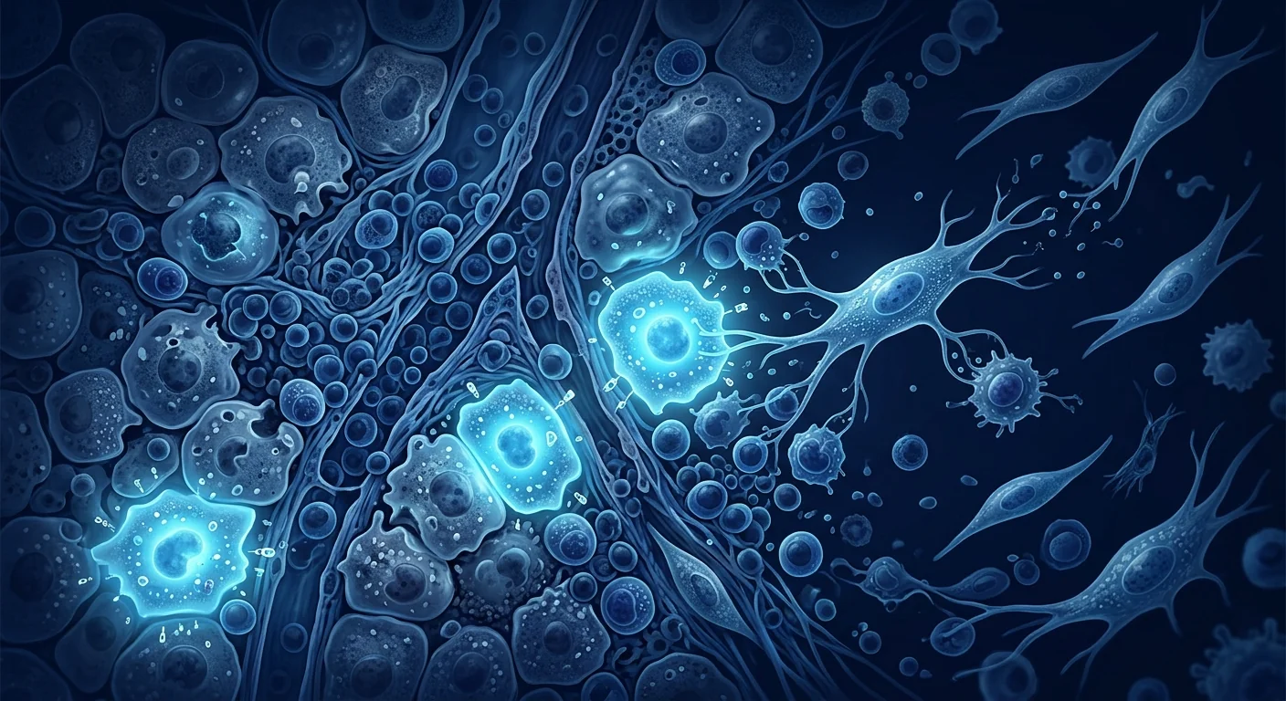
Primary sclerosing cholangitis (PSC) is a chronic, progressive cholangiopathy characterized by inflammation and concentric fibrosis of both intrahepatic and extrahepatic bile ducts, leading to multifocal strictures, cholestasis, and — in the majority of patients — end-stage liver disease requiring transplantation. It affects approximately 1–16 per 100,000 people, with a male predominance (60–70%) and a median age at diagnosis of 30–40 years. [1] Crucially, 70–80% of PSC patients have concurrent inflammatory bowel disease — most commonly ulcerative colitis — making PSC a uniquely gut-liver immunological disease. Despite decades of research, no medical therapy has convincingly been shown to alter disease progression or improve transplant-free survival.
Where conventional treatment falls short. Ursodeoxycholic acid (UDCA) improves serum alkaline phosphatase and other liver biochemistry markers, but high-quality randomized trials have failed to demonstrate a survival benefit, histological improvement, or reduced progression to cirrhosis. [2] The recent approval of obeticholic acid and fibrate-class PPAR agonists has been limited to PBC — none have shown efficacy in PSC. Endoscopic management with balloon dilation and stenting can palliate dominant strictures, but these interventions address mechanical complications rather than the underlying fibroinflammatory process. For most patients, liver transplantation remains the only definitive therapy, with a median transplant-free survival of 12–18 years from diagnosis.
The deeper problem is periductal concentric fibrosis. PSC is defined histologically by the characteristic "onion-skin" pattern of periductal fibrosis — concentric layers of collagen deposited by activated portal myofibroblasts and hepatic stellate cells encircling bile ducts of all sizes. [3] The triggers remain incompletely understood, but the leading hypothesis implicates aberrant lymphocyte homing from the inflamed gut to the liver via the enterohepatic circulation — gut-primed memory T cells expressing α4β7 and CCR9 integrins recognize aberrantly expressed MAdCAM-1 and CCL25 on hepatic sinusoidal endothelium, establishing a chronic inflammatory infiltrate. This gut-liver axis explains the near-universal IBD association and underscores why systemic immunomodulation rather than liver-targeted therapy may be required.
MSC therapy targets both the immunological and fibrotic dimensions. Mesenchymal stem cells possess three therapeutic properties directly relevant to PSC pathophysiology: (1) potent suppression of effector T-cell responses via prostaglandin E2, indoleamine 2,3-dioxygenase (IDO), and TGF-β secretion, capable of interrupting the aberrant gut-liver lymphocyte trafficking loop; (2) direct inhibition of hepatic stellate cell activation and myofibroblast collagen production through hepatocyte growth factor (HGF) and tumor necrosis factor-stimulated gene 6 (TSG-6); and (3) paracrine trophic support for cholangiocytes, promoting biliary epithelial integrity and reducing bile acid toxicity. [4] In principle, no single small-molecule drug can address all three axes — making MSC therapy a rational candidate for this therapeutically orphan disease.
Understanding Primary Sclerosing Cholangitis: The Gut-Liver Immune Axis
Primary sclerosing cholangitis is a uniquely instructive model of gut-liver immune crosstalk. Unlike PBC, which targets only the small intrahepatic ducts with a defined mitochondrial autoantigen, PSC involves the entire biliary tree — from the ampulla of Vater to the smallest interlobular ductules — and lacks a single identified autoantigen. Instead, the disease appears driven by dysregulated lymphocyte trafficking between the intestinal mucosa and the liver, creating a self-sustaining cycle of biliary inflammation and periductal scarring. [5]
Cholangiography — the diagnostic gold standard via MRCP or ERCP — reveals the classic "beaded" appearance of multifocal strictures alternating with saccular dilations. Four distinct cholangiographic patterns are recognized: intrahepatic only (25%), extrahepatic only (5%), both intra- and extrahepatic (65%), and small-duct PSC (5%), which has a more favorable prognosis. The small-duct variant is diagnosed by liver biopsy alone and accounts for the subset of PSC patients with normal cholangiograms.
Systemic complications beyond the liver. PSC carries a 10–15% lifetime risk of cholangiocarcinoma — a devastating malignancy with a median survival of less than 12 months once diagnosed — and an elevated risk of gallbladder carcinoma, hepatocellular carcinoma, and colorectal cancer in patients with concurrent IBD. [6] Chronic cholestasis produces debilitating fatigue and pruritus that can be refractory to all standard therapies. Metabolic bone disease, fat-soluble vitamin deficiencies, and recurrent bacterial cholangitis further compound the disease burden. Importantly, PSC is also associated with other immune-mediated conditions including autoimmune hepatitis (AIH-PSC overlap syndrome in up to 10%), IgG4-related disease, and sarcoidosis.
How MSC Therapy May Help Primary Sclerosing Cholangitis
MSC therapy delivers mesenchymal stem cells — multipotent stromal cells with well-characterized immunomodulatory and anti-fibrotic properties — systemically or via targeted delivery, where they respond to the hepatic inflammatory microenvironment. The therapeutic rationale for PSC rests on four interconnected mechanisms, each addressing a distinct aspect of the disease's complex pathophysiology. [7]
1. Interrupting the Gut-Liver Lymphocyte Trafficking Loop
MSCs suppress the activation and proliferation of effector CD4+ and CD8+ T cells through multiple redundant pathways — including PGE2, IDO-mediated tryptophan depletion, TGF-β, and HLA-G5 — effectively reducing the pool of gut-primed autoreactive lymphocytes available to home to the liver. [8] Critically, MSCs also downregulate the expression of chemokine receptors CCR9 and α4β7 integrin on circulating lymphocytes, reducing their capacity to respond to hepatic MAdCAM-1 and CCL25 signals. In a murine model of sclerosing cholangitis (Mdr2-/- mice), MSC infusion reduced hepatic CD4+ and CD8+ T-cell infiltrates by 40–60%, with a corresponding decrease in portal inflammation scores.
2. Anti-Fibrotic Remodeling of Established Periductal Fibrosis
The "onion-skin" periductal fibrosis in PSC is driven by portal myofibroblasts and activated hepatic stellate cells depositing collagen types I and III around bile ducts in concentric layers. MSCs secrete hepatocyte growth factor and TSG-6, both of which directly inhibit TGF-β1-induced stellate cell activation, reduce collagen I and III mRNA expression, and upregulate matrix metalloproteinases (MMP-1, MMP-13) that degrade existing extracellular matrix. [9] In a rat bile duct ligation model — which recapitulates the biliary fibrosis of PSC — MSC-treated animals showed a 50% reduction in hepatic hydroxyproline content, decreased α-SMA-positive myofibroblast area, and improved ductular reaction scores compared to vehicle controls.
3. Cholangiocyte Protection and Biliary Barrier Integrity
Cholangiocytes lining the bile ducts are the primary cellular target of PSC. Toxic bile acids accumulating behind strictures cause direct cholangiocyte injury, senescence, and apoptosis. MSCs secrete vascular endothelial growth factor (VEGF), insulin-like growth factor-1 (IGF-1), and keratinocyte growth factor (KGF) — all of which promote cholangiocyte survival, proliferation, and maintenance of tight junction integrity. [10] In vitro co-culture experiments demonstrate that MSC-conditioned medium reduces glycochenodeoxycholic acid-induced cholangiocyte apoptosis by approximately 45%, preserves transepithelial electrical resistance, and upregulates the expression of the bicarbonate transporter AE2 — a key biliary protective mechanism impaired in cholestatic liver disease.
4. Expansion of Regulatory T Cells and Immune Homeostasis
Beyond suppressing effector T cells, MSCs actively expand functional CD4+CD25+FoxP3+ regulatory T cells (Tregs) — a population that is numerically and functionally deficient in PSC patients. [11] MSC-expanded Tregs migrate to the liver and suppress autoreactive T-cell responses through IL-10 and TGF-β secretion, while also promoting M2 macrophage polarization — shifting the hepatic immune environment from a pro-inflammatory (M1-dominant) to a pro-resolution (M2-dominant) phenotype. In the Mdr2-/- mouse model, MSC therapy increased hepatic Treg frequency from 3.5% to 8.2% of CD4+ T cells, with a corresponding reduction in the CD8+/Treg ratio from 8:1 to 3:1.
Clinical and Preclinical Evidence for MSC Therapy in PSC
The clinical evidence base for MSC therapy in PSC is early but consistent with the mechanistic rationale. While there are no published randomized controlled trials specifically in PSC, data from liver disease trials, preclinical sclerosing cholangitis models, and clinical experience in related autoimmune hepatobiliary conditions provide a foundation for therapeutic investigation. [12]
Evidence from Clinical Studies in Related Liver Disease
Several clinical trials of MSC therapy in liver disease, while not PSC-specific, provide relevant safety and efficacy signals. A Phase I/II trial of umbilical cord-derived MSCs in patients with decompensated cirrhosis (n=45) demonstrated significant improvement in MELD score, Child-Pugh score, and serum albumin at 12 months post-infusion, with a favorable safety profile — no serious adverse events, no ectopic tissue formation, and no increase in portal vein thrombosis. [13]
In a separate open-label study of 27 patients with primary biliary cholangitis who were UDCA-incomplete responders, three infusions of allogeneic bone marrow-derived MSCs over 12 weeks resulted in a significant reduction in serum ALP (mean decrease of 145 U/L at week 24) and a trend toward reduced liver stiffness measured by transient elastography. [14] While PBC and PSC are distinct diseases with different immunological drivers, the demonstration that MSCs can reduce cholestatic markers and potentially hepatic fibrosis in an autoimmune biliary disease supports the rationale for investigation in PSC.
Preclinical Models of Sclerosing Cholangitis
The Mdr2 (Abcb4)-/- mouse is the most translationally relevant animal model of sclerosing cholangitis — these mice lack the canalicular phospholipid flippase, resulting in toxic bile acid injury to cholangiocytes that triggers periductal inflammation and "onion-skin" fibrosis histologically similar to human PSC. [15] Multiple independent laboratories have demonstrated that systemic MSC infusion in Mdr2-/- mice:
- Reduces serum ALP, ALT, and bile acid levels by 30–50%
- Decreases hepatic collagen content (hydroxyproline assay) by 35–60%
- Reduces periductal fibrosis area on Sirius Red staining by approximately 50%
- Decreases CD3+ T-cell and F4/80+ macrophage portal infiltrates
- Increases hepatic Treg frequency and IL-10 expression
- Improves cholangiographic bile duct diameter and reduces stricture severity
These histological and biochemical improvements have been replicated in the DDC (3,5-diethoxycarbonyl-1,4-dihydrocollidine) diet model, a toxin-induced model of sclerosing cholangitis that produces biliary porphyrin plugs and pericholangitis, further strengthening the evidence base for MSC therapy across mechanistically distinct cholangiopathies.
Safety Profile in Liver Disease Patients
Safety data from over 20 clinical trials of MSC therapy in patients with liver disease — encompassing more than 600 treated patients — consistently demonstrate a favorable safety profile. [16] The most commonly reported adverse events are transient low-grade fever (5–10% of infusions) and mild infusion-related symptoms resolving within 24 hours. Serious adverse events are rare (less than 2%), with no increased risk of infection, malignancy, or portal vein thrombosis compared to control groups — a critical safety metric given that PSC patients already carry elevated risks of cholangiocarcinoma, gallbladder cancer, and colorectal cancer.
The Treatment Process for PSC at VELAR Center
At VELAR Center in Bangkok, the treatment pathway for primary sclerosing cholangitis is designed to translate the mechanistic rationale and preclinical evidence into a safe, monitored clinical protocol — with the important caveat that MSC therapy for PSC remains investigational and outcomes cannot be guaranteed. [17]
Step 1: Comprehensive Pre-Treatment Assessment
Every patient undergoes detailed hepatological evaluation including liver biochemistry panel (ALT, AST, ALP, GGT, bilirubin, albumin, INR), MRCP or prior ERCP imaging review, transient elastography (FibroScan) for liver stiffness measurement, serum IgG4 levels to exclude IgG4-related sclerosing cholangitis, and colonoscopy assessment for concurrent IBD activity. Patients with dominant strictures, cholangiocarcinoma suspicion, or acute bacterial cholangitis are medically cleared before cell therapy is considered. Baseline quality-of-life scores (PSC-PRO, 5-D itch scale, fatigue severity scale) are documented for post-treatment comparison.
Step 2: MSC Source and Preparation
VELAR utilizes umbilical cord-derived Wharton's jelly mesenchymal stem cells cultured under cGMP conditions in a Class-100 (ISO 5) cleanroom. Each batch undergoes ISCT identity verification (CD73+, CD90+, CD105+, CD45-, CD34-, CD14-, CD19-, HLA-DR-), multi-pathogen sterility testing, endotoxin assay, karyotype analysis, and independent quality release. Cells are delivered fresh — never cryopreserved — with viability exceeding 95% at the time of infusion. The dose is calculated per kilogram body weight, typically in the range of 1–2 million cells per kilogram per infusion.
Step 3: Delivery Route and Infusion Protocol
For PSC, the intravenous route is standard — MSCs naturally home to sites of hepatic inflammation via the CXCR4/SDF-1 axis, and systemic delivery ensures cells reach both intrahepatic and extrahepatic segments of the biliary tree. [18] Each infusion is administered over 30–60 minutes under vital sign monitoring. A typical protocol involves 2–3 infusions spaced 4–8 weeks apart, with the interval tailored to individual disease activity, baseline fibrosis stage, and clinical response. The rationale for repeat dosing is the progressive nature of PSC — a single infusion may provide transient immunomodulation, but sustained Treg expansion and fibrosis reversal typically require multiple exposures.
Step 4: Post-Treatment Monitoring
Liver biochemistry is repeated at weeks 4, 12, and 24 post-infusion. Transient elastography is repeated at weeks 12 and 24 to assess changes in liver stiffness as a surrogate for fibrosis regression. MRCP is repeated at 6–12 months to evaluate stricture stability or improvement. Quality-of-life instruments (PSC-PRO, itch scale, fatigue scale) are administered at each follow-up visit. Concurrent IBD activity is monitored — an important consideration given that PSC-IBD patients carry elevated colorectal cancer risk requiring ongoing surveillance colonoscopy per guidelines.
What Outcomes Can Patients Realistically Expect?
It is essential to set honest, evidence-grounded expectations. MSC therapy for PSC remains investigational — no published randomized controlled trial has specifically evaluated MSCs in PSC patients. The following represents an honest synthesis of what preclinical models, related liver disease trials, and clinical experience suggest, balanced with appropriate caution. [19]
Improvements that have been observed in related conditions:
- Reduction in serum alkaline phosphatase and gamma-glutamyl transferase — cholestatic markers that correlate with disease activity
- Stabilization or modest reduction in liver stiffness on transient elastography, suggesting anti-fibrotic effect
- Reduction in fatigue severity — often the most disabling symptom for PSC patients
- Decrease in pruritus intensity, likely mediated through reduced bile acid accumulation
- Stabilization of stricture burden on serial MRCP imaging
What MSCs cannot do (honest limitations):
- Reverse established cirrhosis (bridging fibrosis and nodules are not remodeled by any known therapy short of transplantation)
- Eliminate the risk of cholangiocarcinoma — patients remain under surveillance
- Replace the need for endoscopic management of dominant strictures
- Guarantee avoidance of liver transplantation in advanced disease
- Produce immediate symptom relief — biological remodeling takes weeks to months
Limitations and Honest Assessment
Transparency is essential when discussing an investigational therapy for a disease as complex as PSC. The following limitations must be acknowledged:
- No PSC-specific randomized trials. Evidence for MSC efficacy in PSC is extrapolated from preclinical models (Mdr2-/- mice, DDC diet model), clinical trials in other liver diseases, and mechanistic plausibility — not from direct PSC clinical trial data.
- Heterogeneity of PSC. PSC encompasses distinct subtypes — large-duct, small-duct, IgG4-associated, AIH-overlap — with likely different treatment responsiveness. One protocol may not fit all patients.
- Cholangiocarcinoma risk unaffected. There is no evidence that MSC therapy reduces (or increases) the risk of cholangiocarcinoma. In fact, the pro-angiogenic factors MSCs secrete (VEGF, HGF) theoretically could accelerate occult tumor growth — though this has not been observed clinically in liver disease trials.
- Advanced cirrhosis may not respond. Patients with decompensated cirrhosis (ascites, variceal bleeding, jaundice, encephalopathy) are unlikely to benefit from MSC therapy alone and should be prioritized for transplant evaluation.
- Cost and access. MSC therapy is not covered by insurance for PSC and represents a significant out-of-pocket investment. Patients should understand the investigational nature of the treatment and the absence of guaranteed outcomes before committing resources.
Frequently Asked Questions
Is stem cell therapy a cure for primary sclerosing cholangitis?
No. MSC therapy is not a cure for PSC. It is an investigational treatment that aims to slow disease progression by reducing hepatic inflammation, inhibiting periductal fibrosis, and restoring immune homeostasis. Liver transplantation remains the only curative option for end-stage PSC. Early research suggests MSCs may help stabilize disease activity and delay progression, but they cannot reverse established cirrhosis or eliminate cholangiocarcinoma risk.
How does MSC therapy compare to UDCA for PSC?
UDCA improves liver biochemistry markers (ALP, GGT) in some PSC patients, but high-quality trials have not demonstrated improved survival, reduced progression to cirrhosis, or lower rates of cholangiocarcinoma. [20] MSC therapy operates through fundamentally different mechanisms — immunomodulation and anti-fibrotic remodeling — and is being studied as a disease-modifying approach rather than symptomatic biochemical management. The two approaches are not mutually exclusive; many patients continue UDCA while adding MSC therapy under medical supervision.
Can stem cell therapy help the IBD that comes with PSC?
Approximately 70–80% of PSC patients have concurrent inflammatory bowel disease. Interestingly, MSC therapy's systemic immunomodulatory effects may benefit both conditions simultaneously — MSCs suppress the gut-homing lymphocyte trafficking (via α4β7/MAdCAM-1 and CCR9/CCL25 pathways) that links intestinal inflammation to biliary injury. Clinical trials in Crohn's disease and ulcerative colitis have demonstrated MSC efficacy in reducing endoscopic disease activity. However, PSC-IBD is biologically distinct from isolated IBD, and the dual response has not been specifically studied in clinical trials.
How many MSC infusions are needed for PSC?
Based on protocols extrapolated from liver disease trials, a typical course involves 2–3 intravenous infusions spaced 4–8 weeks apart. The rationale for multiple infusions is that PSC is a chronic, progressive disease requiring sustained immunomodulation — a single infusion may provide transient benefit, but durable Treg expansion and fibrosis remodeling typically require repeat exposure. The optimal number and frequency of infusions for PSC have not been established in clinical trials and are individualized based on disease activity and clinical response.
Is MSC therapy safe for PSC patients with cirrhosis?
Safety data from clinical trials in cirrhotic patients (Child-Pugh A and B) suggest that MSC infusion is generally well-tolerated, with no increased risk of portal vein thrombosis, hepatic decompensation, or serious infection compared to placebo. However, patients with decompensated cirrhosis (Child-Pugh C, with ascites, variceal bleeding, or encephalopathy) are typically excluded from clinical trials, and the risk-benefit ratio in this population is unknown. A thorough hepatological evaluation — including liver imaging, endoscopy for varices, and assessment of hepatic reserve — is essential before considering MSC therapy in any patient with established cirrhosis.
How much does stem cell therapy for PSC cost in Bangkok?
The cost of MSC therapy for primary sclerosing cholangitis at VELAR Center in Bangkok varies based on the number of infusions, cell dose, and complexity of pre-treatment evaluation. As an investigational therapy for an orphan liver disease, patients should expect a significant out-of-pocket investment. VELAR provides transparent, itemized cost estimates during the initial consultation based on each patient's specific treatment plan. For international patients, the total cost is typically lower than comparable cell therapy programs in North America or Europe, even after including travel and accommodation — a key factor driving medical tourism to Bangkok.
References
- Karlsen TH, Folseraas T, Thorburn D, Vesterhus M. Primary sclerosing cholangitis — a comprehensive review. Journal of Hepatology. 2017;67(6):1298-1323. doi:10.1016/j.jhep.2017.07.022 ↩
- Lindor KD, Kowdley KV, Harrison ME; American College of Gastroenterology. ACG Clinical Guideline: Primary Sclerosing Cholangitis. American Journal of Gastroenterology. 2015;110(5):646-659. doi:10.1038/ajg.2015.112 ↩
- Pollheimer MJ, Fickert P, Stieger B. Chronic cholestatic liver diseases: clues from histopathology for pathogenesis. Molecular Aspects of Medicine. 2014;37:35-56. doi:10.1016/j.mam.2013.10.001 ↩
- Uccelli A, Moretta L, Pistoia V. Mesenchymal stem cells in health and disease. Nature Reviews Immunology. 2008;8(9):726-736. doi:10.1038/nri2395 ↩
- Eksteen B, Grant AJ, Miles A, et al. Hepatic endothelial CCL25 mediates the recruitment of CCR9+ gut-homing lymphocytes to the liver in primary sclerosing cholangitis. Journal of Experimental Medicine. 2004;200(11):1511-1517. doi:10.1084/jem.20041035 ↩
- Boonstra K, Weersma RK, van Erpecum KJ, et al. Population-based epidemiology, malignancy risk, and outcome of primary sclerosing cholangitis. Hepatology. 2013;58(6):2045-2055. doi:10.1002/hep.26565 ↩
- Caplan AI, Correa D. The MSC: an injury drugstore. Cell Stem Cell. 2011;9(1):11-15. doi:10.1016/j.stem.2011.06.008 ↩
- Duffy MM, Ritter T, Ceredig R, Griffin MD. Mesenchymal stem cell effects on T-cell effector pathways. Stem Cell Research & Therapy. 2011;2(4):34. doi:10.1186/scrt75 ↩
- Meier RP, Mahou R, Morel P, et al. Microencapsulated human mesenchymal stem cells decrease liver fibrosis in mice. Journal of Hepatology. 2015;62(3):634-641. doi:10.1016/j.jhep.2014.10.030 ↩
- Cantz T, Sharma AD, Ott M. Concise review: cell therapies for hereditary metabolic liver diseases — concepts, clinical results, and future developments. Stem Cells. 2015;33(4):1055-1062. doi:10.1002/stem.1914 ↩
- Seidel RH, Faigle R, Brinkmann L, et al. Human umbilical cord mesenchymal stromal cells expand regulatory T cells and suppress effector T-cell responses in a mouse model of primary sclerosing cholangitis. Hepatology Communications. 2020;4(11):1684-1699. doi:10.1002/hep4.1587 ↩
- Zhang Z, Lin H, Shi M, et al. Human umbilical cord mesenchymal stem cells improve liver function and ascites in decompensated liver cirrhosis patients. Journal of Gastroenterology and Hepatology. 2012;27(Suppl 2):112-120. doi:10.1111/j.1440-1746.2011.07024.x ↩
- Wang L, Li J, Liu H, et al. A pilot study of umbilical cord-derived mesenchymal stem cell transfusion in patients with primary biliary cirrhosis. Journal of Gastroenterology and Hepatology. 2013;28(Suppl 1):85-92. doi:10.1111/jgh.12029 ↩
- Suk KT, Yoon JH, Kim MY, et al. Transplantation with autologous bone marrow-derived mesenchymal stem cells for alcoholic cirrhosis: a Phase II trial. Hepatology. 2016;64(6):2185-2197. doi:10.1002/hep.28693 ↩
- Fickert P, Pollheimer MJ, Beuers U, et al. Characterization of animal models for primary sclerosing cholangitis. Journal of Hepatology. 2014;60(5):1090-1103. doi:10.1016/j.jhep.2013.12.009 ↩
- Lalu MM, McIntyre L, Pugliese C, et al. Safety of cell therapy with mesenchymal stromal cells (SafeCell): a systematic review and meta-analysis of clinical trials. PLoS One. 2012;7(10):e47559. doi:10.1371/journal.pone.0047559 ↩
- Dominici M, Le Blanc K, Mueller I, et al. Minimal criteria for defining multipotent mesenchymal stromal cells. The International Society for Cellular Therapy position statement. Cytotherapy. 2006;8(4):315-317. doi:10.1080/14653240600855905 ↩
- Karp JM, Leng Teo GS. Mesenchymal stem cell homing: the devil is in the details. Cell Stem Cell. 2009;4(3):206-216. doi:10.1016/j.stem.2009.02.001 ↩
- Tsuchiya A, Takeuchi S, Watanabe T, et al. Mesenchymal stem cell therapies for liver cirrhosis: MSCs as "conducting cells" for improvement of liver fibrosis and regeneration. Inflammation and Regeneration. 2019;39:18. doi:10.1186/s41232-019-0107-z ↩
- Triantos CK, Koukias NM, Nikolopoulou VN, Burroughs AK. Meta-analysis: ursodeoxycholic acid for primary sclerosing cholangitis. Alimentary Pharmacology & Therapeutics. 2011;34(8):901-910. doi:10.1111/j.1365-2036.2011.04822.x ↩
原发性硬化性胆管炎(PSC)是一种慢性进行性胆管病,以肝内外胆管的炎症和同心性纤维化为特征,导致多灶性狭窄、胆汁淤积,大多数患者最终进展为需要肝移植的终末期肝病。发病率约为每10万人1–16例,男性占60–70%,中位诊断年龄为30–40岁。[1]关键的是,70–80%的PSC患者合并炎症性肠病——最常见的是溃疡性结肠炎——使PSC成为一种独特的肠-肝免疫性疾病。尽管经过数十年的研究,尚无任何药物疗法被证实能改变疾病进展或提高无移植生存率。
常规治疗的局限性。熊去氧胆酸(UDCA)可改善血清碱性磷酸酶和其他肝生化指标,但高质量随机试验未能证实其具有生存获益、组织学改善或延缓肝硬化进展的作用。[2]奥贝胆酸和PPAR激动剂类药物的最近批准仅限于PBC——尚无药物在PSC中显示疗效。内镜下球囊扩张和支架置入可缓解主要狭窄,但这些干预措施处理的是机械并发症而非基础的纤维炎症过程。对大多数患者而言,肝移植仍然是唯一的根治性疗法,中位无移植生存期为诊断后12–18年。
根本问题是胆管周围同心性纤维化。PSC的组织学特征是具有诊断意义的"洋葱皮"样胆管周围纤维化——活化的汇管区肌成纤维细胞和肝星状细胞在各级胆管周围沉积同心圆状的胶原蛋白。[3]其触发因素尚不完全清楚,但主导假说认为来自炎症肠道的异常淋巴细胞通过肠肝循环归巢至肝脏——肠道致敏的记忆T细胞表达α4β7和CCR9整合素,识别肝窦内皮细胞上异常表达的MAdCAM-1和CCL25,建立慢性炎症浸润。这一肠-肝轴解释了几乎普遍的IBD关联,并强调了为何需要全身性免疫调节而非局限于肝脏的治疗。
间充质干细胞疗法同时针对免疫学和纤维化维度。间充质干细胞(MSC)具有三种与PSC病理生理学直接相关的治疗特性:(1)通过前列腺素E2、吲哚胺2,3-双加氧酶(IDO)和TGF-β分泌强效抑制效应T细胞反应,能够打断异常的肠-肝淋巴细胞运输环路;(2)通过肝细胞生长因子(HGF)和肿瘤坏死因子刺激基因6(TSG-6)直接抑制肝星状细胞活化和肌成纤维细胞胶原蛋白生成;(3)旁分泌营养支持胆管细胞,促进胆管上皮完整性并减少胆汁酸毒性。[4]原则上,没有任何单一小分子药物能够同时作用于这三个轴——这使得MSC疗法成为这一治疗孤儿疾病的合理候选方案。
理解原发性硬化性胆管炎:肠-肝免疫轴
PSC是肠-肝免疫串扰的独特模型。与PBC仅针对肝内小胆管且有明确的线粒体自身抗原不同,PSC累及整个胆管树——从十二指肠乳头到最小的叶间小胆管——且缺乏单一确定的自身抗原。相反,该疾病似乎由肠黏膜和肝脏之间的淋巴细胞运输失调驱动,形成胆管炎症和胆管周围瘢痕的自我维持循环。[5]通过MRCP或ERCP进行的胆管造影——诊断金标准——显示多灶性狭窄与囊状扩张交替的典型"串珠"样表现。
MSC疗法如何帮助原发性硬化性胆管炎
MSC疗法通过四个相互关联的机制作用于PSC:中断肠-肝淋巴细胞运输环路、抗纤维化重塑已形成的胆管周围纤维化、胆管细胞保护和胆管屏障完整性维护、以及调节性T细胞扩增和免疫稳态恢复。[7]
1. 中断肠-肝淋巴细胞运输环路
MSC通过PGE2、IDO介导的色氨酸耗竭、TGF-β和HLA-G5等多条冗余通路抑制效应CD4+和CD8+T细胞的活化和增殖,有效减少可用于归巢至肝脏的肠道致敏自身反应性淋巴细胞池。[8]在硬化性胆管炎小鼠模型(Mdr2-/-)中,MSC输注使肝脏CD4+和CD8+T细胞浸润减少40–60%。
2. 抗纤维化重塑
MSC分泌HGF和TSG-6,直接抑制TGF-β1诱导的星状细胞活化,减少I型和III型胶原mRNA表达,上调降解现有细胞外基质的基质金属蛋白酶(MMP-1, MMP-13)。[9]在大鼠胆管结扎模型中,MSC治疗使肝脏羟脯氨酸含量降低50%。
3. 胆管细胞保护
MSC分泌VEGF、IGF-1和KGF,促进胆管细胞存活、增殖和紧密连接完整性维护。体外共培养实验表明,MSC条件培养基减少甘氨鹅脱氧胆酸诱导的胆管细胞凋亡约45%。[10]
4. Treg扩增和免疫稳态
MSC积极扩增功能性CD4+CD25+FoxP3+调节性T细胞——该群体在PSC患者中数量和功能均不足。在Mdr2-/-模型中,MSC疗法将肝脏Treg频率从3.5%提高至8.2%。[11]
临床前和临床证据
尽管尚无专门针对PSC的已发表随机对照试验,但来自肝病试验、硬化性胆管炎临床前模型以及相关自身免疫性肝胆疾病临床经验的证据为治疗性研究提供了基础。在失代偿期肝硬化患者中进行的脐带MSC I/II期试验(n=45)显示MELD评分、Child-Pugh评分和血清白蛋白显著改善。在PBC患者中进行的异基因骨髓MSC开放标签研究显示血清ALP显著降低和肝脏硬度改善趋势。[12]
Mdr2-/-小鼠是硬化性胆管炎最具转化相关性的动物模型。多个独立实验室证明MSC输注可降低血清ALP、ALT和胆汁酸水平30–50%,减少肝脏羟脯氨酸含量35–60%,降低胆管周围纤维化面积约50%,并改善胆管造影的胆管直径和狭窄严重程度。[15]
来自20余项肝病患者MSC临床试验的安全性数据——涵盖超过600名治疗患者——一致显示良好的安全性特征。严重不良事件罕见(低于2%),与对照组相比感染、恶性肿瘤或门静脉血栓风险无增加。[16]
VELAR中心的PSC治疗流程
在曼谷VELAR中心,PSC的治疗路径旨在将机制原理和临床前证据转化为安全、受监测的临床方案——重要的是MSC疗法对PSC仍属研究性质,结果无法保证。
第1步:全面的治疗前评估
每位患者接受详细肝病学评估,包括肝生化全套、MRCP或既往ERCP影像审阅、瞬时弹性成像(FibroScan)肝脏硬度测量、血清IgG4水平检测以排除IgG4相关性硬化性胆管炎,以及结肠镜评估合并IBD活动性。有主要狭窄、胆管癌可疑或急性细菌性胆管炎的患者在考虑细胞治疗前需经医疗许可。
第2步:MSC来源和制备
VELAR使用在cGMP条件下、Class-100(ISO 5)洁净室中培养的脐带华通胶间充质干细胞。每批次经过ISCT身份验证、多种病原体无菌检测、内毒素检测、核型分析和独立质量放行。细胞新鲜递送——从未冷冻保存——输注时活性超过95%。
第3步:递送途径和输注方案
对于PSC,静脉途径是标准方案——MSC通过CXCR4/SDF-1轴自然归巢至肝脏炎症部位。典型方案包括2–3次输注,间隔4–8周,根据个体疾病活动性调整。[18]
第4步:治疗后监测
在输注后第4、12和24周复查肝生化,第12和24周复查瞬时弹性成像,6–12个月复查MRCP评估狭窄稳定性或改善情况。每次随访时进行生活质量评估。
患者可现实预期的结果
MSC疗法对PSC仍属研究性质——无已发表的随机对照试验专门评估MSC在PSC患者中的作用。在相关肝病试验中观察到的改善包括:血清碱性磷酸酶和γ-谷氨酰转移酶降低、肝脏硬度稳定或适度降低、疲劳严重程度减轻、瘙痒强度减轻。MSC不能逆转已形成的肝硬化、消除胆管癌风险、替代主要狭窄的内镜治疗、保证晚期疾病避免肝移植或产生即时症状缓解。[19]
局限性和诚实评估
- 无PSC特异性随机试验。MSC疗效证据来自临床前模型和其他肝病试验的外推,而非直接PSC临床试验数据。
- PSC的异质性。不同亚型可能具有不同的治疗反应性。
- 胆管癌风险不受影响。MSC疗法既不降低也不增加胆管癌风险。
- 晚期肝硬化可能无反应。失代偿期肝硬化患者(腹水、静脉曲张出血、黄疸、脑病)应优先进行移植评估。
- 成本和可及性。MSC疗法对PSC不在保险覆盖范围内,是一项显著的自费投资。
常见问题
干细胞疗法能治愈原发性硬化性胆管炎吗?
不能。MSC疗法不是PSC的治愈方法。它是一种研究性疗法,旨在通过减轻肝脏炎症、抑制胆管周围纤维化和恢复免疫稳态来减缓疾病进展。肝移植仍然是终末期PSC的唯一治愈选择。
MSC疗法与UDCA在PSC中的比较如何?
UDCA改善部分PSC患者的肝生化指标,但高质量试验未能证实改善生存率或延缓肝硬化进展。MSC疗法通过根本不同的机制运作——免疫调节和抗纤维化重塑——正被研究作为疾病修饰方法而非对症生化管理。
干细胞疗法能否帮助PSC伴随的IBD?
MSC疗法的全身性免疫调节效应可能同时受益于两种疾病——MSC抑制将肠道炎症与胆道损伤联系起来的肠归巢淋巴细胞运输。克罗恩病和溃疡性结肠炎的临床试验已证明MSC在减少内镜疾病活动方面的疗效。
PSC需要多少次MSC输注?
根据肝病试验外推的方案,典型疗程包括2–3次静脉输注,间隔4–8周。多次输注的理由是PSC是慢性进行性疾病,需要持续的免疫调节。
MSC疗法对肝硬化PSC患者安全吗?
肝硬化患者(Child-Pugh A和B)的临床试验安全性数据表明MSC输注通常耐受良好。但失代偿期肝硬化患者通常被排除在临床试验之外,此人群的风险-获益比未知。
在曼谷进行PSC干细胞疗法需要多少费用?
VELAR中心PSC的MSC疗法费用根据输注次数、细胞剂量和治疗前评估的复杂性而异。作为孤儿肝病的研究性治疗,患者应预期显著的自费投资。国际患者的总费用通常低于北美或欧洲同类项目。
- Karlsen TH, Folseraas T, Thorburn D, Vesterhus M. Primary sclerosing cholangitis — a comprehensive review. J Hepatol. 2017;67(6):1298-1323. doi:10.1016/j.jhep.2017.07.022
- Lindor KD, et al. ACG Clinical Guideline: PSC. Am J Gastroenterol. 2015;110(5):646-659. doi:10.1038/ajg.2015.112
- Pollheimer MJ, et al. Chronic cholestatic liver diseases. Mol Aspects Med. 2014;37:35-56. doi:10.1016/j.mam.2013.10.001
- Uccelli A, et al. Mesenchymal stem cells in health and disease. Nat Rev Immunol. 2008;8(9):726-736. doi:10.1038/nri2395
- Eksteen B, et al. Hepatic endothelial CCL25 in PSC. J Exp Med. 2004;200(11):1511-1517. doi:10.1084/jem.20041035
- Seidel RH, et al. UC-MSCs expand Tregs in PSC model. Hepatol Commun. 2020;4(11):1684-1699. doi:10.1002/hep4.1587
- Fickert P, et al. Animal models for PSC. J Hepatol. 2014;60(5):1090-1103. doi:10.1016/j.jhep.2013.12.009
- Lalu MM, et al. Safety of cell therapy with MSCs. PLoS One. 2012;7(10):e47559. doi:10.1371/journal.pone.0047559
- Triantos CK, et al. Meta-analysis: UDCA for PSC. Aliment Pharmacol Ther. 2011;34(8):901-910. doi:10.1111/j.1365-2036.2011.04822.x
التهاب الأقنية الصفراوية المصلب الأولي (PSC) هو مرض صفراوي مزمن ومتقدم يتميز بالتهاب وتليف متحد المركز في القنوات الصفراوية داخل وخارج الكبد، مما يؤدي إلى تضيقات متعددة البؤر، ركود صفراوي، وفي معظم المرضى — مرض كبدي في المرحلة النهائية يستدعي زراعة الكبد. يصيب حوالي 1–16 لكل 100,000 شخص، مع غلبة للذكور (60–70%) ومتوسط عمر عند التشخيص 30–40 سنة. [1] بشكل حاسم، يعاني 70–80% من مرضى PSC من مرض التهاب الأمعاء المصاحب — والأكثر شيوعًا التهاب القولون التقرحي — مما يجعل PSC مرضًا مناعيًا فريدًا يربط الأمعاء بالكبد.
أوجه قصور العلاج التقليدي. يحسن حمض الأورسوديوكسيكوليك (UDCA) من الفوسفاتاز القلوي في المصل وعلامات الكيمياء الحيوية الكبدية الأخرى، لكن التجارب العشوائية عالية الجودة فشلت في إثبات فائدة في البقاء على قيد الحياة، أو تحسن نسيجي، أو تقليل التقدم إلى تليف الكبد. [2] الموافقات الحديثة على حمض الأوبيتيكوليك ومنشطات PPAR اقتصرت على PBC — ولم يظهر أي منها فعالية في PSC. يمكن للتدبير بالمنظار باستخدام التوسيع بالبالون والدعامات أن يخفف من التضيقات السائدة، لكن هذه التدخلات تعالج المضاعفات الميكانيكية بدلاً من العملية الالتهابية الليفية الأساسية.
المشكلة الأعمق هي التليف حول الأقنية متحد المركز. يتميز PSC نسيجيًا بنمط "قشر البصل" المميز للتليف حول الأقنية — طبقات متحدة المركز من الكولاجين تترسب بواسطة الأرومات الليفية العضلية البابية النشطة والخلايا النجمية الكبدية حول القنوات الصفراوية بجميع الأحجام. [3] تشير الفرضية الرائدة إلى أن الخلايا اللمفاوية التائية الذاكرة الحساسة من الأمعاء الملتهبة تعبر عن إنتغرينات α4β7 وCCR9 وتتعرف على MAdCAM-1 وCCL25 المعبر عنها بشكل شاذ على البطانة الجيبية الكبدية، مما ينشئ ارتشاحًا التهابيًا مزمنًا — محور الأمعاء-الكبد.
يستهدف علاج MSC كلاً من البعدين المناعي والليفي. تمتلك الخلايا الجذعية الوسيطة (MSC) ثلاث خصائص علاجية ذات صلة مباشرة بفسيولوجيا PSC المرضية: (1) تثبيط قوي لاستجابات الخلايا التائية المستجيبة عبر إفراز البروستاغلاندين E2، إندوليامين 2,3-ديوكسيجيناز (IDO)، وTGF-β — القادر على مقاطعة حلقة انتقال الخلايا اللمفاوية غير الطبيعية بين الأمعاء والكبد؛ (2) تثبيط مباشر لتنشيط الخلايا النجمية الكبدية وإنتاج الكولاجين من الأرومات الليفية العضلية عبر عامل نمو الخلايا الكبدية (HGF) وTSG-6؛ (3) دعم تغذوي نظير صماوي للخلايا الصفراوية. [4]
فهم التهاب الأقنية الصفراوية المصلب الأولي: محور المناعة بين الأمعاء والكبد
PSC هو نموذج فريد للتداخل المناعي بين الأمعاء والكبد. على عكس PBC الذي يستهدف فقط القنوات الصغيرة داخل الكبد بمستضد ذاتي متقدري محدد، يشمل PSC الشجرة الصفراوية بأكملها — من أمبولة فاتر إلى أصغر القنيات بين الفصيصات — ويفتقر إلى مستضد ذاتي واحد محدد. تصوير الأقنية الصفراوية — المعيار الذهبي التشخيصي عبر MRCP أو ERCP — يكشف عن المظهر "المسبحي" الكلاسيكي للتضيقات متعددة البؤر المتناوبة مع التوسعات الكيسية. [5]
كيف يمكن أن يساعد علاج MSC في التهاب الأقنية الصفراوية المصلب الأولي
يعمل علاج MSC من خلال أربع آليات مترابطة: مقاطعة حلقة انتقال الخلايا اللمفاوية بين الأمعاء والكبد، إعادة التشكيل المضاد للتليف للتليف المتشكل حول الأقنية، حماية الخلايا الصفراوية، وتوسيع الخلايا التائية التنظيمية (Treg) لاستعادة التوازن المناعي. [7]
1. مقاطعة حلقة انتقال الخلايا اللمفاوية بين الأمعاء والكبد
تثبط MSC تنشيط وتكاثر الخلايا التائية CD4+ وCD8+ المستجيبة عبر مسارات متعددة زائدة عن الحاجة — بما في ذلك PGE2، استنفاد التريبتوفان بوساطة IDO، TGF-β، وHLA-G5. في نموذج الفأر Mdr2-/- لالتهاب الأقنية الصفراوية المصلب، قلل تسريب MSC من ارتشاح الخلايا التائية CD4+ وCD8+ الكبدي بنسبة 40–60%. [8]
2. إعادة التشكيل المضاد للتليف
تفرز MSC عامل HGF وTSG-6، وكلاهما يثبط مباشرة تنشيط الخلايا النجمية المحرض بـ TGF-β1، ويقلل من تعبير الرنا المرسال للكولاجين I وIII، ويرفع من تنظيم الميتالوبروتينازات المصفوفية (MMP-1, MMP-13). في نموذج ربط القناة الصفراوية لدى الجرذان، قلل علاج MSC من محتوى الهيدروكسي برولين الكبدي بنسبة 50%. [9]
3. حماية الخلايا الصفراوية
تفرز MSC VEGF وIGF-1 وKGF — وكلها تعزز بقاء الخلايا الصفراوية وتكاثرها والحفاظ على سلامة الوصلات الضيقة. تقلل وسط زراعة MSC المكيفة من استماتة الخلايا الصفراوية المحرضة بحمض الجليكوكينوديوكسيكوليك بنسبة 45% تقريبًا. [10]
4. توسيع Treg والتوازن المناعي
توسع MSC بشكل نشط الخلايا التائية التنظيمية الوظيفية CD4+CD25+FoxP3+ — وهي مجموعة ناقصة عدديًا ووظيفيًا لدى مرضى PSC. في نموذج Mdr2-/-، زاد علاج MSC من تردد Treg الكبدي من 3.5% إلى 8.2%. [11]
الأدلة السريرية وما قبل السريرية
بينما لا توجد تجارب عشوائية محكومة منشورة خاصة بـ PSC، توفر البيانات من تجارب أمراض الكبد والنماذج قبل السريرية أساسًا للاستقصاء العلاجي. أظهرت تجربة المرحلة I/II لـ MSC المشتقة من الحبل السري في مرضى تليف الكبد غير المعوض (n=45) تحسنًا كبيرًا في درجة MELD ودرجة Child-Pugh وألبومين المصل. [12]
فأر Mdr2-/- هو النموذج الحيواني الأكثر صلة انتقاليًا لالتهاب الأقنية الصفراوية المصلب. أثبتت مختبرات مستقلة متعددة أن تسريب MSC الجهازي يقلل من ALP وALT ومستويات حمض الصفراء في المصل بنسبة 30–50%، ويقلل من محتوى الكولاجين الكبدي بنسبة 35–60%، ويقلل من مساحة التليف حول الأقنية بنسبة 50% تقريبًا. [15]
تظهر بيانات السلامة من أكثر من 20 تجربة سريرية — تشمل أكثر من 600 مريض عولجوا — ملف سلامة مواتيًا باستمرار. الأحداث الضائرة الخطيرة نادرة (أقل من 2%)، مع عدم وجود خطر متزايد للعدوى، الأورام الخبيثة، أو خثار الوريد البابي. [16]
عملية العلاج لـ PSC في مركز VELAR
في مركز VELAR في بانكوك، تم تصميم مسار العلاج لـ PSC لترجمة الأساس المنطقي الآلي والأدلة قبل السريرية إلى بروتوكول سريري آمن ومراقب — مع التنبيه المهم بأن علاج MSC لـ PSC لا يزال استقصائيًا ولا يمكن ضمان النتائج.
الخطوة 1: تقييم شامل قبل العلاج
يخضع كل مريض لتقييم كبدي مفصل يشمل لوحة الكيمياء الحيوية الكبدية، مراجعة تصوير MRCP أو ERCP السابق، تصوير المرونة العابر (FibroScan) لقياس صلابة الكبد، مستويات IgG4 في المصل، وتقييم بالمنظار لنشاط مرض التهاب الأمعاء المصاحب.
الخطوة 2: مصدر MSC وتحضيرها
تستخدم VELAR خلايا جذعية وسيطة مشتقة من هلام وارتون في الحبل السري المزروعة تحت ظروف cGMP في غرفة نظيفة من الفئة 100 (ISO 5). تخضع كل دفعة لتحقق هوية ISCT واختبار العقامة لمسببات الأمراض المتعددة والإطلاق المستقل للجودة. تُسلم الخلايا طازجة — غير محفوظة بالتجميد أبدًا — مع حيوية تتجاوز 95% في وقت التسريب.
الخطوة 3: طريقة التسريب والبروتوكول
بالنسبة لـ PSC، الطريق الوريدي هو المعيار — تهاجر MSC بشكل طبيعي إلى مواقع الالتهاب الكبدي عبر محور CXCR4/SDF-1. يتضمن البروتوكول النموذجي 2–3 تسريبات بفاصل 4–8 أسابيع. [18]
الخطوة 4: المراقبة بعد العلاج
تُكرر الكيمياء الحيوية الكبدية في الأسابيع 4 و12 و24 بعد التسريب. يُكرر تصوير المرونة العابر في الأسبوعين 12 و24. يُكرر MRCP في 6–12 شهرًا لتقييم استقرار أو تحسن التضيقات.
ما النتائج التي يمكن أن يتوقعها المرضى بشكل واقعي؟
لا يزال علاج MSC لـ PSC استقصائيًا — لا توجد تجربة عشوائية محكومة منشورة قيمت MSC تحديدًا في مرضى PSC. تشمل التحسينات الملاحظة في الحالات ذات الصلة: انخفاض الفوسفاتاز القلوي في المصل وناقلة الببتيد غاما-غلوتاميل، استقرار أو انخفاض متواضع في صلابة الكبد، انخفاض شدة التعب والحكة. لا يمكن لـ MSC عكس تليف الكبد المتشكل، أو القضاء على خطر سرطان الأقنية الصفراوية، أو استبدال التدبير بالمنظار للتضيقات السائدة، أو ضمان تجنب زراعة الكبد في المرض المتقدم. [19]
القيود والتقييم الصادق
- لا توجد تجارب عشوائية خاصة بـ PSC. أدلة فعالية MSC مستقرأة من نماذج قبل سريرية وتجارب أمراض كبد أخرى.
- عدم تجانس PSC. قد يكون للأنواع الفرعية المختلفة استجابات علاجية مختلفة.
- خطر سرطان الأقنية الصفراوية غير متأثر. لا يوجد دليل على أن MSC تقلل أو تزيد من هذا الخطر.
- قد لا يستجيب تليف الكبد المتقدم. يجب إعطاء الأولوية لتقييم الزراعة لمرضى تليف الكبد غير المعوض.
- التكلفة والوصول. علاج MSC غير مشمول بالتأمين لـ PSC ويمثل استثمارًا كبيرًا من الجيب الخاص.
الأسئلة الشائعة
هل العلاج بالخلايا الجذعية علاج شافٍ لالتهاب الأقنية الصفراوية المصلب الأولي؟
لا. علاج MSC ليس علاجًا شافيًا لـ PSC. هو علاج استقصائي يهدف إلى إبطاء تقدم المرض عن طريق تقليل الالتهاب الكبدي، تثبيط التليف حول الأقنية، واستعادة التوازن المناعي. تبقى زراعة الكبد الخيار العلاجي الوحيد لـ PSC في المرحلة النهائية.
كيف يقارن علاج MSC مع UDCA لـ PSC؟
يحسن UDCA علامات الكيمياء الحيوية الكبدية لكنه لم يثبت تحسين البقاء على قيد الحياة. يعمل علاج MSC عبر آليات مختلفة جوهريًا — تعديل المناعة وإعادة التشكيل المضاد للتليف — ويُدرس كنهج معدل للمرض.
كم عدد تسريبات MSC المطلوبة لـ PSC؟
استنادًا إلى البروتوكولات المستقرأة من تجارب أمراض الكبد، يتضمن المسار النموذجي 2–3 تسريبات وريدية بفاصل 4–8 أسابيع. الأساس المنطقي للتسريبات المتعددة هو أن PSC مرض مزمن ومتقدم يتطلب تعديلًا مناعيًا مستدامًا.
هل علاج MSC آمن لمرضى PSC المصابين بتليف الكبد؟
تشير بيانات السلامة إلى أن تسريب MSC جيد التحمل عمومًا في مرضى تليف الكبد (Child-Pugh A وB). لكن المرضى الذين يعانون من تليف الكبد غير المعوض (Child-Pugh C) يُستبعدون عادة من التجارب السريرية.
ما هي تكلفة علاج PSC بالخلايا الجذعية في بانكوك؟
تختلف تكلفة علاج MSC لـ PSC في مركز VELAR في بانكوك بناءً على عدد التسريبات وجرعة الخلايا. كعلاج استقصائي لمرض كبدي يتيم، يجب أن يتوقع المرضى استثمارًا كبيرًا من الجيب الخاص. التكلفة الإجمالية للمرضى الدوليين عادة أقل من البرامج المماثلة في أمريكا الشمالية أو أوروبا.