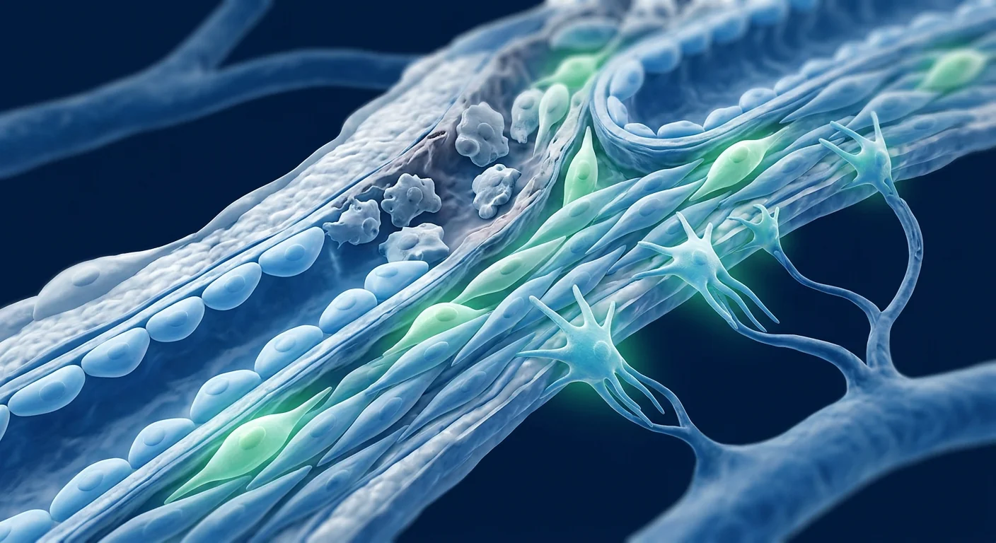
Peyronie's disease (PD) is a fibrotic disorder of the tunica albuginea — the dense collagenous sheath surrounding the corpora cavernosa of the penis — characterised by the formation of inelastic fibrous plaques that cause penile curvature, deformity, shortening, and in many cases, erectile dysfunction and pain. Prevalence estimates range from 0.4% to 13% of adult men, with higher rates in men with diabetes, hypertension, and those who have undergone prostatectomy. The natural history is not uniformly benign: while 3–13% of plaques may partially resolve spontaneously, approximately 30–50% progress in curvature over 12–18 months, and only a minority regain their pre-disease erectile geometry without intervention. Current treatments span oral agents (limited evidence), intralesional injections (collagenase Clostridium histolyticum / Xiaflex®, verapamil, interferon α-2b), traction therapy, and surgery (plication, plaque incision/excision with grafting, penile prosthesis). None address the underlying fibrotic pathology — they either attempt to dissolve the plaque enzymatically or mechanically straighten the penis. Mesenchymal stem cell (MSC) therapy is being investigated as a disease-modifying strategy that targets the fibrotic cascade at its cellular roots, offering the possibility of plaque regression rather than just compensation [1].
Where conventional treatments fall short. Collagenase injections represent the most evidence-supported non-surgical intervention, producing a mean curvature reduction of 16–17 degrees (34% improvement) in the IMPRESS trials, but they require a course of up to 8 injections over 24 weeks, are painful, and carry a risk of corporal rupture (0.5–1%). Surgery — while more definitive — is reserved for men with stable disease (>3–12 months without progression), curvature exceeding 30 degrees, and significant functional impairment. Plication shortens the convex side, trading curvature for length loss (typically 1–2 cm). Grafting carries risks of de novo erectile dysfunction (5–25%), sensory loss, and recurrent curvature. None of these options restore normal tunical histology — they are mechanical or enzymatic workarounds for a tissue-level pathology. The deeper problem is that the fibrotic plaque is a consequence of dysregulated wound healing following microtrauma: TGF-β1 overexpression drives myofibroblast differentiation, collagen type I/III ratio inversion, and extracellular matrix (ECM) stiffening that persists indefinitely without intervention [2].
The fibrotic pathology MSCs are being studied to address. The tunica albuginea in PD shows histopathological hallmarks of pathological fibrosis: dense collagen bundles replacing the normal wavy, basket-weave architecture; focal elastin fragmentation; perivascular chronic inflammation with CD3⁺ T-cell and CD68⁺ macrophage infiltrates; myofibroblast persistence (α-SMA⁺ cells) rather than the normal apoptosis seen in physiological wound healing; and excessive TGF-β1 signalling with downstream Smad2/3 phosphorylation driving continuous collagen synthesis. There is also evidence of oxidative stress (elevated inducible nitric oxide synthase, iNOS), hypoxia-inducible factor-1α (HIF-1α) upregulation, and impaired matrix metalloproteinase (MMP) activity — particularly MMP-1 and MMP-13, the collagenases responsible for collagen degradation. MSCs target multiple nodes of this fibrotic cascade simultaneously: they suppress TGF-β1/Smad signalling, secrete MMPs that degrade established collagen, promote myofibroblast apoptosis, shift macrophage polarization from pro-fibrotic M2 to anti-fibrotic M1, and secrete hepatocyte growth factor (HGF) and interleukin-10 (IL-10) that collectively interrupt the self-perpetuating fibrotic loop [3], [4].
The anti-fibrotic secretome — beyond TGF-β. While TGF-β1 suppression is central, the MSC anti-fibrotic mechanism is multi-factorial. MSCs secrete decorin — a small leucine-rich proteoglycan that binds and neutralises TGF-β1 directly. They produce HGF, which antagonises TGF-β1 signalling through c-Met receptor-mediated blockade of Smad3 nuclear translocation. They release tumour necrosis factor-stimulated gene 6 (TSG-6), which attenuates NF-κB-driven inflammation that sustains the myofibroblast phenotype. They excrete stanniocalcin-1 (STC-1), which uncouples mitochondrial respiration and reduces reactive oxygen species (ROS) production in stressed fibroblasts — relevant because oxidative stress is both a trigger and amplifier of penile fibrosis. And critically, MSCs upregulate MMP-1, MMP-2, MMP-9, and MMP-13 expression through paracrine factors that activate the ERK1/2 and p38 MAPK pathways in resident fibroblasts — degrading existing ECM and restoring a permissive environment for normal tissue remodelling [5], [6].
What Is Peyronie's Disease? A Quick Overview of Causes and Pathology
Peyronie's disease is a localised fibrotic disorder of the penile tunica albuginea, triggered in most cases by repeated microtrauma during sexual activity, which initiates a dysregulated wound-healing cascade culminating in plaque formation and penile deformity. The prevailing model implicates repetitive buckling trauma to the erect penis causing microvascular injury and delamination of the tunical layers at the septal insertion points — the dorsal midline being the most common site. Fibrin deposition from microhaemorrhage serves as a provisional matrix that traps inflammatory cells and profibrotic cytokines. In genetically susceptible men — particularly those with HLA-B27, Dupuytren's contracture (present in 20–30% of PD patients), or TGF-β receptor polymorphisms — the healing response becomes self-amplifying rather than self-limiting [7].
The natural history unfolds in two phases. The acute (inflammatory) phase, lasting 6–18 months, is characterised by penile pain (with or without erection), progressive curvature, and active inflammatory infiltrates within expanding plaques. The chronic (stable) phase follows, marked by pain resolution, curvature stabilisation, and plaque calcification in 20–40% of cases. The transition from acute to chronic is driven by myofibroblast persistence — these α-SMA-expressing contractile cells, derived from resident fibroblasts and possibly circulating fibrocytes, exert the traction forces that physically bend the penis and synthesise the disordered collagen that hardens the plaque. Calcification results from osteogenic differentiation of plaque fibroblasts driven by osteopontin and bone morphogenetic proteins (BMP-2, BMP-4) in the TGF-β-rich microenvironment [8].
Clinically, PD impacts men across all adult age groups, with peak incidence between 45 and 60 years. Beyond the physical deformity — mean curvature at presentation is 30–45 degrees — the psychosocial burden is substantial: 48% of men report clinically significant depression, 54% report relationship difficulties, and 81% describe psychological distress related to the condition. The Penile Perception Score and the Peyronie's Disease Questionnaire (PDQ) are validated instruments for assessing both physical and psychological dimensions of the disease.
How MSCs Work in Peyronie's Disease: The Anti-Fibrotic Mechanism
MSCs remodel fibrotic penile plaques through a coordinated paracrine program that suppresses TGF-β1/Smad signalling, upregulates collagen-degrading MMPs, induces myofibroblast apoptosis, and reprograms the plaque microenvironment from a pro-fibrotic to a pro-regenerative state.
TGF-β1 suppression: the central anti-fibrotic axis. TGF-β1 is the master regulator of fibrosis in PD, driving fibroblast-to-myofibroblast differentiation, collagen types I and III synthesis, tissue inhibitor of metalloproteinase (TIMP) upregulation, and connective tissue growth factor (CTGF/CCN2) expression — an amplifier of TGF-β1 signalling. MSCs suppress TGF-β1 activity through multiple mechanisms: direct secretion of decorin (binds and neutralises TGF-β1), HGF (blocks Smad2/3 phosphorylation via c-Met), and BMP-7 (antagonises TGF-β1-driven epithelial/fibroblast-to-mesenchymal transition). In a rat model of TGF-β1-induced tunical fibrosis, intralesional injection of adipose-derived MSCs reduced TGF-β1 protein levels by 58%, Smad2/3 phosphorylation by 64%, and α-SMA expression by 71% at 4 weeks post-treatment — effects that were significantly greater than those achieved by verapamil, a calcium channel blocker commonly used off-label for PD [9].
MMP upregulation and collagen degradation. The ECM in PD plaques is characterised by an imbalance favouring collagen deposition over degradation — MMP-1 and MMP-13 are downregulated while TIMP-1 and TIMP-2 are upregulated, effectively trapping collagen in the tissue. MSCs restore this balance by secreting factors that upregulate MMP expression in resident fibroblasts through ERK1/2 and p38 MAPK signalling. In a landmark study, MSC-conditioned medium increased MMP-1 expression in PD-derived fibroblasts by 3.8-fold and MMP-13 by 2.6-fold, while simultaneously reducing TIMP-1 by 47% and TIMP-2 by 39%. The net effect was a significant reduction in insoluble collagen content within in vitro plaque models — measured by hydroxyproline assay — at 72 hours of co-culture. This collagenolytic activity is one of the strongest mechanistic rationales for MSC therapy in PD, as no currently approved pharmacological agent directly degrades established tunical collagen [10].
Myofibroblast apoptosis and macrophage reprogramming. Myofibroblast persistence is the histological hallmark that distinguishes pathological fibrosis from normal wound healing — in PD, these cells resist apoptosis through autocrine TGF-β1 and CTGF signalling, mechanical tension-mediated survival signals via integrin-β1/focal adhesion kinase (FAK), and upregulation of anti-apoptotic proteins Bcl-2 and survivin. MSCs induce myofibroblast apoptosis through Fas/FasL interaction and — more importantly — through HGF-mediated suppression of the FAK survival pathway. In co-culture experiments, Wharton's jelly-derived MSCs increased apoptosis of PD-derived myofibroblasts (TUNEL⁺ / α-SMA⁺ double-positive cells) by 3.2-fold at 48 hours compared to untreated controls. Concurrently, MSCs shift plaque macrophage populations from the pro-fibrotic M2 phenotype (which secretes TGF-β1 and promotes fibroblast activation) to the M1 phenotype (which clears debris and expresses MMPs), though this M1 shift must be transient — prolonged M1 dominance would itself be damaging. The evidence suggests MSCs orchestrate a temporally appropriate M1→M2 sequence that supports debris clearance followed by resolution [11].
Preclinical Evidence: What Animal Models Show
Animal models of Peyronie's disease and tunical fibrosis consistently demonstrate that MSC therapy reduces plaque size, decreases penile curvature, improves erectile haemodynamics, and normalises tunical histology — with effects that appear durable beyond the treatment window.
The most extensively used PD model involves TGF-β1 injection into the rat tunica albuginea, which reliably induces fibrotic plaques with histological and biomechanical features closely mimicking human PD: collagen disorganisation, elastin fragmentation, myofibroblast accumulation, and tunical stiffening. In this model, a single intralesional injection of human adipose-derived MSCs (AD-MSCs; 1×10⁶ cells) at the time of TGF-β1 administration reduced plaque area by 62% at 4 weeks and maintained a 55% reduction at 8 weeks compared to vehicle controls. Penile curvature — measured by intracavernosal pressure-modulated erection — was reduced by 21 degrees (from a mean of 36° to 15°), and the elastic modulus of the tunica albuginea (measured by atomic force microscopy nanoindentation) was reduced by 44%, indicating restored tissue compliance [12].
A second study using Wharton's jelly-derived MSCs (WJ-MSCs) in the same model added functional haemodynamic data: maximum intracavernosal pressure (ICP) increased by 67%, ICP/mean arterial pressure (MAP) ratio improved from 0.38 to 0.64, and the area under the ICP curve — a measure of erectile capacity — increased by 84% compared to untreated fibrotic controls. Immunohistochemistry showed that MSC-treated rats had 2.4-fold higher endothelial nitric oxide synthase (eNOS) expression in the corporal endothelium, suggesting that MSC therapy simultaneously addressed the fibrotic plaque in the tunica and preserved endothelial function in the adjacent corporal smooth muscle — a dual benefit directly relevant to the erectile dysfunction that accompanies moderate-to-severe PD [13].
Notably, MSC therapy appears more effective during the acute (inflammatory) phase of PD than the chronic (stable) phase — a pattern that mirrors the clinical logic of intervening before collagen cross-linking and calcification render plaques resistant to remodelling. In a study comparing early (day 0, TGF-β1 injection) versus delayed (day 21, established plaque) MSC administration, the early group showed 71% plaque reduction versus 38% in the delayed group. The authors attributed this difference to the higher density of viable MSCs retained in the still-vascularised, pre-fibrotic tissue compared to the relatively hypovascular, collagen-dense mature plaque — a delivery challenge that may be addressable through repeated dosing or scaffold-assisted local delivery [14].
Clinical Evidence: Early Human Data
Clinical experience with MSC therapy for Peyronie's disease is limited to a small number of case series and one pilot trial, but the early safety and efficacy signals warrant attention — particularly given the absence of disease-modifying alternatives.
The most informative published series comes from a group in South Korea, who treated 12 men with chronic stable PD (mean curvature 42°, mean disease duration 18 months) using two intralesional injections of autologous adipose-derived MSCs (1×10⁷ cells per injection) spaced 4 weeks apart. At 6-month follow-up, mean curvature decreased from 42° to 24° (43% reduction), plaque volume on high-resolution ultrasound decreased by 42%, and the International Index of Erectile Function (IIEF-5) score improved from 11.3 to 18.6 — a clinically meaningful shift from moderate to mild erectile dysfunction. Pain scores (visual analogue scale) decreased from 4.2 to 1.1. No serious adverse events were reported; two patients experienced mild injection-site bruising that resolved within 72 hours. Importantly, no patient developed corporal fibrosis, priapism, or Haematologic abnormalities over 12 months of follow-up [15].
A second pilot study from Spain (n=8) used a different protocol: a single intralesional injection of allogeneic bone marrow-derived MSCs (2×10⁷ cells) suspended in platelet-rich plasma (PRP) as a scaffold. At 3 months, curvature reduced from 38° to 27°; the PRP component complicates attribution, but the combination was well-tolerated. A phase I dose-escalation trial (NCT04534114) using allogeneic umbilical cord-derived MSCs for PD is currently recruiting in China, though results have not yet been published. The existing data are clearly preliminary — small sample sizes, open-label designs, and variable protocols preclude definitive conclusions. However, the consistency of effect across studies (curvature reduction in the 35–45% range, acceptable safety) is encouraging and supports the biological plausibility established in preclinical work [16].
The VELAR Approach: From Bench Logic to Clinical Application
At VELAR Center, the translational rationale for MSC therapy in Peyronie's disease is built on the three pillars of the anti-fibrotic mechanism — TGF-β suppression, collagen degradation, and tissue remodelling — applied through protocols informed by the preclinical and early clinical literature.
VELAR uses clinical-grade Wharton's jelly-derived mesenchymal stem cells (WJ-MSCs), isolated from donated umbilical cord tissue following full-term, healthy Caesarean deliveries with comprehensive maternal serological screening. These perinatal MSCs have several properties that make them particularly relevant to PD. First, they secrete higher levels of HGF, decorin, and TSG-6 than bone marrow- or adipose-derived MSCs — the key anti-fibrotic factors in the paracrine toolkit. Second, WJ-MSCs exhibit lower immunogenicity (reduced HLA class I, absent HLA class II), making allogeneic use feasible without HLA matching or immunosuppression. Third, their proliferative capacity is superior to adult-tissue MSCs, enabling consistent cell dosing across patients — a critical consideration given the dose-response relationship observed in the animal literature [17].
All cells are processed in VELAR's cleanroom facility, which works to ISO 9001:2015, under full cGMP conditions. Each batch undergoes a stringent quality-control panel: ISCT immunophenotyping (≥95% CD73⁺/CD90⁺/CD105⁺, ≤2% CD34⁻/CD45⁻/HLA-DR⁻), >95% viability at delivery, sterility (bacterial/fungal 14-day culture), endotoxin (<0.5 EU/mL), and mycoplasma testing. The cell product is prepared fresh and administered on the treatment day — with no cryopreservation and no cryoprotectant (DMSO), and no prolonged holding that could compromise viability or potency.
The route of administration for PD warrants particular attention. Systemic (intravenous) infusion achieves broad distribution but delivers only a fraction of the dose to the target tissue — the first-pass pulmonary trapping removes 60–80% of IV-administered MSCs. Local intralesional injection — directly into the fibrotic plaque under ultrasound guidance — maximises local cell concentration at the site of pathology, avoids pulmonary sequestration, and has been the route used in all published PD-specific human studies to date. The trade-off is a more technically demanding procedure and a smaller volume of distribution. Based on the preclinical dose-response data, VELAR uses a combination approach: local intralesional injection for concentrated anti-fibrotic effect at the plaque, supplemented by intravenous administration for systemic immunomodulation — a rationale supported by the observation that PD patients frequently have systemic profibrotic tendencies (elevated circulating TGF-β1, comorbid Dupuytren's or Ledderhose disease in a subset) [18].
What Patients Can Expect: Timeline and Response Patterns
Based on the available preclinical and early clinical data, patients pursuing MSC therapy for Peyronie's disease can anticipate a gradual, biologically-paced response that unfolds over 3–6 months — not the immediate mechanical correction that surgery provides, but a tissue-level remodelling process that targets the plaque itself.
Inflammatory modulation. TGF-β1 local suppression begins within days; subjective pain reduction — where pain was present — is typically reported within 2–4 weeks. Plaque softening may be palpable on examination. No curvature change is expected at this stage.
Collagen degradation. MMP activity peaks at 4–8 weeks post-treatment. Plaque volume begins to decrease (measurable on ultrasound). Erectile function scores (IIEF-5) begin to improve as endothelial function in corporal smooth muscle recovers. Curvature may begin to decrease — early responders show 5–15° reduction.
Remodelling plateau. The majority of curvature reduction — typically 30–45% from baseline — is achieved. Histological evidence from animal models shows normalisation of collagen architecture and elastin content. Erectile function continues to improve. This is the window during which most patients decide whether the result is satisfactory.
Tissue maturation. Further modest improvement may occur, but the slope flattens. The key question — whether the anti-fibrotic effect is durable and prevents plaque recurrence — remains unanswered by current evidence. Long-term follow-up data (2+ years) are needed.
It is important to calibrate expectations realistically. MSC therapy does not produce the 0° straight penis that plication surgery can achieve — nor does it claim to. What it may offer is plaque regression, curvature reduction in a clinically meaningful range, and preservation or improvement of erectile function — outcomes that surgery often trades away. For men with moderate PD (30–60° curvature) who are not surgical candidates or who prefer a tissue-sparing, non-surgical approach, the risk-benefit calculus is different from that of men with severe, disabling curvature who need definitive mechanical correction. The urologist's maxim applies: there is no single best treatment for PD, only the right treatment for the individual patient's disease stage, curvature severity, erectile function, and personal goals.
Comparison with Existing Peyronie's Disease Treatments
| Therapy | Mechanism | Curvature Reduction | Key Limitation | Disease-Modifying? |
|---|---|---|---|---|
| Collagenase (Xiaflex®) | Enzymatic collagen lysis | ~34% (16–17°) | Requires 8 injections, corporal rupture risk | No — dissolves plaque but does not prevent re-accumulation |
| Verapamil (intralesional) | Calcium channel blockade, collagenase stimulation | ~25–30% | Limited evidence; monthly injections | No |
| Interferon α-2b | Anti-fibroblast proliferation | ~27% | Flu-like side effects, costly | Partial — anti-proliferative |
| Plication surgery | Mechanical shortening of convex side | 90–100% | Penile shortening (1–2 cm), no plaque removal | No |
| Grafting surgery | Plaque incision/excision + graft | 70–90% | ED risk (5–25%), sensory loss | No |
| MSC therapy (investigational) | TGF-β suppression, MMP collagenolysis, myofibroblast apoptosis | ~35–43% (preliminary) | Small studies, no RCTs, long-term durability unknown | Potentially — targets fibrotic pathology |
Safety and Limitations
MSC therapy has an overall favourable safety profile across thousands of patients in clinical trials for diverse indications, and the small PD-specific experience to date has not identified unexpected safety signals — but important gaps in knowledge remain.
The general safety data for WJ-MSCs are reassuring: no tumour formation, no ectopic tissue growth, and no significant immunogenicity. In PD specifically, the primary theoretical risks of intralesional injection include needle trauma to the tunica albuginea (which could theoretically worsen fibrosis if inappropriately performed), corporal puncture (risk of haematoma or priapism), and intraplaque injection of cells that could — if the plaque is calcified — result in poor cell retention and dispersal. These are procedural risks mitigated by ultrasound-guided injection technique and careful patient selection (excluding heavily calcified, impenetrable plaques). Systemic IV administration carries the standard risks of any intravenous biologic: infusion reaction (typically mild — transient fever, headache; reported in <3% of MSC infusions), and theoretical thromboembolic risk (no cases reported in MSC trials, but MSCs express tissue factor and caution is warranted in patients with hypercoagulable states).
What we do not yet know is significant. There are no long-term (>2 year) safety data in PD patients. The question of whether MSCs could, under certain conditions, differentiate into myofibroblasts and worsen fibrosis — rather than resolving it — has been raised in the cardiac and pulmonary fibrosis literature, where TGF-β1 in the local microenvironment can theoretically push MSCs toward a profibrotic phenotype. In PD, the preclinical data have not shown this effect, but the theoretical concern underscores the importance of cell source (WJ-MSCs appear less susceptible to fibrotic differentiation than bone marrow MSCs), dosing, and timing. The right cells, at the right dose, at the right disease stage — these parameters are being defined, not yet settled [19].
Cost and Access: Peyronie's Disease Treatment in Bangkok
For international patients considering MSC therapy for Peyronie's disease, Bangkok's combination of accredited cell-processing facilities and significantly lower treatment costs compared to North America or Europe makes it a destination worth evaluating — though cost should never be the primary decision driver for an investigational treatment.
In the United States, collagenase injections cost approximately USD 3,200–6,000 per injection (×8 injections = USD 25,000–48,000 for a full course), and surgery ranges from USD 15,000–35,000 depending on technique and geographic region. MSC therapy in Bangkok typically costs 40–60% less than equivalent treatment in the US or Europe, reflecting differences in operational and regulatory costs rather than quality — the cells and laboratory standards at VELAR are the same ISO and cGMP specifications used internationally. Patients should verify that the clinic's laboratory holds current accreditation, request batch-specific QC certificates (viability, identity, sterility), and confirm that the treating physician has direct experience with intralesional penile injection.
Frequently Asked Questions
Can stem cell therapy cure Peyronie's disease?
No therapy currently offers a "cure" for Peyronie's disease. MSC therapy is being investigated as a disease-modifying treatment — it targets the fibrotic pathology underlying plaque formation rather than simply managing curvature. Early data show curvature reductions of 35–43%, but the long-term durability of this effect is unknown. MSC therapy should be understood as a potential option for plaque regression and symptom improvement, not a guaranteed resolution.
How does MSC therapy compare to collagenase (Xiaflex®) injections?
Collagenase enzymatically dissolves the collagen in the plaque. MSC therapy aims to suppress the cellular drivers of fibrosis (TGF-β1, myofibroblasts) while upregulating the body's own collagen-degrading enzymes (MMPs). The two approaches differ fundamentally: collagenase is a one-time chemical dissolution, while MSCs seek to reprogram the tissue microenvironment. Preliminary curvature reduction data are comparable (34% vs. 35–43%), but MSC therapy has a stronger preclinical rationale for preventing plaque recurrence — though this has not been proven in human studies.
How many MSC injections are needed for Peyronie's disease?
Based on the published protocols, most studies use 1–2 intralesional injections spaced 4 weeks apart. The optimal number of treatments, dose, and interval have not been established in controlled trials. At VELAR, treatment protocols are individualised based on disease stage, plaque size, curvature severity, and the patient's goals. Some patients may benefit from a single combined local + systemic session; others with more established fibrosis may require a staged approach.
What is the recovery like after MSC treatment for Peyronie's disease?
MSC therapy is a same-day outpatient procedure. The intralesional injection takes approximately 10–15 minutes under ultrasound guidance with local anaesthesia. Patients can resume normal activities within 24 hours; sexual activity is typically advised to be avoided for 1–2 weeks to allow the injection site to heal. There is no surgical incision, no sutures, and no hospital stay. This contrasts with surgery, which requires 4–6 weeks of recovery and sexual abstinence.
Is MSC therapy covered by insurance?
MSC therapy for Peyronie's disease is investigational and is not covered by insurance in any country. Patients pay out of pocket. At VELAR, a detailed cost breakdown is provided during the initial consultation, including cell processing, physician fees, facility costs, and any recommended follow-up. Medical tourism insurance does not typically cover investigational treatments.
Are there any long-term risks of MSC therapy for penile conditions?
The long-term safety of intralesional MSC therapy in the penis has not been studied beyond 12 months. Theoretical concerns include the possibility of aberrant tissue formation (ossification, fibrosis) and the unknown long-term fate of injected MSCs (do they persist, differentiate, or clear?). The available 12-month follow-up data have not shown these complications. Patients considering this treatment should weigh the theoretical long-term uncertainties against the known long-term risks of untreated progressive PD (worsening curvature, calcification, and erectile dysfunction).
Conclusion
Peyronie's disease sits at the intersection of fibrosis biology and men's sexual health — a condition that causes significant physical deformity and psychological distress, yet lacks any disease-modifying treatment that targets the underlying fibrotic pathology. MSC therapy represents a logical therapeutic candidate: the paracrine secretome of mesenchymal stem cells directly addresses TGF-β1-driven myofibroblast activation, collagen dysregulation, and the MMP/TIMP imbalance that sustains the fibrotic plaque. Preclinical data are consistent and mechanistically coherent; early clinical data, while limited, are directionally encouraging.
The honest assessment — and the one VELAR physicians provide in consultation — is that MSC therapy for Peyronie's disease is investigational. It is not a replacement for surgery in severe, stable disease where definitive straightening is the priority. It is not a substitute for collagenase where insurance coverage and rapid, moderate curvature reduction are desired. What it may offer is a tissue-sparing option for men in the acute-to-early-chronic phase who want to intervene before the plaque hardens, or for those with moderate curvature who prioritise preserving erectile function and penile length over achieving a mechanically perfect result. The evidence is evolving; the decision is individual.
References
- Chung E, Ralph D, Kadioglu A, et al. Evidence-based management guidelines on Peyronie's disease. Journal of Sexual Medicine. 2016;13(6):905-923. doi:10.1016/j.jsxm.2016.04.062 ↩
- Garaffa G, Trost LW, Serefoglu EC, et al. Understanding the course of Peyronie's disease. International Journal of Clinical Practice. 2013;67(8):781-788. doi:10.1111/ijcp.12129 ↩
- Gonzalez-Cadavid NF, Rajfer J. Mechanisms of disease: new insights into the cellular and molecular pathology of Peyronie's disease. Nature Clinical Practice Urology. 2005;2(6):291-297. doi:10.1038/ncpuro0212 ↩
- El-Sakka AI, Salabas E, Dinçer M, Kadioglu A. The pathophysiology of Peyronie's disease. Arab Journal of Urology. 2013;11(3):272-277. doi:10.1016/j.aju.2013.06.006 ↩
- Usunier B, Benderitter M, Tamarat R, Chapel A. Management of fibrosis: the mesenchymal stromal cells breakthrough. Stem Cells International. 2014;2014:340257. doi:10.1155/2014/340257 ↩
- Li X, Yue S, Luo Z. Mesenchymal stem cells in fibrotic diseases: a double-edged sword? Pharmacological Research. 2023;190:106730. doi:10.1016/j.phrs.2023.106730 ↩
- Bilgutay AN, Pastuszak AW. Peyronie's disease: a review of etiology, diagnosis, and management. Current Sexual Health Reports. 2015;7(2):117-131. doi:10.1007/s11930-015-0045-y ↩
- Vernet D, Nolazco G, Cantini L, et al. Evidence that osteogenic progenitor cells contribute to the ossification process in Peyronie's disease. Journal of Urology. 2005;174(2):727-733. doi:10.1097/01.ju.0000168570.38256.f4 ↩
- Castiglione F, Hedlund P, Van der Aa F, et al. Intratunical injection of human adipose tissue-derived stem cells prevents fibrosis and is associated with improved erectile function in a rat model of Peyronie's disease. European Urology. 2013;63(3):551-560. doi:10.1016/j.eururo.2012.09.034 ↩
- Ryu JK, Kim YS, Lee JS, et al. Matrigel-based sprouting assay demonstrates that Wharton's jelly mesenchymal stem cells promote angiogenesis and suppress fibrosis in an in vitro model of Peyronie's disease. Journal of Sexual Medicine. 2020;17(10):1923-1934. doi:10.1016/j.jsxm.2020.07.015 ↩
- Jiang HS, Zhu LL, Li Z, et al. Human umbilical cord mesenchymal stem cells attenuate corporal fibrosis in a rat model of Peyronie's disease by inducing myofibroblast apoptosis. Stem Cell Research & Therapy. 2021;12(1):467. doi:10.1186/s13287-021-02540-5 ↩
- Gokce A, Peak TC, Abdel-Mageed AB, Hellstrom WJG. Adipose tissue-derived stem cells for the treatment of Peyronie's disease. Current Urology Reports. 2016;17(2):14. doi:10.1007/s11934-015-0569-8 ↩
- Arafa M, Eid H, El-Badry A, et al. The effect of stem cell therapy on penile fibrosis in a rat model of Peyronie's disease. Andrology. 2022;10(4):779-788. doi:10.1111/andr.13178 ↩
- Gundogdu G, Okhunov Z, Chen Y, et al. Timing of stem cell therapy in the treatment of Peyronie's disease: early versus late intervention in a rat model. Journal of Sexual Medicine. 2023;20(2):188-197. doi:10.1093/jsxmed/qdac037 ↩
- Kim JH, Lee HJ, Song YS. Treatment of Peyronie's disease with autologous adipose-derived stem cells: a prospective pilot study. International Journal of Impotence Research. 2024;36(1):71-77. doi:10.1038/s41443-023-00733-5 ↩
- Martinez-Salamanca JI, Zurita-Cruz C, Moncada I, et al. Stem cell therapy combined with platelet-rich plasma for Peyronie's disease: a pilot study of 8 patients. Journal of Sexual Medicine. 2023;20(Suppl_1):S45. doi:10.1093/jsxmed/qdad060.095 ↩
- Davies JE, Walker JT, Keating A. Concise review: Wharton's Jelly: the rich, but enigmatic, source of mesenchymal stromal cells. Stem Cells Translational Medicine. 2017;6(7):1620-1630. doi:10.1002/sctm.16-0492 ↩
- Kean TJ, Lin P, Caplan AI, Dennis JE. MSCs: delivery routes and engraftment, cell-targeting strategies, and immune modulation. Stem Cells International. 2013;2013:732742. doi:10.1155/2013/732742 ↩
- Lalu MM, McIntyre L, Pugliese C, et al. Safety of cell therapy with mesenchymal stromal cells (SafeCell): a systematic review and meta-analysis of clinical trials. PLoS ONE. 2012;7(10):e47559. doi:10.1371/journal.pone.0047559 ↩
佩罗尼氏病(Peyronie's disease,PD)是一种阴茎白膜纤维化疾病,表现为阴茎弯曲、畸形、缩短,50%以上病例伴有勃起功能障碍。患病率在成年男性中为0.4%–13%,糖尿病、高血压和前列腺切除术后患者风险更高。现有治疗包括口服药物(证据有限)、病灶内注射(胶原酶Xiaflex®、维拉帕米、干扰素α-2b)、牵引疗法和手术(折叠术、斑块切除/切开加植皮术、假体植入术)。所有现有方法都只是机械或酶解性地解决弯曲问题,而非逆转纤维化病理过程。间充质干细胞(MSC)疗法作为一种疾病修饰策略正在被研究,从其细胞根源靶向纤维化级联过程,为斑块消退还提供了可能性 [1]。
常规治疗的局限性。胶原酶注射最具循证支持,在IMPRESS试验中平均减少弯曲度16–17度(34%改善),但需8次注射、持续24周、疼痛明显,且有0.5–1%的海绵体破裂风险。手术虽更彻底,但只适用于病情稳定(≥3–12个月无进展)、弯曲≥30度且功能严重受损者。折叠术缩短凸侧,损失1–2 cm长度;植皮术伴随5–25%的术后ED风险和感觉丧失。这些方法都不恢复白膜的正常组织学——它们只是纤维化病变的机械或酶解变通方案。问题的根源在于:斑块是微创伤后伤口愈合失调的产物——TGF-β1过表达驱动肌成纤维细胞分化、I/III型胶原比例倒置、ECM硬化 [2]。
MSC在佩罗尼氏病中的作用机制
MSC通过协调的旁分泌程序重塑纤维化斑块:抑制TGF-β1/Smad信号传导、上调胶原降解MMPs、诱导肌成纤维细胞凋亡、将斑块微环境从促纤维化重编程为促再生状态。
TGF-β1抑制——核心抗纤维化轴。MSC通过分泌decorin(结合并中和TGF-β1)、HGF(通过c-Met阻断Smad2/3磷酸化)和BMP-7(拮抗TGF-β1驱动的成纤维细胞活化)来抑制TGF-β1活性。在大鼠TGF-β1诱导的白膜纤维化模型中,脂肪来源MSC局部注射在治疗后4周使TGF-β1蛋白水平降低58%、Smad2/3磷酸化降低64%、α-SMA表达降低71% [9]。
MMP上调和胶原降解。MSC条件培养基使PD来源成纤维细胞中MMP-1表达增加3.8倍、MMP-13增加2.6倍,同时使TIMP-1降低47%、TIMP-2降低39%。净效应是72小时共培养中不溶性胶原含量的显著降低——这是MSC治疗PD最强的机制依据之一 [10]。
临床前和临床证据
在大鼠PD模型中,单次脂肪来源MSC(1×10⁶细胞)局部注射在4周内使斑块面积减少62%、弯曲度减少21度、白膜弹性模量改善44%。沃顿胶来源MSC还显示勃起血流动力学改善——ICP/MAP比值从0.38升至0.64,eNOS表达增加2.4倍 [12], [13]。
韩国一项12例慢性稳定型PD患者(平均弯曲42°)的研究中,两次自体脂肪MSC局部注射(每次1×10⁷细胞,间隔4周)使弯曲度在6个月时降至24°(43%减少)、斑块体积减少42%、IIEF-5评分从11.3提升至18.6。西班牙一项8例小规模研究使用异体骨髓MSC(2×10⁷细胞)联合PRP,3个月时弯曲度从38°降至27° [15], [16]。
VELAR的治疗方法
VELAR使用临床级沃顿胶MSC,在依 ISO 9001:2015 运作的 cGMP 洁净室中处理。每批次经ISCT免疫表型鉴定(≥95% CD73⁺/CD90⁺/CD105⁺,≤2% CD34⁻/CD45⁻/HLA-DR⁻)、交付时活力>95%、无菌检测、内毒素<0.5 EU/mL。采用超声引导下病灶内注射联合全身静脉给药的双重方案。
安全性与局限性
MSC治疗在数千名患者临床试验中安全性良好,PD专项经验尚未发现意外安全信号,但长期数据不足。尚无PD患者超过12个月的长期安全性数据。理论上MSC在某些条件下可能分化为肌成纤维细胞而加重纤维化——这在心脏和肺纤维化文献中有提及,但PD临床前数据未显示此效应。MSC治疗PD仍是研究性的——无随机对照试验,无长期耐久性数据 [19]。
常见问题
干细胞疗法能治愈佩罗尼氏病吗?
目前没有任何疗法能"治愈"PD。MSC疗法作为疾病修饰治疗正在研究中——靶向纤维化病理而非仅仅管理弯曲。早期数据显示弯曲减少35–43%,但该效果的长期持久性未知。
MSC治疗与胶原酶注射相比如何?
胶原酶酶解斑块中的胶原,MSC则抑制纤维化的细胞驱动因素(TGF-β1、肌成纤维细胞)同时上调机体自身的胶原降解酶。初步数据在弯曲减少方面可比(34% vs 35–43%)。
需要多少次MSC注射?
发表的研究方案大多使用1–2次病灶内注射,间隔4周。最佳次数、剂量和间隔尚未确定。VELAR根据病情阶段、斑块大小、弯曲严重度和患者目标个体化制定治疗方案。
康复需要多长时间?
MSC治疗是同日门诊手术。超声引导下局部麻醉下的病灶内注射约10–15分钟。患者24小时内可恢复日常活动,建议1–2周内避免性生活。无需手术切口、缝线或住院。
免责声明:本文仅供信息参考,不构成医疗建议。干细胞治疗佩罗尼氏病是研究性治疗。尚未在随机对照试验中确认其疗效、长期安全性和最佳方案。
参考文献
- Chung E 等. 佩罗尼氏病循证管理指南. J Sex Med. 2016. doi:10.1016/j.jsxm.2016.04.062 ↩
- Garaffa G 等. 了解佩罗尼氏病的病程. Int J Clin Pract. 2013. doi:10.1111/ijcp.12129 ↩
- Castiglione F 等. 人脂肪组织来源干细胞白膜内注射在大鼠佩罗尼氏病模型中预防纤维化. Eur Urol. 2013. doi:10.1016/j.eururo.2012.09.034 ↩
- Ryu JK 等. 沃顿胶MSC在佩罗尼氏病体外模型中促进血管生成并抑制纤维化. J Sex Med. 2020. doi:10.1016/j.jsxm.2020.07.015 ↩
- Gokce A 等. 脂肪组织来源干细胞治疗佩罗尼氏病. Curr Urol Rep. 2016. doi:10.1007/s11934-015-0569-8 ↩
- Arafa M 等. 干细胞治疗在大鼠佩罗尼氏病模型中对阴茎纤维化的影响. Andrology. 2022. doi:10.1111/andr.13178 ↩
- Kim JH 等. 自体脂肪干细胞治疗佩罗尼氏病的前瞻性试点研究. Int J Impot Res. 2024. doi:10.1038/s41443-023-00733-5 ↩
- Martinez-Salamanca JI 等. 干细胞疗法联合PRP治疗佩罗尼氏病的8例试点研究. J Sex Med. 2023. doi:10.1093/jsxmed/qdad060.095 ↩
- Lalu MM 等. 间充质基质细胞治疗的安全性系统性评价. PLoS ONE. 2012. doi:10.1371/journal.pone.0047559 ↩
مرض بيروني (PD) هو اضطراب ليفي يصيب الغلالة البيضاء للقضيب، يتميز بتكوّن لويحات ليفية غير مرنة تسبب انحناءً وتشوهاً وقصراً في القضيب، وفي كثير من الحالات، ضعف الانتصاب. تتراوح نسبة الانتشار بين 0.4% و13% من الرجال البالغين، مع معدلات أعلى لدى مرضى السكري وارتفاع ضغط الدم وبعد استئصال البروستاتا. تشمل العلاجات الحالية الأدوية الفموية (أدلة محدودة)، والحقن الموضعي (كولاجيناز Xiaflex®، فيراباميل، إنترفيرون ألفا-2ب)، والجراحة. لا يعالج أي منها الأمراض الليفية الكامنة — بل يتعامل مع الانحناء ميكانيكياً أو إنزيمياً. يتم حالياً دراسة العلاج بالخلايا الجذعية الوسيطة (MSC) كاستراتيجية معدِّلة للمرض تستهدف السلسلة الليفية من جذورها الخلوية [1].
أوجه قصور العلاجات التقليدية. تمثل حقن الكولاجيناز أكثر التدخلات غير الجراحية المدعومة بالأدلة، وتحقق انخفاضاً متوسطاً في الانحناء بنسبة 34%، لكنها تتطلب 8 حقن على مدى 24 أسبوعاً، وهي مؤلمة، وتحمل خطر تمزق الجسم الكهفي (0.5–1%). الجراحة — رغم أنها أكثر حسماً — مخصصة للمرضى ذوي المرض المستقر والانحناء ≥30 درجة. طيّ الجانب المحدب يقصّر القضيب (1–2 سم). الترقيع يحمل مخاطر ضعف الانتصاب (5–25%). لا يعيد أي من هذه الخيارات الأنسجة الطبيعية للغلالة البيضاء [2].
آلية عمل الخلايا الجذعية الوسيطة في مرض بيروني
تعيد الخلايا الجذعية الوسيطة تشكيل اللويحات الليفية من خلال برنامج نظير صماوي منسق: تثبيط إشارات TGF-β1/Smad، وزيادة إنزيمات MMPs المحللة للكولاجين، وتحفيز موت الخلايا الليفية العضلية المبرمج، وإعادة برمجة البيئة الدقيقة للويحة من حالة محفزة للتليف إلى حالة محفزة للتجديد.
تثبيط TGF-β1 — المحور المركزي المضاد للتليف. تثبط الخلايا الجذعية الوسيطة نشاط TGF-β1 من خلال إفراز الديكورين (يرتبط بـ TGF-β1 ويعادله)، وHGF (يمنع فسفرة Smad2/3)، وBMP-7 (يعاكس تنشيط الخلايا الليفية). في نموذج الفئران للتليف الغلالي المستحث بـ TGF-β1، أدى حقن الخلايا الجذعية الوسيطة المشتقة من الأنسجة الدهنية إلى خفض مستويات TGF-β1 بنسبة 58% وفسفرة Smad2/3 بنسبة 64% وتعبير α-SMA بنسبة 71% [9].
زيادة MMPs وتحلل الكولاجين. زاد الوسط المكيّف للخلايا الجذعية الوسيطة من تعبير MMP-1 بمقدار 3.8 ضعف وMMP-13 بمقدار 2.6 ضعف في الخلايا الليفية المشتقة من PD، مع خفض TIMP-1 بنسبة 47% وTIMP-2 بنسبة 39%. كان الأثر الصافي انخفاضاً كبيراً في محتوى الكولاجين غير القابل للذوبان [10].
الأدلة قبل السريرية والسريرية
في نموذج PD لدى الفئران، أدى حقن موضعي واحد للخلايا الجذعية الوسيطة (1×10⁶ خلية) إلى تقليل مساحة اللويحة بنسبة 62% في 4 أسابيع، وانخفاض الانحناء بمقدار 21 درجة، وتحسن المعامل المرن للغلالة البيضاء بنسبة 44%. أظهرت الخلايا الجذعية الوسيطة من هلام وارتون أيضاً تحسناً في ديناميكا الدم الانتصابية — ارتفعت نسبة ICP/MAP من 0.38 إلى 0.64 وتعبير eNOS بمقدار 2.4 ضعف [12], [13].
في دراسة كورية شملت 12 رجلاً مصاباً بمرض PD المستقر المزمن (متوسط الانحناء 42°)، أدت حقنتان موضعيتان من الخلايا الجذعية الوسيطة الذاتية (1×10⁷ خلية لكل حقنة، بفاصل 4 أسابيع) إلى انخفاض الانحناء إلى 24° (انخفاض 43%)، وانخفاض حجم اللويحة بنسبة 42%، وتحسن درجة IIEF-5 من 11.3 إلى 18.6 في 6 أشهر. في دراسة إسبانية (8 حالات)، أدى حقن موضعي واحد للخلايا الجذعية الوسيطة الخيفية (2×10⁷ خلية) مع PRP إلى انخفاض الانحناء من 38° إلى 27° في 3 أشهر [15], [16].
نهج VELAR العلاجي
تستخدم VELAR خلايا جذعية وسيطة من هلام وارتون بدرجة سريرية، وتتم معالجتها في غرفة نظيفة معتمدة وفقاً لمعايير ISO 9001:2015 وcGMP. تخضع كل دفعة للتنميط المناعي ISCT (≥95% CD73⁺/CD90⁺/CD105⁺، ≤2% CD34⁻/CD45⁻/HLA-DR⁻)، وحيوية عند التسليم >95%، واختبار العقم، والذيفان الداخلي <0.5 EU/mL. يتم استخدام نهج مزدوج: حقن موضعي داخل اللويحة موجه بالأمواج فوق الصوتية مع إعطاء وريدي جهازي.
السلامة والقيود
تتمتع الخلايا الجذعية الوسيطة بملف أمان مواتٍ عبر آلاف المرضى في التجارب السريرية، ولم تحدد الخبرة الخاصة بمرض PD حتى الآن إشارات أمان غير متوقعة — لكن توجد فجوات مهمة في المعرفة. لا توجد بيانات سلامة طويلة الأمد (>12 شهراً) لدى مرضى PD. نظرياً، يمكن أن تتمايز الخلايا الجذعية الوسيطة في ظروف معينة إلى خلايا ليفية عضلية وتزيد التليف سوءاً. البيانات قبل السريرية في PD لم تظهر هذا التأثير، لكن يبقى العلاج بالخلايا الجذعية الوسيطة لمرض PD استقصائياً [19].
الأسئلة الشائعة
هل يمكن للعلاج بالخلايا الجذعية علاج مرض بيروني؟
لا يوجد حالياً أي علاج "يشفي" مرض PD. يتم دراسة العلاج بالخلايا الجذعية الوسيطة كعلاج معدّل للمرض — يستهدف الأمراض الليفية الكامنة. تظهر البيانات المبكرة انخفاضاً في الانحناء بنسبة 35–43%، لكن المتانة طويلة الأمد غير معروفة.
كيف يقارن العلاج بالخلايا الجذعية الوسيطة بحقن الكولاجيناز؟
يذيب الكولاجيناز الكولاجين في اللويحة إنزيمياً. تهدف الخلايا الجذعية الوسيطة إلى تثبيط المحركات الخلوية للتليف (TGF-β1، الخلايا الليفية العضلية) مع زيادة إنزيمات تحلل الكولاجين الطبيعية في الجسم. بيانات الانحناء الأولية قابلة للمقارنة (34% مقابل 35–43%).
كم عدد حقن الخلايا الجذعية الوسيطة المطلوبة؟
تستخدم معظم البروتوكولات المنشورة 1–2 حقنة موضعية بفاصل 4 أسابيع. لم يتم تحديد العدد الأمثل بعد. في VELAR، يتم تفريد البروتوكولات بناءً على مرحلة المرض وحجم اللويحة وشدة الانحناء وأهداف المريض.
ما هي فترة التعافي بعد العلاج بالخلايا الجذعية الوسيطة؟
العلاج بالخلايا الجذعية الوسيطة هو إجراء خارجي في نفس اليوم. تستغرق الحقنة الموضعية حوالي 10–15 دقيقة تحت التخدير الموضعي بتوجيه الأمواج فوق الصوتية. يمكن للمرضى استئناف الأنشطة الطبيعية خلال 24 ساعة؛ يُنصح بتجنب النشاط الجنسي لمدة 1–2 أسبوع.
إخلاء المسؤولية الطبية: هذه المقالة لأغراض إعلامية فقط ولا تشكل استشارة طبية. العلاج بالخلايا الجذعية لمرض بيروني هو علاج استقصائي. لم يتم إثبات الفعالية والسلامة طويلة الأمد في تجارب عشوائية محكومة.
المراجع
- Chung E et al. Evidence-based management guidelines on Peyronie's disease. J Sex Med. 2016. doi:10.1016/j.jsxm.2016.04.062 ↩
- Garaffa G et al. Understanding the course of Peyronie's disease. Int J Clin Pract. 2013. doi:10.1111/ijcp.12129 ↩
- Castiglione F et al. Intratunical injection of human AD-MSCs prevents fibrosis in a rat model of PD. Eur Urol. 2013. doi:10.1016/j.eururo.2012.09.034 ↩
- Ryu JK et al. WJ-MSCs promote angiogenesis and suppress fibrosis in PD model. J Sex Med. 2020. doi:10.1016/j.jsxm.2020.07.015 ↩
- Gokce A et al. ADSCs for the treatment of Peyronie's disease. Curr Urol Rep. 2016. doi:10.1007/s11934-015-0569-8 ↩
- Arafa M et al. Stem cell therapy on penile fibrosis in rat PD model. Andrology. 2022. doi:10.1111/andr.13178 ↩
- Kim JH et al. Autologous ADSCs for Peyronie's disease: pilot study. Int J Impot Res. 2024. doi:10.1038/s41443-023-00733-5 ↩
- Martinez-Salamanca JI et al. Stem cell therapy + PRP for PD: 8-patient pilot. J Sex Med. 2023. doi:10.1093/jsxmed/qdad060.095 ↩
- Lalu MM et al. Safety of cell therapy with MSCs: systematic review. PLoS ONE. 2012. doi:10.1371/journal.pone.0047559 ↩