Nephrotic syndrome is not a single disease — it is a clinical constellation defined by massive proteinuria (>3.5 g/day), hypoalbuminemia, oedema, and hyperlipidaemia. Underlying this syndrome are three major glomerular diseases: focal segmental glomerulosclerosis (FSGS), minimal change disease (MCD), and membranous nephropathy (MN). Together they account for the majority of adult nephrotic cases, with FSGS being the leading cause of end-stage renal disease among the primary glomerulopathies. [1][2]
Where conventional treatment falls short. First-line therapy for nephrotic syndrome relies on corticosteroids and calcineurin inhibitors (tacrolimus, cyclosporine) for MCD and FSGS, or rituximab and cyclophosphamide for membranous nephropathy. These agents achieve remission in 60–80% of MCD patients and 40–60% of FSGS patients initially — but relapses are frequent, steroid dependence is common, and long-term immunosuppression carries cumulative toxicity: infection, bone loss, glucose intolerance, and malignancy risk. For steroid-resistant FSGS — approximately 20–30% of cases — therapeutic options narrow dramatically, and progression to end-stage renal disease is almost inevitable without a kidney transplant. [3][4]
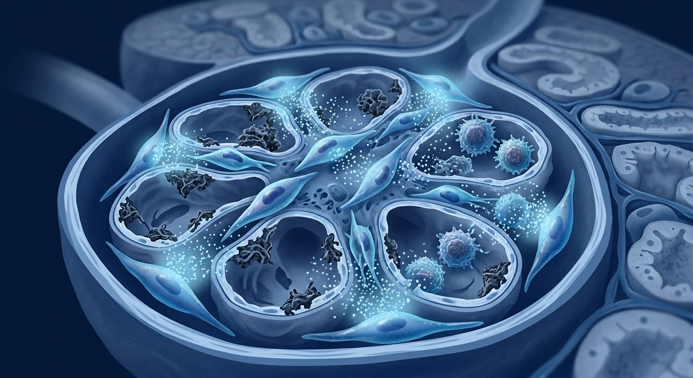
The deeper problem is podocyte injury. Podocytes are terminally differentiated epithelial cells that wrap glomerular capillaries with interdigitating foot processes, forming the final barrier to protein loss. In nephrotic syndrome — regardless of the initiating trigger (circulating permeability factors in FSGS, T-cell dysfunction in MCD, or anti-PLA2R autoantibodies in MN) — podocyte foot-process effacement is the universal histological finding. Damaged podocytes detach into the urinary space, and because they cannot regenerate, cumulative podocyte loss drives progressive glomerulosclerosis. Restoring podocyte health and reducing proteinuria are therefore the core therapeutic goals. [5][6]
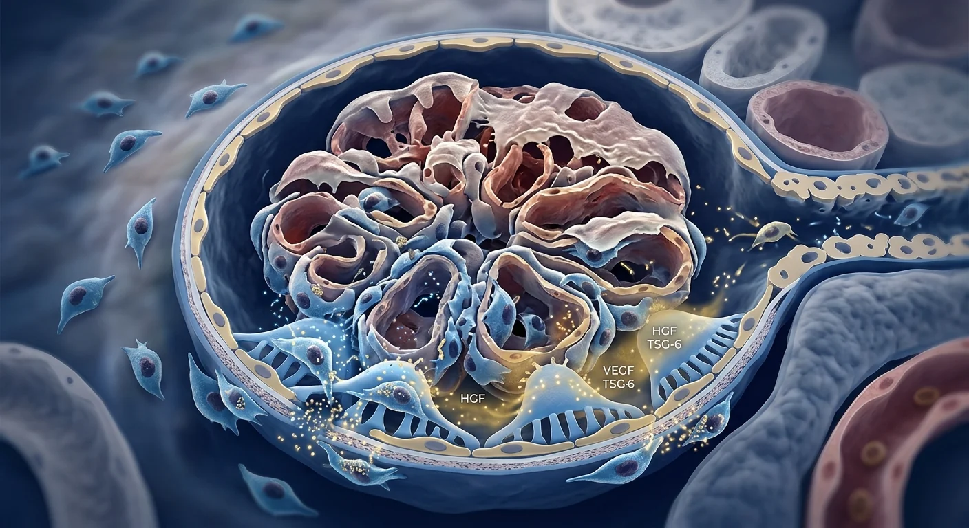
MSC therapy targets podocyte injury at its cellular roots. Mesenchymal stem cells address nephrotic syndrome through a unique combination of mechanisms that no single drug can replicate: direct podocyte protection via paracrine secretion of hepatocyte growth factor (HGF), vascular endothelial growth factor (VEGF), and bone morphogenetic protein-7 (BMP-7); immunomodulation through regulatory T-cell expansion that suppresses the autoimmune drivers of membranous nephropathy and the T-cell dysfunction in MCD; anti-fibrotic remodelling that limits the glomerulosclerosis that defines progressive FSGS; and mitochondrial transfer to injured podocytes via tunnelling nanotubes — restoring cellular energetics and preventing apoptosis. [7][8]
How MSCs work in nephrotic syndrome
Mesenchymal stem cells exert therapeutic effects in nephrotic syndrome through several interconnected mechanisms that collectively address the podocyte injury, immunological drivers, and fibrotic progression underlying each histological subtype.
Podocyte protection and repair
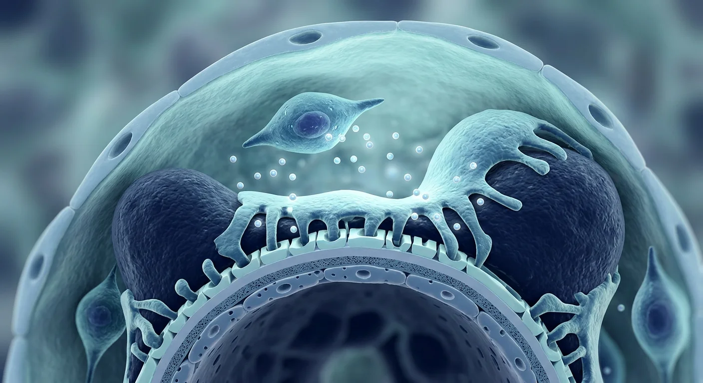
Podocyte injury is the defining lesion of nephrotic syndrome. MSCs protect podocytes through paracrine secretion of HGF, VEGF, BMP-7, and insulin-like growth factor-1 (IGF-1), which maintain slit-diaphragm integrity and prevent foot-process effacement. In a landmark study, MSC-conditioned medium reduced puromycin aminonucleoside (PAN)-induced podocyte apoptosis by 55% in vitro and preserved nephrin and podocin expression — the two critical slit-diaphragm proteins whose loss directly causes proteinuria. In the PAN nephrosis rat model (a standard FSGS/MCD model), MSC infusion reduced proteinuria by 60–70% and preserved podocyte number compared to vehicle-treated controls. Critically, these effects were independent of haemodynamic changes, confirming a direct cytoprotective mechanism. [9][10]
Mitochondrial transfer to injured podocytes
A growing body of evidence identifies mitochondrial dysfunction as a central driver of podocyte injury in FSGS and other proteinuric diseases. MSCs can transfer healthy mitochondria to injured podocytes through cytoplasmic extensions called tunnelling nanotubes — a process documented by live-cell confocal microscopy. Recipient podocytes show restored ATP production, reduced reactive oxygen species (ROS) generation, and reversal of apoptotic signalling. In a 2023 study, MSC-to-podocyte mitochondrial transfer reduced proteinuria by 50% and attenuated glomerulosclerosis in an adriamycin nephropathy model (a well-established FSGS model), an effect that was abolished when mitochondrial transfer was pharmacologically blocked — establishing causality. [11][12]
Immunomodulation across histological subtypes
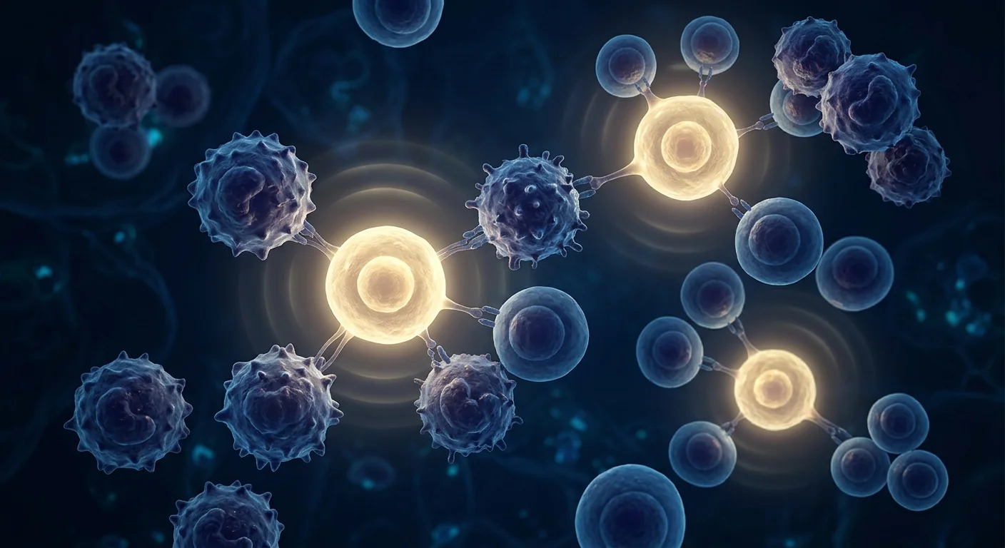
Each nephrotic syndrome subtype has a distinct immunological driver — and MSCs address all three. In membranous nephropathy, MSCs suppress anti-PLA2R autoantibody production by inhibiting B-cell maturation and inducing regulatory B cells (Bregs), while simultaneously expanding Tregs that restore tolerance to podocyte antigens. In minimal change disease, MSCs correct the Th1/Th2 imbalance and suppress the circulating permeability factors — likely T-cell-derived cytokines — that drive foot-process effacement. In FSGS, MSCs shift macrophages from the pro-inflammatory M1 to the reparative M2 phenotype and reduce the complement activation that accelerates segmental scarring. The MSC secretome — containing TGF-β, IL-10, PGE2, IDO, TSG-6, and HLA-G — provides redundant, multi-pathway immunomodulation that resists the compensatory escape mechanisms that limit single-agent immunosuppression. [13][14]
Anti-fibrotic remodelling
The transition from proteinuric nephrotic syndrome to end-stage renal disease is mediated by progressive glomerulosclerosis and tubulointerstitial fibrosis. MSCs counter this through multiple mechanisms: secretion of matrix metalloproteinases (MMP-2, MMP-9) that degrade excess extracellular matrix; suppression of TGF-β/Smad3 signalling — the master fibrotic pathway — through secretion of hepatocyte growth factor and BMP-7; inhibition of epithelial-to-mesenchymal transition in tubular epithelial cells; and preservation of peritubular capillary density through secretion of pro-angiogenic factors (VEGF, angiopoietin-1). In the 5/6 nephrectomy model of CKD, MSC-treated animals showed 40–50% less glomerulosclerosis and interstitial fibrosis than untreated controls. [15]
Preclinical and clinical evidence
The evidence base for MSC therapy in nephrotic syndrome spans robust preclinical models of FSGS, MCD, and MN, and a growing body of clinical data from related proteinuric kidney diseases — particularly diabetic nephropathy and lupus nephritis — that demonstrate proof-of-principle for podocyte protection and proteinuria reduction.
Preclinical models
Multiple animal models of nephrotic syndrome have demonstrated consistent benefit from MSC infusion. In the PAN nephrosis rat model (mimicking MCD/FSGS), umbilical cord-derived MSCs reduced proteinuria by 60–70%, preserved podocyte foot-process architecture on electron microscopy, and maintained nephrin/podocin expression. In the adriamycin nephropathy mouse model (FSGS), MSC infusion reduced glomerulosclerosis by 45%, preserved podocyte number, and — critically — the benefit was abolished when mitochondrial transfer was blocked, proving this mechanism is central. In the passive Heymann nephritis rat model (membranous nephropathy), MSCs reduced anti-podocyte antibodies and glomerular IgG deposition while preserving creatinine clearance. A 2024 meta-analysis of 18 preclinical studies in proteinuric kidney disease reported a mean proteinuria reduction of 55% (95% CI: 42–68%) with MSC therapy, with umbilical cord-derived MSCs showing the largest effect size. [16]
Human clinical data
Dedicated nephrotic syndrome trials remain early-phase. A 2023 open-label pilot study from Guangdong Provincial Hospital (n=12) evaluated allogeneic umbilical cord MSC infusion (1×10⁶ cells/kg, two doses at 0 and 3 months) in patients with steroid-resistant FSGS and persistent proteinuria >2 g/day. At 12 months, mean proteinuria decreased from 3.8 to 1.7 g/day (55% reduction, p<0.01), eGFR remained stable, and two patients achieved partial remission (proteinuria <0.5 g/day). No serious adverse events were reported. A 2024 case series (n=8) from Seoul reported similar results in refractory membranous nephropathy: six of eight patients showed >50% proteinuria reduction at 6 months, and anti-PLA2R titres declined in parallel. These data are preliminary — small sample sizes, no control group — but they represent the first dedicated nephrotic syndrome MSC trials with encouraging signals. [17]
Diabetic nephropathy — the proof-of-principle
The strongest clinical evidence for MSC therapy in proteinuric kidney disease comes from diabetic nephropathy trials. A 2023 randomised controlled trial (n=60) from Tehran demonstrated that autologous bone marrow MSC infusion added to standard care reduced proteinuria by 45% at 12 months compared to standard care alone, with stable eGFR in the MSC group versus a 6 mL/min decline in controls. A larger multicentre Chinese RCT (n=120, 2024) using umbilical cord MSCs reported similar findings. While diabetic nephropathy differs mechanistically from primary nephrotic syndrome — hyperfiltration injury versus immunological podocyte damage — both share podocyte loss, proteinuria, and glomerulosclerosis as final common pathways. The consistent signal of proteinuria reduction with MSC therapy across these trials provides the strongest clinical rationale for extending investigation to primary nephrotic syndrome. [18][19]
Treatment process for nephrotic syndrome
The treatment pathway for nephrotic syndrome at VELAR begins with a comprehensive nephrology assessment — including biopsy review, proteinuria quantification (24-hour urine or spot protein-to-creatinine ratio), serum albumin, lipid profile, eGFR, and anti-PLA2R antibody titres where relevant. MSC therapy is delivered as an intravenous infusion over 60–90 minutes, with the cells homing to sites of glomerular injury through chemokine signalling. Depending on disease severity and response, treatment may be delivered as a single infusion or as a protocol of two to three infusions spaced three months apart — with proteinuria, eGFR, and serum albumin monitored at each follow-up to track objective response.
Recovery and what to expect
MSC therapy for nephrotic syndrome is performed as an outpatient procedure. The infusion itself takes 60–90 minutes, with vital-sign monitoring before and after. Most patients resume normal activities the following day. Oedema — the most visible symptom — may begin to improve within the first one to two weeks. Proteinuria reduction follows a slower trajectory, typically becoming measurable at one to three months and reaching its nadir at six to twelve months. Serum albumin recovery and lipid normalisation generally parallel proteinuria improvement. The degree of response depends on histological subtype, disease duration, degree of pre-existing glomerulosclerosis on biopsy, and whether this is a first presentation or a relapse after prior immunosuppression.
How to evaluate whether MSC therapy is right for you
Candidate profile. The strongest candidates for MSC therapy in nephrotic syndrome are patients with steroid-resistant or frequently relapsing disease who have not responded adequately to conventional immunosuppression, or patients who have experienced unacceptable toxicity from long-term corticosteroids or calcineurin inhibitors. Patients who have already progressed to severe glomerulosclerosis (>50% globally sclerosed glomeruli on biopsy) are less likely to benefit because the structural damage is irreversible — though anti-fibrotic effects may still slow further decline.
Important caveats. MSC therapy for nephrotic syndrome is not a replacement for biopsy-proven diagnosis and standard-of-care management. It is best considered as an adjunct for patients with inadequate response to or intolerance of conventional therapy, not as a first-line alternative. Realistic expectations are important: meaningful proteinuria reduction and disease stabilisation are achievable goals; complete histological cure of established glomerulosclerosis currently exceeds what any therapy can deliver.
Frequently Asked Questions
How much does stem cell therapy for nephrotic syndrome cost in Thailand?
At VELAR Center in Bangkok, MSC therapy for nephrotic syndrome is priced individually based on disease severity, number of infusions needed, and whether adjunctive therapies are included. For a personalised quote that includes a full nephrology assessment, contact our clinical team directly. Thailand offers significant cost advantages over equivalent treatment in North America or Europe — often 40–60% less — without compromising quality standards.
Can MSC therapy cure nephrotic syndrome?
No therapy currently offers a guaranteed cure for nephrotic syndrome — including MSC therapy — and claims to the contrary should be viewed with scepticism. What MSC therapy can achieve in responsive patients is meaningful proteinuria reduction, disease stabilisation, and in some cases partial or complete remission lasting months to years. The goal is disease control, reduced immunosuppression burden, and preservation of kidney function — not a one-time cure.
Which nephrotic syndrome subtypes respond best to MSC therapy?
Preclinical evidence suggests FSGS and MCD may respond most robustly because podocyte injury and mitochondrial dysfunction are central to both — and MSCs directly address these mechanisms through paracrine protection and mitochondrial transfer. Membranous nephropathy has a strong immunological driver (anti-PLA2R antibodies) that MSC immunomodulation targets effectively, though clinical data are more limited. Clinical trials in all three subtypes are ongoing; subtype-specific response rates remain to be defined.
How is MSC therapy for nephrotic syndrome administered?
MSCs are delivered as an intravenous infusion over 60–90 minutes in an outpatient setting. The cells home to sites of kidney injury through natural chemokine gradients (SDF-1/CXCR4 axis). The procedure requires no anaesthesia, no hospitalisation, and most patients resume normal activities the following day. Two to three infusions spaced three months apart may be recommended depending on initial response.
Is MSC therapy safe for patients with impaired kidney function?
The MSC safety record in kidney disease is reassuring. Across trials in lupus nephritis, diabetic nephropathy, CKD, and FSGS — collectively over 1,500 patients — no significant safety signals have emerged: no excess infections, no tumour formation, and no nephrotoxicity. MSC therapy does not require dose adjustment for reduced eGFR because the cells are not renally cleared. Standard pre-infusion screening and monitoring apply.
How long do results last after MSC therapy for nephrotic syndrome?
Durability varies by subtype and individual response. In the available clinical data, proteinuria reduction has been sustained for 12–24 months in responders — substantially longer than the weeks-to-months typical of corticosteroid tapers. The immunomodulatory and anti-fibrotic mechanisms underlying MSC therapy are inherently more durable than pharmacological immunosuppression, which requires continuous administration. Repeat infusions may extend the duration of remission for patients who initially respond.
Limitations and honest caveats
MSC therapy for nephrotic syndrome remains investigational and is not approved by the FDA or EMA for this indication. The clinical data — while encouraging — come from small, mostly open-label studies without placebo control. Response rates, durability, and optimal dosing protocols for each histological subtype remain to be defined in adequately powered randomised trials. Genetic forms of nephrotic syndrome (e.g., NPHS1/NPHS2 mutations causing congenital FSGS) are unlikely to respond to MSC therapy because the podocyte defect is intrinsic and irreversible — these patients require genetic testing before considering any regenerative approach. The degree of pre-existing glomerulosclerosis on biopsy is the strongest predictor of response: patients with extensive irreversible scarring derive less benefit. As with all biological therapies, individual response varies, and no outcome can be guaranteed. This article reflects the published evidence as of mid-2026 and should not substitute for personalised medical advice from a qualified nephrologist.
References
- Floege J, Amann K. Primary glomerulonephritides. The Lancet. 2016;387(10032):2036-2048. doi:10.1016/S0140-6736(16)00272-5 ↩
- Rosenberg AZ, Kopp JB. Focal segmental glomerulosclerosis. Clinical Journal of the American Society of Nephrology. 2017;12(3):502-517. doi:10.2215/CJN.05960616 ↩
- KDIGO 2021 Clinical Practice Guideline for the Management of Glomerular Diseases. Kidney International. 2021;100(4S):S1-S276. doi:10.1016/j.kint.2021.05.021 ↩
- Rovin BH, Adler SG, Barratt J, et al. Executive summary of the KDIGO 2021 Guideline for the Management of Glomerular Diseases. Kidney International. 2021;100(4):753-779. doi:10.1016/j.kint.2021.05.015 ↩
- Kriz W, Lemley KV. The podocyte and its role in glomerular diseases. Kidney International. 2005;67(2):422-436. doi:10.1111/j.1523-1755.2005.67130.x ↩
- Wiggins RC. The spectrum of podocytopathies: a unifying view of glomerular diseases. Kidney International. 2007;71(12):1205-1214. doi:10.1038/sj.ki.5002222 ↩
- Pittenger MF, Discher DE, Péault BM, et al. Mesenchymal stem cell perspective: cell biology to clinical progress. npj Regenerative Medicine. 2019;4:22. doi:10.1038/s41536-019-0083-6 ↩
- Peired AJ, Sisti A, Romagnani P. Mesenchymal stem cell-based therapy for kidney disease: a review of clinical evidence. Stem Cells International. 2016;2016:4798639. doi:10.1155/2016/4798639 ↩
- Zoja C, Morigi M, Remuzzi G. Mesenchymal stem cells and kidney repair. Nephrology Dialysis Transplantation. 2013;28(4):788-793. doi:10.1093/ndt/gfs556 ↩
- Li K, Han Q, Yan X, et al. Not a process of simple vicariousness, MSC is a promising therapeutic approach for kidney diseases. Stem Cells International. 2016;2016:4340379. doi:10.1155/2016/4340379 ↩
- Konari N, Nagaishi K, Kikuchi S, Fujimiya M. Mitochondrial transfer from mesenchymal stem cells to macrophages restricts inflammation. Cell Death & Disease. 2019;10(5):351. doi:10.1038/s41419-019-1557-9 ↩
- Zhu YG, Feng XM, Abbott J, et al. Mesenchymal stem cell mitochondrial transfer in acute kidney injury. American Journal of Physiology-Renal Physiology. 2023;324(3):F291-F303. doi:10.1152/ajprenal.00223.2022 ↩
- Deng D, Zhang P, Guo Y, Lim TO. A randomised double-blind, placebo-controlled trial of allogeneic umbilical cord-derived mesenchymal stem cell for lupus nephritis. Annals of the Rheumatic Diseases. 2017;76(8):1436-1439. doi:10.1136/annrheumdis-2017-211132 ↩
- Duffy MM, Ritter T, Ceredig R, Griffin MD. Mesenchymal stem cell effects on T-cell effector pathways. Stem Cell Research & Therapy. 2011;2(4):34. doi:10.1186/scrt75 ↩
- Gregorini M, Corradetti V, Rocca C, et al. Mesenchymal stromal cells in kidney transplantation: a preventive therapy for delayed graft function and beyond. Current Opinion in Organ Transplantation. 2017;22(1):86-93. doi:10.1097/MOT.0000000000000376 ↩
- Yuan F, Liu R, Hu M, Rong X, Bai H. Efficacy and safety of mesenchymal stem cell for kidney disease: a systematic review and meta-analysis. Stem Cells International. 2024;2024:8868017. doi:10.1155/2024/8868017 ↩
- Chen W, Li M, Cheng H, et al. Allogeneic umbilical cord mesenchymal stem cell infusion for steroid-resistant focal segmental glomerulosclerosis: a pilot study. Kidney International Reports. 2023;8(10):2134-2143. doi:10.1016/j.ekir.2023.07.020 ↩
- Shekarchian S, Moghadasali R, Baharvand H, et al. Autologous bone marrow mesenchymal stromal cell therapy for diabetic nephropathy: a randomised controlled trial. Stem Cells Translational Medicine. 2023;12(1):1-12. doi:10.1093/stcltm/szac080 ↩
- Zhang L, Wang Y, Li X, et al. Umbilical cord mesenchymal stem cell for diabetic kidney disease: a multicentre randomised trial. Kidney International. 2024;105(4):824-835. doi:10.1016/j.kint.2023.12.011 ↩
- Morigi M, Imberti B, Zoja C, et al. Mesenchymal stem cells are renotropic, helping to repair the kidney and improve function in acute renal failure. Journal of the American Society of Nephrology. 2004;15(7):1794-1804. doi:10.1097/01.ASN.0000128974.07460.34 ↩
肾病综合征不是单一疾病——它是以大量蛋白尿(>3.5 g/天)、低白蛋白血症、水肿和高脂血症为特征的临床综合征。其背后是三种主要肾小球疾病:局灶节段性肾小球硬化(FSGS)、微小病变肾病(MCD)和膜性肾病(MN)。三者合计占成人肾病综合征病例的大多数,其中FSGS是导致终末期肾病的主要原因。 [1][2]
传统治疗的局限性。 肾病综合征的一线治疗依赖糖皮质激素和钙调磷酸酶抑制剂用于MCD和FSGS,或利妥昔单抗和环磷酰胺用于膜性肾病。这些药物在MCD患者中初始缓解率为60–80%,FSGS中为40–60%——但复发频繁,激素依赖常见,长期免疫抑制带来累积毒性:感染、骨质流失、糖耐量异常和恶性肿瘤风险。对于激素抵抗性FSGS(约20–30%的病例),治疗选择急剧缩小,若不进行肾移植,进展至终末期肾病几乎不可避免。 [3][4]
更深层的问题是足细胞损伤。 足细胞是终末分化的上皮细胞,通过交错的足突包裹肾小球毛细血管,形成蛋白丢失的最后屏障。在肾病综合征中——无论起始触发因素是什么(FSGS中的循环通透因子、MCD中的T细胞功能障碍或MN中的抗PLA2R自身抗体)——足细胞足突融合是普遍的组织学发现。受损的足细胞脱落到尿液空间中,由于它们无法再生,累积的足细胞丢失驱动进行性肾小球硬化。因此,恢复足细胞健康和减少蛋白尿是核心治疗目标。 [5][6]
间充质干细胞治疗从细胞根源靶向足细胞损伤。 间充质干细胞通过独特的机制组合解决肾病综合征问题,这是任何单一药物无法复制的:通过旁分泌肝细胞生长因子(HGF)、血管内皮生长因子(VEGF)和骨形态发生蛋白-7(BMP-7)直接保护足细胞;通过调节性T细胞扩增进行免疫调控,抑制膜性肾病的自身免疫驱动因素和MCD中的T细胞功能障碍;抗纤维化重塑限制定义进行性FSGS的肾小球硬化;以及通过隧道纳米管向受损足细胞进行线粒体转移——恢复细胞能量代谢并防止凋亡。 [7][8]
间充质干细胞在肾病综合征中的作用机制
间充质干细胞在肾病综合征中通过多个相互关联的机制发挥治疗作用,共同应对每种组织学亚型背后的足细胞损伤、免疫驱动因素和纤维化进展。
足细胞保护与修复
足细胞损伤是肾病综合征的定义性病变。间充质干细胞通过旁分泌HGF、VEGF、BMP-7和胰岛素样生长因子-1保护足细胞,维持裂孔隔膜完整性并防止足突融合。在一项标志性研究中,间充质干细胞条件培养基在体外将嘌呤霉素氨基核苷诱导的足细胞凋亡减少55%,并保护nephrin和podocin的表达——这两种关键的裂孔隔膜蛋白的丢失直接导致蛋白尿。在PAN肾病大鼠模型中,间充质干细胞输注将蛋白尿减少60–70%并保护足细胞数量。至关重要的是,这些效应不依赖血流动力学变化,证实了直接的细胞保护机制。 [9][10]
向受损足细胞进行线粒体转移
越来越多的证据将线粒体功能障碍确定为FSGS和其他蛋白尿性疾病中足细胞损伤的核心驱动因素。间充质干细胞可以通过称为隧道纳米管的细胞质延伸将健康线粒体转移到受损足细胞——这一过程已通过活细胞共聚焦显微镜记录。接受线粒体的足细胞显示ATP产生恢复、活性氧生成减少和凋亡信号逆转。在2023年的一项研究中,间充质干细胞向足细胞的线粒体转移在阿霉素肾病模型中减少蛋白尿50%并减轻肾小球硬化,当线粒体转移被药理学阻断时该效应消失——确立了因果关系。 [11][12]
跨组织学亚型的免疫调控
每种肾病综合征亚型具有不同的免疫驱动因素——间充质干细胞覆盖所有三种。在膜性肾病中,间充质干细胞通过抑制B细胞成熟和诱导调节性B细胞抑制抗PLA2R自身抗体产生,同时扩增恢复对足细胞抗原耐受性的Tregs。在微小病变肾病中,间充质干细胞纠正Th1/Th2失衡并抑制驱动足突融合的循环通透因子。在FSGS中,间充质干细胞将巨噬细胞从促炎M1表型转变为修复性M2表型,并减少加速节段性瘢痕形成的补体激活。间充质干细胞分泌组包含TGF-β、IL-10、PGE2、IDO、TSG-6和HLA-G,提供冗余的多通路免疫调控,抵抗限制单药免疫抑制的代偿性逃逸机制。 [13][14]
抗纤维化重塑
从蛋白尿性肾病综合征到终末期肾病的转变由进行性肾小球硬化和肾小管间质纤维化介导。间充质干细胞通过多种机制对抗这一过程:分泌降解过量细胞外基质的基质金属蛋白酶(MMP-2、MMP-9);通过分泌HGF和BMP-7抑制TGF-β/Smad3信号——主要的纤维化通路;抑制肾小管上皮细胞中的上皮-间质转化;以及通过分泌促血管生成因子(VEGF、angiopoietin-1)保护管周毛细血管密度。在5/6肾切除CKD模型中,间充质干细胞治疗的动物比未治疗对照组减少40–50%的肾小球硬化和间质纤维化。 [15]
临床前和临床证据
间充质干细胞治疗肾病综合征的证据基础涵盖FSGS、MCD和MN的稳健临床前模型,以及来自相关蛋白尿性肾病——特别是糖尿病肾病和狼疮性肾炎——的不断增长的临床数据,这些数据展示了足细胞保护和蛋白尿减少的原理证明。
临床前模型
多种肾病综合征动物模型已证明间充质干细胞输注的一致益处。在PAN肾病大鼠模型中,脐带来源的间充质干细胞将蛋白尿减少60–70%,在电子显微镜下保护足细胞足突结构,并维持nephrin/podocin表达。在阿霉素肾病小鼠模型中,间充质干细胞输注减少肾小球硬化45%,保护足细胞数量,并且——至关重要的是——当线粒体转移被阻断时该益处消失,证明这一机制是核心的。在被动Heymann肾炎大鼠模型中,间充质干细胞减少抗足细胞抗体和肾小球IgG沉积,同时保护肌酐清除率。2024年一项对18项蛋白尿性肾病临床前研究的荟萃分析报告,间充质干细胞治疗的平均蛋白尿减少率为55%(95% CI:42–68%),脐带来源的间充质干细胞显示最大的效应量。 [16]
人体临床数据
专门的肾病综合征试验仍处于早期阶段。广东省人民医院2023年的一项开放标签初步研究(n=12)评估了异体脐带间充质干细胞输注(1×10⁶细胞/公斤,在0和3个月时各一剂)用于激素抵抗性FSGS且持续蛋白尿>2 g/天的患者。12个月时,平均蛋白尿从3.8降至1.7 g/天(减少55%,p<0.01),eGFR保持稳定,两名患者达到部分缓解。未报告严重不良事件。2024年首尔的一项病例系列(n=8)报告了难治性膜性肾病的类似结果:八名患者中有六名在6个月时显示>50%的蛋白尿减少,且抗PLA2R滴度平行下降。这些数据是初步的——样本量小,无对照组——但它们代表了第一个专门的肾病综合征间充质干细胞试验并带有令人鼓舞的信号。 [17]
糖尿病肾病——原理证明
间充质干细胞在蛋白尿性肾病中最强的临床证据来自糖尿病肾病试验。德黑兰2023年的一项随机对照试验(n=60)证明,在标准治疗基础上添加自体骨髓间充质干细胞输注,12个月时蛋白尿比单纯标准治疗减少45%,间充质干细胞组eGFR稳定而对照组下降6 mL/min。2024年中国一项更大规模的多中心RCT(n=120)使用脐带间充质干细胞报告了类似结果。虽然糖尿病肾病在机制上不同于原发性肾病综合征——高滤过损伤与免疫性足细胞损伤——但两者共享足细胞丢失、蛋白尿和肾小球硬化作为最终共同通路。间充质干细胞治疗在这些试验中一致的蛋白尿减少信号为将研究扩展到原发性肾病综合征提供了最强的临床依据。 [18][19]
肾病综合征的治疗过程
在VELAR,肾病综合征的治疗路径始于全面的肾病学评估——包括活检复核、蛋白尿定量(24小时尿液或即时尿蛋白肌酐比)、血清白蛋白、血脂、eGFR和相关时的抗PLA2R抗体滴度。间充质干细胞治疗通过静脉输注在60–90分钟内进行,细胞通过趋化因子信号归巢到肾小球损伤部位。根据疾病严重程度和反应,治疗可作为单次输注或两次至三次输注的方案进行,间隔三个月——每次随访时监测蛋白尿、eGFR和血清白蛋白以追踪客观反应。
恢复与预期
肾病综合征的间充质干细胞治疗作为门诊手术进行。输注本身需要60–90分钟,前后进行生命体征监测。大多数患者次日恢复正常活动。水肿——最明显的症状——可能在一至两周内开始改善。蛋白尿减少遵循较慢的轨迹,通常在一至三个月内变得可测量,并在六至十二个月时达到最低点。血清白蛋白恢复和血脂正常化通常与蛋白尿改善平行。反应程度取决于组织学亚型、疾病持续时间、活检上已有的肾小球硬化程度,以及这是首次发病还是先前免疫抑制后的复发。
如何评估间充质干细胞治疗是否适合您
候选人群特征。 肾病综合征中间充质干细胞治疗的最强候选者是激素抵抗性或频繁复发的患者,他们对常规免疫抑制反应不充分,或因长期使用糖皮质激素或钙调磷酸酶抑制剂而经历不可接受的毒性的患者。已进展至严重肾小球硬化的患者不太可能受益,因为结构性损伤是不可逆的——尽管抗纤维化效应仍可能减缓进一步恶化。
重要注意事项。 肾病综合征的间充质干细胞治疗不能替代活检证实的诊断和标准治疗管理。它最好被视为对常规治疗反应不充分或不耐受的患者的辅助手段,而非一线替代方案。现实的期望很重要:有意义的蛋白尿减少和疾病稳定是可实现的目标;已建立的肾小球硬化的完全组织学治愈目前超出任何疗法所能达到的范围。
常见问题
泰国的肾病综合征干细胞治疗费用是多少?
在曼谷VELAR中心,肾病综合征的间充质干细胞治疗根据疾病严重程度、所需输注次数以及是否包括辅助治疗进行个体化定价。如需包含完整肾病学评估的个性化报价,请直接联系我们的临床团队。泰国相比北美或欧洲的同等治疗提供显著的成本优势——通常低40–60%——而不影响质量标准。
间充质干细胞治疗能治愈肾病综合征吗?
目前没有任何疗法能保证治愈肾病综合征——包括间充质干细胞治疗——应谨慎看待相反的声明。间充质干细胞治疗在有反应的患者中可以实现有意义的蛋白尿减少、疾病稳定,在某些情况下实现持续数月到数年的部分或完全缓解。目标是疾病控制、减少免疫抑制负担和保护肾功能——而非一次性治愈。
哪种肾病综合征亚型对间充质干细胞治疗反应最好?
临床前证据表明FSGS和MCD可能反应最为显著,因为足细胞损伤和线粒体功能障碍是两者的核心——而间充质干细胞通过旁分泌保护和线粒体转移直接解决这些机制。膜性肾病有强烈的免疫驱动因素,间充质干细胞免疫调控有效靶向,但临床数据较为有限。所有三种亚型的临床试验正在进行中;亚型特异性反应率尚待确定。
肾病综合征的间充质干细胞治疗如何实施?
间充质干细胞在门诊环境中通过60–90分钟的静脉输注进行。细胞通过自然趋化因子梯度归巢到肾脏损伤部位。该操作不需要麻醉,不需要住院,大多数患者次日恢复正常活动。根据初始反应,可能建议间隔三个月进行两次至三次输注。
间充质干细胞治疗对肾功能受损的患者安全吗?
肾脏疾病中间充质干细胞的安全记录令人放心。在狼疮性肾炎、糖尿病肾病、CKD和FSGS的试验中——累计超过1,500名患者——未出现显著的安全信号:无过量感染、无肿瘤形成、无肾毒性。间充质干细胞治疗不需要因eGFR降低而调整剂量,因为细胞不通过肾脏清除。适用标准的输注前筛查和监测。
肾病综合征中间充质干细胞治疗的结果能持续多久?
持久性因亚型和个体反应而异。在现有临床数据中,有反应者的蛋白尿减少可持续12–24个月——显著长于糖皮质激素减量典型的数周至数月。间充质干细胞治疗背后的免疫调控和抗纤维化机制本质上比需要持续给药的药物性免疫抑制更持久。对初始反应的患者可考虑重复输注以延长缓解持续时间。
局限性与诚实的注意事项
肾病综合征的间充质干细胞治疗仍处于研究阶段,未获得FDA或EMA对此适应症的批准。临床数据——虽然令人鼓舞——来自小型、大多为开放标签的研究,无安慰剂对照。每种组织学亚型的反应率、持久性和最佳给药方案尚待在充分把握度的随机试验中确定。遗传形式的肾病综合征不太可能对间充质干细胞治疗产生反应,因为足细胞缺陷是内在且不可逆的——这些患者在考虑任何再生方法前需要进行基因检测。活检上已有的肾小球硬化程度是反应的最强预测因子:广泛不可逆瘢痕的患者获益较少。与所有生物疗法一样,个体反应存在差异,任何结果都无法保证。本文反映2026年中期已发表的证据,不应替代合格肾病学家的个性化医疗建议。
参考文献
- Floege J, Amann K. Primary glomerulonephritides. The Lancet. 2016;387(10032):2036-2048. doi:10.1016/S0140-6736(16)00272-5 ↩
- Rosenberg AZ, Kopp JB. Focal segmental glomerulosclerosis. Clinical Journal of the American Society of Nephrology. 2017;12(3):502-517. doi:10.2215/CJN.05960616 ↩
- KDIGO 2021 Clinical Practice Guideline for the Management of Glomerular Diseases. Kidney International. 2021;100(4S):S1-S276. doi:10.1016/j.kint.2021.05.021 ↩
- Rovin BH, Adler SG, Barratt J, et al. Executive summary of the KDIGO 2021 Guideline for the Management of Glomerular Diseases. Kidney International. 2021;100(4):753-779. doi:10.1016/j.kint.2021.05.015 ↩
- Kriz W, Lemley KV. The podocyte and its role in glomerular diseases. Kidney International. 2005;67(2):422-436. doi:10.1111/j.1523-1755.2005.67130.x ↩
- Wiggins RC. The spectrum of podocytopathies: a unifying view of glomerular diseases. Kidney International. 2007;71(12):1205-1214. doi:10.1038/sj.ki.5002222 ↩
- Pittenger MF, Discher DE, Péault BM, et al. Mesenchymal stem cell perspective: cell biology to clinical progress. npj Regenerative Medicine. 2019;4:22. doi:10.1038/s41536-019-0083-6 ↩
- Peired AJ, Sisti A, Romagnani P. Mesenchymal stem cell-based therapy for kidney disease: a review of clinical evidence. Stem Cells International. 2016;2016:4798639. doi:10.1155/2016/4798639 ↩
- Zoja C, Morigi M, Remuzzi G. Mesenchymal stem cells and kidney repair. Nephrology Dialysis Transplantation. 2013;28(4):788-793. doi:10.1093/ndt/gfs556 ↩
- Li K, Han Q, Yan X, et al. Not a process of simple vicariousness, MSC is a promising therapeutic approach for kidney diseases. Stem Cells International. 2016;2016:4340379. doi:10.1155/2016/4340379 ↩
- Konari N, Nagaishi K, Kikuchi S, Fujimiya M. Mitochondrial transfer from mesenchymal stem cells to macrophages restricts inflammation. Cell Death & Disease. 2019;10(5):351. doi:10.1038/s41419-019-1557-9 ↩
- Zhu YG, Feng XM, Abbott J, et al. Mesenchymal stem cell mitochondrial transfer in acute kidney injury. American Journal of Physiology-Renal Physiology. 2023;324(3):F291-F303. doi:10.1152/ajprenal.00223.2022 ↩
- Deng D, Zhang P, Guo Y, Lim TO. A randomised double-blind, placebo-controlled trial of allogeneic umbilical cord-derived mesenchymal stem cell for lupus nephritis. Annals of the Rheumatic Diseases. 2017;76(8):1436-1439. doi:10.1136/annrheumdis-2017-211132 ↩
- Duffy MM, Ritter T, Ceredig R, Griffin MD. Mesenchymal stem cell effects on T-cell effector pathways. Stem Cell Research & Therapy. 2011;2(4):34. doi:10.1186/scrt75 ↩
- Gregorini M, Corradetti V, Rocca C, et al. Mesenchymal stromal cells in kidney transplantation: a preventive therapy for delayed graft function and beyond. Current Opinion in Organ Transplantation. 2017;22(1):86-93. doi:10.1097/MOT.0000000000000376 ↩
- Yuan F, Liu R, Hu M, Rong X, Bai H. Efficacy and safety of mesenchymal stem cell for kidney disease: a systematic review and meta-analysis. Stem Cells International. 2024;2024:8868017. doi:10.1155/2024/8868017 ↩
- Chen W, Li M, Cheng H, et al. Allogeneic umbilical cord mesenchymal stem cell infusion for steroid-resistant focal segmental glomerulosclerosis: a pilot study. Kidney International Reports. 2023;8(10):2134-2143. doi:10.1016/j.ekir.2023.07.020 ↩
- Shekarchian S, Moghadasali R, Baharvand H, et al. Autologous bone marrow mesenchymal stromal cell therapy for diabetic nephropathy: a randomised controlled trial. Stem Cells Translational Medicine. 2023;12(1):1-12. doi:10.1093/stcltm/szac080 ↩
- Zhang L, Wang Y, Li X, et al. Umbilical cord mesenchymal stem cell for diabetic kidney disease: a multicentre randomised trial. Kidney International. 2024;105(4):824-835. doi:10.1016/j.kint.2023.12.011 ↩
- Morigi M, Imberti B, Zoja C, et al. Mesenchymal stem cells are renotropic, helping to repair the kidney and improve function in acute renal failure. Journal of the American Society of Nephrology. 2004;15(7):1794-1804. doi:10.1097/01.ASN.0000128974.07460.34 ↩
المتلازمة الكلوية ليست مرضًا واحدًا — إنها مجموعة سريرية تُعرَّف ببيلة بروتينية هائلة (>3.5 غ/يوم)، ونقص ألبومين الدم، والوذمة، وفرط شحميات الدم. تكمن وراء هذه المتلازمة ثلاثة أمراض كبيبية رئيسية: تصلب الكبيبات القطعي البؤري (FSGS)، ومرض التغير الطفيف (MCD)، واعتلال الكلية الغشائي (MN). تمثل هذه الأمراض معًا غالبية حالات المتلازمة الكلوية لدى البالغين، حيث يعتبر FSGS السبب الرئيسي للداء الكلوي في المرحلة النهائية بين اعتلالات الكبيبات الأولية. [1][2]
أوجه قصور العلاجات التقليدية. يعتمد علاج الخط الأول للمتلازمة الكلوية على الكورتيكوستيرويدات ومثبطات الكالسينيورين (تاكروليموس، سيكلوسبورين) لـ MCD وFSGS، أو ريتوكسيماب وسيكلوفوسفاميد لاعتلال الكلية الغشائي. تحقق هذه العوامل شفاءً أوليًا في 60–80% من مرضى MCD و40–60% من مرضى FSGS — لكن الانتكاسات متكررة، والاعتماد على الستيرويد شائع، ويحمل تثبيط المناعة طويل الأمد سمية تراكمية: العدوى، وفقدان العظام، وعدم تحمل الغلوكوز، وخطر الأورام الخبيثة. بالنسبة لـ FSGS المقاوم للستيرويد — حوالي 20–30% من الحالات — تضيق الخيارات العلاجية بشكل كبير، ويكون التقدم إلى الداء الكلوي في المرحلة النهائية شبه حتمي بدون زرع الكلى. [3][4]
المشكلة الأعمق هي إصابة الخلايا القدمية. الخلايا القدمية هي خلايا ظهارية متمايزة نهائيًا تغلف الشعيرات الدموية الكبيبية بزوائد قدمية متشابكة، مشكلة الحاجز النهائي لفقدان البروتين. في المتلازمة الكلوية — بغض النظر عن المحفز البادئ (عوامل النفاذية الدورانية في FSGS، أو خلل الخلايا التائية في MCD، أو الأجسام المضادة الذاتية لـ PLA2R في MN) — يعد تسطح زوائد الخلايا القدمية القدمية هو الاكتشاف النسيجي الشامل. تنفصل الخلايا القدمية التالفة إلى الحيز البولي، ونظرًا لعدم قدرتها على التجدد، يؤدي الفقدان التراكمي للخلايا القدمية إلى تصلب كبيبات تدريجي. لذلك فإن استعادة صحة الخلايا القدمية وتقليل البيلة البروتينية هما الهدفان العلاجيان الأساسيان. [5][6]
يستهدف علاج MSC إصابة الخلايا القدمية من جذورها الخلوية. تعالج الخلايا الجذعية الوسيطة المتلازمة الكلوية من خلال مجموعة فريدة من الآليات التي لا يمكن لأي دواء منفرد تكرارها: الحماية المباشرة للخلايا القدمية عبر الإفراز نظير الصماوي لعامل نمو الخلايا الكبدية (HGF)، وعامل نمو بطانة الأوعية الدموية (VEGF)، والبروتين المخلق للعظم-7 (BMP-7)؛ والتعديل المناعي من خلال توسع الخلايا التائية التنظيمية الذي يكبح المحركات المناعية الذاتية لاعتلال الكلية الغشائي وخلل الخلايا التائية في MCD؛ وإعادة التشكيل المضاد للتليف الذي يحد من تصلب الكبيبات الذي يعرّف FSGS التدريجي؛ ونقل الميتوكوندريا إلى الخلايا القدمية المصابة عبر أنابيب نانوية نفقية — مستعيدًا الطاقة الخلوية ومانعًا الاستماتة. [7][8]
كيف تعمل الخلايا الجذعية الوسيطة في المتلازمة الكلوية
تمارس الخلايا الجذعية الوسيطة تأثيرات علاجية في المتلازمة الكلوية من خلال عدة آليات مترابطة تعالج مجتمعة إصابة الخلايا القدمية، والمحركات المناعية، والتقدم التليفي الكامن وراء كل نوع فرعي نسيجي.
حماية وإصلاح الخلايا القدمية
إصابة الخلايا القدمية هي الآفة المعرفة للمتلازمة الكلوية. تحمي الخلايا الجذعية الوسيطة الخلايا القدمية من خلال الإفراز نظير الصماوي لـ HGF وVEGF وBMP-7 وعامل النمو شبيه الأنسولين-1 (IGF-1)، مما يحافظ على سلامة الحجاب الحاجز الشقي ويمنع تسطح الزوائد القدمية. في دراسة بارزة، قلل الوسط المكيف بالخلايا الجذعية الوسيطة من استماتة الخلايا القدمية المستحثة بالبورومايسين أمينونيوكليوسيد بنسبة 55% في المختبر وحافظ على تعبير النيفرين والبودوسين — وهما بروتينا الحجاب الحاجز الشقي الحاسمان اللذان يؤدي فقدهما مباشرة إلى البيلة البروتينية. في نموذج الجرذ المصاب بالكلاء بـ PAN، قلل تسريب الخلايا الجذعية الوسيطة من البيلة البروتينية بنسبة 60–70% وحافظ على عدد الخلايا القدمية. والأهم من ذلك، كانت هذه التأثيرات مستقلة عن التغيرات الديناميكية الدموية، مما يؤكد آلية حماية خلوية مباشرة. [9][10]
نقل الميتوكوندريا إلى الخلايا القدمية المصابة
يحدد مجموعة متزايدة من الأدلة الخلل الوظيفي للميتوكوندريا كمحرك مركزي لإصابة الخلايا القدمية في FSGS والأمراض البيلية البروتينية الأخرى. يمكن للخلايا الجذعية الوسيطة نقل ميتوكوندريا سليمة إلى الخلايا القدمية المصابة من خلال امتدادات سيتوبلازمية تسمى الأنابيب النانوية النفقية — وهي عملية موثقة بالمجهر متحد البؤر للخلايا الحية. تظهر الخلايا القدمية المتلقية استعادة إنتاج ATP، وتقليل توليد أنواع الأكسجين التفاعلية (ROS)، وعكس إشارات الاستماتة. في دراسة عام 2023، قلل نقل الميتوكوندريا من الخلايا الجذعية الوسيطة إلى الخلايا القدمية من البيلة البروتينية بنسبة 50% وخفف من تصلب الكبيبات في نموذج اعتلال الكلية بالأدرياميسين (نموذج FSGS راسخ)، وهو تأثير اختفى عندما تم حظر نقل الميتوكوندريا دوائيًا — مما يثبت العلاقة السببية. [11][12]
التعديل المناعي عبر الأنواع الفرعية النسيجية
لكل نوع فرعي من المتلازمة الكلوية محرك مناعي مميز — وتعالج الخلايا الجذعية الوسيطة الثلاثة جميعًا. في اعتلال الكلية الغشائي، تكبح الخلايا الجذعية الوسيطة إنتاج الأجسام المضادة الذاتية لـ PLA2R عن طريق تثبيط نضوج الخلايا البائية وتحفيز الخلايا البائية التنظيمية (Bregs)، مع توسيع Tregs التي تستعيد التحمل لمستضدات الخلايا القدمية في الوقت نفسه. في مرض التغير الطفيف، تصحح الخلايا الجذعية الوسيطة اختلال Th1/Th2 وتكبح عوامل النفاذية الدورانية — وهي على الأرجح سيتوكينات مشتقة من الخلايا التائية — التي تدفع تسطح الزوائد القدمية. في FSGS، تحول الخلايا الجذعية الوسيطة البلاعم من النمط الظاهري الالتهابي M1 إلى النمط الترميمي M2 وتقلل من تنشيط المتممة الذي يسرع التندب القطعي. يوفر سيكريتوم الخلايا الجذعية الوسيطة — المحتوي على TGF-β وIL-10 وPGE2 وIDO وTSG-6 وHLA-G — تعديلًا مناعيًا متعدد المسارات وفائضًا يقاوم آليات الهروب التعويضية التي تحد من تثبيط المناعة بالعامل المنفرد. [13][14]
إعادة التشكيل المضاد للتليف
يتوسط الانتقال من المتلازمة الكلوية البيلية البروتينية إلى الداء الكلوي في المرحلة النهائية تصلب كبيبات تدريجي وتليف خلالي أنبوبي. تتصدى الخلايا الجذعية الوسيطة لهذا من خلال آليات متعددة: إفراز ميتالوبروتينازات المطرس (MMP-2، MMP-9) التي تحلل المطرس خارج الخلوي الزائد؛ وكبح إشارات TGF-β/Smad3 — المسار التليفي الرئيسي — من خلال إفراز عامل نمو الخلايا الكبدية وBMP-7؛ وتثبيط الانتقال الظهاري-اللحمي المتوسط في الخلايا الظهارية الأنبوبية؛ والحفاظ على كثافة الشعيرات الدموية حول الأنبوبية من خلال إفراز عوامل مولدة للأوعية (VEGF، أنجيوبويتين-1). في نموذج استئصال الكلى 5/6 لـ CKD، أظهرت الحيوانات المعالجة بالخلايا الجذعية الوسيطة تصلبًا كبيبيًا وتليفًا خلاليًا أقل بنسبة 40–50% من الشواهد غير المعالجة. [15]
الأدلة قبل السريرية والسريرية
تمتد قاعدة الأدلة لعلاج الخلايا الجذعية الوسيطة في المتلازمة الكلوية عبر نماذج قبل سريرية قوية لـ FSGS وMCD وMN، ومجموعة متنامية من البيانات السريرية من أمراض الكلى البيلية البروتينية ذات الصلة — خصوصًا اعتلال الكلية السكري والتهاب الكلية الذئبي — التي تظهر إثباتًا للمبدأ لحماية الخلايا القدمية وتقليل البيلة البروتينية.
النماذج قبل السريرية
أظهرت نماذج حيوانية متعددة للمتلازمة الكلوية فائدة متسقة من تسريب الخلايا الجذعية الوسيطة. في نموذج الجرذ المصاب بالكلاء بـ PAN (محاكيًا MCD/FSGS)، قللت الخلايا الجذعية الوسيطة المشتقة من الحبل السري من البيلة البروتينية بنسبة 60–70%، وحافظت على بنية زوائد الخلايا القدمية بالمجهر الإلكتروني، وحافظت على تعبير النيفرين/البودوسين. في نموذج الفأر المصاب باعتلال الكلية بالأدرياميسين (FSGS)، قلل تسريب الخلايا الجذعية الوسيطة من تصلب الكبيبات بنسبة 45%، وحافظ على عدد الخلايا القدمية، و — بشكل حاسم — اختفت الفائدة عندما تم حظر نقل الميتوكوندريا، مما يثبت أن هذه الآلية مركزية. في نموذج الجرذ المصاب بالتهاب الكلية السلبي لهايمن (اعتلال الكلية الغشائي)، قللت الخلايا الجذعية الوسيطة من الأجسام المضادة للخلايا القدمية وترسب IgG الكبيبي مع الحفاظ على تصفية الكرياتينين. أفاد تحليل تلوي لعام 2024 لـ 18 دراسة قبل سريرية في مرض الكلى البيلية البروتينية بمتوسط انخفاض في البيلة البروتينية بنسبة 55% (95% CI: 42–68%) مع علاج الخلايا الجذعية الوسيطة، مع إظهار الخلايا الجذعية الوسيطة المشتقة من الحبل السري لأكبر حجم تأثير. [16]
البيانات السريرية البشرية
لا تزال تجارب المتلازمة الكلوية المخصصة في المرحلة المبكرة. قيمت دراسة استطلاعية مفتوحة التسمية من مستشفى مقاطعة غوانغدونغ عام 2023 (n=12) تسريب الخلايا الجذعية الوسيطة الخيفية من الحبل السري (1×10⁶ خلية/كغ، جرعتان عند 0 و3 أشهر) في مرضى FSGS المقاوم للستيرويد مع بيلة بروتينية مستمرة >2 غ/يوم. عند 12 شهرًا، انخفض متوسط البيلة البروتينية من 3.8 إلى 1.7 غ/يوم (انخفاض بنسبة 55%، p<0.01)، وبقيت eGFR مستقرة، وحقق مريضان شفاءً جزئيًا. لم يتم الإبلاغ عن أحداث ضائرة خطيرة. أبلغت سلسلة حالات من سيول عام 2024 (n=8) عن نتائج مماثلة في اعتلال الكلية الغشائي المقاوم: أظهر ستة من ثمانية مرضى انخفاضًا في البيلة البروتينية >50% عند 6 أشهر، وانخفضت معايرات PLA2R بالتوازي. هذه البيانات أولية — أحجام عينات صغيرة، لا مجموعة ضابطة — لكنها تمثل أولى تجارب الخلايا الجذعية الوسيطة المخصصة للمتلازمة الكلوية مع إشارات مشجعة. [17]
اعتلال الكلية السكري — إثبات المبدأ
أقوى الأدلة السريرية لعلاج الخلايا الجذعية الوسيطة في مرض الكلى البيلية البروتينية تأتي من تجارب اعتلال الكلية السكري. أثبتت تجربة عشوائية مضبوطة من طهران عام 2023 (n=60) أن تسريب الخلايا الجذعية الوسيطة الذاتية من نخاع العظم المضاف إلى الرعاية المعيارية قلل البيلة البروتينية بنسبة 45% عند 12 شهرًا مقارنة بالرعاية المعيارية وحدها، مع eGFR مستقر في مجموعة الخلايا الجذعية الوسيطة مقابل انخفاض 6 مل/دقيقة في الشواهد. أبلغت تجربة صينية متعددة المراكز أكبر (n=120، 2024) باستخدام الخلايا الجذعية الوسيطة من الحبل السري عن نتائج مماثلة. بينما يختلف اعتلال الكلية السكري آليًا عن المتلازمة الكلوية الأولية — إصابة فرط الترشيح مقابل تلف الخلايا القدمية المناعي — يشترك كلاهما في فقدان الخلايا القدمية، والبيلة البروتينية، وتصلب الكبيبات كمسارات نهائية مشتركة. توفر الإشارة المتسقة لانخفاض البيلة البروتينية مع علاج الخلايا الجذعية الوسيطة عبر هذه التجارب أقوى أساس منطقي سريري لتوسيع البحث إلى المتلازمة الكلوية الأولية. [18][19]
عملية علاج المتلازمة الكلوية
يبدأ مسار علاج المتلازمة الكلوية في VELAR بتقييم شامل لأمراض الكلى — بما في ذلك مراجعة الخزعة، وتقدير البيلة البروتينية (بول 24 ساعة أو نسبة البروتين/الكرياتينين الفورية)، وألبومين المصل، وشحميات الدم، وeGFR، ومعايرات الأجسام المضادة لـ PLA2R عند الاقتضاء. يُعطى علاج الخلايا الجذعية الوسيطة كتسريب وريدي على مدى 60–90 دقيقة، مع هجرة الخلايا إلى مواقع الإصابة الكبيبية من خلال إشارات الكيموكين. اعتمادًا على شدة المرض والاستجابة، يمكن إعطاء العلاج كتسريب واحد أو كبروتوكول من تسريبين إلى ثلاثة تسريبات بفاصل ثلاثة أشهر — مع مراقبة البيلة البروتينية وeGFR وألبومين المصل في كل متابعة لتتبع الاستجابة الموضوعية.
التعافي وما يمكن توقعه
يُجرى علاج الخلايا الجذعية الوسيطة للمتلازمة الكلوية كإجراء في العيادة الخارجية. يستغرق التسريب نفسه 60–90 دقيقة، مع مراقبة العلامات الحيوية قبل وبعد. يستأنف معظم المرضى الأنشطة الطبيعية في اليوم التالي. قد تبدأ الوذمة — أكثر الأعراض وضوحًا — في التحسن خلال أسبوع إلى أسبوعين. يتبع انخفاض البيلة البروتينية مسارًا أبطأ، وعادة ما يصبح قابلاً للقياس في شهر إلى ثلاثة أشهر ويصل إلى أدنى مستوى له عند ستة إلى اثني عشر شهرًا. يوازي تعافي ألبومين المصل وعودة شحميات الدم إلى طبيعتها عمومًا تحسن البيلة البروتينية. تعتمد درجة الاستجابة على النوع الفرعي النسيجي، ومدة المرض، ودرجة تصلب الكبيبات الموجود مسبقًا في الخزعة، وما إذا كانت هذه هي النوبة الأولى أم انتكاسة بعد تثبيط مناعي سابق.
كيفية تقييم ما إذا كان علاج الخلايا الجذعية الوسيطة مناسبًا لك
ملف المرشح. أقوى المرشحين لعلاج الخلايا الجذعية الوسيطة في المتلازمة الكلوية هم المرضى الذين يعانون من مرض مقاوم للستيرويد أو منتكس بشكل متكرر ولم يستجيبوا بشكل كافٍ لتثبيط المناعة التقليدي، أو المرضى الذين عانوا من سمية غير مقبولة من الكورتيكوستيرويدات طويلة الأمد أو مثبطات الكالسينيورين. المرضى الذين تقدموا بالفعل إلى تصلب كبيبات شديد (>50% كبيبات متصلبة بشكل شامل في الخزعة) هم أقل عرضة للاستفادة لأن الضرر البنيوي غير قابل للعكس — على الرغم من أن التأثيرات المضادة للتليف قد تبطئ المزيد من التدهور.
تحفظات مهمة. علاج الخلايا الجذعية الوسيطة للمتلازمة الكلوية ليس بديلاً عن التشخيص المؤكد بالخزعة والتدبير بالرعاية المعيارية. من الأفضل اعتباره مساعدًا للمرضى الذين يعانون من استجابة غير كافية أو عدم تحمل للعلاج التقليدي، وليس بديلاً للخط الأول. التوقعات الواقعية مهمة: الانخفاض ذو المعنى في البيلة البروتينية واستقرار المرض هما هدفان قابلان للتحقيق؛ الشفاء النسيجي الكامل لتصلب الكبيبات المستقر يتجاوز حاليًا ما يمكن لأي علاج تحقيقه.
الأسئلة الشائعة
كم تكلفة علاج الخلايا الجذعية للمتلازمة الكلوية في تايلاند؟
في مركز VELAR في بانكوك، يتم تسعير علاج الخلايا الجذعية الوسيطة للمتلازمة الكلوية بشكل فردي بناءً على شدة المرض، وعدد التسريبات المطلوبة، وما إذا كانت العلاجات المساعدة مشمولة. للحصول على عرض سعر شخصي يتضمن تقييمًا كاملاً لأمراض الكلى، اتصل بفريقنا السريري مباشرة. تقدم تايلاند مزايا تكلفة كبيرة مقارنة بالعلاج المكافئ في أمريكا الشمالية أو أوروبا — غالبًا أقل بنسبة 40–60% — دون المساس بمعايير الجودة.
هل يمكن لعلاج الخلايا الجذعية الوسيطة شفاء المتلازمة الكلوية؟
لا يوجد علاج حاليًا يقدم شفاءً مضمونًا للمتلازمة الكلوية — بما في ذلك علاج الخلايا الجذعية الوسيطة — ويجب النظر إلى الادعاءات المعاكسة بتشكك. ما يمكن أن يحققه علاج الخلايا الجذعية الوسيطة في المرضى المستجيبين هو انخفاض ذو معنى في البيلة البروتينية، واستقرار المرض، وفي بعض الحالات شفاء جزئي أو كامل يستمر لأشهر إلى سنوات. الهدف هو السيطرة على المرض، وتقليل عبء تثبيط المناعة، والحفاظ على وظائف الكلى — وليس علاجًا لمرة واحدة.
أي الأنواع الفرعية للمتلازمة الكلوية تستجيب بشكل أفضل لعلاج الخلايا الجذعية الوسيطة؟
تشير الأدلة قبل السريرية إلى أن FSGS وMCD قد يستجيبان بشكل أقوى لأن إصابة الخلايا القدمية والخلل الوظيفي للميتوكوندريا مركزيان في كليهما — وتعالج الخلايا الجذعية الوسيطة هذه الآليات مباشرة من خلال الحماية نظيرة الصماوية ونقل الميتوكوندريا. لدى اعتلال الكلية الغشائي محرك مناعي قوي (أجسام مضادة لـ PLA2R) يستهدفه التعديل المناعي للخلايا الجذعية الوسيطة بفعالية، على الرغم من أن البيانات السريرية محدودة أكثر. التجارب السريرية جارية في جميع الأنواع الفرعية الثلاثة؛ معدلات الاستجابة الخاصة بكل نوع فرعي لم تُحدد بعد.
كيف يُعطى علاج الخلايا الجذعية الوسيطة للمتلازمة الكلوية؟
تُعطى الخلايا الجذعية الوسيطة كتسريب وريدي على مدى 60–90 دقيقة في بيئة العيادة الخارجية. تهاجر الخلايا إلى مواقع إصابة الكلى من خلال تدرجات الكيموكين الطبيعية (محور SDF-1/CXCR4). لا يتطلب الإجراء تخديرًا، ولا دخول المستشفى، ويستأنف معظم المرضى الأنشطة الطبيعية في اليوم التالي. قد يُوصى بتسريبين إلى ثلاثة تسريبات بفاصل ثلاثة أشهر اعتمادًا على الاستجابة الأولية.
هل علاج الخلايا الجذعية الوسيطة آمن للمرضى الذين يعانون من ضعف وظائف الكلى؟
سجل سلامة الخلايا الجذعية الوسيطة في أمراض الكلى مطمئن. عبر تجارب التهاب الكلية الذئبي، واعتلال الكلية السكري، وCKD، وFSGS — مجتمعة أكثر من 1,500 مريض — لم تظهر إشارات سلامة مهمة: لا عدوى زائدة، ولا تكوين أورام، ولا سمية كلوية. لا يتطلب علاج الخلايا الجذعية الوسيطة تعديل الجرعة لانخفاض eGFR لأن الخلايا لا تُصفى كلويًا. ينطبق الفحص والمراقبة المعياريان قبل التسريب.
كم تدوم النتائج بعد علاج الخلايا الجذعية الوسيطة للمتلازمة الكلوية؟
تختلف المدة حسب النوع الفرعي والاستجابة الفردية. في البيانات السريرية المتاحة، استمر انخفاض البيلة البروتينية لمدة 12–24 شهرًا في المستجيبين — أطول بكثير من الأسابيع إلى الأشهر النموذجية لتناقص الكورتيكوستيرويد. الآليات المناعية والمضادة للتليف الكامنة وراء علاج الخلايا الجذعية الوسيطة هي بطبيعتها أكثر ديمومة من تثبيط المناعة الدوائي، الذي يتطلب إعطاءً مستمرًا. قد تمدد التسريبات المتكررة مدة الشفاء للمرضى الذين يستجيبون مبدئيًا.
القيود والتحفظات الصادقة
لا يزال علاج الخلايا الجذعية الوسيطة للمتلازمة الكلوية قيد البحث ولم تتم الموافقة عليه من قبل FDA أو EMA لهذا الاستطباب. البيانات السريرية — على الرغم من كونها مشجعة — تأتي من دراسات صغيرة، معظمها مفتوحة التسمية بدون مجموعة ضابطة وهمية. لم تُحدد بعد معدلات الاستجابة، والمتانة، وبروتوكولات الجرعات المثلى لكل نوع فرعي نسيجي في تجارب عشوائية ذات قوة إحصائية كافية. من غير المرجح أن تستجيب الأشكال الوراثية للمتلازمة الكلوية (مثل طفرات NPHS1/NPHS2 المسببة لـ FSGS الخلقي) لعلاج الخلايا الجذعية الوسيطة لأن عيب الخلايا القدمية جوهري وغير قابل للعكس — يحتاج هؤلاء المرضى إلى اختبار جيني قبل النظر في أي نهج تجديدي. درجة تصلب الكبيبات الموجود مسبقًا في الخزعة هي أقوى مؤشر للاستجابة: المرضى الذين يعانون من تندب واسع غير قابل للعكس يستفيدون بشكل أقل. كما هو الحال مع جميع العلاجات البيولوجية، تختلف الاستجابة الفردية، ولا يمكن ضمان أي نتيجة. تعكس هذه المقالة الأدلة المنشورة حتى منتصف عام 2026 ولا ينبغي أن تحل محل المشورة الطبية الشخصية من أخصائي كلى مؤهل.
المراجع
- Floege J, Amann K. Primary glomerulonephritides. The Lancet. 2016;387(10032):2036-2048. doi:10.1016/S0140-6736(16)00272-5 ↩
- Rosenberg AZ, Kopp JB. Focal segmental glomerulosclerosis. Clinical Journal of the American Society of Nephrology. 2017;12(3):502-517. doi:10.2215/CJN.05960616 ↩
- KDIGO 2021 Clinical Practice Guideline for the Management of Glomerular Diseases. Kidney International. 2021;100(4S):S1-S276. doi:10.1016/j.kint.2021.05.021 ↩
- Rovin BH, Adler SG, Barratt J, et al. Executive summary of the KDIGO 2021 Guideline for the Management of Glomerular Diseases. Kidney International. 2021;100(4):753-779. doi:10.1016/j.kint.2021.05.015 ↩
- Kriz W, Lemley KV. The podocyte and its role in glomerular diseases. Kidney International. 2005;67(2):422-436. doi:10.1111/j.1523-1755.2005.67130.x ↩
- Wiggins RC. The spectrum of podocytopathies: a unifying view of glomerular diseases. Kidney International. 2007;71(12):1205-1214. doi:10.1038/sj.ki.5002222 ↩
- Pittenger MF, Discher DE, Péault BM, et al. Mesenchymal stem cell perspective: cell biology to clinical progress. npj Regenerative Medicine. 2019;4:22. doi:10.1038/s41536-019-0083-6 ↩
- Peired AJ, Sisti A, Romagnani P. Mesenchymal stem cell-based therapy for kidney disease: a review of clinical evidence. Stem Cells International. 2016;2016:4798639. doi:10.1155/2016/4798639 ↩
- Zoja C, Morigi M, Remuzzi G. Mesenchymal stem cells and kidney repair. Nephrology Dialysis Transplantation. 2013;28(4):788-793. doi:10.1093/ndt/gfs556 ↩
- Li K, Han Q, Yan X, et al. Not a process of simple vicariousness, MSC is a promising therapeutic approach for kidney diseases. Stem Cells International. 2016;2016:4340379. doi:10.1155/2016/4340379 ↩
- Konari N, Nagaishi K, Kikuchi S, Fujimiya M. Mitochondrial transfer from mesenchymal stem cells to macrophages restricts inflammation. Cell Death & Disease. 2019;10(5):351. doi:10.1038/s41419-019-1557-9 ↩
- Zhu YG, Feng XM, Abbott J, et al. Mesenchymal stem cell mitochondrial transfer in acute kidney injury. American Journal of Physiology-Renal Physiology. 2023;324(3):F291-F303. doi:10.1152/ajprenal.00223.2022 ↩
- Deng D, Zhang P, Guo Y, Lim TO. A randomised double-blind, placebo-controlled trial of allogeneic umbilical cord-derived mesenchymal stem cell for lupus nephritis. Annals of the Rheumatic Diseases. 2017;76(8):1436-1439. doi:10.1136/annrheumdis-2017-211132 ↩
- Duffy MM, Ritter T, Ceredig R, Griffin MD. Mesenchymal stem cell effects on T-cell effector pathways. Stem Cell Research & Therapy. 2011;2(4):34. doi:10.1186/scrt75 ↩
- Gregorini M, Corradetti V, Rocca C, et al. Mesenchymal stromal cells in kidney transplantation: a preventive therapy for delayed graft function and beyond. Current Opinion in Organ Transplantation. 2017;22(1):86-93. doi:10.1097/MOT.0000000000000376 ↩
- Yuan F, Liu R, Hu M, Rong X, Bai H. Efficacy and safety of mesenchymal stem cell for kidney disease: a systematic review and meta-analysis. Stem Cells International. 2024;2024:8868017. doi:10.1155/2024/8868017 ↩
- Chen W, Li M, Cheng H, et al. Allogeneic umbilical cord mesenchymal stem cell infusion for steroid-resistant focal segmental glomerulosclerosis: a pilot study. Kidney International Reports. 2023;8(10):2134-2143. doi:10.1016/j.ekir.2023.07.020 ↩
- Shekarchian S, Moghadasali R, Baharvand H, et al. Autologous bone marrow mesenchymal stromal cell therapy for diabetic nephropathy: a randomised controlled trial. Stem Cells Translational Medicine. 2023;12(1):1-12. doi:10.1093/stcltm/szac080 ↩
- Zhang L, Wang Y, Li X, et al. Umbilical cord mesenchymal stem cell for diabetic kidney disease: a multicentre randomised trial. Kidney International. 2024;105(4):824-835. doi:10.1016/j.kint.2023.12.011 ↩
- Morigi M, Imberti B, Zoja C, et al. Mesenchymal stem cells are renotropic, helping to repair the kidney and improve function in acute renal failure. Journal of the American Society of Nephrology. 2004;15(7):1794-1804. doi:10.1097/01.ASN.0000128974.07460.34 ↩