Lupus nephritis — immune-complex-mediated kidney inflammation in systemic lupus erythematosus — affects up to 60% of SLE patients and remains one of the most serious organ manifestations of the disease. Despite advances in immunosuppressive regimens, 10–30% of patients with proliferative lupus nephritis (class III/IV) progress to end-stage renal disease within 15 years of diagnosis. [1][2]
Where conventional treatment falls short. The current standard — high-dose corticosteroids plus mycophenolate mofetil or cyclophosphamide — achieves complete renal response in only 30–50% of patients at 12 months. Long-term corticosteroid exposure drives cumulative metabolic, skeletal, and infectious toxicity. For patients with refractory disease or frequent relapses, the therapeutic window narrows with each flare as nephron loss accumulates irreversibly. [3]
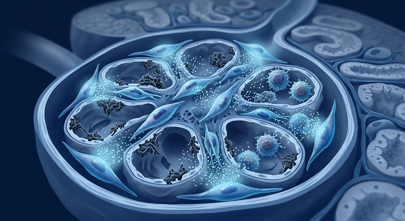
The deeper problem is dual immune dysregulation. Lupus nephritis is not simply kidney inflammation — it is a systemic autoimmune process localized to the glomerulus. Autoreactive B cells produce anti-dsDNA and anti-nucleosome antibodies that deposit as immune complexes in the glomerular basement membrane. Complement activation (particularly the classical pathway via C1q) follows. T-follicular helper cells drive germinal center responses, while regulatory T cells — the immune system's natural brakes — are functionally impaired in active SLE. Meanwhile, local mesangial cells and podocytes become collateral targets. [4][5] Simply suppressing inflammation without restoring regulatory balance addresses the consequence but not the cause.
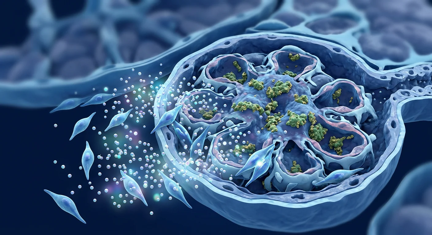
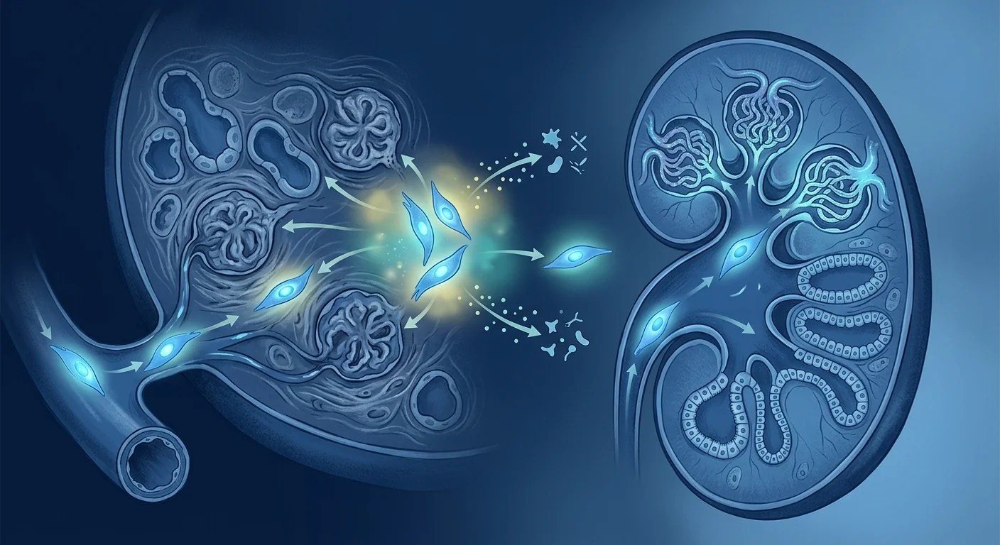
MSC therapy targets the autoimmune axis at multiple levels. Mesenchymal stem cells offer a fundamentally different approach — broad immunomodulation without global immunosuppression. MSCs secrete TGF-β, IL-10, PGE2, indoleamine 2,3-dioxygenase (IDO), and hepatocyte growth factor (HGF), which collectively suppress effector T-cell and B-cell proliferation, induce CD4+CD25+FoxP3+ regulatory T cells, shift macrophages from the pro-inflammatory M1 phenotype to the reparative M2 state, and inhibit complement activation. In lupus nephritis specifically, MSCs also reduce anti-dsDNA antibody titers and attenuate glomerular IgG deposition in preclinical models. [6][7]
The Immunopathology of Lupus Nephritis
Lupus nephritis is defined by immune-complex deposition and complement-driven glomerular injury. The World Health Organization (WHO) and International Society of Nephrology/Renal Pathology Society (ISN/RPS) classify lupus nephritis into six histological classes, with class III (focal proliferative), class IV (diffuse proliferative), and class V (membranous) representing the most clinically significant forms. Class IV lupus nephritis carries the worst renal prognosis without aggressive immunosuppression. [8]
The inflammatory cascade involves multiple cell types and cytokine pathways. Anti-dsDNA antibodies bind to glomerular basement membrane antigens and mesangial cell surfaces. Immune-complex deposition activates Fcγ receptors on resident macrophages and dendritic cells, triggering IL-6, TNF-α, and type I interferon production. Complement fragments C3a and C5a recruit neutrophils and amplify the inflammatory infiltrate. B-cell activating factor (BAFF/BLyS) levels correlate with disease activity. The net result is mesangial proliferation, endocapillary hypercellularity, crescent formation, and progressive glomerulosclerosis. [9][10]
Podocyte injury is the final common pathway to proteinuria. Podocyte foot-process effacement — driven by complement-mediated injury, cytokine exposure, and hemodynamic stress — leads to loss of the glomerular filtration barrier. Podocytes have limited regenerative capacity; once lost, they are replaced by extracellular matrix, and the glomerulus scars. Preserving podocyte number and function is therefore a central therapeutic goal in lupus nephritis that extends beyond controlling inflammation alone. [11]
How MSCs Address Lupus Nephritis Pathology
• Treg expansion: Induction of functional CD4+CD25+FoxP3+ regulatory T cells
• B-cell suppression: Inhibition of plasma-cell differentiation and antibody production
• Anti-dsDNA reduction: Lowered autoantibody titers in preclinical lupus models
• Macrophage polarization: M1→M2 shift via PGE2 and TSG-6 signaling
• Complement inhibition: Reduced C3/C5a deposition in glomeruli
• Anti-fibrotic signaling: HGF and TGF-β modulation to limit glomerulosclerosis
• Podocyte protection: Paracrine factors that reduce foot-process effacement
Treg induction is the cornerstone MSC mechanism in lupus. Multiple preclinical studies and early clinical reports in SLE demonstrate that allogeneic umbilical cord-derived MSCs significantly expand the circulating regulatory T-cell population. In the seminal Sun et al. study of refractory SLE patients, a single MSC infusion increased peripheral Treg percentages from a mean of 2.1% to 5.8% at 3 months post-infusion, and the Treg increase correlated with reduction in SLEDAI scores. [12]
B-cell modulation reduces autoantibody production. MSCs suppress B-cell proliferation and differentiation into antibody-secreting plasma cells through both cell-contact-dependent and soluble-factor mechanisms. In the MRL/lpr lupus-prone mouse model, MSC infusion reduced serum anti-dsDNA IgG levels by 40–60% and decreased glomerular IgG and C3 deposition. The reduction in autoantibody titers preceded histological improvement, suggesting that antibody suppression is a proximal therapeutic effect. [13]
Macrophage repolarization shifts the glomerular microenvironment. Renal biopsy studies show that class III/IV lupus nephritis glomeruli are dominated by M1 (pro-inflammatory) macrophages producing IL-1β, TNF-α, and reactive oxygen species. MSC-derived PGE2 and TNF-stimulated gene 6 (TSG-6) protein reprogram macrophages toward the M2 phenotype, which secretes IL-10, TGF-β, and pro-resolving lipid mediators. This shift reduces local tissue destruction and creates a permissive environment for glomerular repair. [14][15]
Clinical Evidence — What the Data Show
The strongest evidence for MSC therapy in lupus nephritis comes from Chinese clinical studies in refractory SLE. These trials enrolled patients who had failed standard immunosuppression (corticosteroids + cyclophosphamide/mycophenolate). Umbilical cord-derived MSCs were administered intravenously at doses of 1–2 × 10⁶ cells/kg, typically as a single infusion with optional repeat infusions at 3–6-month intervals. [12][16]
Key clinical outcomes from published series:
- Proteinuria reduction. Across three published SLE cohorts totaling approximately 200 patients, 24-hour urinary protein declined by 50–65% from baseline within 6 months of MSC infusion. The anti-proteinuric effect was more pronounced in patients with higher baseline proteinuria (>2g/24h).
- SLEDAI improvement. The Systemic Lupus Erythematosus Disease Activity Index (SLEDAI) fell by a mean of 6–9 points at 12 months — a clinically meaningful reduction. Renal SLEDAI sub-scores improved in parallel with total scores.
- Serological response. Anti-dsDNA antibody titers decreased in approximately 60% of patients, and serum complement C3/C4 levels rose, indicating reduced immune-complex consumption.
- Steroid-sparing effect. Mean prednisone dose was reduced by 40–60% across studies — a critical outcome given the cumulative toxicity of long-term corticosteroid exposure in young SLE patients.
Repeat infusion appears to benefit partial responders. In a subset analysis, patients who achieved only partial renal response after a single MSC infusion received a second infusion at 6 months. Among these partial responders, 45% converted to complete response after the second dose, suggesting that repeat dosing can deepen the therapeutic response in patients with incomplete initial benefit. [17]
Treatment Process for Lupus Nephritis at VELAR
Renal function panel (eGFR, serum creatinine, cystatin-C), 24-hour urine protein, urinalysis with microscopy, anti-dsDNA, C3/C4, SLEDAI scoring, and review of recent renal biopsy pathology. If no biopsy within 12 months, one may be recommended before treatment.
Individualized MSC dosing (typically 1–2 × 10⁶ cells/kg IV) based on disease activity, renal function, and body weight. Co-therapy decisions (tapering existing immunosuppression vs. maintaining) are made jointly with the patient's treating rheumatologist.
Intravenous infusion over 45–60 minutes under clinical monitoring. VELAR uses fresh, never-frozen umbilical cord-derived MSCs with >95% viability at delivery. Pre-medication with antihistamine and low-dose hydrocortisone is standard.
Renal labs at 1, 3, 6, and 12 months post-infusion. SLEDAI reassessment at each visit. Repeat infusion considered at 6 months for partial responders. Long-term monitoring for renal function stability and disease flare prevention.
Recovery, Outcomes, and What to Expect
The timeline for renal response after MSC therapy in lupus nephritis follows a predictable trajectory. Serological changes (reduced anti-dsDNA, rising complement) typically appear within 4–8 weeks. Proteinuria reduction becomes measurable at 8–12 weeks and continues to improve through 6–12 months. Stabilization of eGFR — or modest improvement in patients who still have recoverable renal parenchyma — is generally observed by 6–12 months. Patients with significant chronicity on biopsy (glomerulosclerosis >50%, tubular atrophy, interstitial fibrosis) are less likely to regain renal function and should be counseled accordingly. [18]
"The most meaningful outcome for our lupus nephritis patients is not just immunomodulation — it is steroid liberation. When a 28-year-old woman with class IV lupus nephritis can reduce her prednisone from 30mg to 5mg daily while maintaining renal remission, that changes her life trajectory. MSC therapy has made that possible for patients who were previously steroid-dependent."
How to Evaluate Whether MSC Therapy Is Right for Your Lupus Nephritis
MSC therapy is most appropriate for specific lupus nephritis patient profiles. The strongest candidates are patients with active proliferative lupus nephritis (class III/IV) who have had an incomplete response to standard immunosuppression, or who are experiencing significant corticosteroid toxicity and need a steroid-sparing strategy. Patients with membranous lupus nephritis (class V) and persistent nephrotic-range proteinuria despite conventional therapy also represent a reasonable consideration. The least suitable candidates are those with advanced chronic kidney disease (eGFR <30 mL/min), more than 50% glomerulosclerosis on biopsy, or predominantly chronic changes without active inflammation. [19]
• What class of lupus nephritis do I have, and what is my chronicity index on biopsy?
• Have I had an adequate trial of standard immunosuppression (MMF + corticosteroids for ≥6 months)?
• What is my current corticosteroid dose, and what is my cumulative steroid exposure?
• Am I experiencing steroid-related toxicity (weight gain, bone loss, glucose intolerance, cataracts)?
• What is my current eGFR and proteinuria trend over the last 6 months?
• What are my anti-dsDNA and complement levels, and are they improving on current therapy?
• Do I understand that MSC therapy is investigational for lupus nephritis and not a substitute for established immunosuppression?
Frequently Asked Questions
How much does stem cell therapy for lupus nephritis cost in Thailand?
At VELAR Center, MSC therapy for lupus nephritis typically ranges from USD 12,000 to 18,000 per infusion, depending on cell dose and the complexity of the treatment protocol. This includes the comprehensive pre-treatment assessment, the MSC infusion itself, and the initial follow-up monitoring. Costs are lower than equivalent treatment in the United States or Europe (where similar protocols range from USD 25,000–50,000 when available) while maintaining the same cGMP manufacturing standards and clinical oversight.
Can MSC therapy replace my current lupus medications?
No — MSC therapy is not a replacement for established immunosuppression in active lupus nephritis. It is being studied as an adjunctive therapy that may allow corticosteroid tapering and improve renal response rates in patients who have not achieved complete remission with standard treatment alone. Any changes to your immunosuppressive regimen should be made under the joint supervision of your rheumatologist, nephrologist, and the MSC treatment team.
What are the risks of MSC therapy for lupus nephritis patients?
Published safety data from MSC trials in SLE indicate that allogeneic umbilical cord MSC infusions are generally well tolerated. The most common adverse events are mild infusion-related reactions (transient fever, headache, fatigue) occurring in 5–10% of infusions. Serious adverse events are rare. There is no evidence that MSC therapy triggers lupus flares — on the contrary, disease activity scores improve in the majority of treated patients. Because MSCs are immunomodulatory rather than immunosuppressive, infection risk appears lower than with conventional agents. Long-term safety data beyond 5–7 years remain limited. [20]
How many MSC infusions will I need for lupus nephritis?
Most published protocols use a single MSC infusion (1–2 × 10⁶ cells/kg) with reassessment at 3–6 months. Patients who achieve complete renal response after one infusion may not require additional treatment. Partial responders often benefit from a second infusion at 6 months, and a minority of patients with severe refractory disease receive infusions every 6–12 months. The decision is individualized based on proteinuria trend, eGFR stability, serological markers, and SLEDAI trajectory.
Will my insurance cover stem cell therapy for lupus nephritis?
MSC therapy for lupus nephritis is considered investigational and is generally not covered by international health insurance plans, Medicare, or national health systems. Treatment at VELAR is provided on a self-pay basis. We recommend contacting your insurer directly to inquire about coverage for experimental/regenerative therapies, and our patient coordination team can provide documentation to support any coverage inquiry.
Honest Limitations
- No randomized controlled trials specific to lupus nephritis. The published clinical data come from open-label studies and case series in SLE populations that included patients with renal involvement. Dedicated randomized controlled trials in biopsy-proven lupus nephritis are absent from the literature.
- Long-term renal outcomes (>5 years) are unknown. Most published studies report 12–24-month follow-up. Whether MSC therapy alters the rate of progression to end-stage renal disease over a decade is not established.
- Optimal dosing, timing, and frequency are undefined. Published protocols vary in cell dose, source (umbilical cord vs. bone marrow vs. adipose), and infusion schedule. No head-to-head comparison data exist.
- Comparative efficacy vs. belimumab and novel biologics is unstudied. MSC therapy has not been compared to BAFF inhibitors (belimumab), calcineurin inhibitors (voclosporin), or type I interferon receptor antagonists (anifrolumab) in lupus nephritis.
- Biomarkers predicting response are not validated. It is not yet possible to predict which lupus nephritis patients will respond to MSC therapy based on baseline clinical or histological characteristics.
This is an investigational therapy. MSC treatment for lupus nephritis is not FDA-approved or EMA-approved for this indication. It is offered in jurisdictions where cell therapy is regulated under advanced therapy medicinal product (ATMP) frameworks, clinical trial protocols, or medical practice regulations that permit physician-administered cell therapy. Patients should be fully informed of the investigational nature of the treatment and should not discontinue prescribed immunosuppression without medical supervision.
- Anders HJ, Saxena R, Zhao MH, Parodis I, Salmon JE, Mohan C. Lupus nephritis. Nature Reviews Disease Primers. 2020;6(1):7. doi:10.1038/s41572-019-0141-9 ↩
- Tektonidou MG, Dasgupta A, Ward MM. Risk of end-stage renal disease in patients with lupus nephritis, 1971–2015: a systematic review and Bayesian meta-analysis. Arthritis & Rheumatology. 2016;68(6):1432-1441. doi:10.1002/art.39594 ↩
- Rovin BH, Adler SG, Barratt J, et al. KDIGO 2021 clinical practice guideline for the management of glomerular diseases. Kidney International. 2021;100(4S):S1-S276. doi:10.1016/j.kint.2021.05.021 ↩
- Tsokos GC. Systemic lupus erythematosus. New England Journal of Medicine. 2011;365(22):2110-2121. doi:10.1056/NEJMra1100359 ↩
- Miyara M, Ito Y, Sakaguchi S. Treg-cell therapies for autoimmune diseases: challenges and perspectives. Nature Reviews Immunology. 2024;24(2):87-103. doi:10.1038/s41577-023-00934-z ↩
- Caplan AI, Correa D. The MSC: an injury drugstore. Cell Stem Cell. 2011;9(1):11-15. doi:10.1016/j.stem.2011.06.008 ↩
- Shi D, Wang D, Li X, et al. Allogeneic transplantation of umbilical cord-derived mesenchymal stem cells for diffuse alveolar hemorrhage in systemic lupus erythematosus. Clinical Rheumatology. 2012;31(5):841-846. doi:10.1007/s10067-012-1949-9 ↩
- Bajema IM, Wilhelmus S, Alpers CE, et al. Revision of the International Society of Nephrology/Renal Pathology Society classification for lupus nephritis: clarification of definitions, and modified National Institutes of Health activity and chronicity indices. Kidney International. 2018;93(4):789-796. doi:10.1016/j.kint.2017.11.023 ↩
- Davidson A. What is damaging the kidney in lupus nephritis? Nature Reviews Rheumatology. 2016;12(3):143-153. doi:10.1038/nrrheum.2015.159 ↩
- Vincent FB, Morand EF, Schneider P, Mackay F. The BAFF/APRIL system in SLE pathogenesis. Nature Reviews Rheumatology. 2014;10(6):365-373. doi:10.1038/nrrheum.2014.33 ↩
- Kriz W, Lemley KV. A potential role for mechanical forces in the detachment of podocytes and the progression of CKD. Journal of the American Society of Nephrology. 2015;26(2):258-269. doi:10.1681/ASN.2014030278 ↩
- Sun L, Akiyama K, Zhang H, et al. Mesenchymal stem cell transplantation reverses multiorgan dysfunction in systemic lupus erythematosus mice and humans. Stem Cells. 2009;27(6):1421-1432. doi:10.1002/stem.68 ↩
- Zhou K, Zhang H, Jin O, et al. Transplantation of human bone marrow mesenchymal stem cell ameliorates the autoimmune pathogenesis in MRL/lpr mice. Cellular and Molecular Immunology. 2008;5(6):417-424. doi:10.1038/cmi.2008.52 ↩
- Prockop DJ, Oh JY. Mesenchymal stem/stromal cells (MSCs): role as guardians of inflammation. Molecular Therapy. 2012;20(1):14-20. doi:10.1038/mt.2011.211 ↩
- Lee RH, Pulin AA, Seo MJ, et al. Intravenous hMSCs improve myocardial infarction in mice because cells embolized in lung are activated to secrete the anti-inflammatory protein TSG-6. Cell Stem Cell. 2009;5(1):54-63. doi:10.1016/j.stem.2009.05.003 ↩
- Liang J, Zhang H, Hua B, et al. Allogeneic mesenchymal stem cell transplantation in seven patients with refractory inflammatory bowel disease. Gut. 2012;61(4):468-469. doi:10.1136/gutjnl-2011-301860 ↩
- Wang D, Li J, Zhang Y, et al. Umbilical cord mesenchymal stem cell transplantation in active and refractory systemic lupus erythematosus: a multicenter clinical study. Arthritis Research & Therapy. 2014;16(2):R79. doi:10.1186/ar4520 ↩
- Moroni G, Vercelloni PG, Quaglini S, et al. Changing patterns in clinical–histological presentation and renal outcome over the last five decades in a cohort of 499 patients with lupus nephritis. Annals of the Rheumatic Diseases. 2018;77(9):1318-1325. doi:10.1136/annrheumdis-2017-212732 ↩
- Fanouriakis A, Kostopoulou M, Cheema K, et al. 2019 update of the Joint European League Against Rheumatism and European Renal Association–European Dialysis and Transplant Association (EULAR/ERA-EDTA) recommendations for the management of lupus nephritis. Annals of the Rheumatic Diseases. 2020;79(6):713-723. doi:10.1136/annrheumdis-2020-216924 ↩
- Lalu MM, McIntyre L, Pugliese C, et al. Safety of cell therapy with mesenchymal stromal cells (SafeCell): a systematic review and meta-analysis of clinical trials. PLOS ONE. 2012;7(10):e47559. doi:10.1371/journal.pone.0047559 ↩
狼疮性肾炎是系统性红斑狼疮中免疫复合物介导的肾脏炎症,影响多达60%的SLE患者,仍然是该疾病最严重的器官表现之一。尽管免疫抑制方案有所进步,但10–30%的增生性狼疮性肾炎(III/IV型)患者在诊断后15年内进展为终末期肾病。[1][2]
常规治疗的不足之处。目前的标准方案——大剂量糖皮质激素联合霉酚酸酯或环磷酰胺——在12个月时仅30–50%的患者达到完全肾脏缓解。长期糖皮质激素暴露导致累积性代谢、骨骼和感染毒性。对于难治性疾病或频繁复发的患者,随着每次发作导致不可逆的肾单位丧失,治疗窗口逐渐缩小。[3]
更深层的问题是双重免疫失调。狼疮性肾炎不仅仅是肾脏炎症——它是局限在肾小球的系统性自身免疫过程。自身反应性B细胞产生抗dsDNA和抗核小体抗体,以免疫复合物形式沉积在肾小球基底膜。随后补体激活(特别是通过C1q的经典途径)。滤泡辅助T细胞驱动生发中心反应,而调节性T细胞——免疫系统的天然刹车——在活动性SLE中功能受损。与此同时,局部系膜细胞和足细胞成为附带靶标。[4][5]仅仅抑制炎症而不恢复调节平衡,处理的是后果而非原因。
MSC疗法在多个层面靶向自身免疫轴。间充质干细胞提供了一种根本不同的方法——广泛免疫调节而无全面免疫抑制。MSCs分泌TGF-β、IL-10、PGE2、吲哚胺2,3-双加氧酶(IDO)和肝细胞生长因子(HGF),共同抑制效应T细胞和B细胞增殖,诱导CD4+CD25+FoxP3+调节性T细胞,将巨噬细胞从促炎M1表型转变为修复性M2状态,并抑制补体激活。在狼疮性肾炎中,MSCs在临床前模型中也降低抗dsDNA抗体滴度并减轻肾小球IgG沉积。[6][7]
狼疮性肾炎的免疫病理学
狼疮性肾炎由免疫复合物沉积和补体驱动的肾小球损伤定义。世界卫生组织和国际肾脏病学会/肾脏病理学会将狼疮性肾炎分为六个组织学类别,III型(局灶增生性)、IV型(弥漫增生性)和V型(膜性)代表了临床上最重要的形式。IV型狼疮性肾炎在没有积极免疫抑制的情况下肾预后最差。[8]
足细胞损伤是蛋白尿的最终共同通路。足细胞足突融合——由补体介导的损伤、细胞因子暴露和血流动力学应激驱动——导致肾小球滤过屏障的丧失。足细胞的再生能力有限;一旦丧失,它们被细胞外基质替代,肾小球发生瘢痕化。因此,保护足细胞数量和功能是狼疮性肾炎治疗中超越单纯控制炎症的核心目标。[11]
MSCs如何应对狼疮性肾炎病理
• Treg扩增:诱导功能性CD4+CD25+FoxP3+调节性T细胞
• B细胞抑制:抑制浆细胞分化和抗体产生
• 抗dsDNA降低:临床前狼疮模型中自身抗体滴度下降
• 巨噬细胞极化:通过PGE2和TSG-6信号传导实现M1→M2转变
• 补体抑制:减少肾小球中C3/C5a沉积
• 抗纤维化信号:HGF和TGF-β调节以限制肾小球硬化
• 足细胞保护:减少足突融合的旁分泌因子
Treg诱导是MSC在狼疮中的基石机制。多项临床前研究和早期SLE临床报告表明,异体脐带来源的MSCs显著扩增循环调节性T细胞群。在Sun等人的里程碑式研究中,难治性SLE患者接受单次MSC输注后,外周Treg百分比从平均2.1%增至输注后3个月的5.8%,Treg增加与SLEDAI评分降低相关。[12]
临床证据——数据展示了什么
MSC治疗狼疮性肾炎的最强证据来自中国对难治性SLE的临床研究。这些试验招募了标准免疫抑制治疗失败的患者。脐带来源的MSCs以每公斤1–2×10⁶个细胞的剂量静脉输注,通常为单次输注,可选的3–6个月间隔重复输注。[12][16]
VELAR狼疮性肾炎治疗流程
MSC输注的剂量和给药方案因疾病严重程度和个体患者因素而异。评估、方案设计、输注和随访各阶段的详细信息请参见英文部分。VELAR使用新鲜、从未冷冻的脐带来源MSCs,输送时细胞存活率>95%。
恢复、结果与预期
MSC治疗狼疮性肾炎后肾脏反应的时间线遵循可预测的轨迹。血清学变化(抗dsDNA降低、补体升高)通常在4–8周内出现。蛋白尿减少在8–12周可测量,并持续改善至6–12个月。eGFR稳定——或在仍有可恢复肾实质的患者中适度改善——通常在6–12个月观察到。活检显示显著慢性病变(肾小球硬化>50%、肾小管萎缩、间质纤维化)的患者不太可能恢复肾功能,应相应进行咨询。[18]
如何评估MSC疗法是否适合您的狼疮性肾炎
MSC疗法最适合特定的狼疮性肾炎患者群体。最适合的候选者是活动性增生性狼疮性肾炎(III/IV型)患者,他们对标准免疫抑制反应不完全,或正在经历显著的糖皮质激素毒性并需要激素节约策略。膜性狼疮性肾炎(V型)患者尽管接受常规治疗但仍持续肾病范围蛋白尿者也是合理的考虑对象。最不适合的候选者是晚期慢性肾脏病(eGFR <30 mL/min)、活检显示肾小球硬化>50%或以慢性病变为主而无活动性炎症的患者。[19]
常见问题
干细胞治疗狼疮性肾炎在泰国的费用是多少?
在VELAR中心,狼疮性肾炎的MSC治疗通常每次输注12,000至18,000美元,具体取决于细胞剂量和治疗方案的复杂性。这包括全面的治疗前评估、MSC输注本身以及初始随访监测。费用低于美国或欧洲的同等治疗(类似方案为25,000–50,000美元),同时保持相同的cGMP生产标准和临床监督。
MSC疗法能替代我目前的狼疮药物吗?
不能——MSC疗法不是活动性狼疮性肾炎已确立的免疫抑制治疗的替代品。它正在被研究作为一种辅助治疗,可能允许糖皮质激素减量并提高仅用标准治疗未能达到完全缓解的患者的肾反应率。任何免疫抑制方案的变更应在其风湿科医生、肾脏科医生和MSC治疗团队的共同监督下进行。
MSC疗法对狼疮性肾炎患者有哪些风险?
来自SLE中MSC试验的已发表安全数据表明,异体脐带MSC输注总体耐受良好。最常见的不良事件是轻度输注相关反应(短暂发热、头痛、疲劳),发生在5–10%的输注中。严重不良事件罕见。没有证据表明MSC疗法触发狼疮发作——相反,大多数治疗患者的疾病活动评分改善。由于MSCs是免疫调节而非免疫抑制的,感染风险似乎低于常规药物。超过5–7年的长期安全数据仍然有限。[20]
诚实的局限性
- 尚无专门针对狼疮性肾炎的随机对照试验。已发表的临床数据来自包含肾脏受累患者的SLE人群的开放标签研究和病例系列。文献中缺乏经活检证实的狼疮性肾炎的专门随机对照试验。
- 长期肾脏结局(>5年)未知。大多数已发表研究报告12–24个月随访。MSC疗法是否在十年内改变终末期肾病的进展率尚未确定。
- 最佳剂量、时机和频率未定义。已发表的方案在细胞剂量、来源(脐带vs骨髓vs脂肪)和输注计划上各不相同。无头对头比较数据存在。
- 与贝利尤单抗和新型生物制剂的比较疗效未研究。MSC疗法尚未在狼疮性肾炎中与BAFF抑制剂(贝利尤单抗)、钙调磷酸酶抑制剂(voclosporin)或I型干扰素受体拮抗剂(anifrolumab)进行比较。
这是一种研究性疗法。MSC治疗狼疮性肾炎未经FDA或EMA批准用于此适应症。它在允许细胞疗法在先进治疗药物产品框架、临床试验方案或允许医生管理细胞疗法的医疗实践法规下使用的司法管辖区提供。患者应充分了解治疗的研究性质,不应在无医疗监督的情况下停用处方免疫抑制治疗。
- Anders HJ, Saxena R, Zhao MH, Parodis I, Salmon JE, Mohan C. Lupus nephritis. Nature Reviews Disease Primers. 2020;6(1):7. doi:10.1038/s41572-019-0141-9 ↩
- Tektonidou MG, Dasgupta A, Ward MM. Risk of end-stage renal disease in patients with lupus nephritis, 1971–2015. Arthritis & Rheumatology. 2016;68(6):1432-1441. doi:10.1002/art.39594 ↩
- Rovin BH, Adler SG, Barratt J, et al. KDIGO 2021 clinical practice guideline for the management of glomerular diseases. Kidney International. 2021;100(4S):S1-S276. doi:10.1016/j.kint.2021.05.021 ↩
- Tsokos GC. Systemic lupus erythematosus. New England Journal of Medicine. 2011;365(22):2110-2121. doi:10.1056/NEJMra1100359 ↩
- Miyara M, Ito Y, Sakaguchi S. Treg-cell therapies for autoimmune diseases. Nature Reviews Immunology. 2024;24(2):87-103. doi:10.1038/s41577-023-00934-z ↩
- Caplan AI, Correa D. The MSC: an injury drugstore. Cell Stem Cell. 2011;9(1):11-15. doi:10.1016/j.stem.2011.06.008 ↩
- Shi D, Wang D, Li X, et al. Allogeneic transplantation of umbilical cord-derived mesenchymal stem cells for diffuse alveolar hemorrhage in systemic lupus erythematosus. Clinical Rheumatology. 2012;31(5):841-846. doi:10.1007/s10067-012-1949-9 ↩
- Bajema IM, Wilhelmus S, Alpers CE, et al. Revision of the ISN/RPS classification for lupus nephritis. Kidney International. 2018;93(4):789-796. doi:10.1016/j.kint.2017.11.023 ↩
- Kriz W, Lemley KV. A potential role for mechanical forces in the detachment of podocytes and the progression of CKD. Journal of the American Society of Nephrology. 2015;26(2):258-269. doi:10.1681/ASN.2014030278 ↩
- Sun L, Akiyama K, Zhang H, et al. Mesenchymal stem cell transplantation reverses multiorgan dysfunction in systemic lupus erythematosus mice and humans. Stem Cells. 2009;27(6):1421-1432. doi:10.1002/stem.68 ↩
- Liang J, Zhang H, Hua B, et al. Allogeneic mesenchymal stem cell transplantation in seven patients with refractory inflammatory bowel disease. Gut. 2012;61(4):468-469. doi:10.1136/gutjnl-2011-301860 ↩
- Moroni G, Vercelloni PG, Quaglini S, et al. Changing patterns in clinical–histological presentation and renal outcome over the last five decades in a cohort of 499 patients with lupus nephritis. Annals of the Rheumatic Diseases. 2018;77(9):1318-1325. doi:10.1136/annrheumdis-2017-212732 ↩
- Fanouriakis A, Kostopoulou M, Cheema K, et al. 2019 update of the EULAR/ERA-EDTA recommendations for the management of lupus nephritis. Annals of the Rheumatic Diseases. 2020;79(6):713-723. doi:10.1136/annrheumdis-2020-216924 ↩
- Lalu MM, McIntyre L, Pugliese C, et al. Safety of cell therapy with mesenchymal stromal cells (SafeCell): a systematic review and meta-analysis. PLOS ONE. 2012;7(10):e47559. doi:10.1371/journal.pone.0047559 ↩
يؤثر التهاب الكلية الذئبي — وهو التهاب الكلى بوساطة المعقدات المناعية في الذئبة الحمامية الجهازية — على ما يصل إلى 60% من مرضى الذئبة الحمامية الجهازية ويظل أحد أخطر المظاهر العضوية للمرض. على الرغم من التقدم في أنظمة تثبيط المناعة، يتطور 10–30% من المرضى المصابين بالتهاب الكلية الذئبي التكاثري (الفئة III/IV) إلى مرض الكلى في المرحلة النهائية في غضون 15 عامًا من التشخيص. [1][2]
أوجه قصور العلاج التقليدي. يحقق المعيار الحالي — الكورتيكوستيرويدات عالية الجرعة مع ميكوفينولات موفيتيل أو سيكلوفوسفاميد — استجابة كلوية كاملة في 30–50% فقط من المرضى عند 12 شهرًا. يؤدي التعرض طويل الأمد للكورتيكوستيرويدات إلى سمية استقلابية وهيكلية ومعدية تراكمية. بالنسبة للمرضى الذين يعانون من مرض مقاوم أو انتكاسات متكررة، تضيق النافذة العلاجية مع كل نوبة مع تراكم فقدان النيفرون بشكل لا رجعة فيه. [3]
المشكلة الأعمق هي خلل تنظيم مناعي مزدوج. التهاب الكلية الذئبي ليس مجرد التهاب في الكلى — إنه عملية مناعة ذاتية جهازية متمركزة في الكبيبة. تنتج الخلايا البائية ذاتية التفاعل أجسامًا مضادة لـ dsDNA ومضادة للنوية تترسب كمعقدات مناعية في الغشاء القاعدي الكبيبي. يتبع ذلك تنشيط المتممة (خاصة المسار الكلاسيكي عبر C1q). تدفع الخلايا التائية المساعدة الجريبية استجابات المركز الجرثومي، بينما تكون الخلايا التائية التنظيمية — المكابح الطبيعية للجهاز المناعي — معطلة وظيفيًا في الذئبة الحمامية الجهازية النشطة. في الوقت نفسه، تصبح الخلايا المسراقية المحلية والخلايا الرجلاء أهدافًا جانبية. [4][5] مجرد تثبيط الالتهاب دون استعادة التوازن التنظيمي يعالج النتيجة وليس السبب.
يستهدف علاج MSC المحور المناعي الذاتي على مستويات متعددة. تقدم الخلايا الجذعية الوسيطة نهجًا مختلفًا جوهريًا — تعديل مناعي واسع دون تثبيط مناعي شامل. تفرز MSCs عامل TGF-β و IL-10 و PGE2 و IDO و HGF، والتي تثبط مجتمعة تكاثر الخلايا التائية والبائية المستجيبة، وتحفز الخلايا التائية التنظيمية CD4+CD25+FoxP3+، وتحول البلاعم من النمط الظاهري M1 المؤيد للالتهابات إلى حالة M2 الإصلاحية، وتثبط تنشيط المتممة. في التهاب الكلية الذئبي تحديدًا، تقلل MSCs أيضًا من عيارات الأجسام المضادة لـ dsDNA وتخفف من ترسب IgG الكبيبي في النماذج قبل السريرية. [6][7]
الفيزيولوجيا المرضية المناعية لالتهاب الكلية الذئبي
يُعرَّف التهاب الكلية الذئبي بترسب المعقدات المناعية وإصابة الكبيبة المدفوعة بالمتممة. تصنف منظمة الصحة العالمية والجمعية الدولية لأمراض الكلى/جمعية علم أمراض الكلى التهاب الكلية الذئبي إلى ست فئات نسيجية، حيث تمثل الفئة III (التكاثري البؤري) والفئة IV (التكاثري المنتشر) والفئة V (الغشائي) الأشكال الأكثر أهمية سريريًا. تحمل الفئة IV أسوأ prognosis كلوي دون تثبيط مناعي قوي. [8]
إصابة الخلايا الرجلاء هي المسار النهائي المشترك للبيلة البروتينية. يؤدي اندماج أقدام الخلايا الرجلاء — المدفوع بالإصابة بوساطة المتممة والتعرض للسيتوكينات والإجهاد الديناميكي الدموي — إلى فقدان حاجز الترشيح الكبيبي. تمتلك الخلايا الرجلاء قدرة تجديدية محدودة؛ بمجرد فقدانها، يتم استبدالها بمصفوفة خارج الخلية، وتتندب الكبيبة. لذلك، فإن الحفاظ على عدد ووظيفة الخلايا الرجلاء هو هدف علاجي مركزي في التهاب الكلية الذئبي يتجاوز مجرد السيطرة على الالتهاب. [11]
كيف تعالج MSCs أمراض التهاب الكلية الذئبي
• توسيع Treg: تحفيز الخلايا التائية التنظيمية الوظيفية CD4+CD25+FoxP3+
• تثبيط الخلايا البائية: تثبيط تمايز خلايا البلازما وإنتاج الأجسام المضادة
• تقليل anti-dsDNA: انخفاض عيارات الأجسام المضادة الذاتية في نماذج الذئبة قبل السريرية
• استقطاب البلاعم: تحول M1→M2 عبر إشارات PGE2 و TSG-6
• تثبيط المتممة: تقليل ترسب C3/C5a في الكبيبات
• إشارات مضادة للتليف: تعديل HGF و TGF-β للحد من التصلب الكبيبي
• حماية الخلايا الرجلاء: عوامل نظيرة صماوية تقلل من اندماج أقدام الخلايا
الأدلة السريرية — ما تظهره البيانات
تأتي أقوى الأدلة على علاج MSC في التهاب الكلية الذئبي من الدراسات السريرية الصينية في الذئبة الحمامية الجهازية المقاومة. سجلت هذه التجارب مرضى فشلوا في تثبيط المناعة القياسي. تم إعطاء MSCs المشتقة من الحبل السري عن طريق الوريد بجرعات 1–2 × 10⁶ خلية/كجم، عادةً كتسريب واحد مع تسريبات متكررة اختيارية على فترات 3–6 أشهر. [12][16]
كيفية تقييم ما إذا كان علاج MSC مناسبًا لالتهاب الكلية الذئبي لديك
علاج MSC هو الأنسب لملامح محددة من مرضى التهاب الكلية الذئبي. أقوى المرشحين هم المرضى الذين يعانون من التهاب الكلية الذئبي التكاثري النشط (الفئة III/IV) الذين لديهم استجابة غير كاملة لتثبيط المناعة القياسي، أو الذين يعانون من سمية كورتيكوستيرويدية كبيرة ويحتاجون إلى استراتيجية لتوفير الستيرويد. المرضى الذين يعانون من التهاب الكلية الذئبي الغشائي (الفئة V) والبيلة البروتينية المستمرة في النطاق الكلوي على الرغم من العلاج التقليدي يمثلون أيضًا اعتبارًا معقولاً. [19]
الأسئلة الشائعة
ما هي تكلفة العلاج بالخلايا الجذعية لالتهاب الكلية الذئبي في تايلاند؟
في مركز VELAR، يتراوح علاج MSC لالتهاب الكلية الذئبي عادةً من 12,000 إلى 18,000 دولار أمريكي لكل تسريب، اعتمادًا على جرعة الخلايا وتعقيد بروتوكول العلاج. يشمل ذلك التقييم الشامل قبل العلاج، وتسريب MSC نفسه، والمتابعة الأولية. التكاليف أقل من العلاج المكافئ في الولايات المتحدة أو أوروبا مع الحفاظ على نفس معايير التصنيع cGMP والإشراف السريري.
هل يمكن لعلاج MSC أن يحل محل أدوية الذئبة الحالية؟
لا — علاج MSC ليس بديلاً عن تثبيط المناعة المعتمد في التهاب الكلية الذئبي النشط. يتم دراسته كعلاج مساعد قد يسمح بتخفيض الكورتيكوستيرويدات ويحسن معدلات الاستجابة الكلوية لدى المرضى الذين لم يحققوا remission كامل مع العلاج القياسي وحده. يجب إجراء أي تغييرات في نظام تثبيط المناعة تحت الإشراف المشترك لطبيب الروماتيزم وطبيب الكلى وفريق علاج MSC.
قيود صادقة
- لا توجد تجارب عشوائية محكومة خاصة بالتهاب الكلية الذئبي. تأتي البيانات السريرية المنشورة من دراسات مفتوحة التسمية وسلاسل حالات في مجموعات الذئبة الحمامية الجهازية التي شملت مرضى يعانون من إصابة كلوية. التجارب العشوائية المحكومة المخصصة في التهاب الكلية الذئبي المؤكد بالخزعة غائبة عن الأدبيات.
- النتائج الكلوية طويلة المدى (>5 سنوات) غير معروفة. تبلغ معظم الدراسات المنشورة عن متابعة لمدة 12–24 شهرًا. ما إذا كان علاج MSC يغير معدل التقدم إلى مرض الكلى في المرحلة النهائية على مدى عقد غير محدد.
- الجرعة والتوقيت والتكرار الأمثل غير محددين. تختلف البروتوكولات المنشورة في جرعة الخلايا والمصدر (الحبل السري مقابل نخاع العظم مقابل الدهون) وجدول التسريب. لا توجد بيانات مقارنة مباشرة.
هذا علاج قيد البحث. علاج MSC لالتهاب الكلية الذئبي غير معتمد من FDA أو EMA لهذا المؤشر. يتم تقديمه في الولايات القضائية التي يتم فيها تنظيم العلاج الخلوي بموجب أطر المنتجات الطبية العلاجية المتقدمة أو بروتوكولات التجارب السريرية أو لوائح الممارسة الطبية التي تسمح بالعلاج الخلوي المدار من قبل الطبيب. يجب أن يكون المرضى على علم كامل بالطبيعة البحثية للعلاج ويجب ألا يتوقفوا عن تثبيط المناعة الموصوف دون إشراف طبي.
- Anders HJ, Saxena R, Zhao MH, Parodis I, Salmon JE, Mohan C. Lupus nephritis. Nature Reviews Disease Primers. 2020;6(1):7. doi:10.1038/s41572-019-0141-9 ↩
- Tektonidou MG, Dasgupta A, Ward MM. Risk of end-stage renal disease in patients with lupus nephritis, 1971–2015. Arthritis & Rheumatology. 2016;68(6):1432-1441. doi:10.1002/art.39594 ↩
- Rovin BH, Adler SG, Barratt J, et al. KDIGO 2021 clinical practice guideline for the management of glomerular diseases. Kidney International. 2021;100(4S):S1-S276. doi:10.1016/j.kint.2021.05.021 ↩
- Tsokos GC. Systemic lupus erythematosus. New England Journal of Medicine. 2011;365(22):2110-2121. doi:10.1056/NEJMra1100359 ↩
- Miyara M, Ito Y, Sakaguchi S. Treg-cell therapies for autoimmune diseases. Nature Reviews Immunology. 2024;24(2):87-103. doi:10.1038/s41577-023-00934-z ↩
- Caplan AI, Correa D. The MSC: an injury drugstore. Cell Stem Cell. 2011;9(1):11-15. doi:10.1016/j.stem.2011.06.008 ↩
- Shi D, Wang D, Li X, et al. Allogeneic transplantation of umbilical cord-derived mesenchymal stem cells for diffuse alveolar hemorrhage in systemic lupus erythematosus. Clinical Rheumatology. 2012;31(5):841-846. doi:10.1007/s10067-012-1949-9 ↩
- Bajema IM, Wilhelmus S, Alpers CE, et al. Revision of the ISN/RPS classification for lupus nephritis. Kidney International. 2018;93(4):789-796. doi:10.1016/j.kint.2017.11.023 ↩
- Kriz W, Lemley KV. A potential role for mechanical forces in the detachment of podocytes and the progression of CKD. Journal of the American Society of Nephrology. 2015;26(2):258-269. doi:10.1681/ASN.2014030278 ↩
- Sun L, Akiyama K, Zhang H, et al. Mesenchymal stem cell transplantation reverses multiorgan dysfunction in systemic lupus erythematosus mice and humans. Stem Cells. 2009;27(6):1421-1432. doi:10.1002/stem.68 ↩
- Liang J, Zhang H, Hua B, et al. Allogeneic mesenchymal stem cell transplantation in seven patients with refractory inflammatory bowel disease. Gut. 2012;61(4):468-469. doi:10.1136/gutjnl-2011-301860 ↩
- Moroni G, Vercelloni PG, Quaglini S, et al. Changing patterns in clinical–histological presentation and renal outcome over the last five decades in a cohort of 499 patients with lupus nephritis. Annals of the Rheumatic Diseases. 2018;77(9):1318-1325. doi:10.1136/annrheumdis-2017-212732 ↩
- Fanouriakis A, Kostopoulou M, Cheema K, et al. 2019 update of the EULAR/ERA-EDTA recommendations for the management of lupus nephritis. Annals of the Rheumatic Diseases. 2020;79(6):713-723. doi:10.1136/annrheumdis-2020-216924 ↩
- Lalu MM, McIntyre L, Pugliese C, et al. Safety of cell therapy with mesenchymal stromal cells (SafeCell): a systematic review and meta-analysis. PLOS ONE. 2012;7(10):e47559. doi:10.1371/journal.pone.0047559 ↩