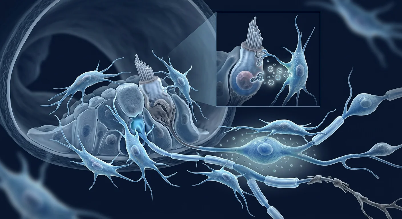
Menière's disease affects roughly 0.2% of the population — approximately 615,000 people in the United States alone — with peak onset between ages 40 and 60. For those living with it, the unpredictable vertigo attacks, progressive sensorineural hearing loss, tinnitus, and aural fullness are not just clinical symptoms; they are a profound erosion of quality of life. Mesenchymal stem cell therapy is being investigated as a novel approach to Menière's disease, targeting the underlying biology — endolymphatic hydrops, cochlear inflammation, and spiral ganglion degeneration — rather than merely suppressing symptoms. [1]
What makes Menière's disease difficult to treat. Current standard-of-care — low-sodium diet, diuretics, betahistine, and in severe cases intratympanic gentamicin or vestibular nerve section — manages symptoms but does not address the underlying pathology. Gentamicin ablation controls vertigo at the cost of vestibular function; surgical options are irreversible and carry their own risks. A therapy that could reduce endolymphatic pressure, dampen cochlear inflammation, and support the survival of auditory and vestibular neurons would represent a genuine advance beyond symptom management. [2]
The biological problem is multi-layered. Menière's disease is pathologically defined by endolymphatic hydrops — an abnormal accumulation of endolymph fluid in the inner ear that distends Reissner's membrane and damages the sensory neuroepithelium. But hydrops alone does not explain the full clinical picture. There is a growing consensus that immune dysregulation, viral triggers, and microvascular dysfunction all contribute to a final common pathway of cochlear and vestibular injury. The key structures at risk are cochlear hair cells (responsible for hearing), spiral ganglion neurons (the primary afferent pathway to the brain), and vestibular hair cells (responsible for balance). [3]
MSC therapy addresses the biology at multiple levels. Rather than targeting a single molecule or pathway, mesenchymal stem cells deploy a broad paracrine program — cytokine modulation, growth factor secretion, extracellular vesicle release, and mitochondrial transfer — that simultaneously addresses inflammation, cellular stress, and tissue repair. This multi-target approach is especially relevant to Menière's disease, where no single drug target has been sufficient to arrest disease progression. [4]
What MSC therapy for Menière's disease means in practice
MSC therapy is not a cure for Menière's disease and is not intended to replace standard medical management. It is an investigational approach being studied for its potential to reduce the frequency and severity of vertigo attacks, slow the progression of hearing loss, and support the long-term health of the auditory-vestibular system — goals that current treatments achieve only partially and often at the cost of function.
How MSC therapy may help Menière's disease: mechanisms of action
Mesenchymal stem cells exert therapeutic effects primarily through their secretome — the complex mixture of bioactive molecules they release — rather than through direct cell replacement. In the context of Menière's disease, four mechanisms are particularly relevant. [5]
Anti-inflammatory modulation of the endolymphatic sac. The endolymphatic sac — the primary site of endolymph resorption and immune surveillance in the inner ear — shows perisaccular fibrosis, immune cell infiltration, and altered cytokine profiles in Menière's patients. MSCs secrete interleukin-10 (IL-10), transforming growth factor-beta (TGF-β), and prostaglandin E2 (PGE2), which collectively shift the local immune environment from a pro-inflammatory Th1/Th17 phenotype toward a regulatory Treg-dominant state. This immunomodulation may reduce the inflammatory component of endolymphatic dysfunction and improve fluid homeostasis. [6]
Strial microvascular protection. The stria vascularis — the ion-transporting epithelium that generates the endocochlear potential essential for hearing — is highly metabolically active and exquisitely sensitive to oxidative stress. MSC-derived vascular endothelial growth factor (VEGF), angiopoietin-1, and hepatocyte growth factor (HGF) support microvascular integrity and reduce endothelial apoptosis, potentially preserving the electrochemical gradient that hair cells depend on for mechanotransduction. [7]
Spiral ganglion neuron survival. Spiral ganglion neurons are the primary sensory neurons of the auditory pathway; their loss is irreversible and correlates directly with speech discrimination deficits. MSCs secrete brain-derived neurotrophic factor (BDNF), glial cell line-derived neurotrophic factor (GDNF), and ciliary neurotrophic factor (CNTF) — neurotrophins with well-established survival-promoting effects on spiral ganglion neurons in vitro and in vivo. Even partial preservation of the spiral ganglion population could translate into clinically meaningful hearing preservation over years of disease progression. [8] [9]
Vestibular hair cell protection. The vestibular end-organs — utricle, saccule, and semicircular canals — are damaged in Menière's disease through the same inflammatory and hydrops-driven mechanisms that affect the cochlea. MSC-derived antioxidant enzymes (superoxide dismutase, catalase) and anti-apoptotic factors (Bcl-2, survivin) may reduce oxidative hair cell death in the vestibular neuroepithelium, potentially contributing to reduced vertigo frequency. [10]
The inner ear is challenging terrain for any therapy
The cochlea and vestibular apparatus are encased in the densest bone in the human body — the otic capsule — and are separated from the systemic circulation by the blood-labyrinthine barrier. These anatomical features protect the inner ear from systemic toxins but also limit access for therapeutic agents. This is why delivery route matters enormously for inner ear MSC therapy, and why much of the current research focuses on optimizing it.
Preclinical evidence: what animal models tell us
The preclinical evidence for MSC therapy in inner ear disorders comes primarily from noise-induced hearing loss, aminoglycoside ototoxicity, and autoimmune inner ear disease models — not from a dedicated Menière's model, which does not exist in rodents. However, the shared mechanisms of cochlear inflammation, oxidative stress, and hair cell/neuron loss make these models informative for understanding how MSCs might behave in the Menière's inner ear. [11]
Noise-induced hearing loss models. In guinea pig and mouse models of acoustic trauma, systemic or intratympanic MSC administration within 24–72 hours of noise exposure significantly reduced auditory brainstem response (ABR) threshold shifts at high frequencies — the region most vulnerable to both noise trauma and Menière's disease. Histological analysis showed reduced outer hair cell loss, decreased cochlear macrophage infiltration, and preserved strial morphology. [12]
Aminoglycoside ototoxicity models. Gentamicin and kanamycin — drugs used clinically to ablate vestibular function in severe Menière's — cause oxidative hair cell death through mitochondrial dysfunction and caspase activation. MSC co-treatment or pretreatment in rodent models reduced hair cell loss by 30–50% and preserved vestibular function as measured by vestibular evoked potentials and swim tests. This is a striking proof-of-concept: MSCs can protect against the very mechanism of injury that current Menière's treatments deliberately inflict. [13]
Autoimmune inner ear disease models. In KLH-induced autoimmune labyrinthitis models, MSC infusion reduced cochlear leukocyte infiltration, lowered perilymph levels of TNF-α and IL-1β, and preserved ABR thresholds compared to untreated controls. The reduction in perilymph cytokine levels correlated with histological preservation of the spiral ganglion and stria vascularis. [14]
Clinical evidence: early human data
Human clinical data on MSC therapy specifically for Menière's disease is extremely limited — there are no randomized controlled trials and only a small number of case reports and open-label series. However, the broader clinical experience with MSC therapy in related auditory conditions (sudden sensorineural hearing loss, noise-induced hearing loss, autoimmune inner ear disease) provides some basis for cautious extrapolation. [15]
What the available data suggests. In published case reports of MSC therapy for inner ear conditions, the most consistent finding is not dramatic hearing improvement but disease stabilization — a halting of the progressive decline that characterizes Menière's. Some patients report reduced vertigo frequency (from 2–4 attacks per month to 0–1), modest pure-tone average improvement (5–15 dB), and subjective reduction in tinnitus severity. These outcomes are modest but clinically meaningful for patients who have been losing ground for years with no other options. [16]
The honest caveat. These reports are uncontrolled, subject to publication bias (positive outcomes are more likely to be written up and published), and involve heterogeneous protocols (different MSC sources, doses, and delivery routes). They should be understood as signals worth investigating — not as evidence of efficacy. Any clinic claiming "proven results" for Menière's disease is making claims the published literature does not support.
Delivery routes: getting MSCs to the inner ear
How MSCs reach the inner ear is arguably the most important variable in therapeutic outcome. Unlike most target organs, the cochlea and vestibular apparatus are not accessible via standard intravenous infusion in sufficient concentrations to reliably exert a therapeutic effect — the blood-labyrinthine barrier, while not as impermeable as the blood-brain barrier, significantly limits passive diffusion of cells and large molecules. Several delivery strategies are under investigation. [17]
Intravenous (IV) infusion. The simplest and least invasive route. MSCs administered intravenously home to sites of inflammation through chemokine receptor-ligand interactions (CXCR4/SDF-1 axis), and systemic immunomodulation may benefit the autoimmune component of Menière's. However, the fraction of infused cells that reach the inner ear is very small — likely well below 1% — and the therapeutic effect is primarily systemic rather than local. Most published case reports use IV delivery, which may explain the modest but consistent outcomes reported.
Intratympanic injection. Delivering MSCs through the tympanic membrane into the middle ear allows diffusion across the round window membrane into the cochlear perilymph. This route achieves much higher local concentrations than IV delivery and bypasses the blood-labyrinthine barrier entirely. Animal studies show robust cell engraftment in the scala tympani and vestibuli following intratympanic MSC injection. The limitation: the round window membrane is thicker in humans than in rodents, and diffusion efficiency is highly variable. Intratympanic delivery remains an active area of research rather than a standardized clinical protocol.
Intra-arterial delivery. Superselective catheterization of the labyrinthine artery — a branch of the anterior inferior cerebellar artery — can deliver MSCs directly to the inner ear circulation. This approach achieves the highest local concentrations of any systemic route and has been used in a small number of neurotology cases. However, the procedure is technically demanding, carries a risk of thromboembolic stroke, and is not suitable for routine clinical use. It remains experimental.
"For Menière's disease, the therapeutic window is probably narrow — you need enough anti-inflammatory and neurotrophic activity in the inner ear to shift the disease trajectory, but not so much that you risk sterile inflammation or vascular compromise. Delivery route is at least as important as cell dose." — Perspective from inner ear regenerative medicine research
Treatment journey: what to expect at VELAR
The Menière's disease treatment journey at VELAR Center follows a structured, assessment-driven protocol designed to match the intervention to the individual patient's disease stage, symptom profile, and treatment goals. [18]
Cost of MSC therapy for Menière's disease in Bangkok
MSC therapy for Menière's disease at VELAR Center is priced individually based on the protocol designed for each patient — typically a structured cycle of 2–4 intravenous sessions. Thailand offers substantially lower treatment costs than equivalent programs in North America, Europe, or Australia while maintaining clinical-grade Wharton's jelly-derived MSCs and ISO laboratory standards. Contact VELAR directly for a personalized assessment and cost breakdown.
Who may benefit most
Based on the mechanisms of action and the pattern of outcomes reported in the limited clinical literature, the patient profile most likely to benefit from MSC therapy for Menière's disease includes individuals who:
- Are in the active, fluctuating stage of Menière's disease — experiencing 2 or more definitive vertigo episodes per month with documented hearing fluctuation. The biology of immunomodulation is most relevant when there is active inflammation to modulate.
- Have bilateral Menière's disease or are at risk of bilateral involvement. Patients with unilateral disease who are candidates for ablative procedures (intratympanic gentamicin) have an alternative treatment pathway; those with bilateral disease or poor hearing in the contralateral ear are not candidates for destructive procedures and are the population for whom a non-ablative approach is most needed.
- Have an identifiable inflammatory or autoimmune component. Patients with elevated systemic inflammatory markers, concurrent autoimmune disease, or a history of rapid bilateral progression may have a stronger biological rationale for immunomodulatory MSC therapy.
- Understand the investigational nature. MSC therapy for Menière's disease is not standard of care. The decision to pursue it should be made with a clear understanding that the evidence is preliminary and that realistic goals are disease stabilization and symptom reduction, not cure.
Not a substitute for standard care
MSC therapy at VELAR is designed to complement — not replace — the standard medical management of Menière's disease. Patients are expected to continue their prescribed low-sodium diet, diuretic therapy, and vestibular rehabilitation as directed by their primary neurotologist. MSC therapy is an adjunctive strategy targeting disease biology that current treatments do not address.
Limitations and honest assessment
MSC therapy for Menière's disease remains investigational. Key limitations to understand before proceeding:
- No randomized controlled trials. The highest-quality evidence for MSC therapy in any inner ear indication comes from preclinical models and small open-label case series. This is a genuine data gap that should inform expectations.
- Variable individual response. Some patients report meaningful vertigo reduction and hearing stabilization; others report minimal change. Predictors of response are not yet established.
- Delivery remains a challenge. The otic capsule and blood-labyrinthine barrier limit the concentration of MSCs that reach the inner ear via systemic delivery. Intratympanic delivery is promising but not yet standardized.
- Long-term durability unknown. The longest published follow-up for MSC therapy in inner ear disease is approximately 12–18 months. Whether benefits are sustained beyond two years or whether repeat dosing is needed is not known.
- Not a replacement for ablative procedures when indicated. For patients with severe, disabling unilateral Menière's who have failed medical management, intratympanic gentamicin or vestibular nerve section remain the evidence-based options for vertigo control — even though they sacrifice vestibular function.
The right question for someone considering MSC therapy for Menière's disease is not "Will this cure my vertigo and restore my hearing?" — the biology does not support a yes. It is "Given the stage of my disease, the frequency of my attacks, and the trajectory of my hearing loss, is there a credible biological rationale for trying to protect the auditory and vestibular cells I still have, and what does the limited evidence actually suggest about the magnitude of benefit I might expect?" That question has an honest answer.
Frequently Asked Questions
How much does stem cell therapy for Menière's disease cost in Thailand?
Treatment costs range depending on the protocol — typically 2–4 IV MSC sessions. Thailand offers pricing substantially below North American and European equivalents while maintaining GMP-grade MSC production and ISO laboratory standards. Contact VELAR directly for a personalized cost estimate after assessment.
Can stem cell therapy cure Menière's disease?
No. MSC therapy is not a cure for Menière's disease. The realistic goal is disease modification — reducing vertigo attack frequency, slowing hearing loss progression, and supporting the long-term health of the auditory-vestibular system — not eliminating the underlying condition.
How are stem cells delivered for inner ear conditions?
At VELAR, systemic intravenous infusion is the primary route. While the blood-labyrinthine barrier limits direct inner ear access, MSCs exert systemic immunomodulatory effects and home to sites of inflammation. Intratympanic delivery (through the eardrum) achieves higher local concentrations but remains investigational.
How many treatments are needed?
Most protocols involve 2–4 sessions over 4–8 weeks, with follow-up audiometry and vestibular assessment at 3, 6, and 12 months. The optimal number and frequency of sessions for Menière's disease specifically has not been established in controlled trials.
Is MSC therapy safe for Menière's disease patients?
The safety profile of MSC therapy is well-characterized across thousands of patients treated for various indications. Serious adverse events are rare. However, inner ear-specific safety data is limited to small case series. Common transient effects include mild fatigue and low-grade fever for 24–48 hours post-infusion.
Can I continue my Menière's medications during MSC therapy?
Yes. MSC therapy is designed as an adjunct to standard medical management, not a replacement. Patients continue their prescribed low-sodium diet, diuretics, betahistine, and vestibular rehabilitation throughout the treatment period.
References
- Lopez-Escamez JA, Carey J, Chung WH, et al. Diagnostic criteria for Menière's disease. Journal of Vestibular Research. 2015;25(1):1-7. doi:10.3233/VES-150549 ↩
- Basura GJ, Adams ME, Monfared A, et al. Clinical practice guideline: Menière's disease. Otolaryngology–Head and Neck Surgery. 2020;162(2_suppl):S1-S55. doi:10.1177/0194599820909438 ↩
- Merchant SN, Adams JC, Nadol JB. Pathophysiology of Meniere's syndrome: are symptoms caused by endolymphatic hydrops? Otology & Neurotology. 2005;26(1):74-81. doi:10.1097/00129492-200501000-00013 ↩
- Caplan AI, Correa D. The MSC: an injury drugstore. Cell Stem Cell. 2011;9(1):11-15. doi:10.1016/j.stem.2011.06.008 ↩
- Pittenger MF, Discher DE, Péault BM, Phinney DG, Hare JM, Caplan AI. Mesenchymal stem cell perspective: cell biology to clinical progress. npj Regenerative Medicine. 2019;4:22. doi:10.1038/s41536-019-0083-6 ↩
- Wei X, Yang X, Han ZP, Qu FF, Shao L, Shi YF. Mesenchymal stem cells: a new trend for cell therapy. Acta Pharmacologica Sinica. 2013;34(6):747-754. doi:10.1038/aps.2013.50 ↩
- Shi Y, Wang Y, Li Q, et al. Immunoregulatory mechanisms of mesenchymal stem and stromal cells in inflammatory diseases. Nature Reviews Nephrology. 2018;14(8):493-507. doi:10.1038/s41581-018-0023-5 ↩
- Géléoc G, Holt JR. Sound strategies for hearing restoration. Science. 2014;344(6184):1241062. doi:10.1126/science.1241062 ↩
- Gillespie LN, Richardson RT, Nayagam BA, Wise AK. Treating hearing disorders with cell and gene therapy. Journal of Neural Engineering. 2014;11(6):065001. doi:10.1088/1741-2560/11/6/065001 ↩
- Warnecke A, Mellott AJ, Römer A, et al. Advances in translational inner ear stem cell research. Hearing Research. 2017;353:76-86. doi:10.1016/j.heares.2017.05.006 ↩
- Mittal R, Nguyen D, Patel AP, et al. Recent advancements in the regeneration of auditory hair cells and hearing restoration. Frontiers in Molecular Neuroscience. 2017;10:236. doi:10.3389/fnmol.2017.00236 ↩
- Kamiya K, Fujinami Y, Goto T, et al. Mesenchymal stem cell transplantation accelerates hearing recovery through the repair of injured cochlear fibrocytes. American Journal of Pathology. 2007;171(1):214-226. doi:10.2353/ajpath.2007.061230 ↩
- Kasagi H, Kuhara T, Okada H, Sueyoshi N, Kurihara H. Mesenchymal stem cell transplantation to the mouse cochlea as a treatment models for sensorineural hearing loss. Regenerative Medicine. 2013;8(5):527-539. doi:10.2217/rme.13.52 ↩
- Yoo TJ, Du X, Kwon SS. Molecular mechanism of autoimmune hearing loss. Acta Oto-Laryngologica. 2002;122(5):502-507. doi:10.1080/00016480260092345 ↩
- Baumgartner WD, Baumgartner JE. Regenerative medicine approaches for inner ear disorders: a review of the current state and future directions. Journal of Clinical Medicine. 2022;11(7):1908. doi:10.3390/jcm11071908 ↩
- Kanzaki S, Toyoda M, Umezawa A, Ogawa K. Application of mesenchymal stem cell therapy and inner ear regeneration. Audiology and Neurotology. 2020;25(1-2):55-63. doi:10.1159/000503893 ↩
- Havenith S, Versnel H, Agterberg MJ, et al. Spiral ganglion cell and inner hair cell protection in the cochlea: a review of current therapeutic strategies. Frontiers in Neurology. 2010;1:152. doi:10.3389/fneur.2010.00152 ↩
- Dominici M, Le Blanc K, Mueller I, et al. Minimal criteria for defining multipotent mesenchymal stromal cells. The International Society for Cellular Therapy position statement. Cytotherapy. 2006;8(4):315-317. doi:10.1080/14653240600855905 ↩
梅尼埃病影响约0.2%的人口——仅在美国就有约615,000人——发病高峰在40至60岁之间。对于患者而言,不可预测的眩晕发作、进行性感音神经性听力损失、耳鸣和耳闷胀感不仅是临床症状,更是对生活质量的严重侵蚀。间充质干细胞疗法正被研究作为梅尼埃病的新型治疗手段,靶向其潜在生物学机制——内淋巴积水、耳蜗炎症和螺旋神经节退化——而非仅仅抑制症状。[1]
梅尼埃病为何难以治疗。目前的标准治疗——低钠饮食、利尿剂、倍他司汀,以及严重病例的鼓室内庆大霉素注射或前庭神经切断术——只能管理症状,无法解决潜在病理。庆大霉素消融术以牺牲前庭功能为代价控制眩晕;手术选择不可逆且存在风险。一种能够减少内淋巴压力、抑制耳蜗炎症、支持听觉和前庭神经元存活的疗法,将代表超越症状管理的真正进步。[2]
生物学问题是多层次的。梅尼埃病的病理学定义为内淋巴积水——内耳内淋巴液异常积聚,使前庭膜扩张并损伤感觉神经上皮。但积水本身不能完全解释临床表现。越来越多的共识认为,免疫失调、病毒触发因素和微血管功能障碍共同促成了耳蜗和前庭损伤的最终共同通路。处于风险中的关键结构是耳蜗毛细胞(负责听力)、螺旋神经节神经元(通往大脑的主要传入通路)和前庭毛细胞(负责平衡)。[3]
MSC疗法在多个层面解决生物学问题。间充质干细胞不针对单一分子或通路,而是部署广泛的旁分泌程序——细胞因子调节、生长因子分泌、细胞外囊泡释放和线粒体转移——同时解决炎症、细胞应激和组织修复。这种多靶点方法对梅尼埃病尤为重要,因为没有单一药物靶点足以阻止疾病进展。[4]
MSC疗法对梅尼埃病意味着什么
MSC疗法不是梅尼埃病的治愈方法,也不旨在替代标准医疗管理。它是一种研究性方法,正在研究其减少眩晕发作频率和严重程度、减缓听力损失进展以及支持听觉-前庭系统长期健康的潜力——这些是当前治疗仅能部分实现且常以功能为代价的目标。
MSC疗法如何帮助梅尼埃病:作用机制
间充质干细胞主要通过其分泌组——它们释放的复杂生物活性分子混合物——发挥治疗作用,而非通过直接细胞替代。在梅尼埃病的背景下,四种机制尤为相关。[5]
内淋巴囊的抗炎调节。内淋巴囊——内耳内淋巴重吸收和免疫监视的主要部位——在梅尼埃病患者中显示出囊周纤维化、免疫细胞浸润和细胞因子谱改变。MSCs分泌白介素-10(IL-10)、转化生长因子-β(TGF-β)和前列腺素E2(PGE2),共同将局部免疫环境从促炎的Th1/Th17表型转向调节性Treg主导状态。这种免疫调节可能减少内淋巴功能障碍的炎症成分并改善液体稳态。[6]
血管纹微血管保护。血管纹——产生对听力至关重要的耳蜗内电位的离子转运上皮——代谢高度活跃,对氧化应激极为敏感。MSC来源的血管内皮生长因子(VEGF)、血管生成素-1和肝细胞生长因子(HGF)支持微血管完整性并减少内皮细胞凋亡,可能保护毛细胞进行机械转导所依赖的电化学梯度。[7]
螺旋神经节神经元存活。螺旋神经节神经元是听觉通路的主要感觉神经元;它们的丧失是不可逆的,与言语辨别缺陷直接相关。MSCs分泌脑源性神经营养因子(BDNF)、胶质细胞系源性神经营养因子(GDNF)和睫状神经营养因子(CNTF)——这些神经营养因子在体外和体内对螺旋神经节神经元具有明确的促存活作用。即使部分保留螺旋神经节群体,也可能在多年的疾病进展中转化为临床上有意义的听力保护。[8] [9]
前庭毛细胞保护。前庭终器——椭圆囊、球囊和半规管——在梅尼埃病中通过与耳蜗相同的炎症和积水驱动机制受损。MSC来源的抗氧化酶(超氧化物歧化酶、过氧化氢酶)和抗凋亡因子(Bcl-2、survivin)可能减少前庭神经上皮中的氧化性毛细胞死亡,可能有助于减少眩晕频率。[10]
内耳对任何疗法都是具有挑战性的领域
耳蜗和前庭器被包裹在人体最致密的骨骼——耳囊——中,并通过血-迷路屏障与体循环分隔。这些解剖特征保护内耳免受全身毒素侵害,但也限制了治疗药物的进入。这就是为什么MSC内耳治疗的递送途径至关重要,也是当前大部分研究集中于优化递送途径的原因。
临床前证据:动物模型告诉我们什么
MSC疗法在内耳疾病中的临床前证据主要来自噪声性听力损失、氨基糖苷类耳毒性和自身免疫性内耳病模型——而非专门的梅尼埃病模型(啮齿动物中不存在)。然而,耳蜗炎症、氧化应激和毛细胞/神经元丧失的共同机制使这些模型对于理解MSCs在梅尼埃病内耳中的行为方式具有参考价值。[11]
噪声性听力损失模型。在豚鼠和小鼠的声创伤模型中,噪声暴露后24-72小时内全身或鼓室内MSC给药显著降低了高频听觉脑干反应(ABR)阈值偏移——该区域对噪声创伤和梅尼埃病都最为脆弱。组织学分析显示外毛细胞损失减少、耳蜗巨噬细胞浸润减少和血管纹形态保留。[12]
氨基糖苷类耳毒性模型。庆大霉素和卡那霉素——临床上用于消融严重梅尼埃病前庭功能的药物——通过线粒体功能障碍和半胱天冬酶激活导致氧化性毛细胞死亡。在啮齿动物模型中,MSC联合治疗或预处理将毛细胞损失减少30-50%,并通过前庭诱发电位和游泳测试保留前庭功能。这是一个引人注目的概念验证:MSCs可以保护免受当前梅尼埃病治疗故意造成的损伤机制。[13]
自身免疫性内耳病模型。在KLH诱导的自身免疫性迷路炎模型中,MSC输注减少了耳蜗白细胞浸润、降低了外淋巴液中TNF-α和IL-1β水平,并与未治疗对照组相比保留了ABR阈值。外淋巴液细胞因子水平的降低与螺旋神经节和血管纹的组织学保留相关。[14]
临床证据:早期人类数据
专门针对梅尼埃病的MSC疗法人类临床数据极为有限——没有随机对照试验,只有少量病例报告和开放标签系列。然而,MSC疗法在相关听觉疾病(突发性感音神经性听力损失、噪声性听力损失、自身免疫性内耳病)中更广泛的临床经验为谨慎外推提供了一些依据。[15]
现有数据的启示。在已发表的内耳疾病MSC疗法病例报告中,最一致的发现不是显著的听力改善,而是疾病稳定——阻止梅尼埃病特征的进行性下降。一些患者报告眩晕频率减少(从每月2-4次发作减少到0-1次)、纯音平均值适度改善(5-15 dB)和耳鸣严重程度主观减轻。这些结果虽然温和,但对多年来一直在失去功能且别无选择的患者而言具有临床意义。[16]
诚实的警告。这些报告无对照、存在发表偏倚(阳性结果更可能被撰写和发表),且涉及异质性方案(不同的MSC来源、剂量和递送途径)。它们应被理解为值得研究的信号——而非疗效证据。任何声称对梅尼埃病有"已验证结果"的诊所都在做出已发表文献不支持的声明。
递送途径:将MSCs送达内耳
MSCs如何到达内耳可以说是治疗结果中最重要的变量。与大多数靶器官不同,耳蜗和前庭器无法通过标准静脉输注以足够浓度到达以可靠发挥治疗作用——血-迷路屏障虽不如血脑屏障那样不通透,但显著限制了细胞和大分子的被动扩散。几种递送策略正在研究中。[17]
静脉(IV)输注。最简单、创伤最小的途径。静脉注射的MSCs通过趋化因子受体-配体相互作用(CXCR4/SDF-1轴)归巢至炎症部位,全身免疫调节可能有益于梅尼埃病的自身免疫成分。然而,到达内耳的输注细胞比例非常小——可能远低于1%——治疗效果主要是全身性的而非局部的。大多数已发表的病例报告使用IV递送,这可能解释了所报告的温和但一致的结果。
鼓室内注射。通过鼓膜将MSCs递送至中耳,允许通过圆窗膜扩散至耳蜗外淋巴。该途径实现比IV递送高得多的局部浓度,并完全绕过血-迷路屏障。动物研究显示鼓室内MSC注射后鼓阶和前庭阶中有强大的细胞植入。局限性:人类圆窗膜比啮齿动物更厚,扩散效率高度可变。鼓室内递送仍是活跃的研究领域而非标准化临床方案。
动脉内递送。迷路动脉——小脑前下动脉的一个分支——的超选择性导管插入可将MSCs直接递送至内耳循环。该方法实现任何全身途径中最高的局部浓度,并已在少数神经耳科病例中使用。然而,该操作技术要求高、存在血栓栓塞性卒中风险,不适合常规临床使用。仍属实验性质。
"对于梅尼埃病,治疗窗口可能很窄——你需要足够的内耳抗炎和神经营养活性来改变疾病轨迹,但不能多到引起无菌性炎症或血管损伤的风险。递送途径至少与细胞剂量同等重要。"——内耳再生医学研究的观点
治疗旅程:在VELAR可以期待什么
VELAR中心梅尼埃病治疗旅程遵循结构化、评估驱动的方案,旨在将干预与个体患者的疾病阶段、症状特征和治疗目标相匹配。[18]
曼谷梅尼埃病MSC疗法费用
VELAR中心的梅尼埃病MSC疗法根据为每位患者设计的方案单独定价——通常为2-4次静脉输注的结构化周期。泰国提供远低于北美、欧洲或澳大利亚同等项目的治疗费用,同时保持临床级华通胶来源MSCs和ISO 实验室标准。请联系VELAR获取个性化评估和费用明细。
谁可能获益最大
基于作用机制和有限临床文献中报告的结果模式,最可能从梅尼埃病MSC疗法中获益的患者特征包括:
- 处于梅尼埃病活动、波动阶段——每月经历2次或更多确诊眩晕发作并有记录的听力波动。当存在活动性炎症需要调节时,免疫调节的生物学原理最为相关。
- 患有双侧梅尼埃病或有双侧受累风险。适合消融手术(鼓室内庆大霉素)的单侧疾病患者有替代治疗途径;双侧疾病或对侧耳听力差的患者不适合破坏性手术,是最需要非消融方法的人群。
- 具有可识别的炎症或自身免疫成分。全身炎症标志物升高、并发自身免疫性疾病或有快速双侧进展病史的患者,可能有更强的免疫调节MSC疗法的生物学依据。
- 了解研究性质。梅尼埃病的MSC疗法不是标准治疗。决定接受治疗应在清楚理解证据是初步的、现实目标是疾病稳定和症状减轻而非治愈的情况下做出。
不替代标准治疗
VELAR的MSC疗法旨在补充——而非替代——梅尼埃病的标准医疗管理。患者应继续遵从其初级神经耳科医生指导的处方低钠饮食、利尿剂治疗和前庭康复。MSC疗法是一种针对当前治疗未解决的疾病生物学的辅助策略。
局限性与诚实评估
梅尼埃病的MSC疗法仍是研究性的。在决定治疗前需了解的关键限制:
- 无随机对照试验。MSC疗法在任何内耳适应症中最高质量的证据来自临床前模型和小型开放标签病例系列。这是一个真正的数据缺口,应当影响期望值。
- 个体反应差异。一些患者报告有意义的眩晕减轻和听力稳定;另一些报告变化很小。反应预测因素尚未建立。
- 递送仍是一大挑战。耳囊和血-迷路屏障限制了通过全身递送到达内耳的MSC浓度。鼓室内递送有前景但尚未标准化。
- 长期持久性未知。内耳疾病MSC疗法的最长已发表随访约为12-18个月。益处是否持续超过两年或是否需要重复给药尚不清楚。
- 不替代有适应症的消融手术。对于药物治疗失败的严重、致残性单侧梅尼埃病患者,鼓室内庆大霉素或前庭神经切断术仍是控制眩晕的循证选择——即使它们牺牲前庭功能。
对于考虑MSC疗法治疗梅尼埃病的人,正确的问题不是"这能治愈我的眩晕并恢复我的听力吗?"——生物学不支持肯定的回答。而是"考虑到我的疾病阶段、发作频率和听力损失的轨迹,是否存在可信的生物学原理来尝试保护我仍然拥有的听觉和前庭细胞,以及有限的证据实际上提示我可以期待的获益程度是什么?"这个问题有一个诚实的答案。
常见问题
泰国梅尼埃病干细胞疗法费用是多少?
治疗费用根据方案而定——通常为2-4次静脉MSC会话。泰国价格远低于北美和欧洲同等项目,同时保持GMP级MSC生产和ISO 实验室标准。请联系VELAR获取评估后的个性化费用估算。
干细胞疗法能治愈梅尼埃病吗?
不能。MSC疗法不是梅尼埃病的治愈方法。现实目标是疾病修饰——减少眩晕发作频率、减缓听力损失进展、支持听觉-前庭系统的长期健康——而非消除潜在疾病。
内耳疾病的干细胞如何递送?
在VELAR,全身静脉输注是主要途径。虽然血-迷路屏障限制直接内耳进入,但MSCs发挥全身免疫调节作用并归巢至炎症部位。鼓室内递送(通过耳膜)实现更高局部浓度但仍为研究性质。
需要多少次治疗?
大多数方案涉及4-8周内2-4次会话,在3、6和12个月时进行随访听力测定和前庭评估。梅尼埃病特异性的最佳会话次数和频率尚未在对照试验中建立。
MSC疗法对梅尼埃病患者安全吗?
MSC疗法的安全性特征已在数千名治疗各种适应症的患者中充分表征。严重不良事件罕见。然而,内耳特异性安全性数据仅限于小型病例系列。常见的短暂效应包括输注后24-48小时的轻度疲劳和低热。
在接受MSC疗法期间可以继续服用梅尼埃病药物吗?
可以。MSC疗法旨在作为标准医疗管理的辅助,而非替代。患者在整个治疗期间继续其处方的低钠饮食、利尿剂、倍他司汀和前庭康复。
参考文献
- Lopez-Escamez JA, Carey J, Chung WH等. 梅尼埃病诊断标准. 前庭研究杂志. 2015;25(1):1-7. doi:10.3233/VES-150549 ↩
- Basura GJ, Adams ME, Monfared A等. 临床实践指南:梅尼埃病. 耳鼻喉-头颈外科. 2020;162(2_suppl):S1-S55. doi:10.1177/0194599820909438 ↩
- Merchant SN, Adams JC, Nadol JB. 梅尼埃综合征的病理生理学:症状是由内淋巴积水引起的吗? 耳科学与神经耳科学. 2005;26(1):74-81. doi:10.1097/00129492-200501000-00013 ↩
- Caplan AI, Correa D. MSC:损伤药房. 细胞干细胞. 2011;9(1):11-15. doi:10.1016/j.stem.2011.06.008 ↩
- Pittenger MF, Discher DE, Péault BM, Phinney DG, Hare JM, Caplan AI. 间充质干细胞视角:从细胞生物学到临床进展. npj再生医学. 2019;4:22. doi:10.1038/s41536-019-0083-6 ↩
- Wei X, Yang X, Han ZP, Qu FF, Shao L, Shi YF. 间充质干细胞:细胞治疗的新趋势. 中国药理学报. 2013;34(6):747-754. doi:10.1038/aps.2013.50 ↩
- Shi Y, Wang Y, Li Q等. 间充质干和基质细胞在炎性疾病中的免疫调节机制. 自然综述肾脏病学. 2018;14(8):493-507. doi:10.1038/s41581-018-0023-5 ↩
- Géléoc G, Holt JR. 听力恢复的声音策略. 科学. 2014;344(6184):1241062. doi:10.1126/science.1241062 ↩
- Gillespie LN, Richardson RT, Nayagam BA, Wise AK. 用细胞和基因疗法治疗听力障碍. 神经工程学杂志. 2014;11(6):065001. doi:10.1088/1741-2560/11/6/065001 ↩
- Warnecke A, Mellott AJ, Römer A等. 转化性内耳干细胞研究进展. 听力研究. 2017;353:76-86. doi:10.1016/j.heares.2017.05.006 ↩
- Mittal R, Nguyen D, Patel AP等. 听觉毛细胞再生和听力恢复的最新进展. 分子神经科学前沿. 2017;10:236. doi:10.3389/fnmol.2017.00236 ↩
- Kamiya K, Fujinami Y, Goto T等. 间充质干细胞移植通过修复受损耳蜗纤维细胞加速听力恢复. 美国病理学杂志. 2007;171(1):214-226. doi:10.2353/ajpath.2007.061230 ↩
- Kasagi H, Kuhara T, Okada H, Sueyoshi N, Kurihara H. 间充质干细胞移植至小鼠耳蜗作为感音神经性听力损失治疗模型. 再生医学. 2013;8(5):527-539. doi:10.2217/rme.13.52 ↩
- Yoo TJ, Du X, Kwon SS. 自身免疫性听力损失的分子机制. 耳鼻喉科学报. 2002;122(5):502-507. doi:10.1080/00016480260092345 ↩
- Baumgartner WD, Baumgartner JE. 内耳疾病的再生医学方法:现状与未来方向综述. 临床医学杂志. 2022;11(7):1908. doi:10.3390/jcm11071908 ↩
- Kanzaki S, Toyoda M, Umezawa A, Ogawa K. 间充质干细胞疗法和内耳再生的应用. 听力学与神经耳科学. 2020;25(1-2):55-63. doi:10.1159/000503893 ↩
- Havenith S, Versnel H, Agterberg MJ等. 耳蜗中螺旋神经节细胞和内毛细胞保护:当前治疗策略综述. 神经病学前沿. 2010;1:152. doi:10.3389/fneur.2010.00152 ↩
- Dominici M, Le Blanc K, Mueller I等. 定义多能间充质基质细胞的最低标准。国际细胞治疗学会立场声明. 细胞治疗. 2006;8(4):315-317. doi:10.1080/14653240600855905 ↩
يؤثر مرض منيير على حوالي 0.2% من السكان — ما يقرب من 615,000 شخص في الولايات المتحدة وحدها — مع ذروة ظهور بين سن 40 و60. بالنسبة للمصابين، فإن نوبات الدوار غير المتوقعة وفقدان السمع الحسي العصبي التدريجي وطنين الأذن والامتلاء الأذني ليست مجرد أعراض سريرية؛ بل هي تآكل عميق لجودة الحياة. يتم دراسة العلاج بالخلايا الجذعية الوسيطة كنهج جديد لمرض منيير، مستهدفًا البيولوجيا الأساسية — الاستسقاء اللمفي الداخلي والتهاب القوقعة وتنكس العقدة الحلزونية — بدلاً من مجرد كبت الأعراض. [1]
ما يجعل مرض منيير صعب العلاج. الرعاية القياسية الحالية — نظام غذائي منخفض الصوديوم ومدرات البول وبيتاهيستين، وفي الحالات الشديدة الجنتاميسين داخل الطبلة أو قطع العصب الدهليزي — تدير الأعراض لكنها لا تعالج المرض الأساسي. استئصال الجنتاميسين يسيطر على الدوار على حساب الوظيفة الدهليزية؛ الخيارات الجراحية لا رجعة فيها وتحمل مخاطرها الخاصة. علاج يمكنه تقليل الضغط اللمفي الداخلي وتثبيط التهاب القوقعة ودعم بقاء الخلايا العصبية السمعية والدهليزية سيمثل تقدمًا حقيقيًا يتجاوز إدارة الأعراض. [2]
المشكلة البيولوجية متعددة المستويات. يُعرَّف مرض منيير مرضيًا بالاستسقاء اللمفي الداخلي — تراكم غير طبيعي لسائل اللمف الداخلي في الأذن الداخلية يؤدي إلى تمدد غشاء رايسنر وإتلاف الظهارة العصبية الحسية. لكن الاستسقاء وحده لا يفسر الصورة السريرية الكاملة. هناك إجماع متزايد على أن خلل التنظيم المناعي والمحفزات الفيروسية والخلل الوعائي الدقيق تساهم جميعًا في مسار نهائي مشترك لإصابة القوقعة والجهاز الدهليزي. الهياكل الرئيسية المعرضة للخطر هي خلايا الشعر القوقعية (المسؤولة عن السمع) والعقدة الحلزونية (المسار الوارد الرئيسي إلى الدماغ) وخلايا الشعر الدهليزية (المسؤولة عن التوازن). [3]
يعالج علاج MSC البيولوجيا على مستويات متعددة. بدلاً من استهداف جزيء أو مسار واحد، تنشر الخلايا الجذعية الوسيطة برنامجًا باراكرينيًا واسعًا — تعديل السيتوكينات وإفراز عوامل النمو وإطلاق الحويصلات خارج الخلية ونقل الميتوكوندريا — يعالج في الوقت نفسه الالتهاب والإجهاد الخلوي وإصلاح الأنسجة. هذا النهج متعدد الأهداف مهم بشكل خاص لمرض منيير، حيث لم يكن أي هدف دوائي واحد كافيًا لإيقاف تقدم المرض. [4]
ما يعنيه علاج MSC لمرض منيير عمليًا
علاج MSC ليس علاجًا شافيًا لمرض منيير وليس المقصود منه استبدال الإدارة الطبية القياسية. إنه نهج بحثي تتم دراسته لقدرته على تقليل تكرار وشدة نوبات الدوار وإبطاء تقدم فقدان السمع ودعم الصحة طويلة المدى للجهاز السمعي الدهليزي — أهداف تحققها العلاجات الحالية جزئيًا فقط وغالبًا على حساب الوظيفة.
كيف يمكن أن يساعد علاج MSC في مرض منيير: آليات العمل
تمارس الخلايا الجذعية الوسيطة تأثيراتها العلاجية بشكل أساسي من خلال سيكريتومها — المزيج المعقد من الجزيئات النشطة بيولوجيًا التي تطلقها — بدلاً من الاستبدال الخلوي المباشر. في سياق مرض منيير، هناك أربع آليات ذات صلة خاصة. [5]
التعديل المضاد للالتهابات للكيس اللمفي الداخلي. الكيس اللمفي الداخلي — الموقع الرئيسي لامتصاص اللمف الداخلي والمراقبة المناعية في الأذن الداخلية — يظهر تليفًا حول الكيس وارتشاحًا للخلايا المناعية وتغيرًا في ملفات السيتوكينات لدى مرضى منيير. تفرز MSCs إنترلوكين-10 (IL-10) وعامل النمو المحول بيتا (TGF-β) وبروستاغلاندين E2 (PGE2)، والتي تحول مجتمعة البيئة المناعية المحلية من النمط الظاهري الالتهابي Th1/Th17 نحو حالة مهيمنة تنظيمية Treg. قد يقلل هذا التعديل المناعي من المكون الالتهابي لخلل اللمف الداخلي ويحسن استتباب السوائل. [6]
حماية الأوعية الدقيقة للسطور الوعائية. السطور الوعائية — الظهارة الناقلة للأيونات التي تولد الجهد القوقعي الداخلي الضروري للسمع — نشطة أيضيًا بشكل كبير وحساسة بشكل استثنائي للإجهاد التأكسدي. عامل نمو بطانة الأوعية الدموية المشتق من MSC (VEGF) وأنجيوبويتين-1 وعامل نمو الخلايا الكبدية (HGF) يدعم سلامة الأوعية الدقيقة ويقلل من موت الخلايا البطانية المبرمج، مما قد يحافظ على التدرج الكهروكيميائي الذي تعتمد عليه خلايا الشعر للنقل الميكانيكي. [7]
بقاء الخلايا العصبية للعقدة الحلزونية. الخلايا العصبية للعقدة الحلزونية هي الخلايا العصبية الحسية الأولية للمسار السمعي؛ فقدانها لا رجعة فيه ويرتبط مباشرة بعجز تمييز الكلام. تفرز MSCs عامل التغذية العصبية المشتق من الدماغ (BDNF) وعامل التغذية العصبية المشتق من خط الخلايا الدبقية (GDNF) وعامل التغذية العصبية الهدبية (CNTF) — عوامل تغذية عصبية ذات تأثيرات معززة للبقاء مثبتة جيدًا على الخلايا العصبية للعقدة الحلزونية في المختبر وفي الجسم الحي. حتى الحفظ الجزئي لمجتمع العقدة الحلزونية يمكن أن يترجم إلى حفظ سمعي ذي معنى سريري على مدى سنوات من تقدم المرض. [8] [9]
حماية خلايا الشعر الدهليزية. الأعضاء الطرفية الدهليزية — القريبة والكييس والقنوات الهلالية — تتضرر في مرض منيير من خلال نفس آليات الالتهاب والاستسقاء التي تؤثر على القوقعة. قد تقلل الإنزيمات المضادة للأكسدة المشتقة من MSC (سوبر أكسيد ديسميوتاز والكتالاز) والعوامل المضادة للموت المبرمج (Bcl-2 وسورفيفين) من موت خلايا الشعر التأكسدي في الظهارة العصبية الدهليزية، مما قد يساهم في تقليل تكرار الدوار. [10]
الأذن الداخلية تضاريس صعبة لأي علاج
القوقعة والجهاز الدهليزي مغلفان بأكثر العظام كثافة في جسم الإنسان — الكبسولة الأذنية — ويفصلان عن الدورة الدموية الجهازية بحاجز الدم-التيه. هذه السمات التشريحية تحمي الأذن الداخلية من السموم الجهازية ولكنها تحد أيضًا من وصول العوامل العلاجية. لهذا السبب فإن مسار التوصيل مهم للغاية لعلاج MSC للأذن الداخلية، ولهذا السبب يركز الكثير من البحث الحالي على تحسينه.
الأدلة قبل السريرية: ما تخبرنا به النماذج الحيوانية
تأتي الأدلة قبل السريرية لعلاج MSC في اضطرابات الأذن الداخلية بشكل أساسي من نماذج فقدان السمع الناجم عن الضوضاء والسمية الأذنية للأمينوغليكوزيدات وأمراض الأذن الداخلية المناعية الذاتية — وليس من نموذج مخصص لمرض منيير، وهو غير موجود في القوارض. ومع ذلك، فإن الآليات المشتركة لالتهاب القوقعة والإجهاد التأكسدي وفقدان خلايا الشعر/الخلايا العصبية تجعل هذه النماذج مفيدة لفهم كيفية تصرف MSCs في أذن منيير الداخلية. [11]
نماذج فقدان السمع الناجم عن الضوضاء. في نماذج الصدمة الصوتية لخنازير غينيا والفئران، قلل إعطاء MSC الجهازي أو داخل الطبلة خلال 24-72 ساعة من التعرض للضوضاء بشكل كبير من تحولات عتبة استجابة جذع الدماغ السمعية (ABR) عند الترددات العالية — المنطقة الأكثر عرضة لكل من صدمة الضوضاء ومرض منيير. أظهر التحليل النسيجي انخفاض فقدان خلايا الشعر الخارجية وانخفاض ارتشاح البلاعم القوقعية والحفاظ على مورفولوجيا السطور الوعائية. [12]
نماذج السمية الأذنية للأمينوغليكوزيدات. الجنتاميسين والكاناميسين — الأدوية المستخدمة سريريًا لاستئصال الوظيفة الدهليزية في منيير الشديد — تسبب موت خلايا الشعر التأكسدي من خلال خلل الميتوكوندريا وتنشيط الكاسباز. قلل العلاج المشترك أو المعالجة المسبقة بـ MSC في نماذج القوارض من فقدان خلايا الشعر بنسبة 30-50% وحافظ على الوظيفة الدهليزية كما تم قياسها بالإمكانات الدهليزية المستثارة واختبارات السباحة. هذا دليل مذهل على المفهوم: يمكن لـ MSCs الحماية من آلية الإصابة ذاتها التي تسببها علاجات منيير الحالية عمدًا. [13]
نماذج أمراض الأذن الداخلية المناعية الذاتية. في نماذج التهاب التيه المناعي الذاتي المستحث بـ KLH، قلل حقن MSC من ارتشاح الكريات البيض القوقعية وخفض مستويات TNF-α و IL-1β في اللمف المحيطي وحافظ على عتبات ABR مقارنة بالضوابط غير المعالجة. ارتبط انخفاض مستويات السيتوكينات في اللمف المحيطي بالحفظ النسيجي للعقدة الحلزونية والسطور الوعائية. [14]
الأدلة السريرية: البيانات البشرية المبكرة
البيانات السريرية البشرية حول علاج MSC تحديدًا لمرض منيير محدودة للغاية — لا توجد تجارب عشوائية محكومة ولا يوجد سوى عدد صغير من تقارير الحالات والسلاسل مفتوحة التسمية. ومع ذلك، فإن الخبرة السريرية الأوسع مع علاج MSC في الحالات السمعية ذات الصلة (فقدان السمع الحسي العصبي المفاجئ وفقدان السمع الناجم عن الضوضاء وأمراض الأذن الداخلية المناعية الذاتية) توفر بعض الأساس للاستقراء الحذر. [15]
ما تشير إليه البيانات المتاحة. في تقارير الحالات المنشورة لعلاج MSC لحالات الأذن الداخلية، فإن النتيجة الأكثر اتساقًا ليست تحسنًا كبيرًا في السمع بل استقرار المرض — إيقاف التدهور التدريجي الذي يميز منيير. أبلغ بعض المرضى عن انخفاض تكرار الدوار (من 2-4 نوبات شهريًا إلى 0-1) وتحسن متواضع في متوسط النغمة النقية (5-15 ديسيبل) وانخفاض شخصي في شدة طنين الأذن. هذه النتائج متواضعة لكنها ذات معنى سريري للمرضى الذين كانوا يفقدون وظائفهم لسنوات بدون خيارات أخرى. [16]
التحذير الصادق. هذه التقارير غير محكومة وتخضع لتحيز النشر (النتائج الإيجابية أكثر احتمالاً للكتابة والنشر) وتتضمن بروتوكولات غير متجانسة (مصادر MSC وجرعات ومسارات توصيل مختلفة). يجب فهمها كإشارات تستحق الدراسة — وليس كدليل على الفعالية. أي عيادة تدعي "نتائج مثبتة" لمرض منيير تقدم ادعاءات لا تدعمها الأدبيات المنشورة.
مسارات التوصيل: إيصال MSCs إلى الأذن الداخلية
كيفية وصول MSCs إلى الأذن الداخلية يمكن القول إنها أهم متغير في النتيجة العلاجية. على عكس معظم الأعضاء المستهدفة، لا يمكن الوصول إلى القوقعة والجهاز الدهليزي عبر التسريب الوريدي القياسي بتركيزات كافية لممارسة تأثير علاجي موثوق — حاجز الدم-التيه، رغم أنه ليس غير منفذ مثل حاجز الدم-الدماغ، يحد بشكل كبير من الانتشار السلبي للخلايا والجزيئات الكبيرة. هناك عدة استراتيجيات توصيل قيد الدراسة. [17]
التسريب الوريدي (IV). أبسط وأقل الطرق توغلاً. MSCs المعطاة وريديًا تتوجه إلى مواقع الالتهاب من خلال تفاعلات مستقبلات-روابط الكيموكين (محور CXCR4/SDF-1)، وقد يفيد التعديل المناعي الجهازي المكون المناعي الذاتي لمرض منيير. ومع ذلك، فإن نسبة الخلايا المحقونة التي تصل إلى الأذن الداخلية صغيرة جدًا — على الأرجح أقل بكثير من 1% — والتأثير العلاجي جهازي في المقام الأول وليس موضعيًا. تستخدم معظم تقارير الحالات المنشورة التوصيل الوريدي، مما قد يفسر النتائج المتواضعة ولكن المتسقة المبلغ عنها.
الحقن داخل الطبلة. توصيل MSCs عبر غشاء الطبلة إلى الأذن الوسطى يسمح بالانتشار عبر غشاء النافذة المستديرة إلى اللمف المحيطي القوقعي. يحقق هذا المسار تركيزات محلية أعلى بكثير من التوصيل الوريدي ويتجاوز حاجز الدم-التيه تمامًا. تظهر الدراسات الحيوانية انغراسًا قويًا للخلايا في السقالة الطبلية والدهليزية بعد حقن MSC داخل الطبلة. القيد: غشاء النافذة المستديرة أكثر سمكًا في البشر منه في القوارض، وكفاءة الانتشار متغيرة بشكل كبير. يظل التوصيل داخل الطبلة منطقة بحث نشطة وليس بروتوكولًا سريريًا موحدًا.
التوصيل داخل الشريان. القسطرة فائقة الانتقائية للشريان التيهي — فرع من الشريان المخيخي السفلي الأمامي — يمكنها توصيل MSCs مباشرة إلى الدورة الدموية للأذن الداخلية. يحقق هذا النهج أعلى تركيزات محلية من أي طريق جهازي وقد تم استخدامه في عدد صغير من حالات طب الأذن العصبي. ومع ذلك، فإن الإجراء متطلب تقنيًا ويحمل خطر السكتة الدماغية الصمية الخثارية وغير مناسب للاستخدام السريري الروتيني. يظل تجريبيًا.
"بالنسبة لمرض منيير، فإن النافذة العلاجية ربما تكون ضيقة — تحتاج إلى نشاط مضاد للالتهابات وتغذية عصبية كافٍ في الأذن الداخلية لتغيير مسار المرض، ولكن ليس كثيرًا بحيث تخاطر بالتهاب عقيم أو إصابة وعائية. مسار التوصيل لا يقل أهمية عن جرعة الخلايا." — منظور من أبحاث الطب التجديدي للأذن الداخلية
رحلة العلاج: ما يمكن توقعه في VELAR
تتبع رحلة علاج مرض منيير في مركز VELAR بروتوكولًا منظمًا قائمًا على التقييم مصممًا لمطابقة التدخل مع مرحلة المرض الفردية وملف الأعراض وأهداف العلاج. [18]
تكلفة علاج MSC لمرض منيير في بانكوك
يتم تسعير علاج MSC لمرض منيير في مركز VELAR بشكل فردي بناءً على البروتوكول المصمم لكل مريض — عادة دورة منظمة من 2-4 جلسات وريدية. تقدم تايلاند تكاليف علاج أقل بكثير من البرامج المكافئة في أمريكا الشمالية أو أوروبا أو أستراليا مع الحفاظ على MSCs من هلام وارتون بدرجة سريرية ومعايير مختبر ISO. اتصل بـ VELAR مباشرة للحصول على تقييم شخصي وتفصيل للتكاليف.
من قد يستفيد أكثر
بناءً على آليات العمل ونمط النتائج المبلغ عنها في الأدبيات السريرية المحدودة، فإن ملف المريض الأكثر احتمالاً للاستفادة من علاج MSC لمرض منيير يشمل الأفراد الذين:
- في المرحلة النشطة والمتقلبة من مرض منيير — يعانون من نوبتي دوار مؤكدتين أو أكثر شهريًا مع تقلب موثق في السمع. بيولوجيا التعديل المناعي أكثر صلة عندما يكون هناك التهاب نشط لتعديله.
- يعانون من مرض منيير ثنائي الجانب أو معرضون لخطر الإصابة الثنائية. المرضى الذين يعانون من مرض أحادي الجانب والمرشحين للإجراءات الاستئصالية (الجنتاميسين داخل الطبلة) لديهم مسار علاجي بديل؛ أولئك الذين يعانون من مرض ثنائي الجانب أو ضعف السمع في الأذن المقابلة ليسوا مرشحين للإجراءات التدميرية وهم الفئة السكانية الأكثر حاجة للنهج غير الاستئصالي.
- لديهم مكون التهابي أو مناعي ذاتي قابل للتحديد. المرضى الذين يعانون من ارتفاع علامات الالتهاب الجهازية أو أمراض مناعية ذاتية متزامنة أو تاريخ من التقدم الثنائي السريع قد يكون لديهم أساس بيولوجي أقوى لعلاج MSC المعدل للمناعة.
- يفهمون الطبيعة البحثية. علاج MSC لمرض منيير ليس رعاية قياسية. يجب اتخاذ قرار المتابعة بفهم واضح أن الأدلة أولية وأن الأهداف الواقعية هي استقرار المرض وتقليل الأعراض، وليس الشفاء.
ليس بديلاً عن الرعاية القياسية
تم تصميم علاج MSC في VELAR ليكمل — وليس ليحل محل — الإدارة الطبية القياسية لمرض منيير. من المتوقع أن يستمر المرضى في نظامهم الغذائي منخفض الصوديوم الموصوف وعلاج مدرات البول وإعادة التأهيل الدهليزي وفقًا لتوجيهات طبيب الأعصاب والأذن الأساسي. علاج MSC هو استراتيجية مساعدة تستهدف بيولوجيا المرض التي لا تعالجها العلاجات الحالية.
القيود والتقييم الصادق
لا يزال علاج MSC لمرض منيير بحثيًا. القيود الرئيسية التي يجب فهمها قبل المتابعة:
- لا توجد تجارب عشوائية محكومة. أعلى جودة من الأدلة لعلاج MSC في أي حالة من حالات الأذن الداخلية تأتي من النماذج قبل السريرية وسلاسل الحالات الصغيرة مفتوحة التسمية. هذه فجوة بيانات حقيقية يجب أن تؤثر على التوقعات.
- استجابة فردية متغيرة. يبلغ بعض المرضى عن انخفاض ذي معنى في الدوار واستقرار السمع؛ ويبلغ آخرون عن تغير طفيف. لم يتم تحديد مؤشرات التنبؤ بالاستجابة بعد.
- لا يزال التوصيل تحديًا. تحد كبسولة الأذن وحاجز الدم-التيه من تركيز MSCs التي تصل إلى الأذن الداخلية عبر التوصيل الجهازي. التوصيل داخل الطبلة واعد لكنه غير موحد بعد.
- المتانة طويلة المدى غير معروفة. أطول متابعة منشورة لعلاج MSC في أمراض الأذن الداخلية هي حوالي 12-18 شهرًا. ما إذا كانت الفوائد مستدامة لأكثر من عامين أو ما إذا كانت هناك حاجة لجرعات متكررة غير معروف.
- ليس بديلاً عن الإجراءات الاستئصالية عند الحاجة. للمرضى الذين يعانون من منيير شديد ومعيق أحادي الجانب والذين فشلت الإدارة الطبية لديهم، يظل الجنتاميسين داخل الطبلة أو قطع العصب الدهليزي الخيارين القائمين على الأدلة للسيطرة على الدوار — حتى لو ضحيا بالوظيفة الدهليزية.
السؤال الصحيح لشخص يفكر في علاج MSC لمرض منيير ليس "هل سيعالج هذا دواري ويستعيد سمعي؟" — البيولوجيا لا تدعم إجابة بنعم. إنه "بالنظر إلى مرحلة مرضي وتكرار نوباتي ومسار فقدان السمع لدي، هل هناك أساس بيولوجي موثوق لمحاولة حماية الخلايا السمعية والدهليزية التي لا تزال لدي، وماذا تشير الأدلة المحدودة فعليًا حول حجم الفائدة التي قد أتوقعها؟" هذا السؤال له إجابة صادقة.
الأسئلة الشائعة
كم تكلفة العلاج بالخلايا الجذعية لمرض منيير في تايلاند؟
تتراوح تكاليف العلاج حسب البروتوكول — عادة 2-4 جلسات MSC وريدية. تقدم تايلاند أسعارًا أقل بكثير من نظيراتها في أمريكا الشمالية وأوروبا مع الحفاظ على إنتاج MSC بدرجة GMP ومعايير مختبر ISO. اتصل بـ VELAR مباشرة للحصول على تقدير تكلفة شخصي بعد التقييم.
هل يمكن للعلاج بالخلايا الجذعية علاج مرض منيير؟
لا. علاج MSC ليس علاجًا شافيًا لمرض منيير. الهدف الواقعي هو تعديل المرض — تقليل تكرار نوبات الدوار وإبطاء تقدم فقدان السمع ودعم الصحة طويلة المدى للجهاز السمعي الدهليزي — وليس القضاء على الحالة الأساسية.
كيف يتم توصيل الخلايا الجذعية لحالات الأذن الداخلية؟
في VELAR، التسريب الوريدي الجهازي هو المسار الأساسي. بينما يحد حاجز الدم-التيه من الوصول المباشر للأذن الداخلية، تمارس MSCs تأثيرات مناعية جهازية وتتوجه إلى مواقع الالتهاب. يحقق التوصيل داخل الطبلة (عبر طبلة الأذن) تركيزات محلية أعلى لكنه لا يزال بحثيًا.
كم عدد العلاجات المطلوبة؟
تتضمن معظم البروتوكولات 2-4 جلسات على مدى 4-8 أسابيع، مع متابعة قياس السمع والتقييم الدهليزي عند 3 و6 و12 شهرًا. لم يتم تحديد العدد والتكرار الأمثل للجلسات لمرض منيير تحديدًا في التجارب المحكومة.
هل علاج MSC آمن لمرضى منيير؟
ملف السلامة لعلاج MSC موثق جيدًا عبر آلاف المرضى الذين عولجوا لمؤشرات مختلفة. الأحداث الضائرة الخطيرة نادرة. ومع ذلك، فإن بيانات السلامة الخاصة بالأذن الداخلية محدودة بسلاسل حالات صغيرة. تشمل التأثيرات العابرة الشائعة التعب الخفيف والحمى منخفضة الدرجة لمدة 24-48 ساعة بعد التسريب.
هل يمكنني الاستمرار في أدوية منيير أثناء علاج MSC؟
نعم. تم تصميم علاج MSC كمساعد للإدارة الطبية القياسية، وليس بديلاً عنها. يستمر المرضى في نظامهم الغذائي منخفض الصوديوم الموصوف ومدرات البول وبيتاهيستين وإعادة التأهيل الدهليزي طوال فترة العلاج.
المراجع
- Lopez-Escamez JA, Carey J, Chung WH, et al. معايير تشخيص مرض منيير. مجلة أبحاث الدهليز. 2015;25(1):1-7. doi:10.3233/VES-150549 ↩
- Basura GJ, Adams ME, Monfared A, et al. دليل الممارسة السريرية: مرض منيير. طب الأنف والأذن والحنجرة — جراحة الرأس والعنق. 2020;162(2_suppl):S1-S55. doi:10.1177/0194599820909438 ↩
- Merchant SN, Adams JC, Nadol JB. الفسيولوجيا المرضية لمتلازمة منيير: هل الأعراض ناجمة عن الاستسقاء اللمفي الداخلي؟ طب الأذن وطب الأعصاب والأذن. 2005;26(1):74-81. doi:10.1097/00129492-200501000-00013 ↩
- Caplan AI, Correa D. MSC: صيدلية الإصابات. الخلايا الجذعية الخلوية. 2011;9(1):11-15. doi:10.1016/j.stem.2011.06.008 ↩
- Pittenger MF, Discher DE, Péault BM, Phinney DG, Hare JM, Caplan AI. منظور الخلايا الجذعية الوسيطة: من بيولوجيا الخلية إلى التقدم السريري. npj الطب التجديدي. 2019;4:22. doi:10.1038/s41536-019-0083-6 ↩
- Wei X, Yang X, Han ZP, Qu FF, Shao L, Shi YF. الخلايا الجذعية الوسيطة: اتجاه جديد للعلاج الخلوي. مجلة علم الأدوية الصينية. 2013;34(6):747-754. doi:10.1038/aps.2013.50 ↩
- Shi Y, Wang Y, Li Q, et al. آليات التنظيم المناعي للخلايا الجذعية الوسيطة والstromal في الأمراض الالتهابية. مراجعات الطبيعة لأمراض الكلى. 2018;14(8):493-507. doi:10.1038/s41581-018-0023-5 ↩
- Géléoc G, Holt JR. استراتيجيات صوتية لاستعادة السمع. العلوم. 2014;344(6184):1241062. doi:10.1126/science.1241062 ↩
- Gillespie LN, Richardson RT, Nayagam BA, Wise AK. علاج اضطرابات السمع بالعلاج الخلوي والجيني. مجلة الهندسة العصبية. 2014;11(6):065001. doi:10.1088/1741-2560/11/6/065001 ↩
- Warnecke A, Mellott AJ, Römer A, et al. التقدم في أبحاث الخلايا الجذعية الترجمية للأذن الداخلية. أبحاث السمع. 2017;353:76-86. doi:10.1016/j.heares.2017.05.006 ↩
- Mittal R, Nguyen D, Patel AP, et al. التطورات الحديثة في تجديد خلايا الشعر السمعية واستعادة السمع. حدود علم الأعصاب الجزيئي. 2017;10:236. doi:10.3389/fnmol.2017.00236 ↩
- Kamiya K, Fujinami Y, Goto T, et al. زرع الخلايا الجذعية الوسيطة يسرع استعادة السمع من خلال إصلاح الخلايا الليفية القوقعية المصابة. المجلة الأمريكية لعلم الأمراض. 2007;171(1):214-226. doi:10.2353/ajpath.2007.061230 ↩
- Kasagi H, Kuhara T, Okada H, Sueyoshi N, Kurihara H. زرع الخلايا الجذعية الوسيطة في قوقعة الفأر كنموذج علاجي لفقدان السمع الحسي العصبي. الطب التجديدي. 2013;8(5):527-539. doi:10.2217/rme.13.52 ↩
- Yoo TJ, Du X, Kwon SS. الآلية الجزيئية لفقدان السمع المناعي الذاتي. مجلة طب الأنف والأذن والحنجرة. 2002;122(5):502-507. doi:10.1080/00016480260092345 ↩
- Baumgartner WD, Baumgartner JE. نهج الطب التجديدي لاضطرابات الأذن الداخلية: مراجعة للحالة الحالية والاتجاهات المستقبلية. مجلة الطب السريري. 2022;11(7):1908. doi:10.3390/jcm11071908 ↩
- Kanzaki S, Toyoda M, Umezawa A, Ogawa K. تطبيق علاج الخلايا الجذعية الوسيطة وتجديد الأذن الداخلية. علم السمع وطب الأعصاب والأذن. 2020;25(1-2):55-63. doi:10.1159/000503893 ↩
- Havenith S, Versnel H, Agterberg MJ, et al. حماية خلايا العقدة الحلزونية وخلايا الشعر الداخلية في القوقعة: مراجعة للاستراتيجيات العلاجية الحالية. حدود طب الأعصاب. 2010;1:152. doi:10.3389/fneur.2010.00152 ↩
- Dominici M, Le Blanc K, Mueller I, et al. المعايير الدنيا لتعريف الخلايا الstromal الوسيطة متعددة القدرات. بيان موقف الجمعية الدولية للعلاج الخلوي. العلاج الخلوي. 2006;8(4):315-317. doi:10.1080/14653240600855905 ↩


