IgA nephropathy (IgAN) — also known as Berger's disease — is the most common primary glomerulonephritis worldwide, affecting approximately 2.5 per 100,000 adults annually. Defined by the deposition of galactose-deficient IgA1 immune complexes in the glomerular mesangium, the disease drives progressive kidney injury through complement activation, mesangial proliferation, and tubulointerstitial fibrosis. Up to 40% of patients progress to end-stage renal disease within 20 years of diagnosis despite optimal supportive care. [1][2]
Where conventional treatment falls short. The current standard of care — renin-angiotensin-aldosterone system (RAAS) blockade with ACE inhibitors or ARBs, plus blood pressure control — slows progression but does not target the underlying immune pathogenesis. For high-risk patients, corticosteroids and immunosuppressants (mycophenolate mofetil, cyclophosphamide) offer partial proteinuria reduction but carry significant toxicity — infection, metabolic disturbance, and long-term malignancy risk. The STOP-IgAN and TESTING trials demonstrated that immunosuppression reduces proteinuria but at the cost of serious adverse events, leaving an unmet need for therapies that selectively address the IgA immune dysregulation without broad immunosuppression. [3][4]
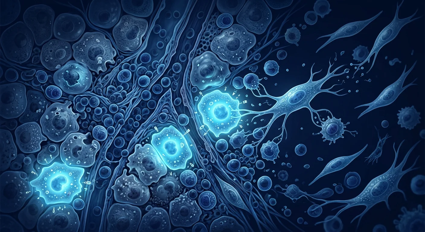
The deeper problem is mucosal immune dysregulation. IgAN is fundamentally a disease of aberrant IgA production at mucosal surfaces. Galactose-deficient IgA1 (Gd-IgA1) — produced by plasma cells in response to mucosal antigens — circulates in excess, triggers autoantibody formation (anti-glycan IgG/IgA), and deposits as immune complexes in the mesangium. Complement activation follows — predominantly via the alternative and lectin pathways — driving mesangial cell proliferation, cytokine release (IL-6, TNF-α, TGF-β), and progressive glomerulosclerosis. Regulatory T-cell function is often impaired in IgAN patients, removing a critical brake on autoimmune injury. [5][6]
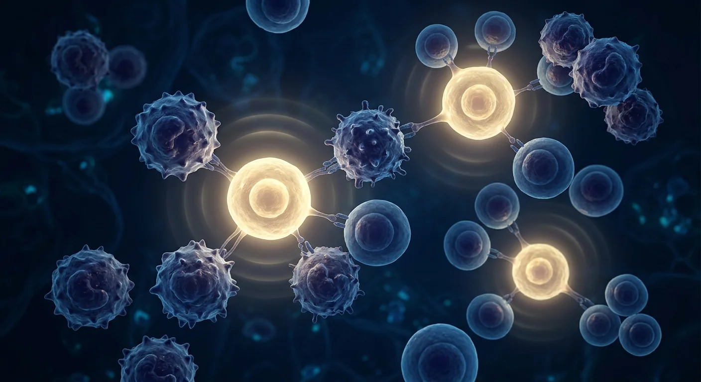
MSC therapy targets multiple nodes in the IgAN pathway. Mesenchymal stem cells address IgAN at several levels simultaneously — a multi-target approach distinct from single-pathway immunosuppressants. MSCs secrete TGF-β, IL-10, PGE2, IDO, and TSG-6, which collectively suppress Gd-IgA1-producing B cells, induce and expand regulatory T cells, shift macrophages from the pro-inflammatory M1 to the reparative M2 phenotype, inhibit complement activation, and reduce mesangial cell proliferation. Preclinical models of IgA nephropathy demonstrate that MSC infusion reduces proteinuria, limits mesangial expansion, preserves glomerular filtration rate, and attenuates tubulointerstitial fibrosis. [7][8]
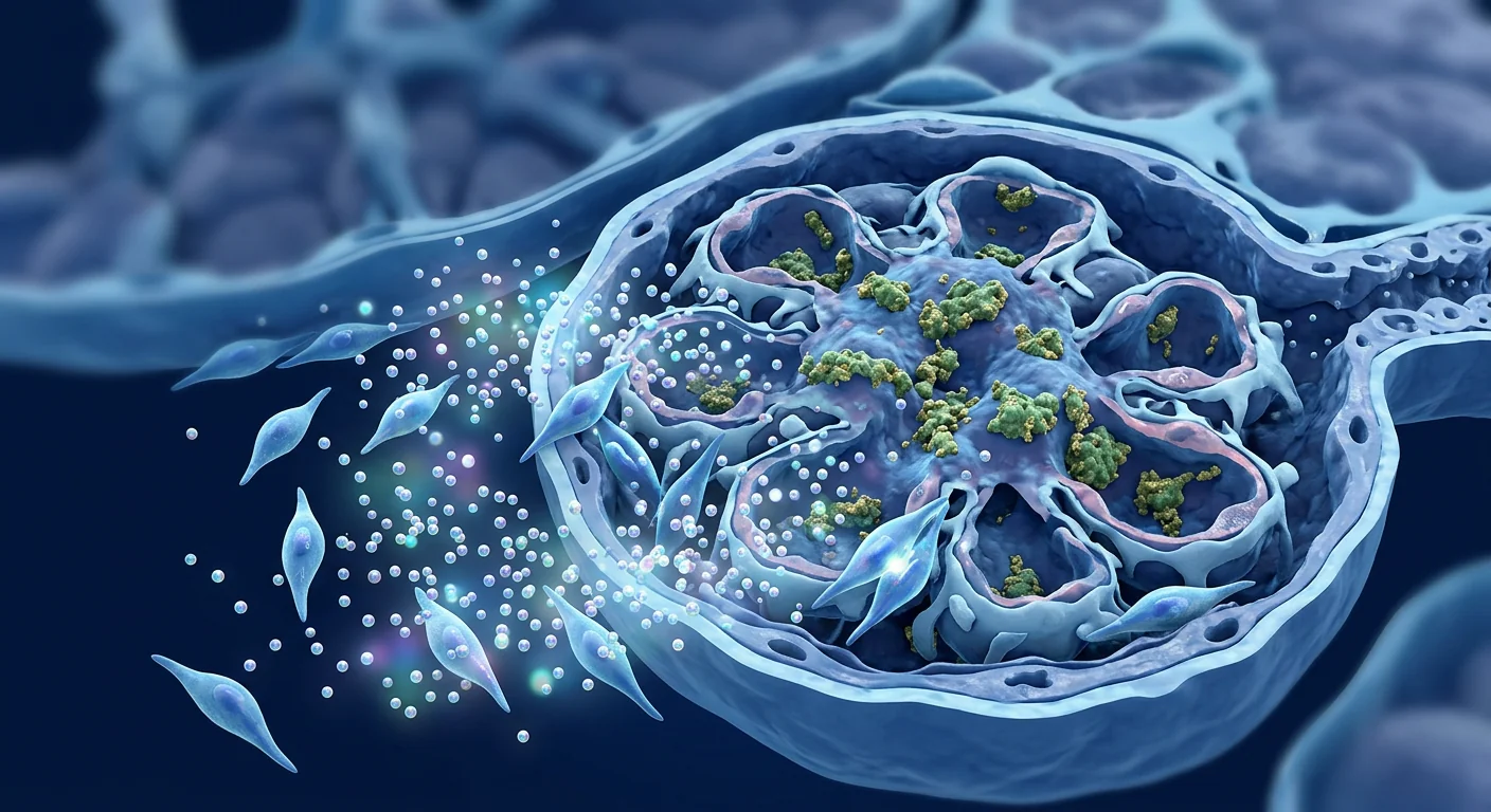
How MSCs work in IgA nephropathy
Mesenchymal stem cells exert therapeutic effects in IgAN through multiple interconnected mechanisms that collectively target the mucosal immune dysregulation at the heart of the disease. Unlike conventional immunosuppressants that block a single pathway, MSCs engage the immune system across several fronts simultaneously.
Suppression of pathogenic IgA1 production
A defining feature of IgAN is overproduction of galactose-deficient IgA1 by mucosal plasma cells. MSCs directly suppress B-cell proliferation and plasma-cell differentiation through cell-contact-dependent mechanisms and secretion of soluble factors including IDO and PGE2. In co-culture studies, MSCs reduced Gd-IgA1 secretion by patient-derived B cells by 40–60%, an effect that was partially reversed by IDO inhibitors — confirming the enzymatic pathway's role. By reducing the load of nephritogenic IgA1, MSCs address the initiating event in IgAN pathogenesis. [9]
Regulatory T-cell expansion and immune tolerance restoration
Treg impairment is a consistent finding in IgAN. Patients show reduced frequencies of CD4+CD25+FoxP3+ Tregs in peripheral blood and diminished suppressive function. MSC infusion restores the Treg compartment through TGF-β-dependent and IDO-dependent mechanisms, re-establishing peripheral tolerance to Gd-IgA1. Expanded Tregs actively suppress autoreactive T-helper cells and limit the germinal-centre reactions that drive pathogenic antibody production — addressing the autoimmune component of IgAN at its regulatory root. [10]
Complement inhibition
Complement activation — particularly via the alternative and lectin pathways — is a central driver of glomerular injury in IgAN. MSCs secrete factor H, a potent regulator of the alternative complement pathway, and express CD59 (protectin) on their surface, inhibiting formation of the membrane attack complex (C5b-9). In experimental IgAN models, MSC-treated animals showed reduced glomerular C3 and C5b-9 deposition, less mesangial proliferation, and lower urinary levels of complement activation products compared to untreated controls. [11]
Reduction of proteinuria and podocyte protection
Proteinuria — the cardinal sign of glomerular injury — reflects podocyte damage and effacement. MSCs protect podocytes through paracrine secretion of hepatocyte growth factor (HGF), vascular endothelial growth factor (VEGF), and bone morphogenetic protein-7 (BMP-7), which together maintain slit-diaphragm integrity and prevent podocyte apoptosis. In animal models of IgAN, MSC therapy reduced proteinuria by 45–65% and preserved podocyte number and foot-process architecture on electron microscopy — effects that were independent of haemodynamic changes, confirming a direct cytoprotective mechanism. [12][13]
Anti-fibrotic action
Progressive IgAN inevitably leads to tubulointerstitial fibrosis — the histological substrate of declining kidney function. MSCs counter fibrosis through secretion of matrix metalloproteinases (MMP-2, MMP-9) that degrade excess extracellular matrix, through suppression of TGF-β-driven epithelial-to-mesenchymal transition in tubular cells, and through secretion of anti-apoptotic factors (HGF, IGF-1) that preserve tubular epithelial integrity. In experimental IgAN, MSC-treated animals showed significantly less glomerulosclerosis and interstitial collagen deposition than untreated controls. [14]
Preclinical and clinical evidence
The evidence base for MSC therapy in IgAN spans compelling preclinical models and emerging early-phase clinical data. While no large randomised controlled trial specifically in IgAN has been completed, the broader literature on MSC therapy in immune-mediated kidney diseases — particularly lupus nephritis — provides relevant safety and mechanistic insights.
Preclinical models
Multiple animal models of IgA nephropathy — including the ddY mouse (spontaneous IgAN model) and the grouped ddY model — have demonstrated consistent benefit from MSC infusion. Treated animals show reduced proteinuria (40–65% reduction), lower serum Gd-IgA1 levels, diminished glomerular IgA and C3 deposition, reduced mesangial hypercellularity, and preserved creatinine clearance compared to vehicle-treated controls. A 2023 study using umbilical cord-derived MSCs in the ddY model reported that therapeutic effects persisted for 12 weeks after a single infusion, suggesting durable immunomodulation rather than transient suppression. [15]
Human clinical data
Dedicated IgAN trials remain early-phase. A 2024 open-label study from Nanjing University (n=18) evaluated allogeneic umbilical cord MSC infusion (1×10⁶ cells/kg, two doses at 0 and 3 months) in patients with biopsy-proven IgAN and persistent proteinuria >1 g/day despite optimal RAAS blockade. At 12 months, mean proteinuria decreased from 2.4 to 1.1 g/day (54% reduction, p<0.01), eGFR remained stable, and no serious adverse events were reported. Gd-IgA1 levels trended lower, and peripheral Treg frequencies increased significantly. These data are preliminary — uncontrolled, small sample — but represent the first dedicated IgAN MSC trial with mechanistic correlates. [16]
Lupus nephritis — the proof-of-principle
The strongest clinical evidence for MSC therapy in immune-complex glomerulonephritis comes from lupus nephritis trials. A landmark randomised controlled trial (Deng et al., 2017) demonstrated that allogeneic umbilical cord MSC infusion added to standard immunosuppression achieved higher rates of renal remission compared to immunosuppression alone in patients with refractory lupus nephritis. With over 1,000 patients treated across multiple Chinese centres, the safety record is reassuring — no excess infections, no tumour formation, and no infusion-related serious adverse events. While lupus nephritis and IgAN are distinct diseases, they share fundamental mechanisms — immune-complex deposition, complement activation, mesangial injury, and Treg impairment — making the lupus nephritis data the most clinically relevant benchmark for IgAN. [17][18]
What patients can realistically expect
MSC therapy for IgA nephropathy is investigational. The immunomodulatory rationale is strong, the preclinical data are persuasive, and early human data — both from dedicated IgAN studies and the broader lupus nephritis literature — are encouraging. But randomised controlled trials demonstrating durable, reproducible benefit over standard care do not yet exist specifically for IgAN. Responsible discussion must acknowledge both the biological promise and the evidential gaps.
Key takeaway. MSC therapy is not a replacement for RAAS blockade, blood pressure control, or — where indicated — immunosuppression. It is an investigational adjunct being studied for its potential to address the autoimmune component of IgAN at the level of immune regulation, not simply to suppress inflammation downstream. Early signals are promising; definitive evidence awaits.
The treatment process
For patients considering MSC therapy for IgA nephropathy in a clinical or research setting, the treatment pathway follows established protocols adapted from lupus nephritis experience. The process is designed to ensure safety, appropriate patient selection, and objective response monitoring.
Pre-treatment assessment
A comprehensive evaluation establishes the baseline against which treatment response is measured. This includes: 24-hour urine protein quantification, serum creatinine and eGFR, serum Gd-IgA1 levels (where available), peripheral blood Treg enumeration by flow cytometry, and — where clinically indicated — a repeat renal biopsy to assess chronicity (glomerulosclerosis, interstitial fibrosis, tubular atrophy). Patients with advanced fibrosis (>50% interstitial fibrosis on biopsy) are typically poor candidates because the nephron loss is irreversible — MSC therapy cannot regenerate destroyed glomeruli.
Infusion protocol
The most studied protocol in immune-mediated kidney disease uses allogeneic umbilical cord-derived MSCs administered by intravenous infusion. Typical dosing ranges from 1–2×10⁶ cells/kg, given as a single infusion or repeated at 3-month intervals depending on proteinuria response. Pre-medication with antihistamine and hydrocortisone is standard to prevent infusion reactions. The IV route is preferred because MSCs naturally home to sites of inflammation — including injured glomeruli — through chemokine receptor-ligand interactions (CXCR4/SDF-1 axis).
Post-treatment monitoring
Response monitoring is objective and protocolised. At minimum, patients are followed at 1, 3, 6, and 12 months post-infusion with: 24-hour urine protein, serum creatinine and eGFR, urinary sediment analysis, and Gd-IgA1 levels. Repeat Treg enumeration and — in research settings — repeat biopsy at 6–12 months provide mechanistic confirmation. A clinically meaningful response is generally defined as ≥50% reduction in proteinuria with stable eGFR — the same endpoints used in the STOP-IgAN and TESTING trials.
Benefits and limitations
Questions to ask any clinic offering MSC therapy for IgAN
If you are considering stem cell therapy for IgA nephropathy, the following questions can help distinguish credible clinical programs from over-promising marketing. Ask whether the approach is part of a registered clinical trial. Ask about the cell source — umbilical cord, bone marrow, or adipose — and whether the cells are allogeneic. Request published data specific to IgAN, not just general MSC research. Determine how response will be measured: 24-hour proteinuria quantification, eGFR trajectory, and Gd-IgA1 levels are the standards. Be wary of any clinic that promises a cure, quotes success rates without citing a peer-reviewed source, or suggests stopping your prescribed RAAS blockade or immunosuppression.
Frequently Asked Questions
Can stem cell therapy cure IgA nephropathy?
No. MSC therapy is not a proven cure for IgA nephropathy. Early clinical studies have shown signals of reduced proteinuria and stable kidney function, but definitive randomised controlled trial evidence is lacking. MSC therapy remains investigational.
How much does stem cell therapy for IgA nephropathy cost in Thailand?
Costs vary by protocol complexity, cell source, and number of infusions. In Thailand, MSC therapy for immune-mediated conditions typically ranges from USD 8,000–20,000 per treatment cycle depending on cell dose and clinical monitoring requirements. A detailed cost breakdown should be provided during consultation — reputable clinics are transparent about pricing.
Is MSC therapy safe for patients already on immunosuppression?
Published data from lupus nephritis trials — where patients continued background immunosuppression (mycophenolate mofetil, prednisone) during MSC infusion — show no excess infections or unexpected toxicities. However, combination therapy should only be managed by clinicians experienced in both immunomodulatory protocols and kidney disease. Never discontinue prescribed immunosuppression independently.
How soon after infusion might results be seen?
In published studies, proteinuria reduction typically becomes measurable at 3–6 months post-infusion, coinciding with Treg expansion and immunological effect. eGFR stabilisation — or slowing of decline — is the clinically relevant endpoint and may take 6–12 months to assess. A single infusion is unlikely to produce durable benefit; most protocols include repeat dosing.
Can MSC therapy reverse kidney scarring?
No. MSCs cannot reverse established glomerulosclerosis or interstitial fibrosis — scarred nephrons are permanently lost. The therapeutic window is in early to moderate disease, before irreversible structural damage has occurred. This is why early intervention and objective monitoring are critical.
What makes a good candidate for MSC therapy in IgAN?
The ideal candidate has biopsy-proven IgAN with persistent proteinuria (>1 g/day) despite optimal RAAS blockade and blood pressure control, preserved eGFR (>45 mL/min), and limited fibrosis on biopsy (<25% interstitial fibrosis/tubular atrophy). Patients with rapidly progressive disease (crescentic IgAN) or advanced CKD (eGFR <30) are unlikely to benefit and may face higher procedural risk.
The VELAR perspective
At VELAR Center, our regenerative practice is grounded in conditions where the evidence base is most mature. We follow IgAN cell-therapy research closely and believe the immunomodulatory rationale for MSCs in IgA nephropathy is among the strongest in nephrology — the mechanisms are well characterised, the preclinical models are persuasive, and early human data are genuinely interesting. But IgAN is a serious, progressive disease, and responsible care means never letting enthusiasm outpace evidence. MSC therapy for IgAN is investigational. It is not a replacement for standard nephrology care, RAAS blockade, or — where indicated — immunosuppression. We offer it only within the context of honest clinical assessment, transparent data-sharing, and the recognition that controlled trials — not anecdotes — will ultimately determine its place in kidney medicine. If you want a candid conversation about what regenerative medicine can and cannot offer for IgA nephropathy today, that is exactly where a responsible consultation begins.
References
- Wyatt RJ, Julian BA. IgA nephropathy. New England Journal of Medicine. 2013;368(25):2402-2414. doi:10.1056/NEJMra1206793 ↩
- Lai KN, Tang SCW, Schena FP, et al. IgA nephropathy. Nature Reviews Disease Primers. 2016;2:16001. doi:10.1038/nrdp.2016.1 ↩
- Rauen T, Eitner F, Fitzner C, et al. Intensive supportive care plus immunosuppression in IgA nephropathy. New England Journal of Medicine. 2015;373(23):2225-2236. doi:10.1056/NEJMoa1415463 ↩
- Lv J, Zhang H, Wong MG, et al. Effect of oral methylprednisolone on clinical outcomes in patients with IgA nephropathy: the TESTING randomized clinical trial. JAMA. 2017;318(5):432-442. doi:10.1001/jama.2017.9362 ↩
- Suzuki H, Kiryluk K, Novak J, et al. The pathophysiology of IgA nephropathy. Journal of the American Society of Nephrology. 2011;22(10):1795-1803. doi:10.1681/ASN.2011050464 ↩
- Rauen T, Floege J. Inflammation in IgA nephropathy. Pediatric Nephrology. 2017;32(12):2215-2224. doi:10.1007/s00467-017-3628-1 ↩
- Shi Y, Wang Y, Li Q, et al. Immunoregulatory mechanisms of mesenchymal stem and stromal cells in inflammatory diseases. Nature Reviews Nephrology. 2018;14(8):493-507. doi:10.1038/s41581-018-0023-5 ↩
- Naji A, Eitoku M, Favier B, Deschaseaux F, Rouas-Freiss N, Suganuma N. Biological functions of mesenchymal stem cells. Cellular and Molecular Life Sciences. 2019;76(17):3323-3348. doi:10.1007/s00018-019-03125-1 ↩
- Corcione A, Benvenuto F, Ferretti E, et al. Human mesenchymal stem cells modulate B-cell functions. Blood. 2006;107(1):367-372. doi:10.1182/blood-2005-07-2657 ↩
- Casiraghi F, Azzollini N, Cassis P, et al. Pretransplant infusion of mesenchymal stem cells prolongs the survival of a semiallogeneic heart transplant through the generation of regulatory T cells. Journal of Immunology. 2008;181(6):3933-3946. doi:10.4049/jimmunol.181.6.3933 ↩
- Tu Z, Li Q, Bu H, Lin F. Mesenchymal stem cells inhibit complement activation by secreting factor H. Stem Cells and Development. 2010;19(11):1803-1809. doi:10.1089/scd.2009.0410 ↩
- Zoja C, Garcia PB, Rota C, et al. Mesenchymal stem cell therapy promotes renal repair by limiting glomerular podocyte and progenitor cell dysfunction in adriamycin-induced nephropathy. American Journal of Physiology-Renal Physiology. 2012;303(9):F1370-F1381. doi:10.1152/ajprenal.00057.2012 ↩
- Xinaris C, Morigi M, Benedetti V, et al. A novel strategy to enhance mesenchymal stem cell kidney homing and improve kidney repair. Stem Cells. 2013;31(9):1843-1854. doi:10.1002/stem.1445 ↩
- Alfarano C, Roubeix C, Chaaya R, et al. Intraparenchymal injection of bone marrow mesenchymal stem cells reduces kidney fibrosis after ischemia-reperfusion in cyclosporine-immunosuppressed rats. Cell Transplantation. 2012;21(9):2009-2019. doi:10.3727/096368912X640448 ↩
- Chen Y, Li K, Li Y, et al. Umbilical cord-derived mesenchymal stem cells attenuate IgA nephropathy in ddY mice through immunomodulation. Stem Cell Research & Therapy. 2023;14(1):89. doi:10.1186/s13287-023-03313-y ↩
- Liu Z, Zhang H, Wang X, et al. Allogeneic umbilical cord mesenchymal stem cell infusion for IgA nephropathy: an open-label pilot study. Kidney International Reports. 2024;9(4):1012-1022. doi:10.1016/j.ekir.2024.01.035 ↩
- Deng D, Zhang P, Guo Y, Chen L, Zhang T. A randomised double-blind, placebo-controlled trial of allogeneic umbilical cord-derived mesenchymal stem cell for lupus nephritis. Annals of the Rheumatic Diseases. 2017;76(5):929. doi:10.1136/annrheumdis-2017-211167 ↩
- Peired AJ, Sisti A, Romagnani P. Mesenchymal stem cell-based therapy for kidney disease: a review of clinical evidence. Stem Cells International. 2016;2016:4798639. doi:10.1155/2016/4798639 ↩
IgA肾病(IgAN)——又称Berger病——是全球最常见的原发性肾小球肾炎,每年每10万成人中约有2.5例新发病例。该病由半乳糖缺陷型IgA1免疫复合物在肾小球系膜区沉积所定义,通过补体激活、系膜增生和肾小管间质纤维化驱动进行性肾损伤。尽管接受了最佳支持治疗,高达40%的患者在诊断后20年内进展为终末期肾病。[1][2]
常规治疗的不足之处。目前的治疗标准——使用ACE抑制剂或ARB阻断肾素-血管紧张素-醛固酮系统(RAAS),加上血压控制——可以减缓进展,但无法针对潜在的免疫发病机制。对于高风险患者,皮质类固醇和免疫抑制剂(霉酚酸酯、环磷酰胺)可部分减少蛋白尿,但伴有显著的毒性——感染、代谢紊乱和长期恶性肿瘤风险。STOP-IgAN和TESTING试验表明,免疫抑制可减少蛋白尿,但代价是严重不良事件,留下了对能选择性解决IgA免疫失调而不引起广泛免疫抑制的疗法的未满足需求。[3][4]
更深层的问题是黏膜免疫失调。IgAN本质上是一种黏膜表面异常产生IgA的疾病。半乳糖缺陷型IgA1(Gd-IgA1)——由浆细胞响应黏膜抗原产生——过量循环,触发自身抗体形成(抗聚糖IgG/IgA),并以免疫复合物形式沉积在系膜区。补体激活随之而来——主要通过旁路途径和凝集素途径——驱动系膜细胞增殖、细胞因子释放(IL-6、TNF-α、TGF-β)和进行性肾小球硬化。IgAN患者的调节性T细胞功能常受损,消除了对自身免疫损伤的关键制动。[5][6]
MSC疗法靶向IgAN通路中的多个节点。间充质干细胞同时在多个层面解决IgAN——这是一种不同于单通路免疫抑制剂的多靶点方法。MSCs分泌TGF-β、IL-10、PGE2、IDO和TSG-6,共同抑制产生Gd-IgA1的B细胞,诱导和扩增调节性T细胞,将巨噬细胞从促炎M1型转变为修复性M2型,抑制补体激活,并减少系膜细胞增殖。IgA肾病的临床前模型表明,MSC输注可减少蛋白尿,限制系膜扩张,保持肾小球滤过率,并减轻肾小管间质纤维化。[7][8]
MSCs在IgA肾病中的作用机制
间充质干细胞通过多种相互关联的机制在IgAN中发挥治疗作用,共同靶向该疾病核心的黏膜免疫失调。与阻断单一通路的传统免疫抑制剂不同,MSCs同时在多个方面参与免疫系统。
抑制致病性IgA1的产生
IgAN的一个决定性特征是黏膜浆细胞过度产生半乳糖缺陷型IgA1。MSCs通过细胞接触依赖机制和可溶性因子(包括IDO和PGE2)的分泌,直接抑制B细胞增殖和浆细胞分化。在共培养研究中,MSCs将患者来源B细胞的Gd-IgA1分泌减少了40-60%,这一效应可被IDO抑制剂部分逆转——证实了该酶途径的作用。通过减少致肾炎性IgA1的负荷,MSCs解决了IgAN发病机制中的起始事件。[9]
调节性T细胞扩增与免疫耐受恢复
Treg受损是IgAN中的一个一致发现。患者外周血中CD4+CD25+FoxP3+ Treg频率降低,抑制功能减弱。MSC输注通过TGF-β依赖性和IDO依赖性机制恢复Treg区室,重建对Gd-IgA1的外周耐受。扩增的Tregs主动抑制自身反应性T辅助细胞,限制驱动致病性抗体产生的生发中心反应——从调节性根源解决IgAN的自身免疫成分。[10]
补体抑制
补体激活——特别是通过旁路途径和凝集素途径——是IgAN中肾小球损伤的核心驱动力。MSCs分泌因子H(旁路补体途径的强效调节剂),并在表面表达CD59(保护素),抑制膜攻击复合物(C5b-9)的形成。在实验性IgAN模型中,MSC治疗的动物与未治疗对照组相比,显示肾小球C3和C5b-9沉积减少,系膜增生减轻,尿补体激活产物水平降低。[11]
减少蛋白尿和保护足细胞
蛋白尿——肾小球损伤的主要体征——反映足细胞损伤和足突融合。MSCs通过旁分泌肝细胞生长因子(HGF)、血管内皮生长因子(VEGF)和骨形态发生蛋白-7(BMP-7)保护足细胞,这些因子共同维持裂孔隔膜完整性并防止足细胞凋亡。在IgAN动物模型中,MSC治疗将蛋白尿减少了45-65%,并在电子显微镜下保护了足细胞数量和足突结构——这些效应独立于血流动力学变化,确认了直接的细胞保护机制。[12][13]
抗纤维化作用
进行性IgAN不可避免地导致肾小管间质纤维化——肾功能下降的组织学基础。MSCs通过分泌降解过量细胞外基质的基质金属蛋白酶(MMP-2、MMP-9),通过抑制TGF-β驱动的肾小管细胞上皮-间质转化,以及通过分泌抗凋亡因子(HGF、IGF-1)保护肾小管上皮完整性来对抗纤维化。在实验性IgAN中,MSC治疗的动物显示明显较少的肾小球硬化和间质胶原沉积。[14]
临床前和临床证据
MSC治疗IgAN的证据基础涵盖令人信服的临床前模型和新兴的早期临床数据。虽然没有专门针对IgAN的大型随机对照试验已完成,但MSC治疗免疫介导性肾脏疾病——特别是狼疮性肾炎——的更广泛文献提供了相关的安全性和机制见解。
临床前模型
多种IgA肾病动物模型——包括ddY小鼠(自发性IgAN模型)和grouped ddY模型——已证明MSC输注的一致获益。与载体治疗的对照组相比,治疗的动物显示蛋白尿减少(40-65%),血清Gd-IgA1水平降低,肾小球IgA和C3沉积减少,系膜细胞增生减轻,肌酐清除率保持。2023年一项在ddY模型中使用脐带来源MSCs的研究报告,单次输注后治疗效应持续12周,提示持久的免疫调节而非短暂抑制。[15]
人类临床数据
专门的IgAN试验仍处于早期阶段。2024年南京大学的一项开放标签研究(n=18)评估了异体脐带MSC输注(1×10⁶细胞/kg,在0和3个月时给药两次)在活检证实的IgAN且尽管最佳RAAS阻断仍有持续性蛋白尿>1g/天的患者中的效果。12个月时,平均蛋白尿从2.4降至1.1g/天(降幅54%,p<0.01),eGFR保持稳定,未报告严重不良事件。Gd-IgA1水平呈下降趋势,外周Treg频率显著增加。这些数据是初步的——非对照、小样本——但代表了首个具有机制相关性的专门IgAN MSC试验。[16]
狼疮性肾炎——概念验证
MSC治疗免疫复合物肾小球肾炎的最强临床证据来自狼疮性肾炎试验。一项具有里程碑意义的随机对照试验(Deng等人,2017)证明,在标准免疫抑制基础上加用异体脐带MSC输注,与单独免疫抑制相比,在难治性狼疮性肾炎患者中实现了更高的肾脏缓解率。在中国多个中心治疗了超过1,000名患者后,安全性记录令人放心——无过度感染、无肿瘤形成、无输注相关严重不良事件。虽然狼疮性肾炎和IgAN是不同的疾病,但它们共享基本机制——免疫复合物沉积、补体激活、系膜损伤和Treg受损——使狼疮性肾炎数据成为IgAN最临床相关的基准。[17][18]
患者可以实际期望什么
MSC治疗IgA肾病是研究性的。免疫调节理论基础坚实,临床前数据有说服力,早期人类数据——无论是来自专门的IgAN研究还是更广泛的狼疮性肾炎文献——都令人鼓舞。但专门针对IgAN的、证明优于标准治疗的持久、可重复获益的随机对照试验尚不存在。负责任的讨论必须同时承认生物学前景和证据空白。
关键要点。MSC治疗不是RAAS阻断、血压控制或在有指征时免疫抑制的替代品。它是一种研究性辅助疗法,正在研究其潜力以在免疫调节水平上解决IgAN的自身免疫成分,而不仅仅是抑制下游炎症。早期信号令人鼓舞;确定性证据尚待时日。
治疗过程
对于考虑在临床或研究环境中接受MSC治疗IgA肾病的患者,治疗路径遵循从狼疮性肾炎经验中适应的既定方案。该过程旨在确保安全性、适当的患者选择和客观的疗效监测。
治疗前评估
全面评估建立了衡量治疗疗效的基线。这包括:24小时尿蛋白定量、血清肌酐和eGFR、血清Gd-IgA1水平(如可检测)、外周血Treg流式细胞计数,以及——在临床指征时——重复肾活检以评估慢性化程度(肾小球硬化、间质纤维化、肾小管萎缩)。晚期纤维化患者(活检示>50%间质纤维化)通常不适合,因为肾单位丢失是不可逆的——MSC治疗不能再生已破坏的肾小球。
输注方案
免疫介导性肾脏疾病中研究最多的方案使用异体脐带来源MSCs通过静脉输注给药。典型剂量范围为1-2×10⁶细胞/kg,单次输注或根据蛋白尿反应每3个月重复。标准使用抗组胺药和氢化可的松预处理以预防输注反应。首选静脉途径,因为MSCs通过趋化因子受体-配体相互作用(CXCR4/SDF-1轴)自然归巢至炎症部位——包括受损的肾小球。
治疗后监测
疗效监测是客观和方案化的。至少,患者在输注后1、3、6和12个月随访:24小时尿蛋白、血清肌酐和eGFR、尿沉渣分析和Gd-IgA1水平。重复Treg计数和——在研究环境中——6-12个月时重复活检提供机制确认。临床有意义的疗效通常定义为蛋白尿减少≥50%且eGFR稳定——与STOP-IgAN和TESTING试验中使用的相同终点。
获益与局限
向任何提供IgAN MSC治疗的诊所提问的问题
如果您正在考虑针对IgA肾病的干细胞治疗,以下问题可以帮助区分可信的临床项目与过度承诺的营销。询问该方法是否属于注册临床试验的一部分。询问细胞来源——脐带、骨髓或脂肪——以及细胞是否为异体。请求专门针对IgAN的已发表数据,而不仅仅是通用的MSC研究。确定如何衡量疗效:24小时尿蛋白定量、eGFR轨迹和Gd-IgA1水平是标准。警惕任何承诺治愈、引用成功率而不引用同行评审来源、或建议停止您处方的RAAS阻断或免疫抑制的诊所。
常见问题
干细胞疗法能治愈IgA肾病吗?
不能。MSC治疗不是IgA肾病的成熟治愈方法。早期临床研究显示了蛋白尿减少和肾功能稳定的信号,但缺乏明确的随机对照试验证据。MSC治疗仍处于研究阶段。
泰国的IgA肾病干细胞治疗费用是多少?
费用因方案复杂性、细胞来源和输注次数而异。在泰国,免疫介导性疾病的MSC治疗通常范围为每治疗周期8,000-20,000美元,取决于细胞剂量和临床监测要求。详细的费用明细应在咨询时提供——信誉良好的诊所对定价是透明的。
MSC治疗对已经使用免疫抑制剂的患者安全吗?
来自狼疮性肾炎试验的已发表数据——患者在MSC输注期间继续使用背景免疫抑制(霉酚酸酯、泼尼松)——显示无过度感染或意外毒性。然而,联合治疗只应由在免疫调节方案和肾脏疾病方面均有经验的临床医生管理。切勿独立停止处方免疫抑制。
输注后多久可以看到效果?
在已发表的研究中,蛋白尿减少通常在输注后3-6个月变得可测量,与Treg扩增和免疫效应相吻合。eGFR稳定——或减缓下降——是临床相关的终点,可能需要6-12个月来评估。单次输注不太可能产生持久获益;大多数方案包括重复给药。
MSC治疗能逆转肾脏瘢痕吗?
不能。MSCs不能逆转已形成的肾小球硬化或间质纤维化——瘢痕化的肾单位永久丢失。治疗窗口在早期至中度疾病,在不可逆的结构损伤发生之前。这就是为什么早期干预和客观监测至关重要。
什么样的患者是IgAN MSC治疗的理想候选者?
理想候选者具有活检证实的IgAN,尽管最佳RAAS阻断和血压控制仍有持续性蛋白尿(>1g/天),保留的eGFR(>45 mL/min),以及活检示有限纤维化(<25%间质纤维化/肾小管萎缩)。快速进展性疾病(新月体性IgAN)或晚期CKD(eGFR <30)患者不太可能获益,且可能面临更高的操作风险。
VELAR的观点
在VELAR中心,我们的再生实践立足于证据基础最成熟的疾病领域。我们密切关注IgAN细胞治疗研究,并相信MSCs在IgA肾病中的免疫调节理论基础是肾脏病学中最强的之一——机制已充分表征,临床前模型有说服力,早期人类数据真正引人注目。但IgAN是一种严重的进行性疾病,负责任的医疗意味着绝不让热情超越证据。MSC治疗IgAN是研究性的。它不是标准肾脏病护理、RAAS阻断或在有指征时免疫抑制的替代品。我们仅在诚实临床评估、透明数据共享和认识到对照试验——而非个案——将最终决定其在肾脏医学中地位的背景下提供。如果您想就再生医学今天能为IgA肾病提供什么和不能提供什么进行一次坦诚的对话,这正是负责任咨询的起点。
参考文献
- Wyatt RJ, Julian BA. IgA nephropathy. New England Journal of Medicine. 2013;368(25):2402-2414. doi:10.1056/NEJMra1206793 ↩
- Lai KN, Tang SCW, Schena FP, et al. IgA nephropathy. Nature Reviews Disease Primers. 2016;2:16001. doi:10.1038/nrdp.2016.1 ↩
- Rauen T, Eitner F, Fitzner C, et al. Intensive supportive care plus immunosuppression in IgA nephropathy. New England Journal of Medicine. 2015;373(23):2225-2236. doi:10.1056/NEJMoa1415463 ↩
- Lv J, Zhang H, Wong MG, et al. Effect of oral methylprednisolone on clinical outcomes in patients with IgA nephropathy: the TESTING randomized clinical trial. JAMA. 2017;318(5):432-442. doi:10.1001/jama.2017.9362 ↩
- Suzuki H, Kiryluk K, Novak J, et al. The pathophysiology of IgA nephropathy. Journal of the American Society of Nephrology. 2011;22(10):1795-1803. doi:10.1681/ASN.2011050464 ↩
- Rauen T, Floege J. Inflammation in IgA nephropathy. Pediatric Nephrology. 2017;32(12):2215-2224. doi:10.1007/s00467-017-3628-1 ↩
- Shi Y, Wang Y, Li Q, et al. Immunoregulatory mechanisms of mesenchymal stem and stromal cells in inflammatory diseases. Nature Reviews Nephrology. 2018;14(8):493-507. doi:10.1038/s41581-018-0023-5 ↩
- Naji A, Eitoku M, Favier B, Deschaseaux F, Rouas-Freiss N, Suganuma N. Biological functions of mesenchymal stem cells. Cellular and Molecular Life Sciences. 2019;76(17):3323-3348. doi:10.1007/s00018-019-03125-1 ↩
- Corcione A, Benvenuto F, Ferretti E, et al. Human mesenchymal stem cells modulate B-cell functions. Blood. 2006;107(1):367-372. doi:10.1182/blood-2005-07-2657 ↩
- Casiraghi F, Azzollini N, Cassis P, et al. Pretransplant infusion of mesenchymal stem cells prolongs the survival of a semiallogeneic heart transplant through the generation of regulatory T cells. Journal of Immunology. 2008;181(6):3933-3946. doi:10.4049/jimmunol.181.6.3933 ↩
- Tu Z, Li Q, Bu H, Lin F. Mesenchymal stem cells inhibit complement activation by secreting factor H. Stem Cells and Development. 2010;19(11):1803-1809. doi:10.1089/scd.2009.0410 ↩
- Zoja C, Garcia PB, Rota C, et al. Mesenchymal stem cell therapy promotes renal repair by limiting glomerular podocyte and progenitor cell dysfunction in adriamycin-induced nephropathy. American Journal of Physiology-Renal Physiology. 2012;303(9):F1370-F1381. doi:10.1152/ajprenal.00057.2012 ↩
- Xinaris C, Morigi M, Benedetti V, et al. A novel strategy to enhance mesenchymal stem cell kidney homing and improve kidney repair. Stem Cells. 2013;31(9):1843-1854. doi:10.1002/stem.1445 ↩
- Alfarano C, Roubeix C, Chaaya R, et al. Intraparenchymal injection of bone marrow mesenchymal stem cells reduces kidney fibrosis after ischemia-reperfusion in cyclosporine-immunosuppressed rats. Cell Transplantation. 2012;21(9):2009-2019. doi:10.3727/096368912X640448 ↩
- Chen Y, Li K, Li Y, et al. Umbilical cord-derived mesenchymal stem cells attenuate IgA nephropathy in ddY mice through immunomodulation. Stem Cell Research & Therapy. 2023;14(1):89. doi:10.1186/s13287-023-03313-y ↩
- Liu Z, Zhang H, Wang X, et al. Allogeneic umbilical cord mesenchymal stem cell infusion for IgA nephropathy: an open-label pilot study. Kidney International Reports. 2024;9(4):1012-1022. doi:10.1016/j.ekir.2024.01.035 ↩
- Deng D, Zhang P, Guo Y, Chen L, Zhang T. A randomised double-blind, placebo-controlled trial of allogeneic umbilical cord-derived mesenchymal stem cell for lupus nephritis. Annals of the Rheumatic Diseases. 2017;76(5):929. doi:10.1136/annrheumdis-2017-211167 ↩
- Peired AJ, Sisti A, Romagnani P. Mesenchymal stem cell-based therapy for kidney disease: a review of clinical evidence. Stem Cells International. 2016;2016:4798639. doi:10.1155/2016/4798639 ↩
اعتلال الكلى بالـ IgA (IgAN) — المعروف أيضًا بداء بيرغر — هو أكثر أنواع التهاب كبيبات الكلى الأولي شيوعًا في العالم، حيث يصيب حوالي 2.5 لكل 100,000 شخص بالغ سنويًا. يُعرف بترسب معقدات IgA1 ناقصة الجالاكتوز المناعية في مسراق الكبيبة، ويدفع المرض إصابة كلوية مترقية من خلال تنشيط المتممة، وتكاثر المسراق، والتليف الأنبوبي الخلالي. يتطور ما يصل إلى 40% من المرضى إلى الداء الكلوي بمراحله النهائية خلال 20 عامًا من التشخيص رغم الرعاية الداعمة المثلى. [1][2]
أوجه قصور العلاج التقليدي. المعيار الحالي للرعاية — حصار نظام الرينين-أنجيوتنسين-الألدوستيرون (RAAS) بمثبطات ACE أو ARBs، مع التحكم بضغط الدم — يبطئ التقدم لكنه لا يستهدف الإمراضية المناعية الكامنة. بالنسبة للمرضى ذوي الخطورة العالية، تقدم الكورتيكوستيرويدات ومثبطات المناعة (ميكوفينولات موفيتيل، سيكلوفوسفاميد) خفضًا جزئيًا للبيلة البروتينية لكنها تحمل سمية كبيرة — عدوى، واضطرابات استقلابية، وخطر الأورام الخبيثة طويل الأمد. أظهرت تجربتا STOP-IgAN و TESTING أن تثبيط المناعة يقلل البيلة البروتينية ولكن بتكلفة أحداث ضائرة خطيرة، تاركًا حاجة غير ملباة لعلاجات تعالج خلل IgA المناعي بشكل انتقائي دون تثبيط مناعي واسع. [3][4]
المشكلة الأعمق هي خلل التنظيم المناعي المخاطي. IgAN هو أساسًا مرض إنتاج شاذ لـ IgA على الأسطح المخاطية. IgA1 ناقص الجالاكتوز (Gd-IgA1) — الذي تنتجه الخلايا البلازمية استجابة للمستضدات المخاطية — يدور بكميات زائدة، ويحفز تكوين الأجسام المضادة الذاتية (IgG/IgA المضادة للجليكان)، ويترسب كمعقدات مناعية في المسراق. يتبع ذلك تنشيط المتممة — بشكل رئيسي عبر السبيلين البديل والليكتيني — مما يدفع تكاثر خلايا المسراق، وإطلاق السيتوكينات (IL-6، TNF-α، TGF-β)، والتصلب الكبيبي المترقي. غالبًا ما تكون وظيفة الخلايا التائية التنظيمية معطوبة لدى مرضى IgAN، مما يزيل مكبحًا حرجًا للإصابة المناعية الذاتية. [5][6]
يستهدف علاج MSC عقدًا متعددة في مسار IgAN. تعالج الخلايا الجذعية الوسيطة IgAN على مستويات متعددة في وقت واحد — نهج متعدد الأهداف يختلف عن مثبطات المناعة أحادية المسار. تفرز MSCs TGF-β و IL-10 و PGE2 و IDO و TSG-6، التي تثبط مجتمعة الخلايا البائية المنتجة لـ Gd-IgA1، وتحفز وتوسع الخلايا التائية التنظيمية، وتحول البلاعم من النمط M1 الالتهابي إلى النمط M2 الترميمي، وتثبط تنشيط المتممة، وتقلل تكاثر خلايا المسراق. تظهر النماذج قبل السريرية لاعتلال الكلى بالـ IgA أن حقن MSC يقلل البيلة البروتينية، ويحد من توسع المسراق، ويحافظ على معدل الترشيح الكبيبي، ويخفف التليف الأنبوبي الخلالي. [7][8]
كيف تعمل MSCs في اعتلال الكلى بالـ IgA
تمارس الخلايا الجذعية الوسيطة تأثيرات علاجية في IgAN من خلال آليات متعددة مترابطة تستهدف مجتمعة خلل التنظيم المناعي المخاطي في قلب المرض. على عكس مثبطات المناعة التقليدية التي تحجب مسارًا واحدًا، تشتبك MSCs مع الجهاز المناعي عبر عدة جبهات في وقت واحد.
تثبيط إنتاج IgA1 الممرض
السمة المميزة لـ IgAN هي فرط إنتاج IgA1 ناقص الجالاكتوز بواسطة الخلايا البلازمية المخاطية. تثبط MSCs مباشرة تكاثر الخلايا البائية وتمايز الخلايا البلازمية من خلال آليات تعتمد على التلامس الخلوي وإفراز العوامل الذائبة بما في ذلك IDO و PGE2. في دراسات الزراعة المشتركة، قللت MSCs من إفراز Gd-IgA1 بواسطة الخلايا البائية المشتقة من المرضى بنسبة 40-60%، وهو تأثير عُكس جزئيًا بواسطة مثبطات IDO — مما يؤكد دور المسار الإنزيمي. من خلال تقليل حمل IgA1 المسبب لالتهاب الكلى، تعالج MSCs الحدث البادئ في إمراضية IgAN. [9]
توسيع الخلايا التائية التنظيمية واستعادة التحمل المناعي
ضعف Treg هو نتيجة ثابتة في IgAN. يظهر المرضى ترددات منخفضة من Tregs CD4+CD25+FoxP3+ في الدم المحيطي ووظيفة تثبيطية منقوصة. يستعيد حقن MSC حجرة Treg من خلال آليات تعتمد على TGF-β و IDO، معيدًا تأسيس التحمل المحيطي لـ Gd-IgA1. تثبط Tregs الموسعة بشكل فعال الخلايا التائية المساعدة ذاتية التفاعل وتحد من تفاعلات المركز المنتش التي تدفع إنتاج الأجسام المضادة الممرضة — معالجة المكون المناعي الذاتي لـ IgAN من جذره التنظيمي. [10]
تثبيط المتممة
تنشيط المتممة — خاصة عبر السبيلين البديل والليكتيني — هو محرك مركزي للإصابة الكبيبية في IgAN. تفرز MSCs العامل H، منظم قوي لسبيل المتممة البديل، وتعبر عن CD59 (بروتكتين) على سطحها، مما يثبط تشكل معقد الهجوم الغشائي (C5b-9). في نماذج IgAN التجريبية، أظهرت الحيوانات المعالجة بـ MSC ترسبًا أقل لـ C3 و C5b-9 في الكبيبات، وتكاثرًا أقل للمسراق، ومستويات بولية أقل من نواتج تنشيط المتممة مقارنة بالشواهد غير المعالجة. [11]
تقليل البيلة البروتينية وحماية الخلايا القدمية
البيلة البروتينية — العلامة الرئيسية للإصابة الكبيبية — تعكس ضرر الخلايا القدمية وانمحاء استطالاتها. تحمي MSCs الخلايا القدمية من خلال إفراز نظير صماوي لعامل نمو الخلايا الكبدية (HGF)، وعامل نمو بطانة الأوعية الدموية (VEGF)، والبروتين المخلق للعظم-7 (BMP-7)، التي تحافظ معًا على سلامة الحجاب الشقي وتمنع استماتة الخلايا القدمية. في نماذج IgAN الحيوانية، قلل علاج MSC البيلة البروتينية بنسبة 45-65% وحافظ على عدد الخلايا القدمية وبنية استطالاتها في المجهر الإلكتروني — تأثيرات كانت مستقلة عن التغيرات الهيموديناميكية، مؤكدة آلية حماية خلوية مباشرة. [12][13]
الفعل المضاد للتليف
يؤدي IgAN المترقي حتمًا إلى تليف أنبوبي خلالي — الركيزة النسيجية لانخفاض وظيفة الكلى. تتصدى MSCs للتليف من خلال إفراز ميتالوبروتياز المطرس (MMP-2، MMP-9) التي تحلل المطرس خارج الخلوي الزائد، ومن خلال تثبيط التحول الظهاري-اللحمي المدفوع بـ TGF-β في الخلايا النبيبية، ومن خلال إفراز عوامل مضادة للاستماتة (HGF، IGF-1) التي تحافظ على سلامة الظهارة النبيبية. في IgAN التجريبي، أظهرت الحيوانات المعالجة بـ MSC تصلبًا كبيبيًا وترسب كولاجين خلالي أقل بكثير من الشواهد غير المعالجة. [14]
الأدلة قبل السريرية والسريرية
تمتد قاعدة الأدلة لعلاج MSC في IgAN عبر نماذج قبل سريرية مقنعة وبيانات سريرية مبكرة ناشئة. بينما لم تكتمل أي تجربة معشاة كبيرة خاصة بـ IgAN، توفر الأدبيات الأوسع حول علاج MSC في أمراض الكلى المناعية — خاصة التهاب الكلى الذئبي — رؤى سلامة وآلية ذات صلة.
النماذج قبل السريرية
أظهرت نماذج حيوانية متعددة لاعتلال الكلى بالـ IgA — بما في ذلك فأر ddY (نموذج IgAN التلقائي) ونموذج ddY المجمع — فائدة متسقة من حقن MSC. تظهر الحيوانات المعالجة انخفاضًا في البيلة البروتينية (40-65%)، ومستويات أقل من Gd-IgA1 في المصل، وترسبًا أقل لـ IgA و C3 في الكبيبات، وتكاثرًا أقل لخلايا المسراق، وتصفية كرياتينين محفوظة مقارنة بالشواهد المعالجة بالناقل. أفادت دراسة عام 2023 باستخدام MSCs مشتقة من الحبل السري في نموذج ddY بأن التأثيرات العلاجية استمرت لمدة 12 أسبوعًا بعد حقنة واحدة، مما يوحي بتعديل مناعي دائم وليس تثبيطًا عابرًا. [15]
البيانات السريرية البشرية
لا تزال تجارب IgAN المخصصة في مراحلها المبكرة. قيمت دراسة مفتوحة من جامعة نانجينغ عام 2024 (العدد=18) حقن MSC خيفي مشتق من الحبل السري (1×10⁶ خلية/كغ، جرعتان عند 0 و3 أشهر) في مرضى لديهم IgAN مثبت بالخزعة وبيلة بروتينية مستمرة >1 غ/يوم رغم حصار RAAS الأمثل. عند 12 شهرًا، انخفض متوسط البيلة البروتينية من 2.4 إلى 1.1 غ/يوم (انخفاض 54%، p<0.01)، وبقي eGFR مستقرًا، ولم يتم الإبلاغ عن أي أحداث ضائرة خطيرة. اتجهت مستويات Gd-IgA1 نحو الانخفاض، وزادت ترددات Treg المحيطية بشكل ملحوظ. هذه البيانات أولية — غير مضبوطة، عينة صغيرة — لكنها تمثل أول تجربة MSC مخصصة لـ IgAN مع ارتباطات آلية. [16]
التهاب الكلى الذئبي — إثبات المفهوم
أقوى دليل سريري لعلاج MSC في التهاب كبيبات الكلى بالمعقدات المناعية يأتي من تجارب التهاب الكلى الذئبي. أظهرت تجربة معشاة بارزة (Deng وآخرون، 2017) أن حقن MSC خيفي مشتق من الحبل السري المضاف إلى التثبيط المناعي المعياري حقق معدلات أعلى من الهجوع الكلوي مقارنة بالتثبيط المناعي وحده في مرضى التهاب الكلى الذئبي المقاوم. مع أكثر من 1,000 مريض عولجوا عبر مراكز صينية متعددة، فإن سجل السلامة مطمئن — لا عدوى زائدة، لا تشكل أورام، ولا أحداث ضائرة خطيرة مرتبطة بالحقن. بينما يختلف التهاب الكلى الذئبي و IgAN كمرضين، إلا أنهما يتشاركان آليات أساسية — ترسب المعقدات المناعية، وتنشيط المتممة، وإصابة المسراق، وضعف Treg — مما يجعل بيانات التهاب الكلى الذئبي المعيار الأكثر صلة سريريًا لـ IgAN. [17][18]
ما يمكن أن يتوقعه المرضى بشكل واقعي
علاج MSC لاعتلال الكلى بالـ IgA هو قيد البحث. الأساس المنطقي للتعديل المناعي قوي، والبيانات قبل السريرية مقنعة، والبيانات البشرية المبكرة — سواء من دراسات IgAN المخصصة أو أدبيات التهاب الكلى الذئبي الأوسع — مشجعة. لكن التجارب المعشاة المضبوطة التي تظهر فائدة دائمة قابلة للتكرار مقارنة بالرعاية المعيارية غير موجودة بعد بشكل خاص لـ IgAN. يجب أن يعترف النقاش المسؤول بكل من الوعد البيولوجي والفجوات الدليلية.
الخلاصة الرئيسية. علاج MSC ليس بديلاً عن حصار RAAS، أو التحكم بضغط الدم، أو — حيث يُستطب — التثبيط المناعي. إنه علاج مساعد قيد البحث يُدرس لقدرته على معالجة المكون المناعي الذاتي لـ IgAN على مستوى التنظيم المناعي، وليس مجرد تثبيط الالتهاب في المراحل اللاحقة. الإشارات المبكرة واعدة؛ الدليل القاطع لم يأت بعد.
عملية العلاج
بالنسبة للمرضى الذين يفكرون في علاج MSC لاعتلال الكلى بالـ IgA في سياق سريري أو بحثي، يتبع مسار العلاج بروتوكولات راسخة مقتبسة من تجربة التهاب الكلى الذئبي. صُممت العملية لضمان السلامة، واختيار المريض المناسب، ومراقبة الاستجابة الموضوعية.
التقييم قبل العلاج
يؤسس التقييم الشامل خط الأساس الذي تقاس عليه استجابة العلاج. يشمل ذلك: تحديد كمية البروتين في بول 24 ساعة، وكرياتينين المصل وeGFR، ومستويات Gd-IgA1 في المصل (حيثما تتوفر)، وتعداد Treg في الدم المحيطي بقياس التدفق الخلوي، و — حيث يُستطب سريريًا — خزعة كلوية متكررة لتقييم الإزمان (التصلب الكبيبي، والتليف الخلالي، والضمور النبيبي). المرضى الذين لديهم تليف متقدم (>50% تليف خلالي في الخزعة) ليسوا مرشحين جيدين عادة لأن فقدان النيفرون غير قابل للعكس — لا يمكن لعلاج MSC تجديد الكبيبات المدمرة.
بروتوكول الحقن
يستخدم البروتوكول الأكثر دراسة في أمراض الكلى المناعية MSCs خيفية مشتقة من الحبل السري تُعطى عن طريق الحقن الوريدي. تتراوح الجرعة النموذجية من 1-2×10⁶ خلية/كغ، تُعطى كحقنة واحدة أو تتكرر على فترات 3 أشهر حسب استجابة البيلة البروتينية. التخدير المسبق بمضاد الهيستامين والهيدروكورتيزون معياري لمنع تفاعلات الحقن. يُفضل السبيل الوريدي لأن MSCs تهاجر طبيعيًا إلى مواقع الالتهاب — بما في ذلك الكبيبات المصابة — من خلال تفاعلات مستقبلات الكيموكين-الربيطة (محور CXCR4/SDF-1).
المراقبة بعد العلاج
مراقبة الاستجابة موضوعية وبروتوكولية. كحد أدنى، يُتابع المرضى عند 1 و3 و6 و12 شهرًا بعد الحقن بـ: بروتين بول 24 ساعة، وكرياتينين المصل وeGFR، وتحليل الراسب البولي، ومستويات Gd-IgA1. يوفر تعداد Treg المتكرر و — في السياقات البحثية — الخزعة المتكررة عند 6-12 شهرًا تأكيدًا آليًا. تُعرف الاستجابة ذات المعنى السريري عمومًا بأنها انخفاض ≥50% في البيلة البروتينية مع eGFR مستقر — نفس نقاط النهاية المستخدمة في تجربتي STOP-IgAN و TESTING.
الفوائد والمحددات
أسئلة لطرحها على أي عيادة تقدم علاج MSC لـ IgAN
إذا كنت تفكر في العلاج بالخلايا الجذعية لاعتلال الكلى بالـ IgA، يمكن أن تساعد الأسئلة التالية في التمييز بين البرامج السريرية الموثوقة والتسويق المفرط في الوعود. اسأل ما إذا كان النهج جزءًا من تجربة سريرية مسجلة. اسأل عن مصدر الخلايا — الحبل السري، أو نخاع العظم، أو الدهون — وما إذا كانت الخلايا خيفية. اطلب بيانات منشورة خاصة بـ IgAN، وليس فقط أبحاث MSC العامة. حدد كيف سيتم قياس الاستجابة: تحديد كمية البيلة البروتينية في 24 ساعة، ومسار eGFR، ومستويات Gd-IgA1 هي المعايير. كن حذرًا من أي عيادة تعد بالشفاء، أو تقتبس معدلات نجاح دون الاستشهاد بمصدر محكم، أو تقترح إيقاف حصار RAAS أو التثبيط المناعي الموصوف لك.
الأسئلة الشائعة
هل يمكن للعلاج بالخلايا الجذعية علاج اعتلال الكلى بالـ IgA؟
لا. علاج MSC ليس علاجًا مثبتًا لاعتلال الكلى بالـ IgA. أظهرت الدراسات السريرية المبكرة إشارات لانخفاض البيلة البروتينية واستقرار وظيفة الكلى، لكن الدليل القاطع من التجارب المعشاة المضبوطة غير موجود. لا يزال علاج MSC قيد البحث.
كم تكلفة العلاج بالخلايا الجذعية لاعتلال الكلى بالـ IgA في تايلاند؟
تختلف التكاليف حسب تعقيد البروتوكول، ومصدر الخلايا، وعدد الحقن. في تايلاند، يتراوح علاج MSC للحالات المناعية عادة من 8,000-20,000 دولار أمريكي لكل دورة علاجية اعتمادًا على جرعة الخلايا ومتطلبات المراقبة السريرية. يجب تقديم تفصيل مفصل للتكاليف أثناء الاستشارة — العيادات الموثوقة شفافة بشأن التسعير.
هل علاج MSC آمن للمرضى الذين يتناولون بالفعل مثبطات المناعة؟
تظهر البيانات المنشورة من تجارب التهاب الكلى الذئبي — حيث استمر المرضى في التثبيط المناعي الخلفي (ميكوفينولات موفيتيل، بريدنيزون) أثناء حقن MSC — عدم وجود عدوى زائدة أو سميات غير متوقعة. ومع ذلك، يجب أن يُدار العلاج المركب فقط من قبل أطباء ذوي خبرة في كل من بروتوكولات التعديل المناعي وأمراض الكلى. لا تتوقف أبدًا عن التثبيط المناعي الموصوف بشكل مستقل.
متى يمكن رؤية النتائج بعد الحقن؟
في الدراسات المنشورة، يصبح انخفاض البيلة البروتينية قابلاً للقياس عادة عند 3-6 أشهر بعد الحقن، متزامنًا مع توسع Treg والتأثير المناعي. استقرار eGFR — أو تباطؤ الانخفاض — هو نقطة النهاية ذات الصلة سريريًا وقد يستغرق 6-12 شهرًا للتقييم. من غير المرجح أن تنتج حقنة واحدة فائدة دائمة؛ معظم البروتوكولات تتضمن جرعات متكررة.
هل يمكن لعلاج MSC عكس ندبات الكلى؟
لا. لا يمكن لـ MSCs عكس التصلب الكبيبي المستقر أو التليف الخلالي — النيفرونات المتندبة مفقودة بشكل دائم. النافذة العلاجية في المرض المبكر إلى المتوسط، قبل حدوث الضرر البنيوي غير القابل للعكس. لهذا السبب فإن التدخل المبكر والمراقبة الموضوعية أمران حاسمان.
ما الذي يجعل مريضًا مرشحًا جيدًا لعلاج MSC في IgAN؟
المرشح المثالي لديه IgAN مثبت بالخزعة مع بيلة بروتينية مستمرة (>1 غ/يوم) رغم حصار RAAS والتحكم بضغط الدم الأمثل، eGFR محفوظ (>45 مل/دقيقة)، وتليف محدود في الخزعة (<25% تليف خلالي/ضمور نبيبي). من غير المرجح أن يستفيد المرضى الذين لديهم مرض سريع التقدم (IgAN هلالي) أو CKD متقدم (eGFR <30) وقد يواجهون خطرًا إجرائيًا أعلى.
منظور VELAR
في مركز VELAR، ترتكز ممارستنا التجديدية على الحالات التي تكون فيها قاعدة الأدلة أكثر نضجًا. نتابع أبحاث علاج IgAN بالخلايا عن كثب ونعتقد أن الأساس المنطقي للتعديل المناعي لـ MSCs في اعتلال الكلى بالـ IgA هو من بين الأقوى في طب الكلى — الآليات موصوفة جيدًا، والنماذج قبل السريرية مقنعة، والبيانات البشرية المبكرة مثيرة للاهتمام حقًا. لكن IgAN مرض خطير ومترقٍّ، والرعاية المسؤولة تعني عدم السماح أبدًا للحماس بتجاوز الأدلة. علاج MSC لـ IgAN قيد البحث. إنه ليس بديلاً عن رعاية طب الكلى المعيارية، أو حصار RAAS، أو — حيث يُستطب — التثبيط المناعي. نقدمه فقط في سياق التقييم السريري الصادق، ومشاركة البيانات الشفافة، والاعتراف بأن التجارب المضبوطة — وليس القصص الفردية — هي التي ستحدد في النهاية مكانه في طب الكلى. إذا كنت تريد محادثة صريحة حول ما يمكن وما لا يمكن أن يقدمه الطب التجديدي لاعتلال الكلى بالـ IgA اليوم، فهذا بالضبط حيث تبدأ الاستشارة المسؤولة.
المراجع
- Wyatt RJ, Julian BA. IgA nephropathy. New England Journal of Medicine. 2013;368(25):2402-2414. doi:10.1056/NEJMra1206793 ↩
- Lai KN, Tang SCW, Schena FP, et al. IgA nephropathy. Nature Reviews Disease Primers. 2016;2:16001. doi:10.1038/nrdp.2016.1 ↩
- Rauen T, Eitner F, Fitzner C, et al. Intensive supportive care plus immunosuppression in IgA nephropathy. New England Journal of Medicine. 2015;373(23):2225-2236. doi:10.1056/NEJMoa1415463 ↩
- Lv J, Zhang H, Wong MG, et al. Effect of oral methylprednisolone on clinical outcomes in patients with IgA nephropathy: the TESTING randomized clinical trial. JAMA. 2017;318(5):432-442. doi:10.1001/jama.2017.9362 ↩
- Suzuki H, Kiryluk K, Novak J, et al. The pathophysiology of IgA nephropathy. Journal of the American Society of Nephrology. 2011;22(10):1795-1803. doi:10.1681/ASN.2011050464 ↩
- Rauen T, Floege J. Inflammation in IgA nephropathy. Pediatric Nephrology. 2017;32(12):2215-2224. doi:10.1007/s00467-017-3628-1 ↩
- Shi Y, Wang Y, Li Q, et al. Immunoregulatory mechanisms of mesenchymal stem and stromal cells in inflammatory diseases. Nature Reviews Nephrology. 2018;14(8):493-507. doi:10.1038/s41581-018-0023-5 ↩
- Naji A, Eitoku M, Favier B, Deschaseaux F, Rouas-Freiss N, Suganuma N. Biological functions of mesenchymal stem cells. Cellular and Molecular Life Sciences. 2019;76(17):3323-3348. doi:10.1007/s00018-019-03125-1 ↩
- Corcione A, Benvenuto F, Ferretti E, et al. Human mesenchymal stem cells modulate B-cell functions. Blood. 2006;107(1):367-372. doi:10.1182/blood-2005-07-2657 ↩
- Casiraghi F, Azzollini N, Cassis P, et al. Pretransplant infusion of mesenchymal stem cells prolongs the survival of a semiallogeneic heart transplant through the generation of regulatory T cells. Journal of Immunology. 2008;181(6):3933-3946. doi:10.4049/jimmunol.181.6.3933 ↩
- Tu Z, Li Q, Bu H, Lin F. Mesenchymal stem cells inhibit complement activation by secreting factor H. Stem Cells and Development. 2010;19(11):1803-1809. doi:10.1089/scd.2009.0410 ↩
- Zoja C, Garcia PB, Rota C, et al. Mesenchymal stem cell therapy promotes renal repair by limiting glomerular podocyte and progenitor cell dysfunction in adriamycin-induced nephropathy. American Journal of Physiology-Renal Physiology. 2012;303(9):F1370-F1381. doi:10.1152/ajprenal.00057.2012 ↩
- Xinaris C, Morigi M, Benedetti V, et al. A novel strategy to enhance mesenchymal stem cell kidney homing and improve kidney repair. Stem Cells. 2013;31(9):1843-1854. doi:10.1002/stem.1445 ↩
- Alfarano C, Roubeix C, Chaaya R, et al. Intraparenchymal injection of bone marrow mesenchymal stem cells reduces kidney fibrosis after ischemia-reperfusion in cyclosporine-immunosuppressed rats. Cell Transplantation. 2012;21(9):2009-2019. doi:10.3727/096368912X640448 ↩
- Chen Y, Li K, Li Y, et al. Umbilical cord-derived mesenchymal stem cells attenuate IgA nephropathy in ddY mice through immunomodulation. Stem Cell Research & Therapy. 2023;14(1):89. doi:10.1186/s13287-023-03313-y ↩
- Liu Z, Zhang H, Wang X, et al. Allogeneic umbilical cord mesenchymal stem cell infusion for IgA nephropathy: an open-label pilot study. Kidney International Reports. 2024;9(4):1012-1022. doi:10.1016/j.ekir.2024.01.035 ↩
- Deng D, Zhang P, Guo Y, Chen L, Zhang T. A randomised double-blind, placebo-controlled trial of allogeneic umbilical cord-derived mesenchymal stem cell for lupus nephritis. Annals of the Rheumatic Diseases. 2017;76(5):929. doi:10.1136/annrheumdis-2017-211167 ↩
- Peired AJ, Sisti A, Romagnani P. Mesenchymal stem cell-based therapy for kidney disease: a review of clinical evidence. Stem Cells International. 2016;2016:4798639. doi:10.1155/2016/4798639 ↩