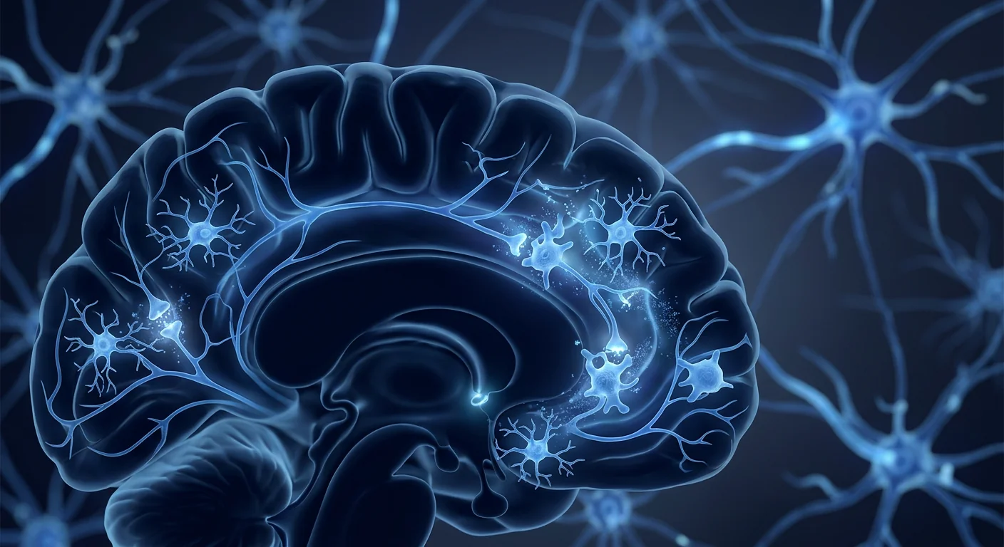
Hypoxic-Ischemic Encephalopathy (HIE) occurs in approximately 1.5 per 1,000 live births in developed countries and at significantly higher rates in low-resource settings. It is a form of neonatal brain injury caused by oxygen deprivation (hypoxia) and reduced blood flow (ischemia) to the brain around the time of birth — during labor, delivery, or the immediate postnatal period. For the past two decades, therapeutic hypothermia (whole-body or selective head cooling initiated within 6 hours of birth) has been the only neuroprotective intervention shown to reduce death and disability in moderate-to-severe HIE. But even with cooling, approximately 40–50% of infants still die or survive with significant neurodevelopmental impairment. Mesenchymal Stem Cell therapy is being studied as a complementary approach — one that extends the therapeutic window beyond the narrow 6-hour cooling window and addresses the secondary inflammatory injury cascade that continues for days to weeks after the initial insult.[1]
What happens in the brain during HIE
HIE is not a single event but a biphasic process. The primary energy failure occurs during the hypoxic-ischemic insult itself — ATP depletion, failure of ion pumps, excitotoxic glutamate release, and acute neuronal necrosis. This is followed by a latent period of partial recovery, and then a secondary energy failure that begins roughly 6–15 hours after the insult and can last for days. This secondary phase is driven by mitochondrial dysfunction, oxidative stress, and a profound neuroinflammatory cascade involving activated microglia, infiltrating peripheral immune cells, and a storm of pro-inflammatory cytokines (IL-1β, IL-6, TNF-α). It is this secondary injury — not the initial oxygen deprivation — that accounts for the majority of long-term brain damage in HIE.[2]
The therapeutic window is wider than previously thought. While therapeutic hypothermia is only effective within 6 hours of birth, the secondary inflammatory cascade continues for days — even weeks. This creates a window of opportunity for MSC therapy: delivery within the first days to weeks after birth could theoretically suppress the inflammatory storm, rescue threatened neurons in the penumbra, and support oligodendrocyte survival and myelination during the critical period of brain development.
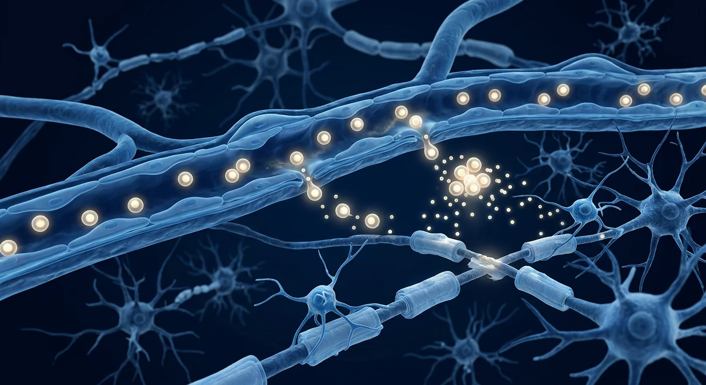
White matter is especially vulnerable. The developing oligodendrocytes that produce myelin in the neonatal brain are exquisitely sensitive to hypoxia-ischemia. Loss of these cells means impaired myelination — the biological basis for the cerebral palsy, cognitive deficits, and epilepsy that frequently follow moderate-to-severe HIE. Protecting and restoring oligodendrocyte function is a central goal of MSC therapy in this population.
How MSC therapy works in the HIE brain
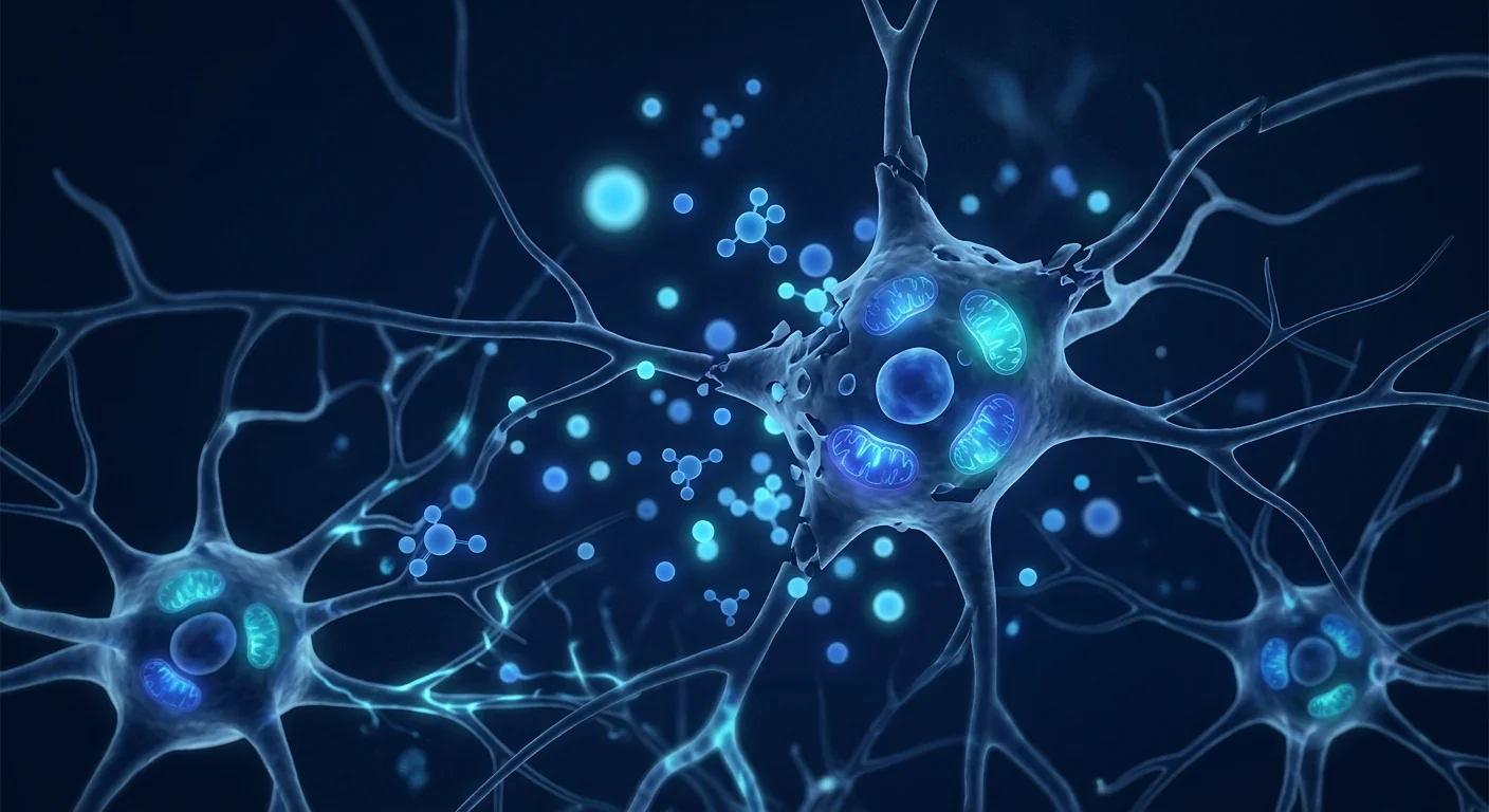
When clinical-grade Mesenchymal Stem Cells are delivered intravenously or intratracheally in the neonatal period, they do not need to engraft permanently in the brain. Their therapeutic effects are overwhelmingly paracrine — mediated by the secretion of bioactive molecules that signal the brain's own protective and repair systems:[3]
1. Neuroinflammation suppression
MSCs respond to the inflammatory signals in the injured neonatal brain by secreting powerful anti-inflammatory mediators — TSG-6 (TNF-stimulated gene 6), PGE2, IL-10, and TGF-β. These molecules shift microglia from a pro-inflammatory (M1-like) to a neuroprotective (M2-like) phenotype, dramatically reducing the cytokine storm that drives secondary brain injury. This is the most well-documented mechanism and the primary rationale for MSC therapy in HIE.[4]
2. Neurotrophic factor secretion
MSCs secrete a rich cocktail of neurotrophic factors: BDNF (brain-derived neurotrophic factor), NGF (nerve growth factor), GDNF (glial-derived neurotrophic factor), and IGF-1. These factors activate pro-survival pathways (PI3K/Akt, MAPK/ERK) in at-risk neurons, suppress apoptotic signaling, and promote synaptic plasticity — the cellular basis for functional recovery.[5]
3. Oligodendrocyte protection and myelination
MSC-derived factors protect oligodendrocyte precursor cells (OPCs) from apoptosis and encourage their differentiation into mature, myelin-producing oligodendrocytes. In rodent and piglet models of neonatal HIE, MSC treatment has been shown to increase myelination, preserve white matter tract integrity on diffusion tensor imaging, and improve long-term motor and cognitive outcomes.[6]
4. Mitochondrial transfer
One of the most intriguing mechanisms involves the direct transfer of healthy mitochondria from MSCs to injured neurons via tunneling nanotubes and extracellular vesicles. This "mitochondrial rescue" restores cellular energy metabolism, reduces oxidative stress, and can salvage neurons that would otherwise undergo apoptosis.[7]
5. Blood-brain barrier stabilization
MSC-derived factors reduce blood-brain barrier permeability by stabilizing tight junction proteins and suppressing matrix metalloproteinases, limiting the influx of peripheral immune cells and neurotoxic plasma proteins into the brain parenchyma.
6. Endogenous repair activation
MSCs stimulate the proliferation and migration of endogenous neural stem/progenitor cells in the subventricular zone and dentate gyrus — effectively recruiting the brain's own repair reserves. This is particularly relevant in neonates, whose brains retain greater neurogenic capacity than adults.
Why cooling alone is not enough
Therapeutic hypothermia is a landmark achievement — it reduces death and disability in moderate-to-severe HIE by approximately 25%. But 40–50% of cooled infants still have poor outcomes. Cooling primarily slows metabolic demand during the primary and early secondary phases. It does not directly suppress neuroinflammation, does not provide trophic support to dying neurons, and does not stimulate repair. MSC therapy is being studied as a complementary treatment — not a replacement for cooling — that addresses the biological gaps cooling leaves behind.[8]
What the clinical evidence says
The clinical evidence for MSC therapy in HIE is emerging but compelling. While the field is earlier-stage than for conditions like osteoarthritis or autoimmune disease, the neonatal brain injury literature is among the most rigorous in regenerative medicine — driven by large animal models (piglets, sheep, non-human primates) that closely mimic human HIE pathophysiology.[9]
- Large animal models: MSC treatment in neonatal piglets and sheep with induced HIE has consistently shown reduced brain lesion volume on MRI, preserved white matter integrity, improved neuromotor scores, and reduced seizure burden. These models are highly translatable because the neonatal piglet brain resembles the term human infant brain in size, myelination stage, and injury patterns.
- Phase I safety trials in human neonates: Small phase I trials have demonstrated that intravenous and intratracheal MSC delivery in term and preterm neonates with HIE is safe and well-tolerated, with no infusion-related adverse events, no tumor formation, and no increased infection risk in short-to-medium follow-up.
- Phase II neurodevelopmental outcomes: Early-phase controlled trials are now reporting neurodevelopmental outcomes at 12–24 months. Results suggest reduced rates of cerebral palsy, improved Bayley Scales of Infant Development scores, and lower seizure frequency compared to cooling-alone historical controls — though numbers remain small and larger randomized trials are ongoing.
- Biomarker evidence: MSC-treated neonates show reduced serum levels of S100B, neuron-specific enolase, and inflammatory cytokines (IL-6, IL-1β) — biomarkers correlated with brain injury severity — within 48–72 hours of treatment.
It is important to be honest: the field is still early. No phase III trial has yet confirmed a statistically significant improvement in death or severe disability at 18–24 months. But the consistency of the preclinical signal, the strong safety profile in neonates, and the compelling mechanistic rationale make HIE one of the most promising applications of neonatal MSC therapy.[10]
Timing and candidacy: when is MSC therapy most relevant?
The evidence suggests several key factors influence candidacy and expected benefit:[11]
- Moderate-to-severe HIE (Sarnat stage II–III): The strongest evidence is for infants with moderate-to-severe HIE — the same population eligible for therapeutic hypothermia. Mild HIE generally has good outcomes with supportive care alone.
- Timing of MSC delivery: Animal studies suggest a therapeutic window of at least 24–72 hours post-insult — and possibly up to 7–10 days — considerably wider than the 6-hour cooling window. The optimal timing remains under investigation, but delivery within the first 3–7 days of life is most commonly studied.
- Cooling + MSC combination: The most realistic clinical scenario is MSC therapy as an adjunct to therapeutic hypothermia, not a replacement. Whether administered during cooling, immediately after rewarming, or in the days following cooling, the combination approach is supported by animal studies showing additive or synergistic benefit.
- Preterm HIE and white matter injury: Preterm infants who develop white matter injury (periventricular leukomalacia) from hypoxia-ischemia or inflammation are a second important population. MSC therapy may be particularly valuable here because cooling is not approved for preterm infants.
- Subacute/chronic HIE (weeks to months after birth): While the strongest evidence is for acute-phase delivery, some studies are exploring later MSC administration (weeks to months after the insult) to support ongoing myelination and neurodevelopment. The rationale is weaker but the safety profile remains favorable.
Realistic timelines: what to expect and when
Neonatal brain repair follows a different trajectory than adult indications, and the timeline for observable benefit reflects the biology of developing brain recovery:[12]
It is critical to understand that in neonatal HIE, the most meaningful outcome is measured at 18–24 months of age — not weeks. The Bayley Scales of Infant Development, the standard neurodevelopmental assessment tool, evaluates cognitive, language, and motor function at this time point because it captures the functional result of brain development during the critical first two years. Families considering MSC therapy for HIE should understand that the goal is to improve the trajectory over months to years, not to produce an immediate visible change.[13]
Safety and what every family should know
When delivered in a clinical-grade setting with proper cell characterization, neonatal MSC therapy has demonstrated an excellent safety profile in phase I and II trials. The most commonly reported adverse events are transient and mild: slight temperature elevation within 24 hours of infusion, mild transient changes in heart rate or oxygen saturation during infusion (self-resolving), and temporary increase in irritability. No increased rates of infection, thrombosis, tumor formation, or graft-versus-host reactions have been reported in neonatal MSC trials.[14]
The most important safeguard is cell quality. In neonatal patients — the most vulnerable population — the stakes for product purity are at their highest. Families should confirm: MSC source (Wharton's Jelly, bone marrow, or adipose — Wharton's Jelly is most commonly used in neonatal trials due to high proliferative capacity and low immunogenicity), GMP-grade manufacturing facility, ISCT identity criteria (≥95% CD73+/CD90+/CD105+, ≤2% CD45-/CD34-/CD14-), viability (>95% at delivery), sterility testing, and endotoxin levels.[15]
Combining MSC therapy with cooling and rehabilitation
The emerging clinical paradigm for HIE management is sequential multimodal therapy: therapeutic hypothermia initiated within hours of birth, MSC infusion delivered during or shortly after rewarming (window: 24 hours to 7 days post-birth), followed by structured neurodevelopmental follow-up and early intervention services (physical therapy, occupational therapy, speech therapy) through the first two years. Each component addresses a different part of the injury-repair timeline: cooling reduces acute metabolic demand, MSCs suppress the secondary inflammatory cascade and support repair biology, and early intervention maximizes the functional expression of preserved and restored neural circuitry.[16]
For families confronting HIE, MSC therapy represents a new chapter in neonatal neurocritical care — not a single intervention but a biological partner to cooling and rehabilitation. The gains are measured across the first two years of life, and they accumulate in the skills a child can demonstrate at 18 and 24 months that were never expected.
— VELAR Clinical Team
Frequently Asked Questions
Can MSC therapy replace therapeutic hypothermia?
No. MSC therapy is being studied as a complementary treatment to cooling, not a replacement. Therapeutic hypothermia remains the standard of care for moderate-to-severe HIE and should be initiated within 6 hours of birth whenever eligibility criteria are met. MSCs address different biological targets — inflammation, trophic support, repair — and are best positioned as an adjunct that extends the therapeutic window beyond the 6-hour cooling limit.
How soon after birth can MSCs be given?
Clinical trials have administered MSCs at time points ranging from within 24 hours to 10 days after birth. The optimal timing remains under investigation, but animal data suggest efficacy within the first 3–7 days — during the secondary inflammatory phase when cytokine levels peak. Delivery during cooling (with careful monitoring) and immediately after rewarming are both being studied.
What route of administration is used for neonates?
Intravenous infusion is the most common route in neonatal trials — it is minimally invasive, well-tolerated, and achieves broad distribution. Intratracheal administration (via the endotracheal tube in ventilated infants) has also been studied and may achieve higher pulmonary and brain concentrations. Intranasal delivery is an emerging non-invasive route under investigation for direct nose-to-brain access.
Is MSC therapy safe for newborns?
Phase I and II trials in term and preterm neonates have demonstrated a strong safety profile: no serious infusion-related adverse events, no tumor formation, no increased infection risk, and no graft-versus-host disease. The safety data in neonates — the most vulnerable patient population — has been reassuring and is one reason the field is advancing toward larger efficacy trials.
What outcomes can families realistically expect?
The goal is to improve neurodevelopmental trajectory over 18–24 months, not to produce immediate visible change. Realistic outcomes include: reduced cerebral palsy severity or incidence, higher Bayley Scales motor and cognitive composite scores, reduced epilepsy risk, and improved functional independence. MSC therapy does not undo all damage — but it aims to narrow the gap between what the injury would have produced and what the brain with biological support can achieve.
How much does MSC therapy for HIE cost in Thailand?
Costs vary based on cell dose, number of infusions, and whether treatment is administered in the NICU or in a specialized regenerative medicine center. At VELAR Center, neonatal protocols are customized based on clinical severity and family preferences. A detailed cost estimate is provided after the initial consultation and medical record review. Contact the VELAR clinical team for a personalized assessment.
References
- Douglas-Escobar M, Weiss MD. Hypoxic-ischemic encephalopathy: a review for the clinician. JAMA Pediatrics. 2015;169(4):397-403. doi:10.1001/jamapediatrics.2014.3269 ↩
- Ferriero DM. Neonatal brain injury. New England Journal of Medicine. 2004;351(19):1985-1995. doi:10.1056/NEJMra041996 ↩
- Ahn SY, Chang YS, Park WS. Mesenchymal stem cells for neonatal brain injury: current perspectives. Neonatal Medicine. 2017;24(2):63-69. doi:10.5385/nm.2017.24.2.63 ↩
- Zhu LH, Bai X, Zhang N, et al. TSG-6 secreted by human umbilical cord mesenchymal stem cells attenuates severe burn-induced excessive inflammation. Stem Cells Translational Medicine. 2016;5(8):1056-1066. doi:10.5966/sctm.2015-0355 ↩
- Park WS, Ahn SY, Sung SI, Ahn JY, Chang YS. Strategies to enhance paracrine potency of transplanted mesenchymal stem cells in intractable neonatal disorders. Pediatric Research. 2018;83(1-2):214-222. doi:10.1038/pr.2017.249 ↩
- van Velthoven CT, Kavelaars A, van Bel F, Heijnen CJ. Mesenchymal stem cell treatment after neonatal hypoxic-ischemic brain injury improves behavioral outcome and induces neuronal and oligodendrocyte regeneration. Brain, Behavior, and Immunity. 2010;24(3):387-393. doi:10.1016/j.bbi.2009.10.017 ↩
- Spees JL, Olson SD, Whitney MJ, Prockop DJ. Mitochondrial transfer between cells can rescue aerobic respiration. Proceedings of the National Academy of Sciences. 2006;103(5):1283-1288. doi:10.1073/pnas.0510511103 ↩
- Jacobs SE, Berg M, Hunt R, Tarnow-Mordi WO, Inder TE, Davis PG. Cooling for newborns with hypoxic ischaemic encephalopathy. Cochrane Database of Systematic Reviews. 2013;(1):CD003311. doi:10.1002/14651858.CD003311.pub3 ↩
- Bennet L, Tan S, van den Heuij L, et al. Cell therapy for neonatal hypoxia-ischemia and cerebral palsy. Annals of Neurology. 2012;71(5):589-600. doi:10.1002/ana.22670 ↩
- Chang YS, Ahn SY, Yoo HS, et al. Mesenchymal stem cells for bronchopulmonary dysplasia: phase 1 dose-escalation clinical trial. Journal of Pediatrics. 2014;164(5):966-972. doi:10.1016/j.jpeds.2013.12.011 ↩
- Cotten CM, Murtha AP, Goldberg RN, et al. Feasibility of autologous cord blood cells for infants with hypoxic-ischemic encephalopathy. Journal of Pediatrics. 2014;164(5):973-979. doi:10.1016/j.jpeds.2013.11.036 ↩
- Tsuji M, Sawada M, Watabe S, et al. Autologous cord blood cell therapy for neonatal hypoxic-ischemic encephalopathy: a pilot study for feasibility and safety. Scientific Reports. 2020;10(1):4603. doi:10.1038/s41598-020-61311-9 ↩
- Saha S, Bjugstad KB, Hung YC, et al. Umbilical cord blood mononuclear cells for hypoxic ischemic encephalopathy. Pediatric Research. 2020;88(2):168-175. doi:10.1038/s41390-020-0874-x ↩
- Ahn SY, Chang YS, Sung DK, et al. Mesenchymal stem cells prevent hydrocephalus after severe intraventricular hemorrhage. Stroke. 2013;44(2):497-504. doi:10.1161/STROKEAHA.112.679092 ↩
- Dominici M, Le Blanc K, Mueller I, et al. Minimal criteria for defining multipotent mesenchymal stromal cells. The International Society for Cellular Therapy position statement. Cytotherapy. 2006;8(4):315-317. doi:10.1080/14653240600855905 ↩
- Serrenho I, Martins C, Rosado ML, et al. Stem cell therapy for neonatal hypoxic-ischemic encephalopathy: a systematic review of preclinical studies. International Journal of Molecular Sciences. 2021;22(6):3146. doi:10.3390/ijms22063146 ↩
缺氧缺血性脑病(HIE)在发达国家的发生率约为每1,000例活产中1.5例,在资源有限地区则显著更高。这是一种由出生前后脑部氧气供应中断(缺氧)和血流量减少(缺血)引起的新生儿脑损伤。过去二十年中,治疗性低温(在出生后6小时内启动的全身或选择性头部降温)一直是唯一被证实可降低中重度HIE死亡率和残疾率的神经保护干预措施。然而,即使采用降温治疗,仍有约40–50%的婴儿死亡或存活时伴有显著的神经发育障碍。间充质干细胞疗法正在被研究作为一种补充性方法——可将治疗窗口从狭窄的6小时降温窗口扩展到数天至数周,针对初始损伤后持续的继发性炎症损伤级联反应。[1]
HIE期间大脑发生了什么
HIE不是一个单一事件,而是一个双相过程。原发性能量衰竭发生在缺氧缺血损伤本身——ATP耗竭、离子泵失效、兴奋性谷氨酸释放和急性神经元坏死。随后是部分恢复的潜伏期,然后是约在损伤后6–15小时开始、可持续数天的继发性能量衰竭。这一继发阶段由线粒体功能障碍、氧化应激和深刻的神经炎症级联反应驱动,涉及活化的小胶质细胞、浸润的外周免疫细胞和促炎细胞因子风暴(IL-1β、IL-6、TNF-α)。正是这种继发性损伤——而非初始的氧气剥夺——造成了HIE中大部分长期脑损伤。[2]
治疗窗口比以往认为的更宽。虽然治疗性低温仅在出生后6小时内有效,但继发性炎症级联反应持续数天甚至数周。这为MSC疗法创造了一个机会窗口:在出生后的最初几天到几周内给药,理论上可以抑制炎症风暴、挽救半暗带中受威胁的神经元,并在大脑发育的关键时期支持少突胶质细胞存活和髓鞘形成。
白质尤其脆弱。新生大脑中产生髓鞘的发育中少突胶质细胞对缺氧缺血极其敏感。这些细胞的丧失意味着髓鞘形成受损——这是中重度HIE后常出现的脑瘫、认知缺陷和癫痫的生物学基础。保护和恢复少突胶质细胞功能是MSC疗法在这一人群中的核心目标。
MSC疗法如何在HIE大脑中发挥作用
当临床级间充质干细胞在新生期通过静脉或气管内输注时,它们不需要在大脑中永久植入。其治疗效果主要是旁分泌的——通过分泌生物活性分子,信号传导大脑自身的保护和修复系统:[3]
1. 抑制神经炎症
MSC通过分泌强大的抗炎介质——TSG-6、PGE2、IL-10和TGF-β——回应受损新生大脑中的炎症信号。这些分子将小胶质细胞从促炎(M1样)表型转变为神经保护(M2样)表型,显著减少驱动继发性脑损伤的细胞因子风暴。这是文献记录最充分的机制,也是MSC治疗HIE的主要理论基础。[4]
2. 神经营养因子分泌
MSC分泌丰富的神经营养因子:BDNF、NGF、GDNF和IGF-1。这些因子激活处于危险中的神经元的促存活通路(PI3K/Akt、MAPK/ERK),抑制凋亡信号,促进突触可塑性——这是功能恢复的细胞基础。[5]
3. 少突胶质细胞保护和髓鞘形成
MSC来源的因子保护少突胶质前体细胞免受凋亡,并促进其分化为成熟的产髓鞘少突胶质细胞。在新生HIE的啮齿类和仔猪模型中,MSC治疗已被证明可增加髓鞘形成、在弥散张量成像上保持白质束完整性,并改善长期运动和认知结果。[6]
4. 线粒体转移
最引人注目的机制之一涉及健康线粒体通过隧道纳米管和细胞外囊泡从MSC直接转移到受损神经元。这种"线粒体救援"恢复细胞能量代谢,减少氧化应激,并可挽救本将凋亡的神经元。[7]
5. 血脑屏障稳定
MSC来源的因子通过稳定紧密连接蛋白和抑制基质金属蛋白酶来降低血脑屏障通透性,限制外周免疫细胞和神经毒性血浆蛋白进入脑实质。
6. 激活内源性修复
MSC刺激脑室下区和齿状回中内源性神经干/祖细胞的增殖和迁移——有效地调动大脑自身的修复储备。这对新生儿的脑尤其重要,因为其神经发生能力比成人更强。
为什么单纯降温是不够的
治疗性低温是一项里程碑式的成就——可将中重度HIE的死亡率和残疾率降低约25%。但40–50%的降温婴儿仍有不良结局。降温主要通过减缓原发和早期继发阶段的代谢需求起作用。它不直接抑制神经炎症,不为濒死神经元提供营养支持,也不刺激修复。MSC疗法被研究作为补充治疗——而非替代降温——以填补降温留下的生物学空白。[8]
临床证据概述
MSC治疗HIE的临床证据正在涌现且令人信服。虽然该领域比骨关节炎或自身免疫性疾病更早期,但新生脑损伤文献是再生医学中最严谨的之一——由紧密模拟人类HIE病理生理学的大动物模型(仔猪、绵羊、非人灵长类)驱动。[9]
- 大动物模型:在诱导HIE的新生仔猪和绵羊中进行MSC治疗一致显示MRI脑损伤体积减小、白质完整性保持、神经运动评分改善和癫痫负担减少。这些模型高度可转化,因为新生仔猪大脑在大小、髓鞘形成阶段和损伤模式上与足月人类婴儿大脑相似。
- 人类新生儿的I期安全性试验:小型I期试验证明,在患有HIE的足月和早产新生儿中静脉和气管内输注MSC是安全且耐受良好的——无输注相关不良事件、无肿瘤形成、短期至中期随访中感染风险无增加。
- II期神经发育结果:早期对照试验正在报告12–24个月的神经发育结果。结果表明,与仅降温的历史对照相比,脑瘫发生率降低、Bayley婴儿发育量表评分提高、癫痫频率降低——尽管样本量仍然较小,大型随机试验正在进行中。
- 生物标志物证据:MSC治疗的新生儿在治疗后48–72小时内血清S100B、神经元特异性烯醇化酶和炎症细胞因子(IL-6、IL-1β)水平降低——这些是与脑损伤严重程度相关的生物标志物。
需要诚实说明的是:该领域仍处于早期阶段。尚无III期试验确认18–24个月时死亡或严重残疾的统计学显著改善。但临床前信号的一致性、新生儿的强大安全性以及令人信服的机制理论使HIE成为新生MSC治疗最有前景的应用之一。[10]
时机和适应症:何时MSC疗法最相关?
证据表明有若干关键因素影响适应症和预期获益:[11]
- 中重度HIE(Sarnat II–III期):最强证据适用于中重度HIE婴儿——与符合治疗性低温条件的人群相同。轻度HIE通常在单独支持治疗下预后良好。
- MSC输注时机:动物研究表明治疗窗口至少为损伤后24–72小时——可能长达7–10天——远宽于6小时降温窗口。最佳时机仍在研究中,但出生后3–7天内输注是最常研究的方案。
- 降温+MSC联合:最现实的临床方案是MSC疗法作为治疗性低温的辅助治疗,而非替代。无论是在降温期间、复温后即刻还是在降温后数天内给药,联合疗法得到动物研究的支持,显示叠加或协同获益。
- 早产HIE和白质损伤:因缺氧缺血或炎症发展为白质损伤的早产儿是第二个重要人群。MSC疗法在此可能特别有价值,因为降温未被批准用于早产儿。
- 亚急性/慢性HIE(出生后数周至数月):虽然最强证据支持急性期给药,但一些研究正在探索后期MSC给药以支持持续的髓鞘形成和神经发育。其理论基础较弱但安全性仍然良好。
现实的时间表:期待什么以及何时
新生脑修复遵循与成人适应症不同的轨迹,可观察获益的时间线反映了发育中大脑恢复的生物学:[12]
理解这一点至关重要:在新生儿HIE中,最有意义的结果是在18–24个月龄测量的——而非数周。Bayley婴儿发育量表是标准的神经发育评估工具,在此时间点评估认知、语言和运动功能,因为它捕捉了关键前两年大脑发育的功能结果。考虑MSC治疗HIE的家庭应理解,目标是改善数月至数年的发育轨迹,而非产生立即可见的变化。[13]
安全性和每个家庭应了解的事项
在临床级环境下进行适当细胞鉴定时,新生MSC疗法在I期和II期试验中展现了优异的安全性。最常报告的不良事件是暂时和轻微的:输注后24小时内轻微体温升高、输注期间心率或氧饱和度的轻微暂时变化(自行恢复)、以及暂时增加的易激惹。在新生MSC试验中未报告感染、血栓形成、肿瘤形成或移植物抗宿主反应的增加。[14]
最重要的保障是细胞质量。在新生儿患者——最脆弱的人群中——产品纯度的风险最高。家庭应确认:MSC来源(沃顿胶、骨髓或脂肪——由于增殖能力强和免疫原性低,沃顿胶是新生试验中最常用的来源)、GMP级生产设施、ISCT身份标准(≥95% CD73+/CD90+/CD105+、≤2% CD45-/CD34-/CD14-)、活力(交付时>95%)、无菌检测和内毒素水平。[15]
MSC疗法与降温和康复相结合
HIE管理的新兴临床范式是序贯多模式治疗:出生后数小时内启动治疗性低温,降温期间或复温后短期内输注MSC(窗口:出生后24小时至7天),然后在头两年进行结构化神经发育随访和早期干预服务(物理治疗、职业治疗、言语治疗)。每个组成部分针对损伤-修复时间线的不同部分:降温减少急性代谢需求,MSC抑制继发性炎症级联反应并支持修复生物学,早期干预最大化保存和恢复的神经回路的功能表达。[16]
对于面对HIE的家庭,MSC疗法代表了新生神经重症监护的新篇章——不是单一干预,而是降温和康复的生物学伙伴。获益在生命头两年中测量,并在18个月和24个月时孩子能够展示的、曾经不被期待掌握的技能中积累。
— VELAR临床团队
参考文献
- Douglas-Escobar M, Weiss MD. Hypoxic-ischemic encephalopathy: a review for the clinician. JAMA Pediatrics. 2015;169(4):397-403. doi:10.1001/jamapediatrics.2014.3269 ↩
- Ferriero DM. Neonatal brain injury. New England Journal of Medicine. 2004;351(19):1985-1995. doi:10.1056/NEJMra041996 ↩
- Ahn SY, Chang YS, Park WS. Mesenchymal stem cells for neonatal brain injury: current perspectives. Neonatal Medicine. 2017;24(2):63-69. doi:10.5385/nm.2017.24.2.63 ↩
- Zhu LH, Bai X, Zhang N, et al. TSG-6 secreted by human umbilical cord mesenchymal stem cells attenuates severe burn-induced excessive inflammation. Stem Cells Translational Medicine. 2016;5(8):1056-1066. doi:10.5966/sctm.2015-0355 ↩
- Park WS, Ahn SY, Sung SI, Ahn JY, Chang YS. Strategies to enhance paracrine potency of transplanted mesenchymal stem cells in intractable neonatal disorders. Pediatric Research. 2018;83(1-2):214-222. doi:10.1038/pr.2017.249 ↩
- van Velthoven CT, Kavelaars A, van Bel F, Heijnen CJ. Mesenchymal stem cell treatment after neonatal hypoxic-ischemic brain injury improves behavioral outcome and induces neuronal and oligodendrocyte regeneration. Brain, Behavior, and Immunity. 2010;24(3):387-393. doi:10.1016/j.bbi.2009.10.017 ↩
- Spees JL, Olson SD, Whitney MJ, Prockop DJ. Mitochondrial transfer between cells can rescue aerobic respiration. Proceedings of the National Academy of Sciences. 2006;103(5):1283-1288. doi:10.1073/pnas.0510511103 ↩
- Jacobs SE, Berg M, Hunt R, Tarnow-Mordi WO, Inder TE, Davis PG. Cooling for newborns with hypoxic ischaemic encephalopathy. Cochrane Database of Systematic Reviews. 2013;(1):CD003311. doi:10.1002/14651858.CD003311.pub3 ↩
- Bennet L, Tan S, van den Heuij L, et al. Cell therapy for neonatal hypoxia-ischemia and cerebral palsy. Annals of Neurology. 2012;71(5):589-600. doi:10.1002/ana.22670 ↩
- Chang YS, Ahn SY, Yoo HS, et al. Mesenchymal stem cells for bronchopulmonary dysplasia: phase 1 dose-escalation clinical trial. Journal of Pediatrics. 2014;164(5):966-972. doi:10.1016/j.jpeds.2013.12.011 ↩
- Cotten CM, Murtha AP, Goldberg RN, et al. Feasibility of autologous cord blood cells for infants with hypoxic-ischemic encephalopathy. Journal of Pediatrics. 2014;164(5):973-979. doi:10.1016/j.jpeds.2013.11.036 ↩
- Tsuji M, Sawada M, Watabe S, et al. Autologous cord blood cell therapy for neonatal hypoxic-ischemic encephalopathy: a pilot study for feasibility and safety. Scientific Reports. 2020;10(1):4603. doi:10.1038/s41598-020-61311-9 ↩
- Saha S, Bjugstad KB, Hung YC, et al. Umbilical cord blood mononuclear cells for hypoxic ischemic encephalopathy. Pediatric Research. 2020;88(2):168-175. doi:10.1038/s41390-020-0874-x ↩
- Ahn SY, Chang YS, Sung DK, et al. Mesenchymal stem cells prevent hydrocephalus after severe intraventricular hemorrhage. Stroke. 2013;44(2):497-504. doi:10.1161/STROKEAHA.112.679092 ↩
- Dominici M, Le Blanc K, Mueller I, et al. Minimal criteria for defining multipotent mesenchymal stromal cells. The International Society for Cellular Therapy position statement. Cytotherapy. 2006;8(4):315-317. doi:10.1080/14653240600855905 ↩
- Serrenho I, Martins C, Rosado ML, et al. Stem cell therapy for neonatal hypoxic-ischemic encephalopathy: a systematic review of preclinical studies. International Journal of Molecular Sciences. 2021;22(6):3146. doi:10.3390/ijms22063146 ↩
يحدث اعتلال الدماغ بنقص التأكسج الإقفاري (HIE) في حوالي 1.5 لكل 1,000 ولادة حية في البلدان المتقدمة وبمعدلات أعلى بكثير في البيئات محدودة الموارد. وهو شكل من إصابات الدماغ الوليدية الناجمة عن الحرمان من الأكسجين (نقص التأكسج) وانخفاض تدفق الدم (الإقفار) إلى الدماغ حول وقت الولادة. على مدى العقدين الماضيين، كان التبريد العلاجي (تبريد الجسم بالكامل أو الرأس الانتقائي خلال 6 ساعات من الولادة) هو التدخل الوحيد الواقي للأعصاب الذي ثبت أنه يقلل الوفاة والإعاقة في HIE المتوسط إلى الشديد. لكن حتى مع التبريد، لا يزال ما يقرب من 40–50% من الرضع يموتون أو ينجون مع إعاقة عصبية نمائية كبيرة. يُدرس علاج الخلايا الجذعية الوسيطة (MSC) كنهج تكميلي — يمدد النافذة العلاجية إلى ما بعد نافذة التبريد الضيقة البالغة 6 ساعات ويعالج شلال الإصابة الالتهابي الثانوي الذي يستمر لأيام إلى أسابيع بعد الإصابة الأولية.[1]
ماذا يحدث في الدماغ خلال HIE
HIE ليس حدثاً واحداً بل عملية ثنائية الطور. يحدث فشل الطاقة الأولي خلال الإصابة بنقص التأكسج الإقفاري نفسها — استنفاد ATP، فشل مضخات الأيونات، إطلاق الغلوتامات الاستثاري، والنخر العصبي الحاد. يتبع ذلك فترة كامنة من التعافي الجزئي، ثم فشل طاقة ثانوي يبدأ بعد حوالي 6–15 ساعة من الإصابة ويمكن أن يستمر لأيام. يُدفع هذا الطور الثانوي بخلل الميتوكوندريا، الإجهاد التأكسدي، وشلال التهابي عصبي عميق يشمل الخلايا الدبقية الصغيرة المنشطة، الخلايا المناعية المحيطية المتسللة، وعاصفة من السيتوكينات الالتهابية (IL-1β، IL-6، TNF-α). هذه الإصابة الثانوية — وليس الحرمان الأولي من الأكسجين — هي المسؤولة عن غالبية تلف الدماغ طويل المدى في HIE.[2]
النافذة العلاجية أوسع مما كان يُعتقد سابقاً. بينما لا يكون التبريد العلاجي فعالاً إلا خلال 6 ساعات من الولادة، يستمر الشلال الالتهابي الثانوي لأيام — حتى أسابيع. هذا يخلق نافذة فرصة لعلاج MSC: يمكن للتوصيل خلال الأيام إلى الأسابيع الأولى بعد الولادة نظرياً أن يثبط العاصفة الالتهابية، ينقذ العصبونات المهددة في منطقة الظل، ويدعم بقاء الخلايا قليلة التغصن والتغطية النخاعية خلال الفترة الحرجة من نمو الدماغ.
المادة البيضاء معرضة بشكل خاص. الخلايا قليلة التغصن النامية التي تنتج الميالين في الدماغ الوليدي حساسة بشكل استثنائي لنقص التأكسج الإقفاري. يعني فقدان هذه الخلايا ضعف التغطية النخاعية — الأساس البيولوجي للشلل الدماغي، العجز المعرفي، والصرع التي تتبع غالباً HIE المتوسط إلى الشديد. حماية واستعادة وظيفة الخلايا قليلة التغصن هو هدف مركزي لعلاج MSC في هذه الفئة.
كيف يعمل علاج MSC في دماغ HIE
عندما تُعطى الخلايا الجذعية الوسيطة ذات الجودة السريرية عن طريق الوريد أو داخل الرغامى في الفترة الوليدية، لا تحتاج إلى الانغراس الدائم في الدماغ. تأثيراتها العلاجية نظير صماوي بشكل ساحق — بوساطة إفراز جزيئات نشطة بيولوجياً تشير إلى أنظمة الحماية والإصلاح الخاصة بالدماغ:[3]
1. تثبيط الالتهاب العصبي
تستجيب MSCs للإشارات الالتهابية في الدماغ الوليدي المصاب بإفراز وسائط قوية مضادة للالتهاب — TSG-6، PGE2، IL-10، وTGF-β. تحول هذه الجزيئات الخلايا الدبقية الصغيرة من النمط الظاهري الالتهابي (شبيه M1) إلى النمط الواقي للأعصاب (شبيه M2)، مما يقلل بشكل كبير من عاصفة السيتوكينات التي تدفع إصابة الدماغ الثانوية. هذه هي الآلية الأكثر توثيقاً والأساس المنطقي الرئيسي لعلاج MSC في HIE.[4]
2. إفراز العوامل العصبية التغذوية
تفرز MSCs مزيجاً غنياً من العوامل العصبية التغذوية: BDNF، NGF، GDNF، وIGF-1. تنشط هذه العوامل مسارات البقاء (PI3K/Akt، MAPK/ERK) في العصبونات المعرضة للخطر، تثبط الإشارات الموتية، وتعزز اللدونة المشبكية — الأساس الخلوي للتعافي الوظيفي.[5]
3. حماية الخلايا قليلة التغصن والتغطية النخاعية
تحمي العوامل المشتقة من MSC الخلايا السليفة قليلة التغصن من الموت المبرمج وتشجع تمايزها إلى خلايا قليلة التغصن ناضجة منتجة للميالين. في نماذج القوارض والخنازير الصغيرة لـ HIE الوليدي، ثبت أن علاج MSC يزيد التغطية النخاعية، يحافظ على سلامة مسالك المادة البيضاء في تصوير الموتر الانتشاري، ويحسن النتائج الحركية والإدراكية طويلة المدى.[6]
4. نقل الميتوكوندريا
إحدى الآليات الأكثر إثارة للاهتمام تتضمن النقل المباشر للميتوكوندريا السليمة من MSCs إلى العصبونات المصابة عبر أنابيب النانو النفقية والحويصلات خارج الخلوية. هذا "الإنقاذ الميتوكوندري" يستعيد استقلاب الطاقة الخلوي، يقلل الإجهاد التأكسدي، ويمكن أن ينقذ العصبونات التي كانت ستموت.[7]
5. استقرار الحاجز الدموي الدماغي
تقلل العوامل المشتقة من MSC نفاذية الحاجز الدموي الدماغي عن طريق تثبيت بروتينات الوصلات المحكمة وتثبيط الميتالوبروتينازات المصفوفية، مما يحد من تدفق الخلايا المناعية المحيطية وبروتينات البلازما السامة للأعصاب إلى النسيج الدماغي.
6. تنشيط الإصلاح الداخلي
تحفز MSCs تكاثر وهجرة الخلايا الجذعية/السليفة العصبية الداخلية في المنطقة تحت البطينية والتلفيف المسنن — مجندةً بشكل فعال احتياطيات الإصلاح الخاصة بالدماغ. هذا ذو صلة خاصة بالولدان، الذين تحتفظ أدمغتهم بقدرة عصبية تولدية أكبر من البالغين.
لماذا التبريد وحده غير كافٍ
التبريد العلاجي إنجاز بارز — يقلل الوفاة والإعاقة في HIE المتوسط إلى الشديد بنسبة 25% تقريباً. لكن 40–50% من الرضع المبردين لا يزال لديهم نتائج سيئة. التبريد يبطئ بشكل أساسي الطلب الاستقلابي خلال الطورين الأولي والثانوي المبكر. وهو لا يثبط الالتهاب العصبي مباشرة، ولا يوفر دعماً تغذوياً للعصبونات المحتضرة، ولا يحفز الإصلاح. يُدرس علاج MSC كعلاج مكمل — وليس بديلاً للتبريد — يعالج الفجوات البيولوجية التي يتركها التبريد.[8]
ماذا تقول الأدلة السريرية
الأدلة السريرية لعلاج MSC في HIE ناشئة لكنها مقنعة. بينما المجال في مرحلة أبكر من حالات مثل الفصال العظمي أو أمراض المناعة الذاتية، فإن أدبيات إصابة الدماغ الوليدية هي من بين الأكثر دقة في الطب التجديدي — مدفوعة بنماذج حيوانية كبيرة (خنازير صغيرة، أغنام، رئيسيات غير بشرية) تحاكي عن كثب الفيزيولوجيا المرضية لـ HIE البشري.[9]
- النماذج الحيوانية الكبيرة: أظهر علاج MSC في الخنازير الصغيرة والأغنام الوليدية مع HIE المستحث باستمرار انخفاض حجم آفة الدماغ في التصوير بالرنين المغناطيسي، الحفاظ على سلامة المادة البيضاء، تحسين درجات الحركة العصبية، وتقليل عبء النوبات.
- تجارب السلامة من المرحلة الأولى في الولدان البشر: أظهرت تجارب المرحلة الأولى الصغيرة أن توصيل MSC عن طريق الوريد وداخل الرغامى في الولدان مكتملي النمو والخدج مع HIE آمن وجيد التحمل، بدون أحداث سلبية مرتبطة بالتسريب، بدون تكوين أورام، وبدون زيادة خطر العدوى في المتابعة القصيرة إلى المتوسطة.
- نتائج النمو العصبي من المرحلة الثانية: بدأت التجارب المبكرة المضبوطة في الإبلاغ عن نتائج النمو العصبي عند 12–24 شهراً. تشير النتائج إلى انخفاض معدلات الشلل الدماغي، تحسن درجات مقياس بايلي لنمو الرضع، وانخفاض تواتر النوبات مقارنةً بالضوابط التاريخية للتبريد وحده.
- أدلة المؤشرات الحيوية: يُظهر الولدان المعالجون بـ MSC انخفاض مستويات S100B في المصل، الإنولاز الخاص بالعصبونات، والسيتوكينات الالتهابية (IL-6، IL-1β) — مؤشرات حيوية مرتبطة بشدة إصابة الدماغ — خلال 48–72 ساعة من العلاج.
من المهم أن نكون صادقين: لا يزال المجال في مرحلة مبكرة. لم تؤكد أي تجربة من المرحلة الثالثة بعد تحسناً ذا دلالة إحصائية في الوفاة أو الإعاقة الشديدة عند 18–24 شهراً. لكن اتساق الإشارة قبل السريرية، ملف السلامة القوي في الولدان، والأساس المنطقي الآلي المقنع تجعل HIE أحد أكثر تطبيقات علاج MSC الوليدي وعداً.[10]
التوقيت والأهلية: متى يكون علاج MSC أكثر أهمية؟
تشير الأدلة إلى عدة عوامل رئيسية تؤثر على الأهلية والفائدة المتوقعة:[11]
- HIE المتوسط إلى الشديد (مرحلة Sarnat II–III): أقوى الأدلة للرضع الذين يعانون من HIE المتوسط إلى الشديد — نفس الفئة المؤهلة للتبريد العلاجي. HIE الخفيف عموماً له نتائج جيدة بالرعاية الداعمة وحدها.
- توقيت توصيل MSC: تشير الدراسات الحيوانية إلى نافذة علاجية لا تقل عن 24–72 ساعة بعد الإصابة — وربما تصل إلى 7–10 أيام — أوسع بكثير من نافذة التبريد البالغة 6 ساعات. يبقى التوقيت الأمثل قيد التحقيق، لكن التوصيل خلال 3–7 أيام من الحياة هو الأكثر دراسة.
- التبريد + MSC مجتمعين: السيناريو السريري الأكثر واقعية هو علاج MSC كمساعد للتبريد العلاجي، وليس بديلاً. سواء أُعطي أثناء التبريد، بعد إعادة التدفئة مباشرة، أو في الأيام التالية للتبريد، يُدعم النهج المشترك بدراسات حيوانية تُظهر فائدة إضافية أو تآزرية.
- HIE الخدج وإصابة المادة البيضاء: الرضع الخدج الذين يصابون بإصابة المادة البيضاء من نقص التأكسج الإقفاري أو الالتهاب هم فئة ثانية مهمة. قد يكون علاج MSC ذا قيمة خاصة هنا لأن التبريد غير معتمد للرضع الخدج.
- HIE تحت الحاد/المزمن (أسابيع إلى أشهر بعد الولادة): بينما أقوى الأدلة للتوصيل في الطور الحاد، تستكشف بعض الدراسات إعطاء MSC المتأخر لدعم التغطية النخاعية المستمرة والنمو العصبي. الأساس المنطقي أضعف لكن ملف السلامة يبقى مواتياً.
جداول زمنية واقعية: ماذا تتوقع ومتى
يتبع إصلاح الدماغ الوليدي مساراً مختلفاً عن دواعي البالغين، ويعكس الجدول الزمني للفائدة الملحوظة بيولوجيا تعافي الدماغ النامي:[12]
من الضروري فهم أنه في HIE الوليدي، تُقاس النتيجة الأكثر أهمية عند عمر 18–24 شهراً — وليس أسابيع. تقيم مقاييس بايلي لنمو الرضع، أداة التقييم العصبي النمائي القياسية، الوظيفة الإدراكية واللغوية والحركية في هذه النقطة الزمنية لأنها تلتقط النتيجة الوظيفية لنمو الدماغ خلال أول عامين حرجين. يجب أن تفهم الأسر التي تفكر في علاج MSC لـ HIE أن الهدف هو تحسين المسار على مدى أشهر إلى سنوات، وليس إنتاج تغيير فوري مرئي.[13]
السلامة وما يجب أن تعرفه كل أسرة
عند التوصيل في بيئة ذات جودة سريرية مع توصيف خلوي مناسب، أظهر علاج MSC الوليدي ملف أمان ممتاز في تجارب المرحلتين الأولى والثانية. الأحداث السلبية الأكثر شيوعاً عابرة وخفيفة: ارتفاع طفيف في درجة الحرارة خلال 24 ساعة من التسريب، تغيرات عابرة خفيفة في معدل ضربات القلب أو تشبع الأكسجين أثناء التسريب (تزول تلقائياً)، وزيادة مؤقتة في الهياج. لم تُبلغ عن معدلات متزايدة من العدوى، الخثار، تكوين الأورام، أو تفاعلات داء الطعم حيال الثوي في تجارب MSC الوليدية.[14]
أهم ضمانة هي جودة الخلايا. في المرضى الولدان — الفئة الأكثر ضعفاً — تكون مخاطر نقاء المنتج في أعلى مستوياتها. يجب على الأسر تأكيد: مصدر MSC (هلام وارتون، نخاع العظم، أو الدهون — هلام وارتون هو الأكثر استخداماً في التجارب الوليدية بسبب القدرة التكاثرية العالية والمناعة المنخفضة)، منشأة تصنيع بمعيار GMP، معايير هوية ISCT (≥95% CD73+/CD90+/CD105+، ≤2% CD45-/CD34-/CD14-)، الحيوية (>95% عند التسليم)، اختبار العقامة، ومستويات الذيفان الداخلي.[15]
الجمع بين علاج MSC والتبريد والتأهيل
النموذج السريري الناشئ لإدارة HIE هو العلاج متعدد الوسائط المتسلسل: التبريد العلاجي خلال ساعات من الولادة، تسريب MSC خلال أو بعد إعادة التدفئة بقليل (النافذة: 24 ساعة إلى 7 أيام بعد الولادة)، متبوعاً بمتابعة عصبية نمائية منظمة وخدمات التدخل المبكر (العلاج الطبيعي، العلاج الوظيفي، علاج النطق) خلال أول عامين. كل مكون يعالج جزءاً مختلفاً من الجدول الزمني للإصابة والإصلاح: التبريد يقلل الطلب الاستقلابي الحاد، MSCs تثبط الشلال الالتهابي الثانوي وتدعم بيولوجيا الإصلاح، والتدخل المبكر يعظم التعبير الوظيفي للدوائر العصبية المحفوظة والمستعادة.[16]
للأسر التي تواجه HIE، يمثل علاج MSC فصلاً جديداً في الرعاية العصبية المركزة الوليدية — ليس تدخلاً واحداً بل شريكاً بيولوجياً للتبريد والتأهيل. تُقاس المكاسب عبر أول عامين من الحياة، وتتراكم في المهارات التي يمكن للطفل إظهارها في 18 و24 شهراً والتي لم تكن متوقعة أبداً.
— فريق VELAR السريري
المراجع
- Douglas-Escobar M, Weiss MD. Hypoxic-ischemic encephalopathy: a review for the clinician. JAMA Pediatrics. 2015;169(4):397-403. doi:10.1001/jamapediatrics.2014.3269 ↩
- Ferriero DM. Neonatal brain injury. New England Journal of Medicine. 2004;351(19):1985-1995. doi:10.1056/NEJMra041996 ↩
- Ahn SY, Chang YS, Park WS. Mesenchymal stem cells for neonatal brain injury: current perspectives. Neonatal Medicine. 2017;24(2):63-69. doi:10.5385/nm.2017.24.2.63 ↩
- Zhu LH, Bai X, Zhang N, et al. TSG-6 secreted by human umbilical cord mesenchymal stem cells attenuates severe burn-induced excessive inflammation. Stem Cells Translational Medicine. 2016;5(8):1056-1066. doi:10.5966/sctm.2015-0355 ↩
- Park WS, Ahn SY, Sung SI, Ahn JY, Chang YS. Strategies to enhance paracrine potency of transplanted mesenchymal stem cells in intractable neonatal disorders. Pediatric Research. 2018;83(1-2):214-222. doi:10.1038/pr.2017.249 ↩
- van Velthoven CT, Kavelaars A, van Bel F, Heijnen CJ. Mesenchymal stem cell treatment after neonatal hypoxic-ischemic brain injury improves behavioral outcome and induces neuronal and oligodendrocyte regeneration. Brain, Behavior, and Immunity. 2010;24(3):387-393. doi:10.1016/j.bbi.2009.10.017 ↩
- Spees JL, Olson SD, Whitney MJ, Prockop DJ. Mitochondrial transfer between cells can rescue aerobic respiration. Proceedings of the National Academy of Sciences. 2006;103(5):1283-1288. doi:10.1073/pnas.0510511103 ↩
- Jacobs SE, Berg M, Hunt R, Tarnow-Mordi WO, Inder TE, Davis PG. Cooling for newborns with hypoxic ischaemic encephalopathy. Cochrane Database of Systematic Reviews. 2013;(1):CD003311. doi:10.1002/14651858.CD003311.pub3 ↩
- Bennet L, Tan S, van den Heuij L, et al. Cell therapy for neonatal hypoxia-ischemia and cerebral palsy. Annals of Neurology. 2012;71(5):589-600. doi:10.1002/ana.22670 ↩
- Chang YS, Ahn SY, Yoo HS, et al. Mesenchymal stem cells for bronchopulmonary dysplasia: phase 1 dose-escalation clinical trial. Journal of Pediatrics. 2014;164(5):966-972. doi:10.1016/j.jpeds.2013.12.011 ↩
- Cotten CM, Murtha AP, Goldberg RN, et al. Feasibility of autologous cord blood cells for infants with hypoxic-ischemic encephalopathy. Journal of Pediatrics. 2014;164(5):973-979. doi:10.1016/j.jpeds.2013.11.036 ↩
- Tsuji M, Sawada M, Watabe S, et al. Autologous cord blood cell therapy for neonatal hypoxic-ischemic encephalopathy: a pilot study for feasibility and safety. Scientific Reports. 2020;10(1):4603. doi:10.1038/s41598-020-61311-9 ↩
- Saha S, Bjugstad KB, Hung YC, et al. Umbilical cord blood mononuclear cells for hypoxic ischemic encephalopathy. Pediatric Research. 2020;88(2):168-175. doi:10.1038/s41390-020-0874-x ↩
- Ahn SY, Chang YS, Sung DK, et al. Mesenchymal stem cells prevent hydrocephalus after severe intraventricular hemorrhage. Stroke. 2013;44(2):497-504. doi:10.1161/STROKEAHA.112.679092 ↩
- Dominici M, Le Blanc K, Mueller I, et al. Minimal criteria for defining multipotent mesenchymal stromal cells. The International Society for Cellular Therapy position statement. Cytotherapy. 2006;8(4):315-317. doi:10.1080/14653240600855905 ↩
- Serrenho I, Martins C, Rosado ML, et al. Stem cell therapy for neonatal hypoxic-ischemic encephalopathy: a systematic review of preclinical studies. International Journal of Molecular Sciences. 2021;22(6):3146. doi:10.3390/ijms22063146 ↩