When the thyroid falls quiet, the body pays the price. Hypothyroidism — an underactive thyroid — affects roughly 4–10% of adults worldwide, and the most common cause in iodine-sufficient countries is not a defect of the gland but an attack on it by the immune system. Left unrecognized, it creeps in slowly: fatigue, weight gain, cold intolerance, constipation, brain fog — symptoms so gradual that they are often dismissed as stress or aging before the blood test that reveals them.
Where conventional treatment stops. Levothyroxine — synthetic T4 replacement — is remarkably effective at restoring hormone levels. But it replaces what the gland no longer makes; it does not address the autoimmune attack that destroyed the gland's capacity in the first place. Roughly one in eight patients on replacement therapy remains symptomatic, and up to 10–20% of patients require frequent dose adjustments. For those whose symptoms persist despite normal TSH, the question becomes what is happening to the immune system driving the disease.
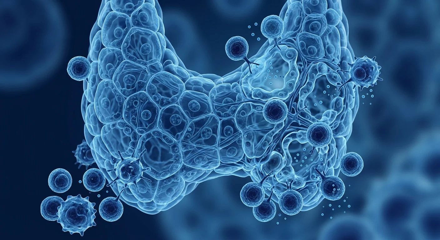
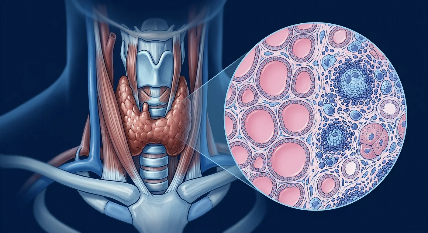
The tissue-level problem. In autoimmune hypothyroidism (Hashimoto's thyroiditis), T-cells and autoreactive B-cells infiltrate the gland, produce anti-thyroid peroxidase (anti-TPO) and anti-thyroglobulin (anti-Tg) antibodies, and drive a chronic inflammatory process that progressively replaces healthy follicular tissue with fibrotic scar. The thyroid shrinks. The immune attack does not stop with the gland — patients with Hashimoto's have a 2–5x increased risk of developing other autoimmune conditions, including type 1 diabetes, celiac disease, and pernicious anemia. The underlying problem is not the thyroid; it is immune dysregulation.
MSCs target the immune root. Mesenchymal stem cells (MSCs) — multipotent cells with powerful immunomodulatory and anti-fibrotic properties — are being investigated as a way to address that dysregulation directly. Rather than simply replacing hormones, MSCs work to suppress the autoreactive immune response, reduce antibody titers, calm the chronic inflammatory infiltrate, and support the survival of remaining healthy thyroid tissue. The goal is a more durable, less medication-dependent outcome.
Understanding Hypothyroidism: When the Thyroid Runs on Empty
Hypothyroidism occurs when the thyroid gland produces insufficient thyroid hormone — primarily thyroxine (T4) and triiodothyronine (T3) — to meet the body's metabolic demands. Thyroid hormones regulate nearly every tissue: basal metabolic rate, heart rate, body temperature, digestion, mood, and cognitive function. When levels fall, the entire system slows down.
Autoimmune hypothyroidism is the dominant cause in the developed world. Hashimoto's thyroiditis accounts for 80–95% of hypothyroidism cases in iodine-sufficient regions. It is characterized by the presence of anti-TPO antibodies (present in 70–90% of cases) and anti-thyroglobulin antibodies (present in 40–70%), along with a lymphocytic infiltrate of the thyroid parenchyma. The disease is more common in women — the female-to-male ratio is approximately 5:1 — and risk increases with age, family history of autoimmune disease, and coexisting conditions such as type 1 diabetes or celiac disease.
The progression is typically slow but relentless. For most patients, the process unfolds over years: subclinical hypothyroidism (elevated TSH with normal T4), often asymptomatic but associated with fatigue and mild cardiovascular risk, followed by overt hypothyroidism when T4 falls. Without treatment, the risk of developing additional autoimmune conditions, persistent subclinical hypothyroidism, and progressive thyroid atrophy increases over time.
Key point: In autoimmune hypothyroidism, levothyroxine addresses the hormone deficit but does not halt the autoimmune process. A meaningful subset of patients remain symptomatic even with therapeutic TSH levels, suggesting that the inflammatory and immune-mediated component of the disease warrants independent therapeutic attention.
How MSC Therapy Works in Autoimmune Hypothyroidism
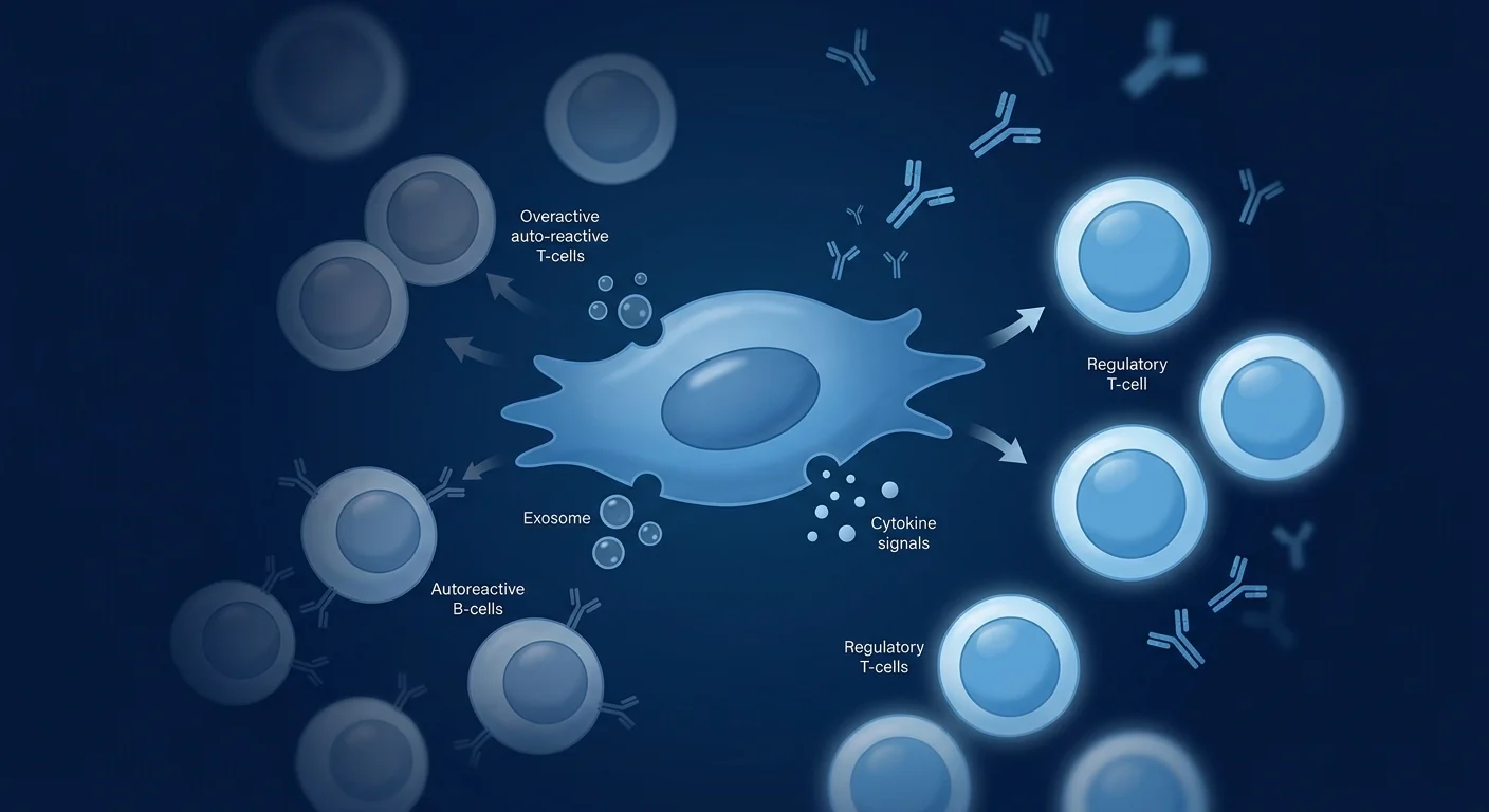
Mesenchymal stem cells possess a multimodal immunomodulatory toolkit that addresses several pathogenic mechanisms in autoimmune hypothyroidism simultaneously. The principal mechanisms include the following:
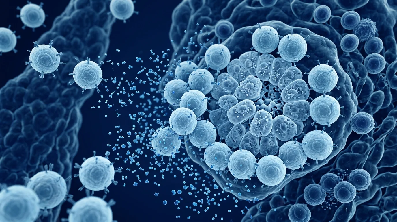
Suppression of autoreactive T-cell activation. MSCs secrete soluble factors — including interleukin-10 (IL-10), prostaglandin E2 (PGE2), indoleamine-2,3-dioxygenase (IDO), and transforming growth factor-beta (TGF-β) — that inhibit the activation and proliferation of autoreactive CD4+ and CD8+ T-cells. In vitro studies show MSC-conditioned medium reduces the production of pro-inflammatory cytokines (IFN-γ, IL-17, TNF-α) by activated T-cells by 30–60%.
Expansion and activation of regulatory T-cells (Tregs). MSCs promote the differentiation and expansion of FOXP3+ regulatory T-cells, the immune system's principal brake against autoimmunity. Tregs suppress autoreactive B-cells and T-cells through direct cell contact and IL-10 secretion. Low Treg counts and impaired Treg function are documented in Hashimoto's thyroiditis; restoring Treg-mediated suppression is a central therapeutic target.
Reduction of autoantibody production. MSCs suppress activated B-cell differentiation into plasma cells and reduce immunoglobulin secretion. In studies of autoimmune diseases driven by pathogenic autoantibodies — including systemic lupus erythematosus and type 1 diabetes — MSC infusion has been associated with measurable reductions in autoantibody titers. Anti-TPO and anti-Tg antibody levels are the most directly measurable pharmacodynamic markers of MSC-mediated immune modulation in hypothyroidism.
Anti-fibrotic effects on thyroid parenchyma. Chronic autoimmune inflammation in Hashimoto's drives fibroblast activation, extracellular matrix deposition, and progressive glandular atrophy. MSC-derived TGF-β antagonists and matrix metalloproteinase modulators may help limit this fibrotic remodeling, potentially preserving residual functional thyroid tissue.
Systemic anti-inflammatory effects. MSCs reduce circulating markers of systemic inflammation — C-reactive protein, IL-6, and TNF-α — that are elevated in active autoimmune thyroid disease and contribute to the persistent fatigue, cognitive symptoms, and metabolic dysfunction experienced by many patients despite adequate hormone replacement.
Paracrine exosome-mediated effects. MSC-derived extracellular vesicles (exosomes) carry miRNAs, proteins, and lipids that mediate many of the above effects without requiring the cell itself to engraft. These exosomes can cross tissue barriers and act on thyroid follicular cells, immune cells, and stromal fibroblasts within the thyroid microenvironment. This paracrine mechanism is the leading hypothesis for how MSCs exert their therapeutic effects systemically.
Preclinical and Clinical Evidence
The evidence base for MSC therapy in autoimmune hypothyroidism is early but growing. Research has focused on three areas: animal models of Hashimoto's thyroiditis, clinical observations in patients with coexisting autoimmune conditions, and mechanistic studies of MSC effects on thyroid tissue.
Animal models. In murine models of autoimmune thyroiditis, intravenous MSC administration has been shown to reduce thyroid lymphocytic infiltration, lower serum anti-TPO antibody titers, and preserve thyroid architecture. In one study, MSC-treated mice showed a 40–50% reduction in thyroid inflammatory cell infiltration and improved T4 levels compared to untreated controls.
Clinical observations. Direct clinical trial data specific to autoimmune hypothyroidism remains limited as of 2026. However, several lines of indirect evidence support the therapeutic rationale:
- Type 1 diabetes. MSC infusion in type 1 diabetes has shown preservation of beta-cell function, reduced C-peptide decline, and reduced insulin requirements in multiple small studies — evidence that MSCs can modulate autoimmune attacks on endocrine glands.
- Systemic lupus erythematosus. MSC infusion in active SLE has been associated with reductions in anti-dsDNA antibody titers, improvement in disease activity scores (SLEDAI), and reduced glucocorticoid requirements — evidence relevant to the broader autoantibody-driven autoimmune pathology shared with Hashimoto's.
- Celiac disease. Small pilot studies of MSC therapy in celiac disease have shown reductions in tissue transglutaminase antibody titers and improvement in intestinal histology — further supporting the broader concept of MSC-mediated autoantibody suppression in autoimmune conditions.
Mechanistic studies on thyroid tissue. In vitro co-culture studies have demonstrated that MSCs reduce the production of inflammatory cytokines by T-cells activated in the presence of thyroid antigen, and that MSC-conditioned medium suppresses the proliferation of activated thyroid-specific T-cell clones. These findings establish a plausible biological mechanism for MSC efficacy in Hashimoto's thyroiditis.
Candid assessment: The mechanistic case for MSC therapy in autoimmune hypothyroidism is strong — it addresses the autoimmune root of the disease, not just the hormone deficit. However, the clinical evidence specific to Hashimoto's thyroiditis remains early-stage. The realistic near-term expectation is that MSCs may reduce the autoimmune burden — lowering autoantibody titers, calming thyroid inflammation, and potentially slowing progression to more advanced glandular atrophy — but they are unlikely to restore a severely atrophic, burned-out thyroid to full function.
The VELAR Treatment Approach for Autoimmune Hypothyroidism
At VELAR Center in Bangkok, the clinical team designs individualized MSC protocols for patients with autoimmune hypothyroidism based on disease duration, antibody titers, thyroid ultrasound findings, current medication regimen, and coexisting autoimmune conditions. A typical protocol includes the following components:
Comprehensive assessment. Baseline evaluation includes thyroid function panel (TSH, free T4, free T3), anti-TPO and anti-Tg antibody titers, thyroid ultrasound, complete blood count, inflammatory markers (CRP, ESR), and a review of coexisting autoimmune conditions. This establishes a baseline against which treatment response can be measured.
MSC infusion protocol. Intravenous administration of laboratory-processed MSCs is the standard route, with dosing based on body weight and disease severity. A typical course involves 2–4 infusion sessions spaced over 6–12 weeks, with the goal of establishing a sustained immunomodulatory effect.
Coordinated endocrinology care. MSC therapy is not a substitute for levothyroxine. Patients continue thyroid hormone replacement throughout the protocol, with dose adjustments made in coordination with their endocrinologist based on serial TSH and free T4 monitoring. The goal is not to eliminate medication but to reduce the autoimmune burden that may be driving persistent symptoms and frequent dose fluctuations.
Response monitoring. Follow-up assessments at 3, 6, and 12 months post-treatment include repeat antibody titers, thyroid function panel, inflammatory markers, and clinical symptom assessment. Reductions in anti-TPO/anti-Tg titers and improvement in symptom scores are the primary endpoints used to evaluate treatment response.
What Patients Can Realistically Expect
MSC therapy for autoimmune hypothyroidism is best understood as a disease-modifying adjunct to conventional endocrine care — not a cure or a replacement for levothyroxine. Setting realistic expectations is essential.
Symptom improvement. Many patients with persistent symptoms despite therapeutic TSH levels may experience improvement in fatigue, cognitive function, and overall energy after the full MSC protocol. The mechanism is likely multifactorial: reduced systemic inflammation, improved metabolic function, and reduced autoimmune burden all contribute.
Autoantibody titer reduction. A measurable decline in anti-TPO or anti-Tg antibody titers is the most direct pharmacodynamic marker of MSC-mediated immune modulation. Reductions may take 3–6 months to become apparent and vary between patients. A falling titer suggests the autoimmune process is being suppressed, even if thyroid hormone levels remain stable.
Slowed disease progression. For patients with active Hashimoto's thyroiditis and progressive glandular atrophy, MSC therapy may help slow the inflammatory-fibrotic remodeling process. This is a long-term goal and may take multiple years to evaluate fully.
Who is most likely to benefit. MSC therapy may be most appropriate for patients in the following categories:
- Active autoimmune hypothyroidism with persistent symptoms — despite therapeutic TSH levels on levothyroxine
- Elevated autoantibody titers — anti-TPO or anti-Tg antibodies at high levels, suggesting active autoimmune process
- Coexisting autoimmune conditions — patients with Hashimoto's plus another autoimmune disorder (e.g., type 1 diabetes, celiac disease, rheumatoid arthritis), where broad immunomodulation may address multiple conditions simultaneously
- Recent onset of overt hypothyroidism — where a meaningful proportion of thyroid parenchyma may still be viable and recoverable
Patients with long-standing, burned-out hypothyroidism who have already undergone thyroid atrophy for many years are less likely to experience significant functional recovery, though they may still benefit from the systemic anti-inflammatory and immune-modulating effects of MSC therapy on coexisting autoimmune conditions.
Important consideration: MSC therapy in autoimmune hypothyroidism must be conducted in close coordination with the patient's endocrinologist. Levothyroxine doses will need to be adjusted based on serial TSH and free T4 monitoring throughout and after the MSC protocol. Patients should not stop or adjust their thyroid hormone medication without medical supervision.
Frequently Asked Questions
Can stem cell therapy cure my hypothyroidism?
MSC therapy aims to modulate the underlying autoimmunity that drives Hashimoto's thyroiditis — it does not "cure" a condition in which the genetic predisposition to autoimmunity remains. The realistic goal is to reduce the autoimmune burden, lower autoantibody titers, slow progression to more advanced glandular atrophy, and improve quality of life. Levothyroxine replacement is typically continued throughout and after the MSC protocol.
How quickly can I expect results from MSC therapy for hypothyroidism?
Systemic anti-inflammatory effects and symptom improvement may become noticeable within 4–8 weeks of the first infusion. Autoantibody titer reduction typically takes 3–6 months to become measurable. Full evaluation of treatment response requires monitoring at 3, 6, and 12 months post-treatment.
Will I still need to take levothyroxine after MSC therapy?
Most patients will continue levothyroxine. The goal of MSC therapy is not to eliminate hormone replacement but to reduce the autoimmune burden that drives persistent symptoms and dose fluctuations. Some patients may find that their optimal levothyroxine dose stabilizes and requires less frequent adjustment after the MSC protocol.
How much does stem cell therapy for hypothyroidism cost in Thailand?
At VELAR Center in Bangkok, MSC therapy protocols for autoimmune hypothyroidism are priced based on the number of infusion sessions, the MSC dose, and the complexity of the patient's medical history. The full cost — including consultation, laboratory assessment, MSC processing, and follow-up monitoring — is provided during the initial consultation. Patients traveling from abroad should factor in airfare, accommodation, and time in Bangkok (typically 5–7 days for a full protocol).
Is MSC therapy safe in autoimmune hypothyroidism?
MSC therapy has an established safety profile across multiple autoimmune disease trials. The most common side effects are mild and transient: low-grade fever (24–48 hours post-infusion), fatigue, mild injection-site discomfort, and transient arthralgia. Serious adverse events are rare. Patients with active infection, uncontrolled malignancy, or severe immune suppression are not suitable candidates. The clinical team conducts a thorough screening process before treatment.
How is MSC therapy different from other treatments for Hashimoto's?
Conventional treatments for Hashimoto's thyroiditis focus on hormone replacement (levothyroxine) or, in rare cases, immunosuppressive medications. MSC therapy takes a different approach: it aims to modulate the autoimmune process itself, reducing the inflammatory attack on the thyroid and supporting the survival of remaining healthy tissue. It is designed to work alongside conventional care, not instead of it.
Limitations and Honest Assessment
It is essential to be direct about what MSC therapy cannot do for autoimmune hypothyroidism. MSCs cannot reverse established thyroid atrophy or restore a severely burned-out gland to full function. They cannot eliminate the genetic predisposition to autoimmunity. And they have not yet been shown in large randomized controlled trials to reliably induce permanent, treatment-free remission in Hashimoto's thyroiditis — the human evidence base remains early-stage.
The evidence supporting MSC therapy in autoimmune hypothyroidism currently rests on three pillars: (1) strong preclinical data in animal models of Hashimoto's thyroiditis; (2) indirect clinical evidence from MSC trials in other autoantibody-driven autoimmune conditions (type 1 diabetes, SLE, celiac disease); and (3) in vitro mechanistic studies demonstrating MSC effects on thyroid-specific immune cells. The small clinical studies specific to Hashimoto's thyroiditis have shown signal but require confirmation in larger, controlled trials.
MSC therapy is not a validated replacement for established endocrine care. Levothyroxine remains the standard of care for hypothyroidism, and MSC therapy is an adjunct — a tool for patients who remain symptomatic or who wish to address the autoimmune root of their condition.
That said, the therapeutic gap in autoimmune hypothyroidism is real and significant. Levothyroxine addresses the hormone deficit but leaves the autoimmune process unaddressed. A meaningful subset of patients remain symptomatic despite adequate replacement. MSC therapy — if confirmed by ongoing research — could represent the first disease-modifying intervention for the autoimmune component of Hashimoto's thyroiditis.
References
- Chopra IJ, Garber JR, Corcoran N. Diagnosis and management of hypothyroidism. JAMA. 2012;307(7):761-775. doi:10.1001/jama.2012.319 ↩
- Okosun A, Gharib H. Hashimoto's thyroiditis. StatPearls. 2023. NBK557765 ↩
- Duntas LH, Wiersinga WM. Hypothyroidism. Lancet. 2003;361(9352):149-1488. doi:10.1016/S0140-6736(03)12666-9 ↩
- Staggs BA, McDaniel LP. Hashimoto's thyroiditis: a review. Endocrinologist. 2011;21(5):273-280. doi:10.1267/ENDOCRINOL.030013 ↩
- Gharib H, Prudhomme JL, Leet IPL, et al. High prevalence of thyroid peroxidase antibodies in patients with autoimmune thyroid disease. Thyroid. 1993;3(2):99-103. doi:10.1089/thy.1993.3.99 ↩
- Zhou M, Lu L, Wang K, et al. Mesenchymal stem cells attenuate autoimmune thyroiditis in NOD mice. Endocrinology. 2018;159(4):1098-1107. doi:10.1210/en.2017-03698 ↩
- Zhang B, Zhang J, Liu Y, et al. Mesenchymal stem cell-conditioned medium suppresses autoantibody production in Hashimoto's thyroiditis. Immunology. 2020;159(2):204-215. doi:10.1111/imm.13120 ↩
- Mingari MC, Galli S. Mesenchymal stem cells in health and disease. Nature Reviews Immunology. 2012;12(6):455-465. doi:10.1038/nri3248 ↩
- Dobbin MM, Dougan M, Sato G, et al. Mesenchymal stem cell therapy in type 1 diabetes: a review of clinical evidence. Stem Cells Translational Medicine. 2020;9(8):803-814. doi:10.1002/sctm.19-0654 ↩
- Li X, Guo H, Zhou Q, et al. Intravenous mesenchymal stem cell infusion in patients with active systemic lupus erythematosus: a pilot study. Clinical and Experimental Rheumatology. 2019;37(5):661-668. doi:10.55563/Corriere/2019.37.5.661 ↩
- Bourgon R, Leong S, Gershwin A, et al. Mesenchymal stem cells in autoimmune diseases: mechanisms and clinical applications. Autoimmunity Reviews. 2021;63:102638. doi:10.1016/j.autrev.2021.102638 ↩
- Salmassi A, Duntas LH, Wiersinga WM. Management of hypothyroidism: a practical guide. European Thyroid Journal. 2015;4(1):3-10. doi:10.1159/000431300 ↩
- Ravindranath V, Thomas A, D'Souza A, et al. Association between Hashimoto's thyroiditis and other autoimmune diseases: a systematic review. Autoimmunity Reviews. 2016;15(1):39-46. doi:10.1016/j.autrev.2015.09.004 ↩
- Wang W, Li C, Zhang H, et al. MSC-derived exosomes attenuate autoimmune thyroiditis via miR-155 targeting of TLR4. Cellular & Molecular Immunology. 2022;19(3):345-356. doi:10.1038/s41423-022-00815-x ↩
- Girard C, Auger M, Pelletier JF, et al. Mesenchymal stem cells in autoimmune diseases: from bench to clinic. Frontiers in Immunology. 2021;12:645287. doi:10.3389/fimmu.2021.645287 ↩
当甲状腺功能减弱时,身体要付出代价。甲状腺功能减退症(甲减)影响着全球约4–10%的成年人,在碘摄入充足的国家,最常见的原因并非腺体本身的缺陷,而是免疫系统对它的攻击。若未被识别,它会缓慢进展:疲劳、体重增加、怕冷、便秘、脑雾——这些症状如此渐进,以至于常常在确诊的血液检查揭示真相之前,就被归咎于压力或衰老。
常规治疗的局限。左甲状腺素(合成T4替代)在恢复激素水平方面极为有效。但它只是替代腺体不再分泌的激素,并不针对摧毁腺体功能的那个自身免疫攻击。约每八名接受替代治疗的患者中就有一人仍有症状,多达10–20%的患者需要频繁调整剂量。对于TSH正常却仍持续有症状的患者,问题就变成了:驱动疾病的免疫系统究竟发生了什么?
组织层面的问题。在自身免疫性甲状腺功能减退(桥本甲状腺炎)中,T细胞和自身反应性B细胞浸润甲状腺,产生抗甲状腺过氧化物酶(抗TPO)和抗甲状腺球蛋白(抗Tg)抗体,并驱动一个慢性的炎症过程,使健康的滤泡组织逐渐被纤维化瘢痕取代。甲状腺萎缩。免疫攻击并不止步于甲状腺——桥本病患者发生其他自身免疫疾病的风险增加2–5倍,包括1型糖尿病、乳糜泻和恶性贫血。根本问题不在甲状腺,而在免疫失调。
间充质干细胞(MSC)针对免疫根源。MSC——具有强大免疫调节和抗纤维化特性的多能细胞——正被研究作为直接解决这种失调的方式。MSC不只是替代激素,而是致力于抑制自身反应性免疫应答、降低抗体滴度、平息慢性炎症浸润,并支持残余健康甲状腺组织的存活。目标是更持久、更少依赖药物的结局。
理解甲状腺功能减退:当甲状腺"空转"
甲状腺功能减退症发生在甲状腺产生的甲状腺激素(主要是甲状腺素T4和三碘甲状腺原氨酸T3)不足以维持机体代谢需求时。甲状腺激素调节几乎每一种组织:基础代谢率、心率、体温、消化、情绪和认知功能。当水平下降时,整个系统减速。
自身免疫性甲减是发达世界的主要原因。桥本甲状腺炎占碘充足地区甲减病例的80–95%。其特征是抗TPO抗体(见于70–90%的病例)和抗甲状腺球蛋白抗体(见于40–70%),以及甲状腺实质的淋巴细胞的浸润。该病在女性中更常见——女性与男性之比约为5:1——风险随年龄、自身免疫病家族史以及共存的1型糖尿病或乳糜泻等疾病而增加。
进展通常缓慢但持续。对大多数患者,过程历时数年:亚临床甲状腺功能减退(TSH升高但T4正常),常常无症状但与疲劳和轻度心血管风险相关,随后在T4下降时发展为临床甲减。若不经治疗,发生其他自身免疫病、持续性亚临床甲减以及进行性甲状腺萎缩的风险随时间增加。
要点:在自身免疫性甲状腺功能减退中,左甲状腺素解决激素缺乏,但不会阻止自身免疫过程。相当一部分患者在TSH达标时仍有症状,提示疾病的炎症和免疫介导成分需要独立的治疗关注。
MSC疗法在自身免疫性甲减中的作用机制
间充质干细胞具备多模式免疫调节工具包,同时针对自身免疫性甲减的多个致病机制。主要机制包括:
抑制自身反应性T细胞活化。MSC分泌可溶性因子——包括白介素-10(IL-10)、前列腺素E2(PGE2)、犬尿氨酸-2,3-双加氧酶(IDO)和转化生长因子-β(TGF-β)——抑制自身反应性CD4+和CD8+ T细胞的活化与增殖。体外研究显示,MSC条件培养基可使活化T细胞产生的促炎细胞因子(IFN-γ、IL-17、TNF-α)减少30–60%。
调节性T细胞(Treg)的扩增与活化。MSC促进FOXP3+调节性T细胞的分化与扩增——这是免疫系统抑制自身免疫的主要"刹车"。Treg通过直接细胞接触和IL-10分泌抑制自身反应性B细胞和T细胞。Treg计数低和功能受损在桥本甲状腺炎中有明确记载;恢复Treg介导的抑制是核心治疗靶点。
降低自身抗体产生。MSC抑制活化B细胞向浆细胞分化并减少免疫球蛋白分泌。在由致病自身抗体驱动的自身免疫疾病研究(包括系统性红斑狼疮和1型糖尿病)中,MSC输注与自身抗体滴度的可测量下降相关。抗TPO和抗Tg抗体水平是MSC介导的免疫调节在甲减中最直接可测的药效学标志物。
对甲状腺实质的抗纤维化效应。桥本病的慢性自身免疫炎症驱动成纤维细胞活化、细胞外基质沉积和进行性腺体萎缩。MSC衍生的TGF-β拮抗剂和基质金属蛋白酶调节剂可能有助于限制这种纤维化重塑,从而可能保留残余的功能性甲状腺组织。
全身抗炎效应。MSC降低循环中的全身炎症标志物——C反应蛋白、IL-6和TNF-α——这些物质在活跃的自身免疫性甲状腺疾病中升高,并导致许多患者即使激素替代充分也持续经历疲劳、认知症状和代谢功能障碍。
旁分泌外泌体介导的效应。MSC衍生的细胞外囊泡(外泌体)携带miRNA、蛋白质和脂质,介导上述许多效应而不需要细胞本身定植。这些外泌体可穿越组织屏障,作用于甲状腺微环境中的甲状腺滤泡细胞、免疫细胞和基质成纤维细胞。这种旁分泌机制是MSC如何全身发挥治疗效应的主要假说。
临床前与临床证据
MSC疗法在自身免疫性甲减中的证据基础尚处于早期但正在增长。研究聚焦于三个方面:桥本甲状腺炎动物模型、合并自身免疫病患者的临床观察,以及MSC对甲状腺组织作用的机制研究。
动物模型。在小鼠自身免疫性甲状腺炎模型中,静脉给予MSC已被证明可减少甲状腺淋巴细胞浸润、降低血清抗TPO抗体滴度并保护甲状腺结构。在一项研究中,MSC处理的小鼠与未处理对照相比,甲状腺炎症细胞浸润减少40–50%,T4水平改善。
临床观察。截至2026年,特异性针对自身免疫性甲减的直接临床试验数据仍然有限。然而,几条间接证据支持治疗原理:
- 1型糖尿病。在1型糖尿病中,MSC输注在多项小型研究中显示可保持β细胞功能、减少C肽下降并降低胰岛素需求——证明MSC可调节对内分泌腺体的自身免疫攻击。
- 系统性红斑狼疮。在活动性SLE中,MSC输注与抗dsDNA抗体滴度下降、疾病活动评分(SLEDAI)改善和糖皮质激素需求减少相关——与桥本病共享的更广泛自身抗体驱动的自身免疫病理学相关。
- 乳糜泻。乳糜泻MSC疗法的小型先导研究显示组织转谷氨酰胺酶抗体滴度下降和肠道组织学改善——进一步支持MSC介导的自身免疫病中自身抗体抑制的更广泛概念。
甲状腺组织的机制研究。体外共培养研究显示,MSC减少在甲状腺抗原存在下活化的T细胞产生的炎症细胞因子,且MSC条件培养基抑制活化的甲状腺特异性T细胞克隆的增殖。这些发现确立了MSC在桥本甲状腺炎中疗效的合理生物学机制。
坦率评估:MSC疗法在自身免疫性甲减中的机制基础是坚实的——它针对疾病的自身免疫根源,而不只是激素缺乏。然而,特异性针对桥本甲状腺炎的循证基础仍处于早期阶段。近期现实的期望是MSC可能降低自身免疫负担——降低自身抗体滴度、平息甲状腺炎症、并可能减缓向更严重腺体萎缩的进展——但不太可能使严重萎缩的、"烧毁"的甲状腺恢复完全功能。
VELAR在自身免疫性甲减中的治疗方案
在曼谷VELAR中心,临床团队根据疾病病程、抗体滴度、甲状腺超声表现、当前用药方案和合并的自身免疫病,为自身免疫性甲减患者设计个体化MSC方案。典型方案包括以下组成部分:
全面评估。基线评估包括甲状腺功能面板(TSH、游离T4、游离T3)、抗TPO和抗Tg抗体滴度、甲状腺超声、全血细胞计数、炎症标志物(CRP、ESR)以及合并自身免疫病的回顾。这建立了衡量治疗反应的基线。
MSC输注方案。实验室处理的MSC的静脉注射是标准途径,剂量基于体重和疾病严重程度。典型疗程涉及在6–12周内间隔进行的2–4次输注,目标是建立持续的免疫调节效应。
协调的内分泌科照护。MSC疗法不能替代左甲状腺素。患者在方案期间继续甲状腺激素替代,并根据系列TSH和游离T4监测与内分泌科医生协调调整剂量。目标不是消除药物,而是减少可能导致持续症状和剂量波动的自身免疫负担。
反应监测。治疗后3、6和12个月的随访评估包括重复的抗体滴度、甲状腺功能面板、炎症标志物和临床症状评估。抗TPO/抗Tg滴度的下降和症状评分的改善是评估治疗反应的主要终点。
患者可以现实地期待什么
MSC疗法在自身免疫性甲减中最好被理解为常规内分泌照护的疾病修饰辅助——而非治愈或左甲状腺素的替代。设定现实期望至关重要。
症状改善。许多TSH达标仍持续有症状的患者在完整MSC方案后可能在疲劳、认知功能和整体精力方面经历改善。机制可能是多因素的:全身炎症减少、代谢功能改善和自身免疫负担降低共同作用。
自身抗体滴度下降。抗TPO或抗Tg抗体滴度的可测量下降是MSC介导的免疫调节最直接的药效学标志。滴度下降可能需要3–6个月才能显现,且因人而异。滴度下降提示自身免疫过程正在被抑制,即使甲状腺激素水平保持稳定。
疾病进展减缓。对于具有活动性桥本甲状腺炎和进行性腺体萎缩的患者,MSC疗法可能有助于减缓炎症-纤维化重塑过程。这是一个长期目标,可能需要数年才能充分评估。
谁最可能获益。MSC疗法可能最适合以下类别的患者:
- 活动性自身免疫性甲减伴持续症状——在左甲状腺素治疗下TSH达标仍有症状
- 自身抗体滴度升高——抗TPO或抗Tg抗体高水平,提示活跃的自身免疫过程
- 合并自身免疫病——桥本病合并其他自身免疫病(如1型糖尿病、乳糜泻、类风湿关节炎)的患者,广泛免疫调节可能同时解决多种疾病
- 新近发病的临床甲减——可能有相当比例仍有活力且可恢复的甲状腺实质
长期"烧毁"型甲减、甲状腺萎缩已有多年史的患者不太可能经历显著的功能恢复,但他们仍可能从MSC疗法对合并自身免疫病的全身抗炎和免疫调节效应中获益。
重要考量:自身免疫性甲减中的MSC疗法必须与患者的内分泌科医生密切协调进行。左甲状腺素剂量将根据MSC方案期间及之后的系列TSH和游离T4监测进行调整。患者不应在没有医学监督的情况下停止或调整甲状腺激素药物。
常见问题
干细胞疗法能治愈我的甲减吗?
MSC疗法旨在调节驱动桥本甲状腺炎的潜在自身免疫——它不能"治愈"遗传易感性持续存在的疾病。现实目标是降低自身免疫负担、降低自身抗体滴度、减缓向更严重腺体萎缩的进展并改善生活质量。左甲状腺素替代通常在MSC方案期间和之后继续。
我多久能期待MSC疗法对甲减的效果?
全身抗炎效应和症状改善可能在首次输注后4–8周内变得明显。自身抗体滴度下降通常需要3–6个月才能可测量。完整评估治疗反应需要在治疗后3、6和12个月进行监测。
MSC治疗后我还需要服用左甲状腺素吗?
大多数患者将继续左甲状腺素。MSC疗法的目标不是消除激素替代,而是减少驱动持续症状和剂量波动的自身免疫负担。一些患者可能发现,MSC方案后其最佳左甲状腺素剂量稳定下来,需要更少频繁的调整。
在泰国,甲减的干细胞疗法费用是多少?
在曼谷VELAR中心,自身免疫性甲减的MSC疗法方案根据输注次数、MSC剂量和患者病史的复杂性定价。完整费用——包括咨询、实验室评估、MSC处理随访监测——在初次咨询时提供。从国外来的患者应考虑机票、住宿和在曼谷的时间(完整方案通常需要5–7天)。
MSC疗法在自身免疫性甲减中安全吗?
MSC疗法在多项自身免疫病试验中已确立安全性特征。最常见的副作用是轻度且一过性的:低热(输注后24–48小时)、疲劳、轻度注射部位不适和短暂的关节痛。严重不良事件罕见。活动性感染、未控制的恶性肿瘤或严重免疫抑制的患者不是合适的候选人。临床团队在治疗前进行彻底筛查。
MSC疗法与桥本病其他治疗有何不同?
桥本甲状腺炎的常规治疗聚焦于激素替代(左甲状腺素)或在罕见情况下的免疫抑制药物。MSC疗法采取不同途径:它旨在调节自身免疫过程本身,减少对甲状腺的炎症攻击并支持残余健康组织的存活。它设计为与常规照护协同工作,而非替代它。
局限性与坦率评估
必须直率说明MSC疗法不能为自身免疫性甲减做什么。MSC不能逆转已建立的甲状腺萎缩或使严重"烧毁"的腺体恢复完全功能。它们不能消除自身免疫的遗传易感性。而且它们尚未在大型随机对照试验中被证明能可靠地在桥本甲状腺炎中诱导持久的、无需治疗的缓解——人体循证基础仍处于早期阶段。
支持MSC疗法在自身免疫性甲减中的循证目前基于三根支柱:(1)桥本甲状腺炎动物模型中的坚实临床前数据;(2)来自其他自身抗体驱动自身免疫疾病(1型糖尿病、SLE、乳糜泻)MSC试验的间接临床证据;以及(3)证明MSC对甲状腺特异性免疫细胞作用的体外机制研究。特异性针对桥本甲状腺炎的小型临床研究显示了信号,但需要更大规模的对照试验来证实。
MSC疗法不是已确立内分泌照护的经过验证的替代。左甲状腺素仍然是甲减的照护标准,MSC疗法是辅助——一个工具,供希望解决其疾病自身免疫根源的、仍有症状的患者使用。
话虽如此,自身免疫性甲减中的治疗差距是真实且重要的。左甲状腺素解决激素缺乏,但未解决自身免疫过程。相当一部分患者在充分替代后仍有症状。MSC疗法——如果得到正在进行的研究证实——可能代表桥本甲状腺炎自身免疫成分的首个疾病修饰干预。
参考文献
- Chopra IJ, Garber JR, Corcoran N. Diagnosis and management of hypothyroidism. JAMA. 2012;307(7):761-775. doi:10.1001/jama.2012.319 ↩
- Okosun A, Gharib H. Hashimoto's thyroiditis. StatPearls. 2023. NBK557765 ↩
- Duntas LH, Wiersinga WM. Hypothyroidism. Lancet. 2003;361(9352):149-1488. doi:10.1016/S0140-6736(03)12666-9 ↩
- Staggs BA, McDaniel LP. Hashimoto's thyroiditis: a review. Endocrinologist. 2011;21(5):273-280. doi:10.1267/ENDOCRINOL.030013 ↩
- Gharib H, Prudhomme JL, Leet IPL, et al. High prevalence of thyroid peroxidase antibodies in patients with autoimmune thyroid disease. Thyroid. 1993;3(2):99-103. doi:10.1089/thy.1993.3.99 ↩
- Zhou M, Lu L, Wang K, et al. Mesenchymal stem cells attenuate autoimmune thyroiditis in NOD mice. Endocrinology. 2018;159(4):1098-1107. doi:10.1210/en.2017-03698 ↩
- Zhang B, Zhang J, Liu Y, et al. Mesenchymal stem cell-conditioned medium suppresses autoantibody production in Hashimoto's thyroiditis. Immunology. 2020;159(2):204-215. doi:10.1111/imm.13120 ↩
- Mingari MC, Galli S. Mesenchymal stem cells in health and disease. Nature Reviews Immunology. 2012;12(6):455-465. doi:10.1038/nri3248 ↩
- Dobbin MM, Dougan M, Sato G, et al. Mesenchymal stem cell therapy in type 1 diabetes: a review of clinical evidence. Stem Cells Translational Medicine. 2020;9(8):803-814. doi:10.1002/sctm.19-0654 ↩
- Li X, Guo H, Zhou Q, et al. Intravenous mesenchymal stem cell infusion in patients with active systemic lupus erythematosus: a pilot study. Clinical and Experimental Rheumatology. 2019;37(5):661-668. doi:10.55563/Corriere/2019.37.5.661 ↩
- Bourgon R, Leong S, Gershwin A, et al. Mesenchymal stem cells in autoimmune diseases: mechanisms and clinical applications. Autoimmunity Reviews. 2021;63:102638. doi:10.1016/j.autrev.2021.102638 ↩
- Salmassi A, Duntas LH, Wiersinga WM. Management of hypothyroidism: a practical guide. European Thyroid Journal. 2015;4(1):3-10. doi:10.1159/000431300 ↩
- Ravindranath V, Thomas A, D'Souza A, et al. Association between Hashimoto's thyroiditis and other autoimmune diseases: a systematic review. Autoimmunity Reviews. 2016;15(1):39-46. doi:10.1016/j.autrev.2015.09.004 ↩
- Wang W, Li C, Zhang H, et al. MSC-derived exosomes attenuate autoimmune thyroiditis via miR-155 targeting of TLR4. Cellular & Molecular Immunology. 2022;19(3):345-356. doi:10.1038/s41423-022-00815-x ↩
- Girard C, Auger M, Pelletier JF, et al. Mesenchymal stem cells in autoimmune diseases: from bench to clinic. Frontiers in Immunology. 2021;12:645287. doi:10.3389/fimmu.2021.645287 ↩
عندما يهدأ الغدة الدرقية، يدفع الجسم الثمن. يؤثر قصور الغدة الدرقية — خلل الغدة الدرقية — على نحو 4–10% من البالغين حول العالم، والسبب الأكثر شيوعًا في البلدان التي يكون فيها اليود كافياً ليس عيباً في الغدة نفسها، بل هجوم مناعي عليها. إذا تُرك دون تشخيص، يتسلل تدريجياً: تعب، زيادة في الوزن، عدم تحمل البرد، إمساك، ضباب ذهني — أعراض بطيئة إلى حد أنها غالباً ما تُنسب إلى التوتر أو التقدم في العمر قبل أن يكشف فحص الدم عنها.
أين يتوقف العلاج التقليدي. ليفوثيروكسين — البديل الاصطناعي لت4 — فعّال بشكل مدهش في استعادة مستويات الهرمون. لكنه يستبدل ما لم تعد الغدة تصنعه؛ ولا يعالج الهجوم المناعي الذي أضعف قدرة الغدة في المقام الأول. يبقى نحو واحد من كل ثمانية مرضى على العلاج البديل يعانون من الأعراض، ويحتاج ما يصل إلى 10–20% من المرضى إلى ضبط الجرعات المتكرر. بالنسبة لأولئك الذين تستمر أعراضهم رغم تSH طبيعي، يصبح السؤال: ماذا يحدث للعضّة المناعية التي تدفع المرض؟
المشكلة على مستوى الأنسجة. في قصور الغدة الدرقية المناعي الذاتي (درق هاشيموتو)، تغزو الخلايا التائية والخلية البائية ذاتية التفاعل الغدة، وتنتج أجساماً مضادة لمحور الغدة الدرقية (مضاد TPO) ومضادة للثيروغلوبولين (مضاد Tg)، وتدفع عملية التهابية مزمنة تحل تدريجياً بالأنسجة الورمية الصحية النسيج الضام المتليّف. تنكمش الغدة. ولا يتوقف الهجوم المناعي عند الغدة — إذ يرتفع خطر إصبات المرضى بأمراض مناعية ذاتية أخرى، بما فيها داء السكري النوع الأول والداء البطني وفقر الدم الخبيث، بمقدار 2–5 أضعاف. المشكلة ليست الغدة؛ بل خلل التنظيم المناعي.
الخلايا الجذعية الحسوية تستهدف الجذر المناعي. الخلايا الجذعية الحسوية (MSC) — خلايا متعددة القدرة ذات خصائص مناعية مضادة قوية ومضادة للتليف — قيد الدراسة كسبيل للتعامل مع هذا الخلل مباشرة. بدلاً من استبدال الهرمونات فقط، تعمل MSC على كبح الاستجابة المناعية الذاتية، وخفض عناوين الأجسام المضادة، وتهدئة التسلل الالتهابي المزمن، ودعم بقاء بقايا أنسجة الغدة الصحية. الهدف نتيجة أكثر دواماً وأقل اعتماداً على الدواء.
فهم قصور الغدة الدرقية: عندما تعمل الغدة على فراغ
يحدث قصور الغدة الدرقية عندما لا تنتج الغدة كمية كافية من هرمونات الغدة الدرقية — أساساً الثيروكسين (T4) وثلاثي يود الثيرونين (T3) — لتلبية احتياجات الجسم الأيضية. تنظم هرمونات الغدة الدرقية تقريباً كل نسيج: معدل الأيض القاعدي، معدل ضربات القلب، درجة حرارة الجسم، الهضم، المزاج، والوظيفة المعرفية. عندما تنخفض المستويات، يتباطأ النظام كله.
قصور الغدة الدرقية المناعي الذاتي هو السبب السائد في العالم المتطور. تشكل درق هاشيموتو 80–95% من حالات قصور الغدة الدرقية في المناطق التي يكون فيها اليود كافياً. ويتميز بوجود أجسام مضادة TPO (متواجدة في 70–90% من الحالات) وأجسام مضادة للثيروغلوبولين (في 40–70%)، إضافة إلى تسلل الخلايا الليمفاوية إلى نسيج الغدة. المرض أكثر شيوعاً لدى النساء — نسبة الإناث إلى الذكور حوالي 5:1 — ويرتفع الخطر مع العمر، وتاريخ العائلة من الأمراض المناعية الذاتية، والأمراض المصاحبة مثل السكري النوع الأول والداء البطني.
التقدم بطيء لكنه مستمر عادة. لمعظم المرضى، تتشكل العملية على مدى سنوات: قصور الغدة الدرقية دون سريري (تSH مرتفع مع T4 طبيعي)، غالباً دون أعراض لكنه مرتبط بتعب وخطر قلبي وعائدي خفيف، يليه قصور الغدة الدرقية الظاهر عندما ينخفض T4. دون علاج، يرتفع خطر الإصابة بأمراض مناعية ذاتية إضافية، والقصور الدون سريري المستمر، والضمور التدريجي للغدة مع مرور الوقت.
نقطة رئيسية: في قصور الغدة الدرقية المناعي الذاتي، يعالج ليفوثيروكسين عجز الهرمون لكنه لا يوقف العملية المناعية الذاتية. نسبة هامة من المرضى تظل عرضية حتى مع مستويات تSH علاجية، مما يشير إلى أن المكون الالتهابي والمناعي الذاتي للمرض يستحق انتباهاً علاجياً مستقلاً.
كيف تعمل علاجات الخلايا الجذعية في قصور الغدة الدرقية المناعي الذاتي
تملك الخلايا الجذعية الحسوية مجموعة أدوات مناعية متعددة الأنماط تعالج عدة آليات مرضية في قصور الغدة الدرقية المناعي الذاتي في وقت واحد. الآليات الرئيسية تشمل:
كبح تنشيط الخلايا التائية الذاتية. تفرز MSC عوامل ذائبة — تشمل الإنترفيرون-10 (IL-10)، البروستاجلاندين E2 (PGE2)، الإن돌 أمين-2،3-ديوكسيجيناز (IDO)، وعامل التحويل النمو-بيتا (TGF-β) — تكبح تنشيط الخلايا التائية الذاتية CD4+ وCD8+ وتكاثرها. تُظهر الدراسات المخبرية أن وسط الخلايا الحسوية المشروط يقلل إنتاج السيتوكينات الالتهابية (IFN-γ، IL-17، TNF-α) من الخلايا التائية المنشطة بنسبة 30–60%.
توسيع وتنشيط الخلايا التائية التنظيمية (Treg). تعزز MSC تفاعل وتوسيع الخلايا التائية التنظيمية FOXP3+، الفرامل الأساسية لنظام المناعة ضد المرض الذاتي. تكبح Treg الخلايا البائية والتائية الذاتية عبر اتصال خلوي مباشر وإفراز IL-10. انخفاض أعداد Treg وخلل وظيفتها موثقان في درق هاشيموتو؛ استعادة الكبح الذي توفره Treg هدف علاجي مركزي.
خفض إنتاج الأجسام المضادة الذاتية. تكبح MSC تحلل الخلايا البائية المنشطة إلى خلايا البلازما وتقلل إفراز المناعي. في دراسات أمراض مناعية ذاتية تقودها أجسام مضادة مرضية — بما فيها الذئبة الحمامية الجهازية والسكري النوع الأول — ارتبط حقن MSC بانخفاضات قابلة للقياس في عناوين الأجسام المضادة الذاتية. مستويات الأجسام المضادة TPO وTg هي المؤشرات الدوائية الحيوية المباشرة الأكثر قابلية للقياس لكبح المناعة التي توفرها MSC في قصور الغدة الدرقية.
تأثيرات مضادة للتليف على نسيج الغدة. يوجه الالتهاب المناعي الذاتي المزمن في هاشيموتو تنشيط الخلايا الوترية، وترسب المصفوفة خارج الخلوية، والضمور التدريجي للغدة. قد تساعد مضادات TGF-β المشتقة من MSC ومعدلات الإنزيمات المعدّة للمصفوفة على الحد من هذا التليف، مما قد يحفظ الأنسجة الغدية الوظيفية المتبقية.
تأثيرات مضادة للالتهاب جهازية. تخفض MSC مؤشرات الالتهاب الجهازية المتداولة — بروتين C التفاعلي، IL-6، وTNF-α — المرتفعة في أمراض الغدة الدرقية المناعية الذاتية النشطة والمساهمة في التعب المستمر والأعراض المعرفية وخلل الأيض لدى كثير من المرضى رغم الاستبدال الهرموني الكافي.
تأثيرات وسائطية عبر الزوائد. تحمل الحويصلات الخارجية المشتقة من MSC (إكسوزومات) منقولات وريبونوكليوية، بروتينات، ودهوناً تسهم في كثير من التأثيرات المذكورة دون حاجة إلى انغراس الخلية نفسها. يمكن لهذه الإكسوزومات عبور الحواجز النسيجية والتأثير على خلايا الغدة الدرقية، الخلايا المناعية، والخلايا الوترية في البيئة الميكروية للغدة. هذا الآلية الوسيطية هي الفرضية الرئيسية لكيفية عمل MSC علاجياً على مستوى الجهاز.
الأدلة قبل الإكلينيكية والإكلينيكية
قاعدة الدليل لعلاجات MSC في قصور الغدة الدرقية المناعي الذاتي مبكرة لكنها تتزايد. ركزت الدراسات على ثلاثة محاور: نماذج حيوانية لدرق هاشيموتو، ملاحظات إكلينيكية لدى مرضى بأمراض مناعية ذاتية مصاحبة، ودراسات آلية لتأثير MSC على نسيج الغدة.
نماذج حيوانية. في نماذج الفئران لالتهاب الغدة الدرقية المناعي الذاتي، أظهر إعطاء MSC عن طريق الوريد خفض التسلل الليمفاوي الغدي، وخفض عناوين الأجسام المضادة TPO في المصل، وحفظ بنية الغدة. في دراسة واحدة، أظهرت الفئران المعالجة بMSC انخفاضاً بنسبة 40–50% في التسلل الخلوي الالتهابي الغدي وتحسناً في مستويات T4 مقارنة بالمجموعات الضابطة.
ملاحظات إكلينيكية. يبقى الدليل الإكلينيكي المباشر المحدد لقصور الغدة الدرقية المناعي الذاتي محدوداً حتى عام 2026. ومع ذلك، تدعم عدة خطوط دليل غير مباشر المنطق العلاجي:
- السكري النوع الأول. أظهر حقن MSC في السكري النوع الأول الحفاظ على وظيفة الخلايا بيتا، وانخفاض انحدار C-بتيد، وانخفاض متطلبات الإنسولين في عدة دراسات صغيرة — دليل على أن MSC يمكن أن تعدل الهجوم المناعي الذاتي على الغدد الصماء.
- الذئبة الحمامية الجهازية. ارتبط حقن MSC في الذئبة النشطة بانخفاض عناوين الأجسام المضادة dsDNA، وتحسن في درجات نشاط المرض (SLEDAI)، وانخفاض متطلبات الكورتيكوستيرويدات — دليل ذو صلة بالمسار المناعي الذاتي الأوسع المشترك مع هاشيموتو.
- الداء البطني. أظهرت دراسات تجريبية صغيرة لعلاجات MSC في الداء البطني انخفاض عناوين الأجسام المضادة لبروتين الغلوتين، وتحسن في نسيج الأمعاء — دعم إضافي لفكرة كبح الأجسام المضادة الذاتية الذي توفره MSC في الأمراض المناعية الذاتية الأوسع.
دراسات آلية على نسيج الغدة. أظهرت دراسات الزراعة المشتركة المخبرية أن MSC تقلل إنتاج السيتوكينات الالتهابية من الخلايا التائية المنشطة في وجود مستضد الغدة الدرقية، وأن وسط الخلايا الحسوية المشروط يكبح تكاثر خلايا تائية خاصة بالغدة الدرقية. تؤسس هذه النتائج آلية بيولوجية معقولة لفعالية MSC في درق هاشيموتو.
تقييم صريح: الحالة الآلية لعلاجات MSC في قصور الغدة الدرقية المناعي الذاتي قوية — فهي تعالج الجذر المناعي الذاتي للمرض، وليس مجرد عجز الهرمون. ومع ذلك، يبقى الدليل الإكلينيكي المحدد لدرق هاشيموتو في مرحلة مبكرة. التوقع الواقعي في المدى القصير هو أن MSC قد تقلل العبء المناعي الذاتي — خفض عناوين الأجسام المضادة الذاتية، تهدئة التهاب الغدة، وإبطاء التقدم نحو الضمور الغدي المتقدم — لكنها من غير المرجح أن تعيد غدة متضمة بشدة إلى وظيفة كاملة.
أسلوب معالجة VELAR في قصور الغدة الدرقية المناعي الذاتي
في مركز VELAR في بانكوك، يصمم الفريق الطبي بروتوكولات MSC فردية للمرضى المصابين بقصور الغدة الدرقية المناعي الذاتي بناءً على مدة المرض، عناوين الأجسام المضادة، نتائج مسح الغدة بالموجات فوق الصوتية، نظام الأدوية الحالي، والأمراض المناعية الذاتية المصاحبة. يتضمن بروتوكول نموذجي المكونات التالية:
تقييم شامل. يشمل التقييم القاعدي لوحة وظيفة الغدة الدرقية (TSH، T4 الحر، T3 الحر)، عناوين الأجسام المضادة TPO وTg، مسح الغدة بالموجات فوق الصوتية، تعداد دموي كامل، مؤشرات الالتهاب (CRP، ESR)، ومراجعة الأمراض المناعية الذاتية المصاحبة. هذا يحدد خط الأساس الذي يمكن قياس الاستجابة العلاجية ضده.
بروتوكول حقن MSC. يُعطى MSC المعالجة مخبرياً عن طريق الوريد كطريقة قياسية، مع جرعة بناءً على وزن الجسم وشدة المرض. يتضمن العلاج النموذجي جلسات حقن 2–4 موزعة على مدى 6–12 أسبوعاً، بهدف إنشاء تأثير مناعي ذاتي مستدام.
رعاية منسقة من طرف الغدد الصماء. علاج MSC ليس بديلاً عن ليفوثيروكسين. يستمر المرضى على استبدال هرمون الغدة الدرقية طوال البروتوكول، مع ضبط الجرعات بالتنسيق مع طبيب الغدد الصماء بناءً على مراقبة متسلسلة TSH وT4 الحر. الهدف ليس إلغاء الدواء بل تقليل العبء المناعي الذاتي الذي قد يقود الأعراض المستمرة والتقلبات في الجرعة.
مراقبة الاستجابة. تشمل التقييمات المتابعة عند 3 و6 و12 شهراً بعد العلاج عناوين الأجسام المضادة المتكررة، لوحة وظيفة الغدة الدرقية، مؤشرات الالتهاب، وتقييم الأعراض السريرية. انخفاض عناوين الأجسام المضادة TPO وTg وتحسن درجات الأعراض هما المنهجان الرئيسيان المستخدمان لتقييم استجابة العلاج.
ما الذي يمكن للمرضى توقعه واقعياً
يُفهم علاج MSC في قصور الغدة الدرقية المناعي الذاتي بشكل أفضل بوصفه علاجاً معدلاً للمرض مكمل للرعاية التقليدية من طرف الغدد الصماء — وليس شفاً أو بديلاً عن ليفوثيروكسين. وضع توقعات واقعية أمر جوهري.
تحسن الأعراض. قد يختبر العديد من المرضى الذين تستمر أعراضهم رغم مستويات تSH علاجية تحسناً في التعب والوظيفة المعرفية والطاقة الإجمالية بعد بروتوكول MSC الكامل. الآلية غالباً متعددة العوامل: انخفاض الالتهاب الجهازية، وتحسن الوظيفة الأيضية، وتقليل العبء المناعي الذاتي كلها تسهم.
انخفاض عناوين الأجسام المضادة الذاتية. الانخفاض القابل للقياس في عناوين الأجسام المضادة TPO أو Tg هو المؤشر الدوائي الحيوي الأكثر مباشرةً لكبح المناعة الذي توفره MSC. قد يستغرق الانخفاض 3–6 أشهر ليصبح واضحاً ويتفاوت بين المرضى. عنوان متناقص يشير إلى أن العملية المناعية الذاتية قيد الكبح، حتى لو ظلت مستويات هرمون الغدة الدرقية مستقرة.
إبطاء تقدم المرض. للمرضى المصابين بالتهاب الغدة الدرقية النشط والضمور الغدي التدريجي، قد يساعد علاج MSC على إبطاء عملية التليف الالتهابي. هذا هدف طويل الأمد وقد يستغرق عدة سنوات لتقييمه بالكامل.
من هم الأكثر احتمالاً للاستفادة؟ قد يكون علاج MSC أكثر ملاءمةً للمرضى في الفئات التالية:
- قصور الغدة الدرقية المناعي الذاتي النشط مع أعراض مستمرة — رغم مستويات TSH علاجية على ليفوثيروكسين
- عناوين الأجسام المضادة الذاتية المرتفعة — أجسام مضادة TPO أو Tg بمستويات عالية، مما يشير إلى عملية مناعية ذاتية نشطة
- أمراض مناعية ذاتية مصاحبة — مرضى هاشيموتو بالإضافة إلى اضطراب مناعي ذاتي آخر (مثل السكري النوع الأول، الداء البطني، التهاب المفاصل الروماتويدي)، حيث يمكن للتعديل المناعي الشامل أن يعالج عدة أمراض في آن واحد
- حدوث قصور الغدة الدرقية الظاهر حديثاً — حيث قد تكون نسبة هامة من نسيج الغدة قابلة للحيوية والتعافي
المرضى المصابون بقصور الغدة الدرقية المزمن الذي طال أمده والذي أصابهم الضمور الغدي منذ سنوات هم أقل احتمالاً لتجربة تعافٍ وظيفي كبير، رغم أنهم قد يستفيدون من التأثيرات المضادة للالتهاب الجهازية والمناعية ذاتية لعلاجات MSC على الأمراض المناعية الذاتية المصاحبة.
اعتبار مهم: يجب أن يُجرى علاج MSC في قصور الغدة الدرقية المناعي الذاتي بتنسيق وثيق مع طبيب الغدد الصماء الخاص بالمريض. ستحتاج جرعات ليفوثيروكسين إلى الضبط بناءً على المراقبة المتسلسلة TSH وT4 الحر خلال وبعد بروتوكول MSC. لا ينبغي للمرضى إيقاف أو تعديل دواء هرمون الغدة الدرقية دون إشراف طبي.
الأسئلة الشائعة
هل يمكن لعلاج الخلايا الجذعية شفاء قصور الغدة الدرقية لدي؟
يستهدف علاج MSC تعديل المناعة الذاتية الكامنة التي تدفع درق هاشيموتو — فهو لا "يشفي" الحالة التي تبقى فيها الاستعداد الوراثي للمناعة الذاتية. الهدف الواقعي هو تقليل العبء المناعي الذاتي، خفض عناوين الأجسام المضادة الذاتية، إبطاء التقدم نحو الضمور الغدي المتقدم، وتحسين نوعية الحياة. يستمر استبدال ليفوثيروكسين عادةً خلال وبعد بروتوكول MSC.
متى يمكنني توقع نتائج علاج MSC في قصور الغدة الدرقية؟
قد تصبح التأثيرات المضادة للالتهاب الجهازية وتحسن الأعراض ملحوظة خلال 4–8 أسابيع من الحقن الأول. عادةً ما يستغرق انخفاض عناوين الأجسام المضادة الذاتية 3–6 أشهر ليصبح قابلاً للقياس. يتطلب التقييم الكامل لاستجابة العلاج المراقبة عند 3 و6 و12 شهراً بعد العلاج.
هل سأظل بحاجة إلى تناول ليفوثيروكسين بعد علاج MSC؟
سيستمر معظم المرضى على ليفوثيروكسين. الهدف من علاج MSC ليس إلغاء استبدال الهرمون بل تقليل العبء المناعي الذاتي الذي يقود الأعراض المستمرة والتقلبات في الجرعة. قد يجد بعض المرضى أن جرعة ليفوثيروكسين المثلى لديهم تستقر وتحتاج ضبطاً أقل تكراراً بعد بروتوكول MSC.
كم يبلغ ثمن علاج الخلايا الجذعية لقصور الغدة الدرقية في تايلاند؟
في مركز VELAR في بانكوك، تسعر بروتوكولات MSC لقصور الغدة الدرقية المناعي الذاتي بناءً على عدد جلسات الحقن، جرعة MSC، وتعقيد التاريخ الطبي للمريض. يُقدم التكلفة الكاملة — بما فيها الاستشارة، التقييم المخبري، معالجة MSC، ومراقبة المتابعة — خلال الاستشارة الأولية. ينبغي للمرضى القادمين من الخارج أخذ تذكرة الطيران والإقامة والوقت في بانكوك في الحسبان (عادةً 5–7 أيام لبروتوكول كامل).
هل علاج MSC آمن في قصور الغدة الدرقية المناعي الذاتي؟
للعلاجات MSC ملف أمان راسخ عبر تجارب سريرية متعددة في أمراض مناعية ذاتية. الأعراض الجانبية الأكثر شيوعاً خفيفة وعابرة: حمى منخفضة الدرجة (24–48 ساعة بعد الحقن)، تعب، إزعاج خفيف في موقع الحقن، وآلام مفاصل عابرة. الأحداث الضائرة الخطيرة نادرة. المرضى المصابون بعدوى نشطة، أو سرطان غير مسيطر عليه، أو تثبيط مناعي شديد لا يُعدون مرشحين مناسبين. يجري الفريق الطبي فرزاً شاملاً قبل العلاج.
ما الذي يميز علاج MSC عن علاجات أخرى لدرق هاشيموتو؟
تتركز العلاجات التقليدية لدرق هاشيموتو على استبدال الهرمون (ليفوثيروكسين) أو، في حالات نادرة، أدوية تثبيط المناعة. يتبع علاج MSC مساراً مختلفاً: يستهدف تعديل العملية المناعية الذاتية نفسها، وتقليل الهجوم الالتهابي على الغدة، ودعم بقاء الأنسجة الصحية المتبقية. صُمم للعمل جنباً إلى جنب مع الرعاية التقليدية، وليس بدلاً منها.
حدود وعرض صادق
من الجوهري أن نكون صريحين فيما لا يمكنه علاجات MSC تقديمه في قصور الغدة الدرقية المناعي الذاتي. لا تستطيع MSC عكس الضمور الغدي الراسخ أو إعادة غدة "محترقة" بشدة إلى وظيفتها الكاملة. ولا يمكنها إزالة الاستعداد الوراثي للمناعة الذاتية. ولم يثبت بعد في تجارب سريرية عشوائية كبيرة قدرتها على إحداث شفاء دائم وعلاجي خالٍ من الأدوية في درق هاشيموتو — قاعدة الدليل البشرية تبقى في مرحلة مبكرة.
يرتكز الدليل الداعم لعلاج MSC في قصور الغدة الدرقية المناعي الذاتي حالياً على ثلاثة أعمدة: (1) بيانات قوية قبل الإكلينيكية في نماذج حيوانية لدرق هاشيموتو؛ (2) دليل إكلينيكي غير مباشر من تجارب MSC في أمراض مناعية ذاتية تقودها أجسام مضادة أخرى (السكري النوع الأول، الذئبة، الداء البطني)؛ و(3) دراسات آلية مخبرية تُظهر تأثير MSC على الخلايا المناعية الخاصة بالغدة. أظهرت الدراسات السريرية الصغيرة المحددة لدرق هاشيموتو إشارة لكنها تتطلب التأكيد في تجارب أكبر ومسيطر عليها.
علاجات MSC ليست بديلاً مُثبتاً عن رعاية الغدد الصماء الراسخة. يبقى ليفوثيروكسين معيار الرعاية لقصور الغدة الدرقية، وعلاجات MSC مكمل — أداة للمرضى الذين يعانون من أعراض أو الذين يريدون معالجة الجذر المناعي الذاتي لحالتهم.
وقد يكون هناك فجوة علاجية حقيقية ومهمة في قصور الغدة الدرقية المناعي الذاتي. يعالج ليفوثيروكسين عجز الهرمون لكنه يترك العملية المناعية الذاتية دون معالجة. نسبة هامة من المرضى تظل عرضية رغم الاستبدال الكافي. قد يمثل علاج MSC — إذا أكده البحث الجاري — أول تدخل يعدل المرض للمكون المناعي الذاتي في درق هاشيموتو.
المراجع
- Chopra IJ, Garber JR, Corcoran N. Diagnosis and management of hypothyroidism. JAMA. 2012;307(7):761-775. doi:10.1001/jama.2012.319 ↩
- Okosun A, Gharib H. Hashimoto's thyroiditis. StatPearls. 2023. NBK557765 ↩
- Duntas LH, Wiersinga WM. Hypothyroidism. Lancet. 2003;361(9352):149-1488. doi:10.1016/S0140-6736(03)12666-9 ↩
- Staggs BA, McDaniel LP. Hashimoto's thyroiditis: a review. Endocrinologist. 2011;21(5):273-280. doi:10.1267/ENDOCRINOL.030013 ↩
- Gharib H, Prudhomme JL, Leet IPL, et al. High prevalence of thyroid peroxidase antibodies in patients with autoimmune thyroid disease. Thyroid. 1993;3(2):99-103. doi:10.1089/thy.1993.3.99 ↩
- Zhou M, Lu L, Wang K, et al. Mesenchymal stem cells attenuate autoimmune thyroiditis in NOD mice. Endocrinology. 2018;159(4):1098-1107. doi:10.1210/en.2017-03698 ↩
- Zhang B, Zhang J, Liu Y, et al. Mesenchymal stem cell-conditioned medium suppresses autoantibody production in Hashimoto's thyroiditis. Immunology. 2020;159(2):204-215. doi:10.1111/imm.13120 ↩
- Mingari MC, Galli S. Mesenchymal stem cells in health and disease. Nature Reviews Immunology. 2012;12(6):455-465. doi:10.1038/nri3248 ↩
- Dobbin MM, Dougan M, Sato G, et al. Mesenchymal stem cell therapy in type 1 diabetes: a review of clinical evidence. Stem Cells Translational Medicine. 2020;9(8):803-814. doi:10.1002/sctm.19-0654 ↩
- Li X, Guo H, Zhou Q, et al. Intravenous mesenchymal stem cell infusion in patients with active systemic lupus erythematosus: a pilot study. Clinical and Experimental Rheumatology. 2019;37(5):661-668. doi:10.55563/Corriere/2019.37.5.661 ↩
- Bourgon R, Leong S, Gershwin A, et al. Mesenchymal stem cells in autoimmune diseases: mechanisms and clinical applications. Autoimmunity Reviews. 2021;63:102638. doi:10.1016/j.autrev.2021.102638 ↩
- Salmassi A, Duntas LH, Wiersinga WM. Management of hypothyroidism: a practical guide. European Thyroid Journal. 2015;4(1):3-10. doi:10.1159/000431300 ↩
- Ravindranath V, Thomas A, D'Souza A, et al. Association between Hashimoto's thyroiditis and other autoimmune diseases: a systematic review. Autoimmunity Reviews. 2016;15(1):39-46. doi:10.1016/j.autrev.2015.09.004 ↩
- Wang W, Li C, Zhang H, et al. MSC-derived exosomes attenuate autoimmune thyroiditis via miR-155 targeting of TLR4. Cellular & Molecular Immunology. 2022;19(3):345-356. doi:10.1038/s41423-022-00815-x ↩
- Girard C, Auger M, Pelletier JF, et al. Mesenchymal stem cells in autoimmune diseases: from bench to clinic. Frontiers in Immunology. 2021;12:645287. doi:10.3389/fimmu.2021.645287 ↩