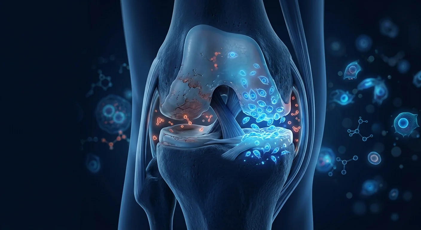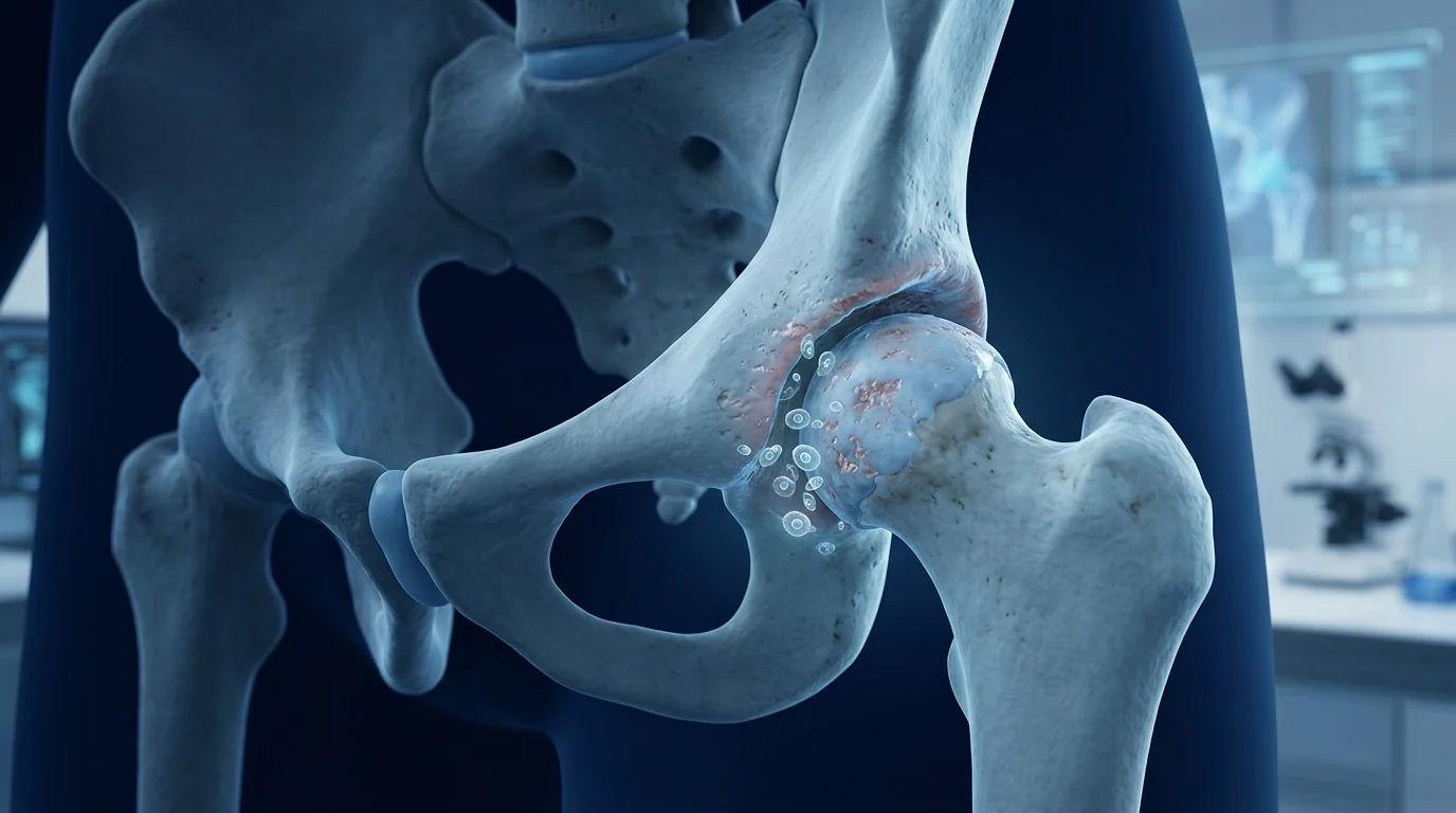
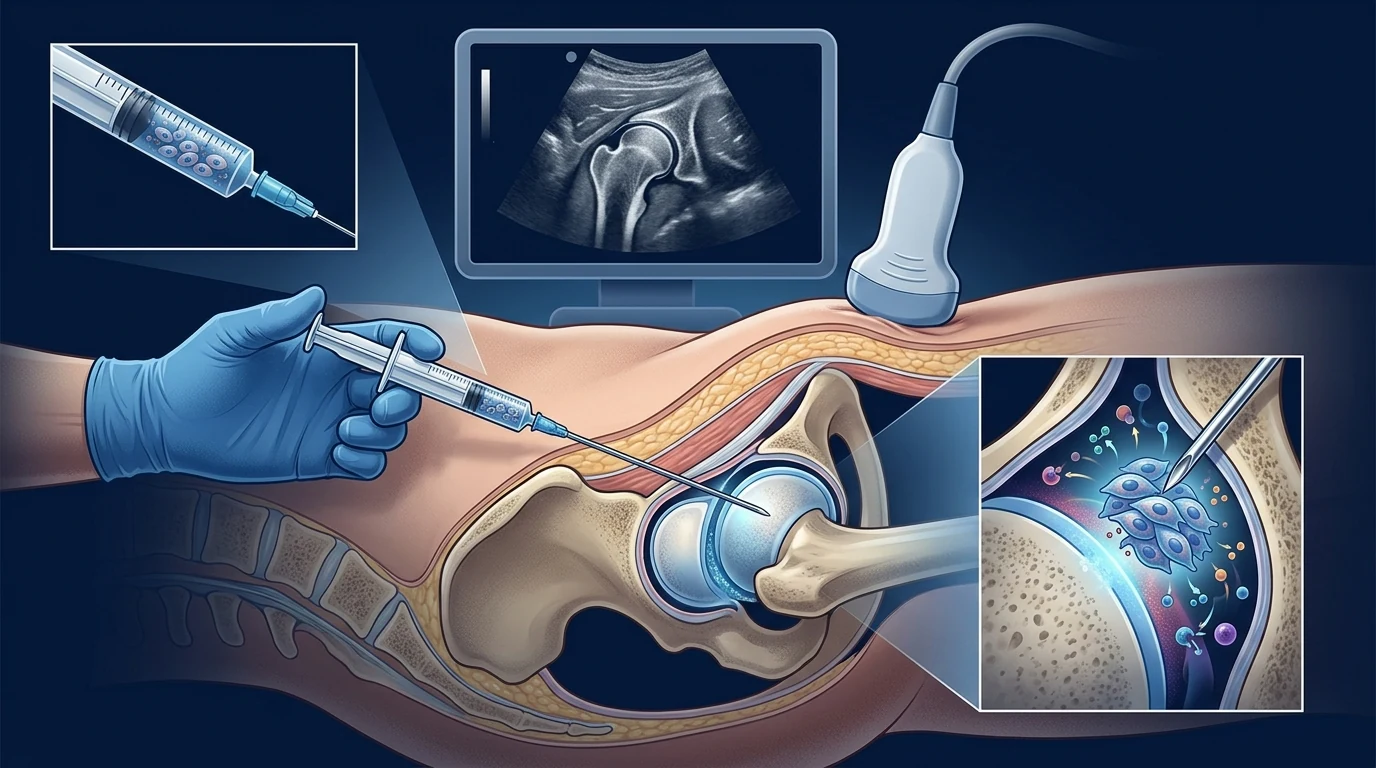
Stem cell therapy for hip osteoarthritis means injecting mesenchymal stem cells (MSCs) directly into the hip joint\u2014under fluoroscopic or ultrasound guidance\u2014with the goal of reducing the chronic low-grade inflammation that drives cartilage loss, and supporting the joint\u2019s own repair mechanisms. Unlike total hip replacement, it is a biologic intervention aimed at hips that still have cartilage worth preserving; unlike corticosteroid injections, it attempts to modify the disease biology rather than temporarily suppress symptoms.
Why the hip is different from the knee. Hip osteoarthritis presents unique challenges that make extrapolating from the larger knee OA MSC literature problematic. The hip is a deep ball-and-socket joint; access requires image guidance; the joint space is smaller, the cartilage geometry more complex, and the mechanical loads higher. The evidence base for MSC therapy in hip OA is smaller and earlier-stage than for the knee, but the available data are directionally consistent\u2014and the clinical need is just as large [1].
The honest frame. As of 2026, no MSC product is approved by the FDA or EMA for hip osteoarthritis. The appropriate description is early-stage investigational therapy with promising but limited evidence\u2014not a hip replacement alternative for end-stage disease. We explain exactly what that means below.
What this article covers. The unique biology of hip OA, why MSCs are a rational candidate for this joint, what the clinical studies show on pain and function, how the evidence compares with the knee OA literature, and what any responsible practitioner should tell you before you proceed.
What is hip osteoarthritis\u2014and why the knee evidence does not automatically transfer
Hip osteoarthritis is a degenerative joint disease in which the articular cartilage of the femoral head and acetabulum progressively thins, the underlying bone remodels, and the synovium becomes inflamed\u2014the same core processes as knee OA, but in a joint with fundamentally different anatomy and loading. Globally, hip OA affects approximately 8\u201310% of adults over 40, with prevalence reaching 25\u201335% in those over 75 [2]. The Global Burden of Disease Study 2021 projected that hip OA cases will increase by 78.6% by 2050, driven by aging populations and rising obesity rates [3].
Where knee evidence transfers. The basic mechanisms\u2014chondrocyte senescence, synovial inflammation, subchondral bone remodelling\u2014are identical. MSC immunomodulation, trophic support, and anti-fibrotic signalling should operate similarly in any diarthrodial joint [4].
Where it does not. Hip OA has distinct risk factor profiles (femoroacetabular impingement, acetabular dysplasia, occupational loading) that affect which patients present at which stage. The hip\u2019s deep location makes injection accuracy more technically demanding, and imaging guidance is considered essential for any serious therapeutic protocol. These differences mean the hip OA MSC literature must be evaluated on its own terms\u2014not assumed from the knee data.
What happens inside an osteoarthritic hip
Three parallel processes define the pathology, and a rational cell therapy must address each.
Cartilage failure begins with chondrocyte senescence. As in the knee, resident chondrocytes in the femoral head and acetabulum become senescent, secrete matrix-degrading enzymes (MMP-13, ADAMTS-5), and lose their ability to maintain the proteoglycan-collagen matrix [5]. The cartilage surface fibrillates, fissures, and eventually delaminates\u2014exposing bone.
Synovial inflammation is compartmentalised within the joint capsule. Synovial macrophages release IL-1\u03b2, TNF-\u03b1, and IL-6 into the joint fluid. This compartmentalised nature is precisely what makes local delivery of MSCs rational: the cells are deposited directly into the inflammatory milieu rather than relying on systemic distribution [6].
Subchondral bone remodelling and osteophyte formation. The bone beneath the degenerating cartilage becomes sclerotic, and marginal osteophytes form\u2014the structural changes visible on X-ray that define the Kellgren-Lawrence grade. These bony changes contribute significantly to pain and stiffness and are largely irreversible once established.
Why MSCs are a candidate for hip osteoarthritis
The rationale mirrors the knee, but with delivery considerations that make the hip distinct.
Immunomodulation of the joint microenvironment. MSCs suppress M1-polarised synovial macrophages and promote M2 polarisation, reducing intra-articular IL-1\u03b2 and TNF-\u03b1 levels [7]. This is the mechanism with the strongest preclinical support across all OA joints, and it maps directly onto the inflammatory component of hip OA.
Chondroprotection via trophic factors. MSCs secrete TGF-\u03b2, IGF-1, BMP-2, and BMP-7, which support chondrocyte survival and matrix production. Co-culture studies demonstrate reduced chondrocyte apoptosis and preserved proteoglycan content in cartilage explants exposed to inflammatory stimuli [8].
Delivery must be image-guided. Unlike the easily accessible knee, the hip requires fluoroscopic or ultrasound guidance for accurate intra-articular placement. Studies confirm that blind hip injections miss the joint space 25\u201340% of the time\u2014a critical quality consideration when evaluating clinical results [9].
What the clinical evidence for hip OA shows
The hip OA MSC literature is smaller than the knee\u2019s but directionally consistent. Several recent reviews summarise the available data.
A 2024 systematic review of intra-articular MSC injections for hip OA identified five eligible clinical studies and concluded that MSC infiltration provides significant improvements in pain (VAS) and function (Harris Hip Score, WOMAC) with a favourable safety profile, though the evidence was limited by small sample sizes and heterogeneous protocols [10]. A 2023 scoping review of stem cell-based therapies for hip OA similarly reported short- to mid-term improvements in pain and function across most included studies [11].
Key individual studies define the evidence floor. In a 2015 long-term follow-up study, Emadedin and colleagues treated 18 patients with hip OA using autologous bone-marrow MSCs\u2014intra-articular injection under fluoroscopic guidance\u2014and observed statistically significant improvements in VAS pain scores and WOMAC function scores that were maintained at 12 months. No serious adverse events were reported [12].
A 2019 preliminary case series by Dall\u2019Oca et al. treated six patients with fluoroscopy-guided injections of autologous micro-fragmented adipose tissue and reported significant improvements in the Harris Hip Score (from 67.2 at baseline to 84.6 at follow-up), WOMAC, and VAS scores at 6 months [13]. A 2023 study of autologous micro-fragmented adipose tissue in hip OA found durable improvements at 3 years in patients with early-to-moderate disease, suggesting that stage-appropriate patient selection may be a key determinant of outcomes [14].
Cell sources studied include bone marrow, adipose tissue (micro-fragmented and culture-expanded), and umbilical cord\u2014with adipose-derived approaches dominating the hip literature partly because of the relatively simple harvesting procedure compared with bone-marrow aspiration [15].
The honest headline
No MSC product is approved by the FDA or EMA for hip osteoarthritis as of 2026. The evidence base is consistent and promising but small: a handful of prospective studies, controlled comparisons limited, and long-term data beyond 2\u20133 years sparse. Any clinic promising guaranteed femoral head preservation or complete pain resolution is misrepresenting the data.
How hip OA outcomes are measured
The functional gold standard for hip OA is the Harris Hip Score (a composite of pain, function, deformity, and range of motion) plus WOMAC (pain, stiffness, function) and the VAS pain score (0\u2013100 mm). Radiographic endpoints include joint-space width on plain X-ray and cartilage composition via MRI with delayed gadolinium-enhanced imaging (dGEMRIC) or T2 mapping [16].
The pattern across the hip OA studies mirrors the knee: MSC-treated patients show clinically meaningful pain reduction (mean VAS improvement of 20\u201335 mm) and functional improvement at 6\u201312 months, with durability signals extending to 24\u201336 months in some adipose-tissue protocols. Importantly, structural imaging data for the hip are even more preliminary than for the knee\u2014no study to date has demonstrated definitive cartilage regeneration on MRI, only maintained or slowed progression in early-stage patients.
What the evidence supports\u2014and what it does not
A fair reading supports four conclusions. Intra-articular MSC therapy for hip OA is safe across all published studies, with adverse events limited to transient post-injection pain and swelling. It consistently reduces pain and improves function in the available prospective studies and case series. The data are directionally consistent with the larger knee OA MSC literature, which lends credibility to the mechanism. And imaging-guided delivery is non-negotiable for any serious hip OA protocol.
Equally important is what the evidence does not yet support:
- It does not establish disease modification. Structural data for the hip are minimal; no imaging endpoint has convincingly shown cartilage regeneration.
- It does not confirm efficacy for end-stage (Kellgren-Lawrence grade 4) disease. The strongest signals come from early-to-moderate OA.
- It does not define an optimal protocol. Cell source, dose, frequency, and delivery method vary; head-to-head comparisons are absent.
- It does not replace total hip arthroplasty for patients with advanced structural damage and disabling symptoms. Arthroplasty remains the definitive option.
MSC therapy for hip OA is best understood as a potential joint-preservation strategy\u2014a bridge that may extend the functional life of a native hip and delay arthroplasty in appropriately selected patients\u2014not as a replacement for physiotherapy, weight management, or eventual surgical care.
How to evaluate a provider
Because the evidence is smaller than for knee OA, diligence is particularly important. Ask:
- Do they use imaging guidance for the injection? Ultrasound or fluoroscopy is the standard of care; blind injections are not acceptable for the hip.
- What cell type and source? Micro-fragmented adipose tissue, bone-marrow MSCs, and umbilical-cord MSCs are different products with different evidence bases and regulatory pathways.
- Can they cite published outcome data for their specific protocol in the hip? Knee data do not automatically transfer.
- Do they track validated outcome scores? Harris Hip Score, WOMAC, and VAS before and after treatment indicate a data-driven approach.
- What quality data exist for the cells? Identity testing (CD73+, CD90+, CD105+, CD45-), viability above 95% at delivery, and sterility release are the baseline.
Be sceptical of guarantees, of claims that end-stage hips can avoid replacement, and of any provider who cannot separate what the knee literature shows from what the hip literature shows.
The VELAR perspective
At VELAR Center, we approach hip osteoarthritis with the same evidence-based rigor we apply to every condition\u2014which means we are more cautious, not less, where the data are thinner. We use umbilical-cord-derived MSCs manufactured under cGMP conditions with full ISCT-compliant characterisation and third-party sterility release, delivered fresh and never frozen. Our hip OA consultations include a review of the patient\u2019s imaging (X-ray and MRI where available) and a transparent discussion: MSC therapy is a joint-preservation strategy for early-to-moderate hip OA in appropriate candidates, not a replacement for arthroplasty in end-stage disease. Where the evidence does not support a cell-based approach, we say so\u2014and where it might, we structure the conversation around what outcomes the data suggest are actually achievable.
Frequently Asked Questions
Is stem cell therapy for hip osteoarthritis effective?
The available evidence\u2014from small prospective studies and case series\u2014consistently reports pain reduction and functional improvement at 6 to 24 months. Effect sizes are clinically meaningful, but the evidence base is considerably smaller than for the knee, and no definitive conclusion can yet be drawn. Larger randomised controlled trials are needed.
How does MSC therapy for the hip differ from the knee?
The hip is a deeper joint that requires image-guided injection (ultrasound or fluoroscopy). The evidence base is earlier-stage for the hip than the knee. The mechanisms of MSC action are the same, but the margin for injection accuracy and the patient selection criteria differ.
How long do the effects last?
Most studies report improvements at 12 months. Several adipose-tissue series suggest durability to 24\u201336 months, particularly in patients with early-stage disease. Data beyond 3 years are limited.
Can MSC therapy avoid the need for hip replacement?
In patients with mild-to-moderate hip OA, MSC therapy may slow progression and defer the need for arthroplasty. There is no evidence that it reverses end-stage structural damage or eliminates the eventual need for surgery in advanced disease. The appropriate framing is joint preservation, not replacement avoidance.
Is the procedure painful?
Patients typically report mild-to-moderate discomfort during and immediately after the injection, comparable to a diagnostic hip arthrogram. Transient post-injection pain and swelling lasting 24\u201372 hours are common. Serious adverse events in the published literature are rare.
Who is a good candidate?
The best evidence supports patients with early-to-moderate hip OA\u2014Kellgren-Lawrence grades 1\u20133\u2014with preserved joint space and clear inflammatory symptoms. Advanced cartilage loss (grade 4), severe dysplasia, or active joint infection are relative contraindications.
Limitations of this review
MSC therapy for hip osteoarthritis remains investigational. The clinical evidence comes from small prospective studies and case series with heterogeneous cell sources, doses, and follow-up durations. Randomised controlled trials specific to the hip are very limited. Structural imaging endpoints are preliminary, and long-term durability is not yet established. This article summarises published research for educational purposes and does not constitute medical advice; treatment decisions should be made with a qualified clinician after imaging and clinical assessment.
References
- Giorgino R, Alessandri Bonetti M, Migliorini F, Nannini A, Mangiavini L, Peretti GM, Randelli PS. Management of hip osteoarthritis: harnessing the potential of mesenchymal stem cells\u2014a systematic review. European Journal of Orthopaedic Surgery & Traumatology. 2024;34(8):3847-3857. doi:10.1007/s00590-024-04089-0 \u21a9
- Fan Z, Yan L, Liu H, et al. The prevalence of hip osteoarthritis: a systematic review and meta-analysis. Arthritis Research & Therapy. 2023;25(1):51. doi:10.1186/s13075-023-03033-7 \u21a9
- GBD 2021 Osteoarthritis Collaborators. Global, regional, and national burden of osteoarthritis, 1990\u20132020 and projections to 2050: a systematic analysis for the Global Burden of Disease Study 2021. The Lancet Rheumatology. 2023;5(9):e508-e522. doi:10.1016/S2665-9913(23)00163-7 \u21a9
- Muller J, Roehrich S, Riehn D, et al. Mesenchymal stem cell therapy for inflammatory and degenerative diseases. Journal of Internal Medicine. 2021;289(6):649-669. doi:10.1111/joim.13149 \u21a9
- Lehman AM, Kishida CF, Martin JA, et al. Chondrocyte senescence: a target for the treatment of osteoarthritis. Current Opinion in Biotechnology. 2016;37:59-64. doi:10.1016/j.copbio.2016.01.005 \u21a9
- Buckley EL, Fuchs PK, Kraus VB, Loeser RF. The role of inflammation in osteoarthritis pain. Seminars in Arthritis and Rheumatism. 2015;45(1):85-88. doi:10.1016/j.semarthrit.2014.12.007 \u21a9
- Zhao X, Zhao Y, Sun X, Xing Y, Wang X, Yang Q. Immunomodulation of MSCs and MSC-derived extracellular vesicles in osteoarthritis. Frontiers in Bioengineering and Biotechnology. 2020;8:575057. doi:10.3389/fbioe.2020.575057 \u21a9
- Kusabuchi H, Nakagawa K, Nakashima M, et al. Human mesenchymal stem cells ameliorate immune-mediated inflammation and tissue destruction in rheumatoid arthritis. Arthritis & Rheumatism. 2007;56(11):3473-3482. doi:10.1002/art.22873 \u21a9
- Natali S, Screpis D, Romeo M, et al. Is intra-articular injection of autologous micro-fragmented adipose tissue effective in hip osteoarthritis? A three year follow-up. International Orthopaedics. 2023;47:1487-1492. doi:10.1007/s00264-022-05611-x \u21a9
- Giorgino R, et al. (2024). Eur J Orthop Surg Traumatol 34(8):3847-3857. doi:10.1007/s00590-024-04089-0 \u21a9
- Entessari M, et al. Current evidence on mesenchymal stem cells for hip osteoarthritis: a narrative review. Regenerative Medicine. 2023;18(20):1-12. doi:10.2217/rme-2023-0071 \u21a9
- Emadedin M, Ghorbani Liastani M, Fazeli R, et al. Long-term follow-up of intra-articular injection of autologous mesenchymal stem cells in patients with knee, ankle, or hip osteoarthritis. Archives of Iranian Medicine. 2015;18(6):336-344. PMID: 26058927 \u21a9
- Dall\u2019Oca C, Barboni E, Roberto V, Elena MS, Bruno M. Mesenchymal stem cells injection in hip osteoarthritis: preliminary results. Acta Bio Medica Atenei Parm. 2019;90(Suppl 1):75-80. doi:10.1007/s00167-018-4955-x \u21a9
- Natali S, et al. (2023). Int Orthop 47:1487-1492. doi:10.1007/s00264-022-05611-x \u21a9
- Freitag J, Bates D, Boyd R, et al. Mesenchymal stem cell therapy in the treatment of osteoarthritis: reparative pathways, safety and efficacy\u2014a review. BMC Musculoskeletal Disorders. 2016;17:230. doi:10.1186/s12891-016-1085-9 \u21a9
- Cao Y, Ou Y, Sheng W, et al. Efficacy and safety of mesenchymal stem cells in knee osteoarthritis: a systematic review and meta-analysis of randomized controlled trials. Stem Cell Research & Therapy. 2025;16(1):122. doi:10.1186/s13287-025-04252-2 \u21a9
髋关节骨关节炎的干细胞治疗,是指将间充质干细胞(MSC)在透视或超声引导下直接注射到髋关节腔内——目的是减轻驱动软骨丢失的慢性低度炎症,并支持关节自身的修复机制。与全髋置换不同,这是一种生物干预,针对的是仍有值得保留的软骨的髋关节;与皮质类固醇注射不同,它试图改变疾病生物学,而非暂时压制症状。
为什么髋关节不同于膝关节。髋关节骨关节炎在临床研究上存在独特挑战,使膝关节MSC文献的经验不能直接推广。髋关节是深部球窝关节,需要影像引导方可穿刺;关节腔更小,软骨形态更复杂,机械负荷更高。髋关节OA的MSC证据基础比膝关节更小、更早期,但现有数据的方向是一致的——而且临床需求同样巨大 [1]。
诚实的定位。截至2026年,没有任何MSC产品被FDA或EMA批准用于髋关节骨关节炎。恰当的表述是有前景但证据有限的早期研究性疗法——而非终末期疾病的髋关节置换替代方案。下文将解释这具体意味着什么。
本文内容。髋关节OA的独特生物学、MSC为何是该关节的合理候选、临床研究在疼痛与功能上显示了什么、证据与膝关节MSC文献如何比较,以及任何负责任的从业者在您决定前应告诉您的信息。
什么是髋关节骨关节炎——为什么膝关节证据不能自动迁移
髋关节骨关节炎是一种退行性关节疾病:股骨头和髋臼的关节软骨逐渐变薄,其下骨重塑,滑膜发炎——核心过程与膝关节OA相同,但关节的解剖与负荷根本不同。全球范围内,髋关节OA影响约8-10%的40岁以上成人,75岁以上人群患病率达25-35% [2]。全球疾病负担研究2021预测,到2050年髋关节OA病例将增加78.6%,由老龄化与肥胖率上升驱动 [3]。
膝关节证据可迁移的部分。基本机制——软骨细胞衰老、滑膜炎症、软骨下骨重塑——是相同的。MSC的免疫调节、营养支持和抗纤维化信号应在任何活动关节中类似地发挥作用 [4]。
不可迁移的部分。髋关节OA有其独特的风险因子(股骨髋臼撞击、髋臼发育不良、职业性负荷),影响患者在哪个阶段就诊。髋关节的深部位置使注射准确性技术要求更高,任何严肃治疗方案的影像引导被认为是必需的。这些差异意味着髋关节OA的MSC文献必须在其自身条件下进行评估——不能从膝关节数据推断。
骨关节炎的髋关节内发生了什么
三个平行过程定义了病理,而理性的细胞疗法必须应对每一过程。
软骨失败始于软骨细胞衰老。与膝关节一样,股骨头和髋臼的驻留软骨细胞变得衰老,分泌基质降解酶(MMP-13、ADAMTS-5),失去维持蛋白聚糖-胶原基质的能力 [5]。软骨表面纤维化、开裂,最终剥脱——暴露骨质。
滑膜炎症局限于关节囊内。滑膜巨噬细胞向关节液中释放IL-1β、TNF-α和IL-6。这种局限性特征正是局部给药MSC合理的原因:细胞被直接投放到炎症环境中,而非依赖全身分布 [6]。
软骨下骨重塑与骨赘形成。退变软骨下的骨变得硬化,形成边缘骨赘——X线上可见的、定义Kellgren-Lawrence分级的结构变化。这些骨性变化对疼痛和僵硬有显著贡献,一旦建立基本不可逆。
为什么MSC是髋关节骨关节炎的候选
理论依据与膝关节相似,但给药考虑使髋关节不同。
关节微环境的免疫调节。MSC抑制M1极化的滑膜巨噬细胞,促进M2极化,降低关节内IL-1β和TNF-α水平 [7]。这是所有OA关节中临床前支持最强的机制,直接对应髋关节OA的炎症成分。
通过营养因子的软骨保护。MSC分泌TGF-β、IGF-1、BMP-2和BMP-7,支持软骨细胞存活与基质生产。共培养研究显示,暴露于炎症刺激的软骨外植体中的软骨细胞凋亡减少、蛋白聚糖含量得以保持 [8]。
给药必须影像引导。与容易穿刺的膝关节不同,髋关节需要透视或超声引导才能准确放置关节内。研究证实盲目髋关节注射错过关节腔的概率为25-40%——这是评估临床结果时的关键质量考虑 [9]。
髋关节OA的临床证据显示了什么
髋关节OA的MSC文献比膝关节小,但方向一致。几篇近期综述总结了可用数据。
2024年一项关于髋关节OA关节内MSC注射的系统评价识别了五项符合条件的临床研究,结论是MSC浸润可显著改善疼痛(VAS)和功能(Harris髋关节评分、WOMAC),安全性特征良好,但证据受限于小样本量和异质性方案 [10]。2023年一项髋关节OA干细胞治疗的范围综述也报告了大多数纳入研究中短期至中期疼痛与功能的改善 [11]。
关键个体研究定义了证据基础。在2015年的长期随访研究中,Emadedin等使用自体骨髓MSC——透视引导下关节内注射——治疗18名髋关节OA患者,观察到的VAS疼痛评分和WOMAC功能评分在12个月时保持统计学显著改善。未报告严重不良事件 [12]。
2019年Dall'Oca等的初步病例系列使用透视引导下自体微碎片化脂肪组织注射治疗6名患者,报告Harris髋关节评分(从基线的67.2改善至随访时的84.6)、WOMAC和VAS评分在6个月时显著改善 [13]。2023年一项髋关节OA自体微碎片化脂肪组织研究在早中期疾病患者中发现改善持续达3年,提示分期适当的患者选择可能是结果的关键决定因素 [14]。
研究的细胞来源包括骨髓、脂肪组织(微碎片化和培养扩增)和脐带——其中脂肪来源方法在髋关节文献中占主导地位,部分原因是与骨髓抽吸相比采集程序相对简单 [15]。
诚实的结论
截至2026年,没有任何MSC产品被FDA或EMA批准用于髋关节骨关节炎。证据基础一致且有前景但规模小:少量前瞻性研究,对照比较有限,超过2-3年的长期数据稀疏。任何承诺保证股骨头保留或完全消除疼痛的机构都在歪曲数据。
髋关节OA结果如何衡量
髋关节OA的功能金标准是Harris髋关节评分(疼痛、功能、畸形和活动范围的复合指标),加上WOMAC(疼痛、僵硬、功能)和VAS疼痛评分(0-100 mm)。影像学终点包括平片上关节间隙宽度以及通过钆增强延迟成像(dGEMRIC)或T2映射的MRI软骨成分分析 [16]。
髋关节OA研究的模式与膝关节相似:MSC治疗患者在6-12个月时显示临床有意义的疼痛减轻(平均VAS改善20-35 mm)和功能改善,某些脂肪组织方案中的持久性信号延伸至24-36个月。重要的是,髋关节的结构影像数据甚至比膝关节更初步——迄今没有研究在MRI上证明了明确的软骨再生,仅早期患者中可见维持或减缓的进展。
证据支持什么——又不支持什么
公允的解读支持四点结论。髋关节OA的关节内MSC治疗在所有已发表研究中是安全的,不良事件限于短暂的注射后疼痛和肿胀。它在可用的前瞻性研究和病例系列中持续减轻疼痛并改善功能。数据与更大的膝关节OA-MSC文献方向一致,增加了机制的可信度。对于任何严肃的髋关节OA方案,影像引导下的递送是不可或缺的。
同样重要的是证据目前尚未支持的:
- 未确立疾病修饰。髋关节的结构数据极少;没有影像学终点令人信服地显示软骨再生。
- 未确认对终末期(Kellgren-Lawrence 4级)疾病的有效性。最强信号来自早中期OA。
- 未确定最佳方案。细胞来源、剂量、频率和递送方式各异;缺乏头对头比较。
- 不能替代全髋关节置换术用于结构性损害严重且症状致残的患者。关节置换仍是确定的选择。
髋关节OA的MSC治疗最好被理解为一种潜在的关节保存策略——可能延长原生髋关节功能寿命、在适当选择患者中推迟关节置换的桥梁——而非物理治疗、体重管理或最终手术护理的替代品。
如何评估医疗机构
由于证据比膝关节少,审慎格外重要。要问:
- 是否使用影像引导进行注射?超声或透视是标准;盲目注射对髋关节不可接受。
- 何种细胞类型与来源?微碎片化脂肪组织、骨髓MSC和脐带MSC是不同产品,证据基础和监管路径各异。
- 能否为其在髋关节的具体方案引用已发表的结局数据?膝关节数据不能自动迁移。
- 是否跟踪验证过的结局评分?治疗前后Harris髋关节评分、WOMAC和VAS表明数据驱动的方法。
- 细胞有什么质量数据?按ISCT标准(CD73+、CD90+、CD105+、CD45-)的身份检测,交付时活性高于95%,无菌放行是基线。
对保证、对终末期髋关节可避免置换的说法、以及任何不能区分膝关节文献与髋关节文献的机构,都要深度怀疑。
VELAR的观点
在VELAR中心,我们以与所有疾病相同的循证严谨态度对待髋关节骨关节炎——这意味着在数据较薄之处我们更加谨慎,而非放松标准。我们使用cGMP条件下制造、ISCT合规鉴定、第三方无菌放行的脐带来源MSC,新鲜交付从不冷冻。我们的髋关节OA咨询包括审阅患者影像(X线和可用的MRI),并进行透明讨论:MSC治疗对于适当候选者的早中期髋关节OA是一种关节保存策略,而非终末期疾病关节置换的替代。在证据不支持细胞治疗之处,我们如实告知——在可能支持之处,我们围绕数据表明实际可达到的结局来构建对话。
常见问题
髋关节骨关节炎的干细胞治疗有效吗?
现有证据——来自小型前瞻性研究和病例系列——一致报告6至24个月的疼痛减轻和功能改善。效应量具有临床意义,但证据基础比膝关节小得多,尚不能得出确定性结论。需要更大规模的随机对照试验。
髋关节的MSC治疗与膝关节有何不同?
髋关节是更深的关节,需要影像引导注射(超声或透视)。髋关节的证据基础比膝关节更早期。MSC的作用机制相同,但注射准确度的裕度和患者选择标准不同。
效果能持续多久?
大多数研究报告12个月时的改善。几个脂肪组织系列提示早中期疾病患者的持久性可达24-36个月。超过3年的数据有限。
MSC治疗能避免髋关节置换吗?
在轻中度髋关节OA患者中,MSC治疗可减缓进展、推迟置换需求。没有证据表明它能逆转终末期结构性损害或消除晚期疾病最终的手术需求。恰当的定位是关节保存,而非避免置换。
这个程序痛吗?
患者在注射期间和注射后通常报告与诊断性髋关节造影相似的中轻度不适。注射后短暂疼痛和肿胀持续24-72小时是常见的。已发表文献中严重不良事件罕见。
谁适合作为候选?
最佳证据支持早期至中度髋关节OA患者——Kellgren-Lawrence 1-3级——关节间隙尚存且有明确炎症症状。严重的软骨丢失(4级)、严重发育不良或活动性关节感染是相对禁忌症。
本综述的局限性
髋关节骨关节炎的MSC治疗仍属研究性。临床证据来自小型前瞻性研究和病例系列,细胞来源、剂量和随访时长各异。针对髋关节的随机对照试验极为有限。结构影像终点是初步的,长期持久性尚未确立。本文总结已发表研究用于教育目的,不构成医疗建议;治疗决定应在影像与临床评估后与合格医生共同做出。
参考文献
- Giorgino R, et al. Management of hip osteoarthritis: harnessing the potential of mesenchymal stem cells\u2014a systematic review. Eur J Orthop Surg Traumatol. 2024;34(8):3847-3857. doi:10.1007/s00590-024-04089-0 \u21a9
- Fan Z, et al. The prevalence of hip osteoarthritis: a systematic review and meta-analysis. Arthritis Res Ther. 2023;25(1):51. doi:10.1186/s13075-023-03033-7 \u21a9
- GBD 2021 Osteoarthritis Collaborators. Global burden of osteoarthritis. Lancet Rheumatol. 2023;5(9):e508-e522. doi:10.1016/S2665-9913(23)00163-7 \u21a9
- Muller J, et al. MSC therapy for inflammatory and degenerative diseases. J Intern Med. 2021;289(6):649-669. doi:10.1111/joim.13149 \u21a9
- Lehman AM, et al. Chondrocyte senescence. Curr Opin Biotechnol. 2016;37:59-64. doi:10.1016/j.copbio.2016.01.005 \u21a9
- Buckley EL, et al. Role of inflammation in OA pain. Semin Arthritis Rheum. 2015;45(1):85-88. doi:10.1016/j.semarthrit.2014.12.007 \u21a9
- Zhao X, et al. Immunomodulation of MSCs in OA. Front Bioeng Biotechnol. 2020;8:575057. doi:10.3389/fbioe.2020.575057 \u21a9
- Kusabuchi H, et al. Human MSCs ameliorate inflammation in RA. Arthritis Rheum. 2007;56(11):3473-3482. doi:10.1002/art.22873 \u21a9
- Natali S, et al. Intra-articular MFAT in hip OA: 3-year follow-up. Int Orthop. 2023;47:1487-1492. doi:10.1007/s00264-022-05611-x \u21a9
- Giorgino R, et al. (2024). Eur J Orthop Surg Traumatol 34(8):3847-3857. doi:10.1007/s00590-024-04089-0 \u21a9
- Entessari M, et al. MSC for hip OA: narrative review. Regen Med. 2023;18(20). doi:10.2217/rme-2023-0071 \u21a9
- Emadedin M, et al. Long-term follow-up intra-articular autologous MSCs in OA. Arch Iran Med. 2015;18(6):336-344. PMID: 26058927 \u21a9
- Dall\u2019Oca C, et al. MSC injection in hip OA: preliminary. Acta Biomed. 2019;90(Suppl 1):75-80. PMID: 30715002 \u21a9
- Natali S, et al. (2023). Int Orthop 47:1487-1492. doi:10.1007/s00264-022-05611-x \u21a9
- Freitag J, et al. MSC therapy in OA: review. BMC Musculoskelet Disord. 2016;17:230. doi:10.1186/s12891-016-1085-9 \u21a9
- Cao Y, et al. Efficacy and safety MSCs knee OA: meta-analysis. Stem Cell Res Ther. 2025;16(1):122. doi:10.1186/s13287-025-04252-2 \u21a9
يعني علاج فصال الورك بالخلية الجذعية حقن خلايا جذعية وسيتة (MSCs) تحت توجيه التنظير التألقي أو الموجات فوق الصوتية مباشرةً في جوف مفصل الورك \u2014 بهدف تقليل الالتهاب المزمن منخفض الدرجة الذي يقود فقدان الغضروف، ودعم آليات إصلاح المفصل الذاتية. وعلى خلاف استبدال الورك كليًا، فهو تدخّل بيولوجي يستهدف أوراكًا لا يزال فيها غضروف يستحق الحفظ؛ وعلى خلاف حقن الكورتيكوستيرويد، فهو يحاول معالجة بيولوجيا المرض لا مجرد كبت الأعراض مؤقتًا.
لماذا الورك يختلف عن الركبة. يطرح فصال الورك تحديات فريدة تجعل استقراء أدبيات الخلايا الجذعية الوسيطة الأكبر حجماً الخاصة بالركبة إشكالياً. الورك هو مفصل كروي حق عميق؛ يحتاج الوصول إليه توجيهًا تصويريًا؛ حيزه المفصلي أصغر، وهندسة غضروفه أكثر تعقيدًا، وأحماله الميكانيكية أعلى. قاعدة أدلة فصال الورك للخلايا الجذعية الوسيطة أصغر وأبكر مرحلةً من تلك الخاصة بالركبة، لكن البيانات المتاحة متسقة الاتجاه \u2014 والحاجة السريرية لا تقل ضخامة [1].
الإطار الصريح. حتى عام 2026، لا يوجد أي منتج للخلايا الجذعية الوسيطة معتمد من إدارة الغذاء والدواء الأمريكية (FDA) أو وكالة الأدوية الأوروبية (EMA) لفصال الورك. الوصف المناسب هو علاج تجريبي مبكر بأدلة واعدة لكن محدودة \u2014 لا بديل عن استبدال الورك في المرض المتقدم. سنشرح بالضبط ما يعنيه ذلك أدناه.
ما يغطيه هذا المقال. بيولوجيا فصال الورك الفريدة، ولماذا الخلايا الجذعية الوسيطة مرشحة عقلية لهذا المفصل، وما الذي تظهره الدراسات السريرية من ألم ووظيفة، وكيف تقارن الأدلة بأدبيات الركبة، وما الذي يجب أن يخبرك به أي ممارس مسؤول قبل أن تمضي.
ما هو فصال الورك \u2014 ولماذا لا تنتقل أدلة الركبة آليًا
فصال الورك هو مرض مفصلي تنكسي يفقد فيه الغضروف المفصلي لرأس الفخذ والحق سمكه تدريجيًا، ويُعاد تشكيل العظم التحتي، ويلتهب الغشاء الزليلي \u2014 نفس العمليات الأساسية لفصال الركبة، لكن في مفصل بتشريح وحمل مختلفين جوهريًا. عالميًا، يؤثر فصال الورك على حوالي 8\u201310% من البالغين فوق سن 40، وتصل نسبة الانتشار إلى 25\u201335% لدى من تجاوزوا 75 عامًا [2]. توقع دراسة العبء العالمي للأمراض 2021 أن حالات فصال الورك ستزداد بنسبة 78.6% بحلول 2050، بفعل شيخوخة السكان وارتفاع معدلات السمنة [3].
حيث تنتقل أدلة الركبة. الآليات الأساسية \u2014 شيخوخة الخلايا الغضروفية، والتهاب الزليل، وإعادة تشكيل العظم تحت الغضروفي \u2014 متطابقة. يجب أن تعمل مناعة الخلايا الجذعية الوسيطة ودعمها الغذائي وإشاراتها المضادة للتليف بطريقة مماثلة في أي مفصل زليلي [4].
حيث لا تنتقل. لفصال الورك عوامل خطر مميزة (انحشار الفخذ-الحقي، خلل تنسج الحُق، التحميل المهني) تؤثر على أي المرضى يقدمون بأي مرحلة. موقع الورك العميق يجعل دقة الحقن أكثر تطلبًا من الناحية التقنية، والتوجيه التصويري يُعتبر أساسيًا لأي بروتوكول علاجي جاد. هذه الفروق تعني أن أدبيات الخلايا الجذعية الوسيطة لفصال الورك يجب أن تُقيّم بشروطها الخاصة \u2014 لا أن تُفترض من بيانات الركبة.
ما الذي يحدث داخل ورك مصاب بفصال العظم
ثلاث عمليات متوازية تحدد علم الأمراض، ويجب أن يتصدى علاج الخلية العقلية لكل منها.
فشل الغضروف يبدأ بشيوخة الخلايا الغضروفية. كما في الركبة، تصبح الخلايا الغضروفية المقيمة في رأس الفخذ والحق شائخة، وتفرز إنزيمات محلّة للنسيج (MMP-13، ADAMTS-5)، وتفقد قدرتها على صيانة نسيج البروتيوغليكان-كولاجين [5]. يتشقق سطح الغضروف ويتفلّق وينفصل في النهاية \u2014 مكشّفًا العظم.
التهاب الزليل محصور ضمن كيس المفصل. تفرز البلاعم الزليلية IL-1β وTNF-α وIL-6 في السائل المفصلي. هذه الطبيعة المحصورة هي بالضبط ما يجعل التوصيل الموضعي للخلايا الجذعية الوسيطة عقلانيًا: تُودع الخلايا مباشرةً في المحيط الالتهابي لا أن تعتمد على التوزيع الجهازي [6].
إعادة تشكيل العظم تحت الغضروفي وتشكّل النوابت العظمية. يصبح العظم تحت الغضروف المتدهور متصلبًا، وتتشكل نوابت عظمية حافية \u2014 التغييرات البنيوية الظاهرة على الأشعة السينية التي تحدّد درجة Kellgren-Lawrence. تساهم هذه التغييرات العظمية إسهامًا كبيرًا في الألم والتيبُّس، وهي غير قابلة للعكس إلى حد كبير بمجرد تأسيسها.
لماذا الخلايا الجذعية الوسيطة مرشحة لفصال الورك
الأساس المنطقي يعكس الركبة، لكن مع اعتبارات توصيل تجعل الورك متميزًا.
تعديل مناعة البيئة الدقيقة للمفصل. تكبح الخلايا الجذعية الوسيطة البلاعم الزليلية المستقطبة نحو M1 وتُعزز استقطاب M2، فتُخفّض مستويات IL-1β وTNF-α داخل المفصل [7]. هذه هي الآلية الأكثر دعمًا قبل السريري عبر جميع مفاصل فصال العظم، وهي تطابق مباشرةً المكون الالتهابي لفصال الورك.
حماية الغضروف عبر عوامل التغذية. تفرز الخلايا الجذعية الوسيطة TGF-β وIGF-1 وBMP-2 وBMP-7، التي تدعم بقاء الخلايا الغضروفية وإنتاج النسيج. تُظهر دراسات الزرع المشترك انخفاض استماتة الخلايا الغضروفية والحفاظ على محتوى البروتيوغليكان في رقاقات الغضروف المعرّضة لمنبهات التهابية [8].
التوصيل يجب أن يكون موجهًا تصويريًا. بخلاف الركبة سهلة الوصول، يحتاج الورك إلى توجيه تنظير تألقي أو موجات فوق صوتية لوضع دقيق داخل المفصل. تؤكد الدراسات أن حقن الورك الأعمى يخطئ الحيز المفصلي بنسبة 25\u201340% من المرات \u2014 اعتبار جودة حاسم عند تقييم النتائج السريرية [9].
ماذا تُظهر الأدلة السريرية لفصال الورك
أدبيات فصال الورك للخلايا الجذعية الوسيطة أصغر من أدبيات الركبة لكنها متسقة الاتجاه. وتُجمِل عدة مراجعات حديثة البيانات المتاحة.
خلصت مراجعة منهجية عام 2024 لحقن الخلايا الجذعية الوسيطة داخل المفصل لفصال الورك إلى أن تسلل الخلايا الجذعية يحسّن الألم (VAS) والوظيفة (درجة Harris للورك، WOMAC) إحصائياً وسريرياً مع ملمح أمان موات، رغم أن الأدلة محدودة بصغر حجم العينة وتباين البروتوكولات [10]. وبالمثل، أبلغت مراجعة نطاقية عام 2023 عن تحسن على المدى القصير والمتوسط في الألم والوظيفة عبر معظم الدراسات المشمولة للعلاجات القائمة على الخلايا الجذعية لفصال الورك [11].
دراسات فردية رئيسية تحدد أرضية الأدلة. في دراسة متابعة طويلة الأمد عام 2015، عالج Emadedin وزملاؤه 18 مريضًا بفصال الورك باستخدام خلايا جذعية وسيطية ذاتية من نخاع العظم \u2014 حقن داخل المفصل بتوجيه تنظير تألقي \u2014 ورصدوا تحسنات ذات دلالة إحصائية في درجات ألم VAS ودرجات وظيفة WOMAC ظلت محفوظة عند 12 شهرًا. لم تُبلّغ حوادث سلبية خطيرة [12].
وسلسلة حالات أولية عام 2019 لـ Dall\u2019Oca وآخرين عالجت ستة مرضى بحقن نسيج دهني مفتّت دقيق ذاتي بتوجيه تنظير تألقي، وأبلغت عن تحسنات ذات دلالة في درجة الورك Harris (من 67.2 عند الأساس إلى 84.6 عند المتابعة) وWOMAC وVAS عند 6 أشهر [13]. ووجدت دراسة عام 2023 للنسيج الدهني المفتّت الدقيق الذاتي في فصال الورك تحسنات دائمة عند 3 سنوات لدى مرضى المرحلة المبكرة-المتوسطة، مما يشير إلى أن اختيار المرضى الملائم للمرحلة قد يكون محددًا رئيسيًا للنتائج [14].
تتباين مصادر الخلايا المدروسة \u2014 نخاع العظم، النسيج الدهني (المفتّت دقيقًا والمُزْرع)، والحبل السري \u2014 مع هيمنة النهج المشتقة من الدهن على أدبيات الورك جزئيًا بسبب سهولة إجراء الجمع نسبيًا مقارنة بشفط نخاع العظم [15].
الخلاصة الصريحة
لا يوجد أي منتج للخلايا الجذعية الوسيطة معتمد من FDA أو EMA لعلاج فصال الورك حتى 2026. قاعدة الأدلة متسقة وواعدة لكنها صغيرة: حفنة من الدراسات المستقبلية، والمقارنات المضبوطة محدودة، والبيانات طويلة المدى بعد 2\u20133 سنوات متناثرة. أي عيادة تعد بحفظ رأس الفخذ المضمون أو بزوال الألم الكامل تُشوّه البيانات.
كيف تُقاس نتائج فصال الورك
المعيار الوظيفي الذهبي لفصال الورك هو درجة Harris للورك (مركبة من الألم والوظيفة والتشوه ونطاق الحركة) بالإضافة إلى WOMAC (الألم والتيبّس والوظيفة) ودرجة VAS للألم (0\u2013100 مم). تشمل نقاط النهاية الشعاعية عرض حيز المفصل على الأشعة السينية العادية وتركيب الغضروف عبر التصوير بالرنين المغناطيسي مع التصوير المعزّز بالغادولينيوم المتأخر (dGEMRIC) أو تخطيط T2 [16].
النمط عبر دراسات فصال الورك يعكس الركبة: يُظهر المرضى المعالجون بالخلايا الجذعية الوسيطة انخفاضًا ذا معنى سريري في الألم (متوسط تحسّن VAS 20\u201335 مم) وتحسّنًا وظيفيًا عند 6\u201312 شهرًا، مع إشارات دوام تمتد إلى 24\u201336 شهرًا في بعض بروتوكولات النسيج الدهني. والأهم أن بيانات التصوير البنيوي للورك أكثر أولية حتى من بيانات الركبة \u2014 لم تُظهر أي دراسة حتى الآن تجددًا قاطعًا للغضروف على الرنين المغناطيسي، فقط الحفاظ على التقدم أو إبطائه في مرضى المرحلة المبكرة.
ما تدعمه الأدلة \u2014 وما لا تدعمه
قراءة عادلة تدعم أربعة استنتاجات. علاج الخلايا الجذعية الوسيطة داخل المفصل لفصال الورك آمن في جميع الدراسات المنشورة، مع حوادث سلبية محدودة بألم وتورم طائفين بعد الحقن. إنه يقلل الألم باستمرار ويحسن الوظيفة في الدراسات المستقبلية وسلاسل الحالات المتاحة. البيانات متسقة الاتجاه مع أدبيات فصال الركبة الأكبر، مما يمنح المصداقية للآلية. والتوصيل الموجه تصويريًا غير قابل للتفاوض لأي بروتوكول فصال ورك جاد.
ومما لا تقل أهمية، ما لا تدعمه الأدلة بعد:
- لا تثبت تعديل المرض. البيانات البنيوية للورك ضئيلة؛ لا توجد نقطة نهاية تصويرية أظهرت بشكل مقنع تجدد الغضروف.
- لا تؤكد الفعالية للمرحلة النهائية (درجة 4 Kellgren-Lawrence). أقوى الإشارات من فصال مبكر-متوسط.
- لا تحدد بروتوكولًا مثاليًا. مصدر الخلية والجرعة والتكرار وطريقة التوصيل تختلف؛ مقارنات الرأس-مقابل-الرأس غائبة.
- لا تستبدل استبدال الورك الكلي للمرضى ذوي الضرر البنيوي المتقدم والأعراض المعطلة. يبقى استبدال المفصل الخيار المحدد.
علاج الخلايا الجذعية الوسيطة لفصال الورك يُفهم على أفضل وجه كـاستراتيجية محتملة لحفظ المفصل \u2014 جسر قد يطيل العمر الوظيفي لورك طبيعي ويؤجل استبدال المفصل في مرضى مختارين بشكل مناسب \u2014 لا كبديل عن العلاج الطبيعي وإدارة الوزن أو الرعاية الجراحية المحتومة.
كيف تقيّم مزوّدًا
بما أن الأدلة أصغر من أدلة الركبة، فإن الاجتهاد مهم بشكل خاص. اسأل:
- هل يستخدمون توجيهًا تصويريًا للحقن؟ الموجات فوق الصوتية أو التنظير التألقي هو معيار الرعاية؛ الحقن الأعمى غير مقبول للورك.
- ما نوع الخلية ومصدرها؟ النسيج الدهني المفتّت دقيقًا، وخلايا نخاع العظم الوسيطة، وخلايا الحبل السري هي منتجات مختلفة بقواعد أدلة ومسارات تنظيمية مختلفة.
- هل يستطيعون الاستشهاد بنتائج منشورة لبروتوكولهم الخاص في الورك؟ بيانات الركبة لا تنتقل آليًا.
- هل يتتبعون نتائج مُقَيَّمة بمقاييس مُصادَق عليها؟ درجة Harris للورك وWOMAC وVAS قبل العلاج وبعده تشير إلى نهج قائم على البيانات.
- ما بيانات الجودة الموجودة للخلايا؟ اختبار الهوية (CD73+، CD90+، CD105+، CD45-)، وحيوية أعلى من 95% عند التسليم، وإطلاق خلو من الجراثيم \u2014 هي خط الأساس.
كن شكّاكًا في الضمانات، وفي الادعاءات بأن الأوراك المنتهية يمكنها تجنب الاستبدال، وفي أي مزوّد لا يستطيع التفريق بين ما تظهره أدبيات الركبة وما تظهره أدبيات الورك.
رؤية VELAR
في مركز VELAR، نتعامل مع فصال الورك بنفس الدقة القائمة على الأدلة التي نطبقها على كل حالة \u2014 مما يعني أننا أكثر حذرًا، لا أقل، حيث البيانات أرق. نستخدم خلايا وسيطة من الحبل السري مصنّعة في ظروف cGMP مع ترشيح كامل متوافق مع معايير ISCT وإطلاق خلو من الجراثيم من طرف ثالث، نقدّمها طازجة لا مجمدة أبدًا. تتضمن استشارات فصال الورك لدينا مراجعة تصوير المريض (أشعة سينية ورنين مغناطيسي حيثما متاح) ونقاشًا شفافًا: علاج الخلايا الجذعية الوسيطة هو استراتيجية حفظ مفصل لفصال الورك المبكر-المتوسط في مرشحين مناسبين، لا بديل عن استبدال المفصل في المرض النهائي. حيث لا تدعم الأدلة نهجًا خلويًا، نقول ذلك \u2014 وحيث قد تدعمه، نبني المحادثة حول النتائج التي تشير البيانات إلى أنها قابلة للتحقيق فعليًا.
الأسئلة الشائعة
هل علاج الخلايا الجذعية الوسيطة فعال لفصال الورك؟
الأدلة المتاحة \u2014 من دراسات مستقبلية صغيرة وسلاسل حالات \u2014 تبلغ باستمرار عن تخفيف الألم وتحسن وظيفي عند 6 إلى 24 شهرًا. مقاييس التأثير ذات أهمية سريرية، لكن قاعدة الأدلة أصغر بكثير من الركبة، ولا يمكن استخلاص استنتاج حاسم بعد. هناك حاجة إلى تجارب عشوائية مضبوطة أكبر.
كيف يختلف علاج الخلايا الجذعية الوسيطة للورك عن الركبة؟
الورك مفصل أعمق يتطلب حقنًا موجهًا تصويريًا (موجات فوق صوتية أو تنظير تألقي). قاعدة الأدلة أبكر للورك منها للركبة. آليات عمل الخلايا الجذعية الوسيطة هي نفسها، لكن هامش دقة الحقن ومعايير اختيار المرضى تختلف.
كم تدوم التأثيرات؟
تبلغ معظم الدراسات عن تحسن عند 12 شهرًا. وتشير العديد من سلاسل النسيج الدهني إلى دوام يصل إلى 24\u201336 شهرًا، خصوصًا لدى مرضى المرحلة المبكرة. البيانات بعد 3 سنوات محدودة.
هل يمكن لعلاج الخلايا الجذعية تجنب الحاجة إلى استبدال الورك؟
في المرضى ذوي فصال الورك الخفيف-المتوسط، قد يبطئ علاج الخلايا الجذعية الوسيطة التقدم ويؤجل الحاجة إلى استبدال المفصل. لا يوجد دليل على أنه يعكس الضرر البنيوي النهائي أو يلغي الحاجة المحتومة للجراحة في المرض المتقدم. الإطار المناسب هو حفظ المفصل، لا تجنب الاستبدال.
هل الإجراء مؤلم؟
يبلغ المرضى عادةً عن انزعاج خفيف-متوسط أثناء الحقن وبعده مباشرة، مماثل لتصوير الورك التشخيصي. ألم وتورم طائفان بعد الحقن يستمران 24\u201372 ساعة شائعان. الحوادث السلبية الخطيرة في الأدبيات المنشورة نادرة.
من هو المرشح الجيد؟
أفضل الأدلة تدعم المرضى ذوي فصال الورك المبكر-المتوسط \u2014 درجات Kellgren-Lawrence 1\u20133 \u2014 مع حيز مفصلي محفوظ وأعراض التهابية واضحة. فقدان الغضروف المتقدم (درجة 4) أو خلل التنسج الشديد أو التهاب المفصل النشط هي موانع نسبية.
قيود هذه المراجعة
يبقى علاج فصال الورك بالخلايا الجذعية الوسيطة تجريبيًا. الأدلة السريرية تأتي من دراسات مستقبلية صغيرة وسلاسل حالات بمصادر خلوية وجرعات ومدد متابعة متباينة. التجارب العشوائية المضبوطة الخاصة بالورك محدودة جدًا. نقاط النهاية التصويرية البنيوية أولية، والدوام الطويل المدى غير مؤكد بعد. يلخص هذا المقال أبحاثًا منشورة لأغراض تعليمية ولا يشكل نصيحة طبية؛ يجب اتخاذ قرارات العلاج مع طبيب مؤهل بعد التصوير والتقييم السريري.
المراجع
- Giorgino R, et al. Management of hip osteoarthritis and MSCs. Eur J Orthop Surg Traumatol. 2024;34(8):3847-3857. doi:10.1007/s00590-024-04089-0 ↩
- Fan Z, et al. Prevalence of hip OA. Arthritis Res Ther. 2023;25(1):51. doi:10.1186/s13075-023-03033-7 ↩
- GBD 2021 OA Collaborators. Lancet Rheumatol. 2023;5(9):e508-e522. doi:10.1016/S2665-9913(23)00163-7 ↩
- Muller J, et al. MSC therapy inflammatory diseases. J Intern Med. 2021;289(6):649-669. doi:10.1111/joim.13149 ↩
- Lehman AM, et al. Chondrocyte senescence. Curr Opin Biotechnol. 2016;37:59-64. doi:10.1016/j.copbio.2016.01.005 ↩
- Buckley EL, et al. Inflammation in OA pain. Semin Arthritis Rheum. 2015;45(1):85-88. doi:10.1016/j.semarthrit.2014.12.007 ↩
- Zhao X, et al. MSC immunomodulation OA. Front Bioeng Biotechnol. 2020;8:575057. doi:10.3389/fbioe.2020.575057 ↩
- Kusabuchi H, et al. Human MSCs inflammation RA. Arthritis Rheum. 2007;56(11):3473-3482. doi:10.1002/art.22873 ↩
- Natali S, et al. MFAT in hip OA 3yr. Int Orthop. 2023;47:1487-1492. doi:10.1007/s00264-022-05611-x ↩
- Giorgino R, et al. (2024). Eur J Orthop Surg Traumatol. doi:10.1007/s00590-024-04089-0 ↩
- Entessari M, et al. MSC hip OA narrative review. Regen Med. 2023. doi:10.2217/rme-2023-0071 ↩
- Emadedin M, et al. Long-term follow-up intra-articular MSCs OA. Arch Iran Med. 2015;18(6):336-344. PMID: 26058927 ↩
- Dall'Oca C, et al. MSC hip injection preliminary. Acta Biomed. 2019;90(Suppl 1):75-80. PMID: 30715002 ↩
- Natali S, et al. (2023). Int Orthop 47:1487-1492. doi:10.1007/s00264-022-05611-x ↩
- Freitag J, et al. MSC therapy OA review. BMC Musculoskelet Disord. 2016;17:230. doi:10.1186/s12891-016-1085-9 ↩
- Cao Y, et al. Efficacy meta-analysis MSCs knee OA. Stem Cell Res Ther. 2025;16(1):122. doi:10.1186/s13287-025-04252-2 ↩