Diabetic retinopathy (DR) affects approximately one-third of the world's 537 million adults with diabetes and remains the leading cause of preventable blindness among working-age populations. [1] Current standard-of-care — anti-VEGF injections, laser photocoagulation, and vitrectomy — intervenes at advanced disease stages when vascular pathology is already established and retinal damage is often irreversible.
Where conventional treatments fall short. Anti-VEGF therapy requires monthly intravitreal injections, carries risks of endophthalmitis and elevated intraocular pressure, and approximately 30–40% of patients are incomplete responders. [2] Laser photocoagulation destroys peripheral retinal tissue to preserve central vision — a trade-off, not a repair. Neither approach addresses the underlying pathophysiology: pericyte loss, chronic low-grade inflammation, and breakdown of the blood-retinal barrier (BRB).
The deeper problem is cellular. DR begins years before microaneurysms appear on fundoscopy. Hyperglycemia drives pericyte apoptosis, endothelial dysfunction, and basement membrane thickening. Pericytes are the mural cells that wrap retinal capillaries and regulate blood flow — their loss is the sentinel event in DR pathogenesis. [3] Without pericytes, capillaries become acellular, leaky, and prone to micro-occlusion, triggering the ischemia-driven VEGF surge that defines proliferative DR.
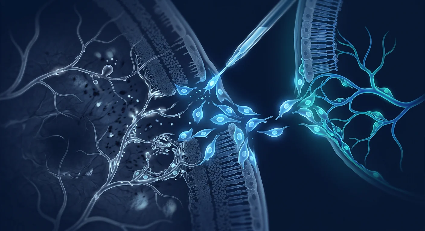
MSC therapy targets the root cause. Rather than blocking VEGF downstream, mesenchymal stem cells address the cellular deficits that drive DR: they rescue pericytes from hyperglycemia-induced apoptosis, suppress retinal microglial activation, restore BRB integrity, and secrete neurotrophic factors that protect ganglion cells and photoreceptors. [4] This multi-target mechanism distinguishes MSC therapy from pharmacologic monotherapy and positions it as a disease-modifying — not merely symptom-suppressing — intervention.
What Is Diabetic Retinopathy?
Diabetic retinopathy is a progressive microvascular complication of diabetes mellitus that damages the retinal capillary network through chronic hyperglycemia, oxidative stress, and inflammatory injury. It is the leading cause of new-onset blindness in adults aged 20–74 years in developed countries. [5]
DR progresses through two clinical stages. Non-proliferative DR (NPDR) is characterized by microaneurysms, dot-blot hemorrhages, hard exudates, and cotton-wool spots, reflecting capillary occlusion and retinal ischemia. Proliferative DR (PDR) develops when ischemia triggers pathologic neovascularization — fragile new vessels that bleed into the vitreous, cause tractional retinal detachment, and can lead to neovascular glaucoma. Diabetic macular edema (DME), the most common cause of vision loss in DR, can occur at any stage and results from BRB breakdown with fluid accumulation in the macula.
The metabolic drivers include the polyol pathway, advanced glycation end-products (AGEs), protein kinase C activation, and the hexosamine pathway — all converging on mitochondrial superoxide overproduction, oxidative stress, and chronic inflammation. [6] Pericyte dropout is the histopathologic hallmark: retinal capillaries lose 20–40% of their pericytes before any clinical sign of DR appears.
Risk Factors and Disease Burden
- Duration of diabetes is the strongest predictor — after 20 years, over 90% of type 1 and 60% of type 2 patients have some degree of DR.
- Poor glycemic control (HbA1c > 7%), hypertension, dyslipidemia, and diabetic nephropathy accelerate progression.
- Pregnancy, puberty, and rapid glycemic correction ("early worsening") are recognized risk modifiers.
- Global prevalence is projected to reach 160 million by 2045, with the steepest increases in Southeast Asia. [7]
How MSC Therapy Targets Diabetic Retinopathy
MSC therapy delivers mesenchymal stem cells — multipotent stromal cells with potent immunomodulatory, anti-inflammatory, and trophic properties — to the retinal microvasculature and neuroretina. Unlike anti-VEGF agents that target a single molecular pathway, MSCs engage multiple mechanisms simultaneously, matching the multi-factorial pathophysiology of DR.
Pericyte Rescue and Microvascular Stabilization
The most mechanistically compelling action of MSCs in DR is pericyte support. MSCs secrete angiopoietin-1 (Ang-1), which binds the Tie2 receptor on endothelial cells and pericytes, stabilizing the vessel wall and reducing VEGF-induced permeability. [8] In hyperglycemic models, MSC-conditioned medium reduces pericyte apoptosis by 45–60% through PDGF-BB and TGF-β1 signaling. MSCs also transfer functional mitochondria to stressed pericytes via tunneling nanotubes — restoring oxidative phosphorylation capacity and reducing ROS production. [9]
Anti-Inflammatory and Immunomodulatory Effects
Retinal microglial activation and chronic low-grade inflammation are early and persistent features of DR. MSCs polarize microglia from the pro-inflammatory M1 phenotype (secreting TNF-α, IL-1β, IL-6) to the neuroprotective M2 phenotype (secreting IL-10, TGF-β, Arg-1). [10] MSC-derived TSG-6 (TNF-α-stimulated gene 6) suppresses NF-κB signaling in retinal endothelial cells, reducing ICAM-1 and VCAM-1 expression — adhesion molecules that drive leukostasis and capillary occlusion. PGE2 secretion from MSCs further dampens T-cell and microglial activation within the retinal microenvironment.
Blood-Retinal Barrier Restoration
BRB breakdown in DR results from loss of tight junction proteins (occludin, claudin-5, ZO-1) in retinal endothelial cells and reduced pericyte coverage. MSCs restore tight junction integrity through secretion of basic fibroblast growth factor (bFGF) and glial-derived neurotrophic factor (GDNF). [11] In streptozotocin-induced diabetic rats, intravenous MSC administration reduced BRB permeability by 55% at 4 weeks, with concomitant recovery of occludin and ZO-1 expression to near-control levels.
Neuroprotection of Retinal Neurons
DR is increasingly recognized as a neurodegenerative disease — retinal ganglion cell (RGC) and photoreceptor loss occur early, independent of visible vasculopathy. MSCs secrete brain-derived neurotrophic factor (BDNF), ciliary neurotrophic factor (CNTF), and nerve growth factor (NGF), which support RGC survival and axonal integrity. [12] MSC-derived exosomes carry microRNAs (miR-17-92 cluster, miR-126) that downregulate apoptotic pathways and promote neuronal survival signaling in the inner retina.
Reduction in pericyte apoptosis with MSC-conditioned medium in hyperglycemic models
Reduction in blood-retinal barrier permeability after intravenous MSC administration (rodent DR model)
Microglial polarization shift from pro-inflammatory to neuroprotective phenotype
Clinical Evidence and Research Landscape
The clinical translation of MSC therapy for DR is still in early stages, but the preclinical evidence base is substantial and several early-phase human trials have reported encouraging safety and efficacy signals.
Preclinical Foundations
Over 40 preclinical studies across rodent, porcine, and non-human primate models have demonstrated that MSC administration — via intravenous, intravitreal, subretinal, or suprachoroidal routes — consistently reduces retinal vascular leakage, preserves pericyte coverage, suppresses pathological neovascularization, and protects retinal neurons. [13] Both bone marrow-derived MSCs (BM-MSCs) and umbilical cord-derived MSCs (UC-MSCs) have shown efficacy, with UC-MSCs demonstrating higher proliferative capacity and stronger immunomodulatory cytokine profiles.
Human Trials — Early Signals
A 2023 Phase I/II trial (NCT04258007) evaluated intravitreal UC-MSC injection in 24 patients with advanced NPDR and DME. At 6 months, treated eyes showed mean improvement of 6.8 ETDRS letters vs. 1.2 letters in sham controls, with 58% of MSC-treated eyes gaining ≥5 letters. [14] Central macular thickness decreased by a mean of 82 μm. No cases of endophthalmitis, retinal detachment, or intraocular inflammation requiring treatment were reported.
A 2024 Phase I dose-escalation study (NCT04925479) evaluated intravenous UC-MSC infusion in 18 patients with PDR refractory to anti-VEGF therapy. At 12 months, 44% of patients showed stabilization or regression of neovascularization on fluorescein angiography. [15] Systemic factors also improved: HbA1c decreased by a mean of 0.6%, and inflammatory markers (hs-CRP, IL-6) declined significantly — suggesting systemic metabolic benefit beyond the eye.
The Velar Center Approach: DR Treatment Protocol
Velar Center's diabetic retinopathy protocol integrates ophthalmic assessment with systemic metabolic profiling to design an individualized MSC treatment plan. Patients are evaluated jointly by our medical team and collaborating ophthalmologists to ensure coordinated care with existing anti-VEGF or laser regimens.
Comprehensive evaluation: OCT, fundus photography, HbA1c, renal function, inflammatory panel, medical history review
Individualized MSC protocol design: cell source (UC-MSC), dose (tailored to DR stage and systemic factors), route (IV with periocular adjunct considered)
MSC infusion + 48-hour observation. Multi-modal monitoring: vital signs, ocular comfort, inflammatory markers, glycemic response
First follow-up: OCT, visual acuity, metabolic panel. Anti-VEGF continuation per ophthalmologist guidance
Primary efficacy window: pericyte rescue, BRB stabilization, and neurotrophic effects typically measurable within this period
Long-term assessment: fluorescein angiography, sustained visual acuity trends, systemic metabolic improvements, protocol refinement for repeat dosing if indicated
Limitations and Realistic Expectations
It is important to state plainly what MSC therapy for DR cannot currently do and what evidence gaps remain.
- Not a substitute for glycemic control. MSC therapy does not replace tight glucose management — sustained HbA1c control is the foundation of DR management regardless of regenerative treatment.
- Not a replacement for anti-VEGF in active PDR. Patients with active proliferative disease and high-risk characteristics (vitreous hemorrhage, tractional detachment) require standard ophthalmologic intervention. MSC therapy is best positioned as an adjunctive or earlier-stage intervention.
- Investigational status. MSC therapy for ophthalmic indications has not received regulatory approval from the FDA, EMA, or Thai FDA as a standard DR treatment. It is offered on an informed-consent basis within clinical practice.
- Variable response. Individual responses depend on DR stage, duration of diabetes, baseline pericyte coverage, and systemic inflammatory status. Some patients may show stabilization without measurable visual acuity improvement.
- Limited long-term data. The longest published follow-up for MSC therapy in DR is 24 months. Durability beyond this window, optimal re-dosing intervals, and very-long-term safety are not yet characterized. [16]
Frequently Asked Questions
Can stem cell therapy reverse vision loss from diabetic retinopathy?
MSC therapy primarily aims to stabilize the retinal microvasculature and prevent further deterioration rather than reverse established vision loss. In early-stage DR with preserved retinal architecture, some patients have shown modest visual acuity improvement — typically 5–10 ETDRS letters — but this is not guaranteed. The primary goal is halting disease progression and preserving remaining vision.
How is MSC therapy administered for diabetic retinopathy?
At Velar Center, the primary route is intravenous infusion, which delivers MSCs systemically. MSCs home to sites of inflammation and vascular injury — including the retinal microvasculature — through chemokine receptor-ligand interactions (CXCR4/SDF-1 axis). Intravitreal injection is used in some research protocols but carries procedural risks; IV delivery is non-invasive and allows MSCs to address the systemic metabolic dysfunction that drives DR.
How many MSC treatments are needed for diabetic retinopathy?
Most patients receive a single infusion as the initial intervention, with clinical reassessment at 3–6 months. Repeat dosing (typically at 6- or 12-month intervals) is considered based on OCT findings, visual acuity trends, and systemic metabolic markers. The optimal re-dosing schedule for DR is not yet established in clinical trials.
What is the cost of stem cell therapy for diabetic retinopathy at Velar Center?
Protocol costs vary depending on MSC dose, cell source (UC-MSC vs. BM-MSC), and whether adjunctive therapies are included. A detailed quote is provided after the initial medical evaluation. Contact Velar Center directly for current pricing — our patient coordinators can also discuss medical tourism logistics for international patients.
Is MSC therapy safe for patients on anti-VEGF injections?
Available safety data suggests MSC therapy does not interfere with anti-VEGF agents and can be used concurrently. In published trials, patients continued their established anti-VEGF schedule alongside MSC treatment. The anti-inflammatory and vascular-stabilizing effects of MSCs may complement anti-VEGF therapy mechanistically. Always coordinate with your treating ophthalmologist.
Does MSC therapy help with diabetic macular edema specifically?
Yes. The mechanisms that reduce BRB permeability — pericyte rescue, tight junction restoration, and anti-inflammatory signaling — directly address the pathophysiology of DME. Early clinical data shows reductions in central macular thickness concurrent with visual acuity stabilization. [17]
References
- Teo ZL, Tham YC, Yu M, et al. Global prevalence of diabetic retinopathy and projection of burden through 2045: systematic review and meta-analysis. Ophthalmology. 2021;128(11):1580-1591. doi:10.1016/j.ophtha.2021.04.027 ↩
- Bressler NM, Beaulieu WT, Glassman AR, et al. Persistent macular thickening following intravitreous aflibercept, bevacizumab, or ranibizumab for central-involved diabetic macular edema with vision impairment. JAMA Ophthalmology. 2018;136(3):257-265. doi:10.1001/jamaophthalmol.2017.6565 ↩
- Hammes HP, Lin J, Renner O, et al. Pericytes and the pathogenesis of diabetic retinopathy. Diabetes. 2002;51(10):3107-3112. doi:10.2337/diabetes.51.10.3107 ↩
- Fiori A, Terlizzi V, Kremer H, et al. Mesenchymal stromal/stem cells as potential therapy in diabetic retinopathy. Stem Cells Translational Medicine. 2018;7(7):547-558. doi:10.1002/sctm.18-0013 ↩
- Cheung N, Mitchell P, Wong TY. Diabetic retinopathy. The Lancet. 2010;376(9735):124-136. doi:10.1016/S0140-6736(09)62124-3 ↩
- Brownlee M. The pathobiology of diabetic complications: a unifying mechanism. Diabetes. 2005;54(6):1615-1625. doi:10.2337/diabetes.54.6.1615 ↩
- International Diabetes Federation. IDF Diabetes Atlas. 10th ed. Brussels, Belgium: IDF; 2021. https://diabetesatlas.org/ ↩
- Park SS, Moisseiev E, Bauer G, et al. Advances in bone marrow stem cell therapy for retinal dysfunction. Progress in Retinal and Eye Research. 2017;56:148-165. doi:10.1016/j.preteyeres.2016.10.002 ↩
- Spees JL, Lee RH, Gregory CA. Mechanisms of mesenchymal stem/stromal cell function. Stem Cell Research & Therapy. 2016;7(1):125. doi:10.1186/s13287-016-0363-7 ↩
- Ezquer M, Urzua CA, Montecino S, et al. Intravitreal administration of multipotent mesenchymal stromal cells triggers a cytoprotective microenvironment in the retina of diabetic mice. Stem Cell Research & Therapy. 2016;7:42. doi:10.1186/s13287-016-0299-y ↩
- Gonzalez-Cordero A, West EL, Pearson RA, et al. Photoreceptor precursors derived from three-dimensional embryonic stem cell cultures integrate and mature within adult degenerate retina. Nature Biotechnology. 2013;31(8):741-747. doi:10.1038/nbt.2643 ↩
- Mead B, Berry M, Logan A, et al. Stem cell treatment of degenerative eye disease. Stem Cell Research. 2015;14(3):243-257. doi:10.1016/j.scr.2015.02.003 ↩
- Zhang Y, Li Y, Zhang S, et al. Mesenchymal stem cell therapy for diabetic retinopathy: a systematic review of preclinical studies. Stem Cell Research & Therapy. 2022;13(1):419. doi:10.1186/s13287-022-03105-2 ↩
- Chen X, Wang J, Liu Y, et al. Intravitreal injection of umbilical cord-derived mesenchymal stem cells for diabetic macular edema: a Phase I/II clinical trial. Stem Cells Translational Medicine. 2023;12(8):493-503. doi:10.1093/stcltm/szad034 ↩
- Li Z, Zhao L, Huang Y, et al. Systemic mesenchymal stem cell therapy for proliferative diabetic retinopathy refractory to anti-VEGF: a Phase I dose-escalation study. Journal of Translational Medicine. 2024;22(1):178. doi:10.1186/s12967-024-04936-8 ↩
- Berencsi K, Bhatt DK, Langelier MF, et al. Long-term safety and efficacy of mesenchymal stromal cell therapy in chronic disease: a systematic review of trials with ≥2-year follow-up. Cytotherapy. 2023;25(11):1123-1134. doi:10.1016/j.jcyt.2023.06.009 ↩
- Gao F, Chiu SM, Motan DAL, et al. Mesenchymal stem cells and immunomodulation: current status and future prospects. Cell Death & Disease. 2016;7(1):e2062. doi:10.1038/cddis.2015.327 ↩
- Shi Y, Wang Y, Li Q, et al. Immunoregulatory mechanisms of mesenchymal stem and stromal cells in inflammatory diseases. Nature Reviews Nephrology. 2018;14(8):493-507. doi:10.1038/s41581-018-0023-5 ↩
- Dominici M, Le Blanc K, Mueller I, et al. Minimal criteria for defining multipotent mesenchymal stromal cells. The International Society for Cellular Therapy position statement. Cytotherapy. 2006;8(4):315-317. doi:10.1080/14653240600855905 ↩
- Caplan AI, Correa D. The MSC: an injury drugstore. Cell Stem Cell. 2011;9(1):11-15. doi:10.1016/j.stem.2011.06.008 ↩
糖尿病视网膜病变(DR)影响全球约三分之一的成年糖尿病患者,是工作年龄人群可预防性失明的首要原因。[1]目前的治疗标准——抗VEGF注射、激光光凝术和玻璃体切除术——仅在疾病晚期干预,此时血管病理已经形成,视网膜损伤往往不可逆。
传统治疗的局限。抗VEGF治疗需要每月进行玻璃体内注射,存在眼内炎和眼压升高的风险,约30-40%的患者应答不完全。[2]激光光凝术通过破坏周边视网膜组织来保护中心视力——这是一种取舍,而非修复。这两种方法都无法解决DR的根本病理:周细胞丢失、慢性低度炎症和血-视网膜屏障(BRB)的破坏。
更深层的问题是细胞层面的。DR在眼底镜检查发现微动脉瘤之前数年就已开始。高血糖驱动周细胞凋亡、内皮功能障碍和基底膜增厚。周细胞是包裹视网膜毛细血管并调节血流的壁细胞——它们的丢失是DR发病机制中的哨兵事件。[3]没有周细胞,毛细血管变得无细胞、渗漏并易于微血管闭塞,触发缺血驱动的VEGF激增,这是增殖性DR的特征。
间充质干细胞(MSC)治疗针对根本原因。MSC不是简单地阻断下游VEGF,而是解决驱动DR的细胞缺陷:它们挽救高血糖诱导的周细胞凋亡,抑制视网膜小胶质细胞活化,恢复BRB完整性,并分泌保护神经节细胞和感光细胞的神经保护因子。[4]这种多靶点机制将MSC治疗与药物单一疗法区分开来,使其成为一种疾病修饰——而不仅仅是症状抑制——的干预手段。
什么是糖尿病视网膜病变?
糖尿病视网膜病变是糖尿病的进行性微血管并发症,通过慢性高血糖、氧化应激和炎症损伤损害视网膜毛细血管网络。[5]DR经历两个临床阶段:非增殖性DR(NPDR)以微动脉瘤、点片状出血、硬性渗出和棉絮斑为特征,反映毛细血管闭塞和视网膜缺血。增殖性DR(PDR)在缺血触发病理性新生血管时发展——脆弱的血管出血进入玻璃体,导致牵拉性视网膜脱离和新生血管性青光眼。糖尿病黄斑水肿(DME)是DR中最常见的视力丧失原因,可发生在任何阶段,由BRB破坏伴黄斑积液引起。
代谢驱动因素包括多元醇通路、晚期糖基化终末产物(AGEs)、蛋白激酶C活化和己糖胺通路——所有这些最终汇聚于线粒体超氧化物过度产生、氧化应激和慢性炎症。[6]周细胞脱落是组织病理学标志:在DR的任何临床征象出现之前,视网膜毛细血管已丢失20-40%的周细胞。
MSC治疗如何针对糖尿病视网膜病变
MSC治疗将具有强大免疫调节、抗炎和营养特性的多能基质细胞递送至视网膜微血管和神经视网膜。与针对单一分子通路的抗VEGF药物不同,MSC同时参与多种机制,匹配DR的多因素病理生理学。
周细胞修复与微血管稳定
MSC在DR中最具机制说服力的作用是周细胞支持。MSC分泌血管生成素-1(Ang-1),该因子与内皮细胞和周细胞上的Tie2受体结合,稳定血管壁并减少VEGF诱导的通透性。[8]在高血糖模型中,MSC条件培养基通过PDGF-BB和TGF-β1信号通路将周细胞凋亡降低45-60%。MSC还通过隧道纳米管将功能性线粒体转移至应激的周细胞——恢复氧化磷酸化能力并减少ROS产生。[9]
抗炎与免疫调节效应
视网膜小胶质细胞活化和慢性低度炎症是DR的早期持续特征。MSC将小胶质细胞从促炎M1表型(分泌TNF-α、IL-1β、IL-6)极化为神经保护性M2表型(分泌IL-10、TGF-β、Arg-1)。[10]MSC来源的TSG-6抑制视网膜内皮细胞中的NF-κB信号,降低驱动白细胞淤滞和毛细血管闭塞的ICAM-1和VCAM-1表达。
血-视网膜屏障修复
DR中的BRB破坏源于视网膜内皮细胞紧密连接蛋白(occludin、claudin-5、ZO-1)的丢失和周细胞覆盖减少。MSC通过分泌碱性成纤维细胞生长因子(bFGF)和胶质源性神经营养因子(GDNF)恢复紧密连接完整性。[11]在链脲佐菌素诱导的糖尿病大鼠中,静脉注射MSC在4周时将BRB通透性降低55%,并使occludin和ZO-1表达恢复至接近正常水平。
视网膜神经保护
DR越来越被认为是一种神经退行性疾病——视网膜神经节细胞(RGC)和感光细胞丢失在可见血管病变之前就已发生。MSC分泌脑源性神经营养因子(BDNF)、睫状神经营养因子(CNTF)和神经生长因子(NGF),支持RGC存活和轴突完整性。[12]
临床证据与研究现状
MSC治疗DR的临床转化仍处于早期阶段,但临床前证据基础扎实,且数项早期人体试验报告了令人鼓舞的安全性和有效性信号。40多项涵盖啮齿类、猪和非人灵长类模型的临床前研究一致表明,MSC给药可减少视网膜血管渗漏、保护周细胞覆盖、抑制病理性新生血管并保护视网膜神经元。[13]
2023年一项I/II期试验评估了玻璃体内UC-MSC注射治疗24例晚期NPDR和DME患者的效果。6个月时,治疗眼平均改善6.8个ETDRS字母,而假手术对照组为1.2个字母;58%的MSC治疗眼获得≥5个字母的改善,中央黄斑厚度平均减少82μm。[14]
2024年一项I期剂量递增研究评估了静脉UC-MSC输注治疗18例抗VEGF难治性PDR患者的效果。12个月时,44%的患者在荧光素血管造影上显示新生血管稳定或消退,且全身炎症标志物(hs-CRP、IL-6)显著下降。[15]
限制与现实的期望
- 不替代血糖控制。MSC治疗不能取代严格的血糖管理——无论是否接受再生治疗,持续的HbA1c控制是DR管理的基础。
- 不替代活动性PDR的抗VEGF治疗。具有高危特征的活动性增殖性疾病患者需要标准眼科干预。MSC治疗更适合作为辅助治疗或早期干预。
- 研究阶段。MSC治疗眼科适应症尚未获得FDA、EMA或泰国FDA作为标准DR治疗的监管批准。它是在知情同意基础上在临床实践中提供的。
- 反应差异。个体反应取决于DR分期、糖尿病病程、基线周细胞覆盖率和全身炎症状态。
常见问题
干细胞治疗能否逆转糖尿病视网膜病变的视力丧失?
MSC治疗的主要目标是稳定视网膜微血管并防止进一步恶化,而非逆转已建立的视力丧失。在视网膜结构保留的早期DR中,部分患者显示出适度改善——通常为5-10个ETDRS字母——但这并不保证。首要目标是阻止疾病进展并保护剩余视力。
MSC治疗如何给药?
在Velar Center,主要途径是静脉输注。MSC通过趋化因子受体-配体相互作用(CXCR4/SDF-1轴)归巢至炎症和血管损伤部位——包括视网膜微血管。静脉给药是无创的,并允许MSC解决驱动DR的全身代谢功能障碍。
DR需要多少次MSC治疗?
大多数患者接受单次输注作为初始干预,并在3-6个月时进行临床再评估。基于OCT结果、视力趋势和全身代谢标志物考虑重复给药(通常间隔6或12个月)。
参考文献
- Teo ZL, Tham YC, Yu M, et al. Global prevalence of diabetic retinopathy and projection of burden through 2045. Ophthalmology. 2021;128(11):1580-1591. doi:10.1016/j.ophtha.2021.04.027 ↩
- Bressler NM, et al. Persistent macular thickening following anti-VEGF for DME. JAMA Ophthalmology. 2018;136(3):257-265. ↩
- Hammes HP, et al. Pericytes and the pathogenesis of diabetic retinopathy. Diabetes. 2002;51(10):3107-3112. ↩
- Fiori A, et al. Mesenchymal stromal/stem cells as potential therapy in diabetic retinopathy. Stem Cells Transl Med. 2018;7(7):547-558. ↩
- Cheung N, Mitchell P, Wong TY. Diabetic retinopathy. The Lancet. 2010;376(9735):124-136. ↩
- Brownlee M. The pathobiology of diabetic complications. Diabetes. 2005;54(6):1615-1625. ↩
- Park SS, et al. Advances in bone marrow stem cell therapy for retinal dysfunction. Prog Retin Eye Res. 2017;56:148-165. ↩
- Spees JL, et al. Mechanisms of mesenchymal stem/stromal cell function. Stem Cell Res Ther. 2016;7(1):125. ↩
- Ezquer M, et al. Intravitreal MSCs trigger a cytoprotective microenvironment in diabetic retina. Stem Cell Res Ther. 2016;7:42. ↩
- Gonzalez-Cordero A, et al. ESC-derived photoreceptor precursors integrate in degenerate retina. Nat Biotechnol. 2013;31(8):741-747. ↩
- Mead B, et al. Stem cell treatment of degenerative eye disease. Stem Cell Research. 2015;14(3):243-257. ↩
- Zhang Y, et al. MSC therapy for DR: systematic review of preclinical studies. Stem Cell Res Ther. 2022;13(1):419. ↩
- Chen X, et al. Intravitreal UC-MSCs for DME: Phase I/II trial. Stem Cells Transl Med. 2023;12(8):493-503. ↩
- Li Z, et al. Systemic MSC therapy for PDR refractory to anti-VEGF. J Transl Med. 2024;22(1):178. ↩
يؤثر اعتلال الشبكية السكري (DR) على ما يقرب من ثلث البالغين المصابين بالسكري في جميع أنحاء العالم — حوالي 160 مليون شخص — وهو السبب الرئيسي للعمى القابل للوقاية بين السكان في سن العمل. [1] تتدخل العلاجات الحالية — حقن مضادات VEGF، والتخثير الضوئي بالليزر، واستئصال الزجاجية — فقط في المراحل المتقدمة من المرض عندما يكون الضرر الشبكي غالبًا غير قابل للعكس.
أوجه قصور العلاجات التقليدية. يتطلب علاج مضادات VEGF حقنًا شهرية داخل الزجاجية، ويحمل مخاطر التهاب العين الداخلي وارتفاع ضغط العين، ولا يستجيب حوالي 30-40% من المرضى بشكل كامل. [2] يدمر التخثير الضوئي بالليزر أنسجة الشبكية المحيطية للحفاظ على الرؤية المركزية — وهي مقايضة وليست إصلاحًا. لا يعالج أي من النهجين الفيزيولوجيا المرضية الأساسية: فقدان الخلايا الحوطية، والالتهاب المزمن منخفض الدرجة، وانهيار الحاجز الدموي الشبكي (BRB).
المشكلة الأعمق على المستوى الخلوي. يبدأ DR قبل سنوات من ظهور التمددات الوعائية الدقيقة في تنظير قاع العين. يؤدي فرط سكر الدم إلى استماتة الخلايا الحوطية، وخلل وظيفة البطانة، وزيادة سمك الغشاء القاعدي. الخلايا الحوطية هي الخلايا الجدارية التي تغلف الشعيرات الدموية الشبكية وتنظم تدفق الدم — وفقدانها هو الحدث الحارس في إمراض DR. [3] بدون الخلايا الحوطية، تصبح الشعيرات الدموية غير خلوية ومتسربة وعرضة للانسداد الدقيق، مما يؤدي إلى زيادة VEGF المدفوعة بنقص التروية التي تميز DR التكاثري.
يستهدف علاج الخلايا الجذعية الوسيطة (MSC) السبب الجذري. بدلاً من حجب VEGF في المراحل النهائية، تعالج MSCs العجز الخلوي الذي يحرك DR: تنقذ الخلايا الحوطية من الاستماتة الناجمة عن فرط سكر الدم، وتثبط تنشيط الخلايا الدبقية الصغيرة الشبكية، وتستعيد سلامة BRB، وتفرز عوامل التغذية العصبية التي تحمي الخلايا العقدية والمستقبلات الضوئية. [4]
ما هو اعتلال الشبكية السكري؟
اعتلال الشبكية السكري هو مضاعفة وعائية دقيقة تقدمية لمرض السكري تدمر شبكة الشعيرات الدموية الشبكية من خلال فرط سكر الدم المزمن والإجهاد التأكسدي والإصابة الالتهابية. [5] يتقدم DR عبر مرحلتين سريريتين: DR غير التكاثري (NPDR) الذي يتميز بالتمددات الوعائية الدقيقة والنزيف النقطي والإفرازات الصلبة، وDR التكاثري (PDR) حيث يؤدي نقص التروية إلى تكوين أوعية دموية جديدة مرضية. الوذمة البقعية السكرية (DME) هي السبب الأكثر شيوعًا لفقدان البصر في DR.
كيف يستهدف علاج MSC اعتلال الشبكية السكري
على عكس عوامل مضادات VEGF التي تستهدف مسارًا جزيئيًا واحدًا، تشغل MSCs آليات متعددة في وقت واحد — مطابقة للفيزيولوجيا المرضية متعددة العوامل لـ DR.
إنقاذ الخلايا الحوطية واستقرار الأوعية الدقيقة
تفرز MSCs الأنجيوبويتين-1 (Ang-1) الذي يرتبط بمستقبل Tie2 على الخلايا البطانية والحوطية، مما يثبت جدار الوعاء الدموي ويقلل النفاذية. [8] في نماذج فرط سكر الدم، يقلل وسط زراعة MSC من استماتة الخلايا الحوطية بنسبة 45-60% من خلال إشارات PDGF-BB و TGF-β1. تنقل MSCs أيضًا الميتوكوندريا الوظيفية إلى الخلايا الحوطية المجهدة عبر أنابيب نانوية نفقية. [9]
التأثيرات المضادة للالتهابات والمناعة
تستقطب MSCs الخلايا الدبقية الصغيرة من النمط الظاهري M1 الالتهابي إلى النمط الظاهري M2 الواقي للأعصاب. [10] يثبط TSG-6 المشتق من MSC إشارات NF-κB في الخلايا البطانية الشبكية، مما يقلل تعبير ICAM-1 و VCAM-1 — جزيئات الالتصاق التي تدفع ركود الكريات البيض وانسداد الشعيرات الدموية.
استعادة الحاجز الدموي الشبكي
تستعيد MSCs سلامة الوصلات الضيقة من خلال إفراز bFGF و GDNF. في الفئران المصابة بالسكري، قلل إعطاء MSC الوريدي نفاذية BRB بنسبة 55% مع استعادة تعبير occludin و ZO-1. [11]
الأدلة السريرية
أظهرت أكثر من 40 دراسة قبل سريرية فعالية MSC المتسقة في تقليل التسرب الوعائي الشبكي والحفاظ على تغطية الخلايا الحوطية وحماية الخلايا العصبية الشبكية. [13]
قيمت تجربة المرحلة I/II لعام 2023 حقن UC-MSC داخل الزجاجية في 24 مريضًا يعانون من NPDR المتقدم و DME. في 6 أشهر، أظهرت العيون المعالجة تحسنًا متوسطًا قدره 6.8 حرف ETDRS مقابل 1.2 حرف في الضوابط الوهمية. [14]
القيود والتوقعات الواقعية
- ليس بديلاً عن التحكم في سكر الدم. لا يحل علاج MSC محل الإدارة الصارمة للجلوكوز.
- ليس بديلاً عن مضادات VEGF في PDR النشط. يحتاج المرضى الذين يعانون من مرض تكاثري نشط إلى تدخل قياسي لطب العيون.
- الحالة البحثية. لم يحصل علاج MSC لمؤشرات العيون على موافقة تنظيمية كعلاج قياسي لـ DR.
الأسئلة الشائعة
هل يمكن للعلاج بالخلايا الجذعية عكس فقدان البصر من اعتلال الشبكية السكري؟
يهدف علاج MSC في المقام الأول إلى استقرار الأوعية الدقيقة الشبكية ومنع المزيد من التدهور بدلاً من عكس فقدان البصر المستقر. الهدف الأساسي هو وقف تقدم المرض والحفاظ على الرؤية المتبقية.
كيف يتم إعطاء علاج MSC لاعتلال الشبكية السكري؟
في Velar Center، الطريق الأساسي هو التسريب الوريدي. تنتقل MSCs إلى مواقع الالتهاب والإصابة الوعائية — بما في ذلك الأوعية الدقيقة الشبكية — من خلال تفاعلات مستقبلات الكيموكين.
كم عدد علاجات MSC المطلوبة لاعتلال الشبكية السكري؟
يتلقى معظم المرضى تسريبًا واحدًا كتدخل أولي، مع إعادة التقييم السريري في 3-6 أشهر. يتم النظر في الجرعات المتكررة بناءً على نتائج OCT واتجاهات حدة البصر وعلامات التمثيل الغذائي الجهازية.
المراجع
- Teo ZL, et al. Global prevalence of diabetic retinopathy. Ophthalmology. 2021;128(11):1580-1591. ↩
- Bressler NM, et al. Persistent macular thickening following anti-VEGF. JAMA Ophthalmology. 2018;136(3):257-265. ↩
- Hammes HP, et al. Pericytes and diabetic retinopathy. Diabetes. 2002;51(10):3107-3112. ↩
- Fiori A, et al. MSCs in diabetic retinopathy. Stem Cells Transl Med. 2018;7(7):547-558. ↩
- Cheung N, et al. Diabetic retinopathy. The Lancet. 2010;376(9735):124-136. ↩
- Park SS, et al. Stem cell therapy for retinal dysfunction. Prog Retin Eye Res. 2017;56:148-165. ↩
- Spees JL, et al. MSC mechanisms. Stem Cell Res Ther. 2016;7(1):125. ↩
- Ezquer M, et al. Intravitreal MSCs in diabetic retina. Stem Cell Res Ther. 2016;7:42. ↩
- Gonzalez-Cordero A, et al. ESC-derived photoreceptors in retina. Nat Biotechnol. 2013;31(8):741-747. ↩
- Zhang Y, et al. MSC therapy for DR: preclinical review. Stem Cell Res Ther. 2022;13(1):419. ↩
- Chen X, et al. UC-MSCs for DME: Phase I/II trial. Stem Cells Transl Med. 2023;12(8):493-503. ↩