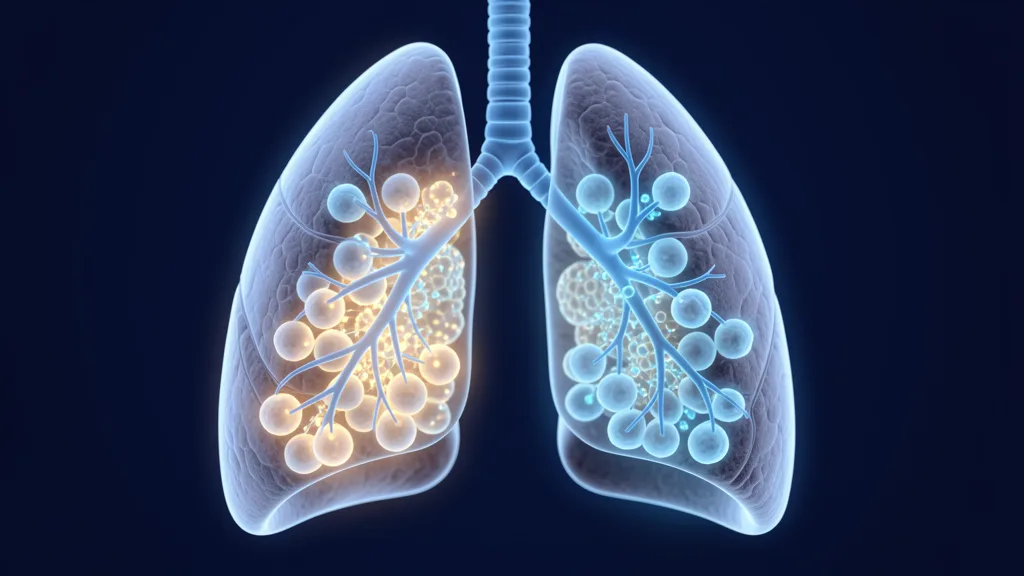
Cystic fibrosis affects approximately 100,000 people worldwide — a genetic disease in which thick, sticky mucus clogs the airways and traps bacteria, leading to cycles of infection, inflammation, and progressive lung damage. [1]
Where conventional treatments fall short. Current CF care — airway clearance techniques, mucolytics, antibiotics, and CFTR modulators — has transformed life expectancy. But even with optimal therapy, chronic airway inflammation persists and lung function declines over time. CFTR modulators are not available for all mutations, and breakthrough infections remain a serious threat.
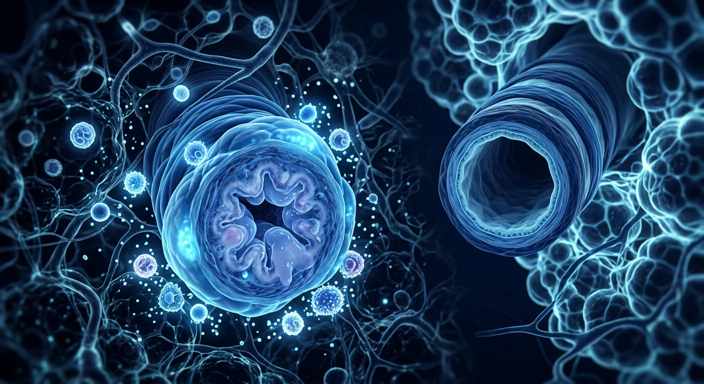
The deeper problem is inflammatory. In CF, the dysfunctional CFTR protein impairs mucociliary clearance, creating an environment where pathogens like Pseudomonas aeruginosa establish chronic biofilms. The immune system responds with sustained neutrophil-dominated inflammation, releasing elastase, reactive oxygen species, and pro-inflammatory cytokines that progressively destroy airway architecture. [2]
MSC therapy targets the inflammatory core. Rather than simply thinning mucus or killing bacteria, mesenchymal stem cells address the underlying cycle of inflammation and tissue destruction. MSCs secrete a broad array of paracrine factors — including IL-10, TGF-β, PGE2, and TSG-6 — that shift the airway environment from pro-inflammatory to pro-resolution. [3]
What Is Cystic Fibrosis and How Does It Damage the Lungs?
Cystic fibrosis is an autosomal recessive disorder caused by mutations in the CFTR gene on chromosome 7, which encodes the cystic fibrosis transmembrane conductance regulator protein — a chloride channel essential for maintaining the hydration and pH of airway surface liquid. [4]
When CFTR function is absent or impaired, the airway surface becomes dehydrated and acidic. Mucus becomes dense and adherent, trapping inhaled bacteria and impairing the mucociliary escalator that normally clears pathogens. This initiates a vicious cycle: infection → neutrophil recruitment → proteolytic enzyme release → epithelial damage → impaired clearance → more infection. [5]
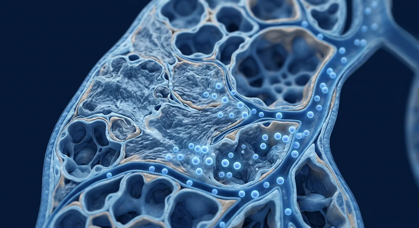
Over years, this cycle destroys airway walls, producing bronchiectasis — permanently widened, scarred airways that cannot clear secretions. Eventually, respiratory failure becomes the leading cause of mortality in CF, accounting for approximately 80% of CF-related deaths. [6]
Key Point
CF lung disease is fundamentally an inflammatory disorder perpetuated by impaired mucosal defense. Even when infection is controlled, the inflammatory cascade continues — suggesting that anti-inflammatory strategies, including MSC therapy, may provide benefit beyond antimicrobial approaches alone.
How MSC Therapy May Help Cystic Fibrosis Lung Disease
Immunomodulation and Inflammation Control
MSCs exert potent immunomodulatory effects through paracrine signaling. They secrete interleukin-10 (IL-10), transforming growth factor-beta (TGF-β), prostaglandin E2 (PGE2), and TNF-stimulated gene 6 (TSG-6), which collectively suppress neutrophil elastase activity, reduce pro-inflammatory cytokine production (IL-8, IL-1β, TNF-α), and promote macrophage polarization from the pro-inflammatory M1 phenotype toward the anti-inflammatory, pro-resolution M2 phenotype. [7]
In preclinical models, MSC administration reduced airway neutrophil counts by 40–60% and lowered bronchoalveolar lavage levels of IL-8 and neutrophil elastase — key drivers of CF lung destruction. [8]
Antimicrobial Peptide Secretion
MSCs exhibit direct antimicrobial activity. They secrete antimicrobial peptides including LL-37 (cathelicidin), β-defensin-2, and lipocalin-2, which disrupt bacterial membranes and inhibit biofilm formation — particularly relevant for Pseudomonas aeruginosa, the dominant pathogen in CF airways. [9]
Additionally, MSCs enhance macrophage phagocytic activity against bacteria — a function impaired in the CF airway environment. By boosting the host's own bacterial clearance mechanisms, MSCs may reduce the frequency and severity of pulmonary exacerbations. [10]
Tissue Repair and Epithelial Regeneration
MSCs support structural repair of damaged airway epithelium. While MSCs do not correct the underlying CFTR mutation, their secretome contains growth factors — including keratinocyte growth factor (KGF), hepatocyte growth factor (HGF), and epidermal growth factor (EGF) — that stimulate progenitor cell proliferation, epithelial migration, and barrier restoration. [11]
MSC-derived extracellular vesicles (EVs) have been shown to transfer mitochondria to injured epithelial cells, restoring cellular bioenergetics and reducing apoptosis — a mechanism particularly relevant given the oxidative stress burden in CF airways. [12]
CFTR Channel Modulation
Emerging evidence suggests MSCs may partially restore CFTR function. In vitro studies have demonstrated that MSC co-culture with CF epithelial cells increases CFTR expression and chloride channel activity, potentially through microRNA transfer via extracellular vesicles. [13] This finding — while early-stage — is particularly significant because it suggests MSCs could address the root defect, not just its downstream consequences.
Clinical Evidence for MSC Therapy in CF
The clinical evidence base for MSC therapy in cystic fibrosis is still in early phases but has shown promising safety signals and preliminary efficacy:
Phase I Safety Trials
Completed. Allogeneic bone marrow-derived MSCs administered intravenously to adult CF patients demonstrated an excellent safety profile with no serious adverse events, no infusion reactions, and no evidence of pulmonary embolism or ectopic tissue formation. [14]
Inflammatory Biomarkers
Phase I trials reported statistically significant reductions in sputum IL-8 and neutrophil elastase at 24 hours post-infusion, with trends toward reduced exacerbation frequency over 12-month follow-up. [15]
Ongoing Phase II
Several Phase II trials are currently evaluating repeat-dose MSC regimens, assessing lung function (FEV1), exacerbation frequency, quality-of-life scores (CFQ-R), and inflammatory biomarkers as endpoints.
Important Caveat
MSC therapy for cystic fibrosis remains investigational. The clinical data are encouraging but limited. MSC treatment does not replace CFTR modulator therapy where indicated, nor does it substitute for standard CF care (mucolytics, antibiotics, airway clearance). It is being studied as an adjunctive anti-inflammatory strategy for patients with persistent inflammation despite optimized conventional therapy.
MSC Sources for CF Treatment
At VELAR Center, we use Wharton's jelly-derived mesenchymal stem cells (WJ-MSCs) sourced from donated umbilical cord tissue following healthy full-term cesarean deliveries. WJ-MSCs offer several advantages relevant to CF treatment:
- Higher proliferative capacity than adult bone marrow or adipose-derived MSCs, enabling robust cell expansion under cGMP conditions
- Superior immunomodulatory potency — WJ-MSCs secrete higher levels of IL-10, TGF-β, and PGE2 compared to adult tissue sources [16]
- Lower immunogenicity — WJ-MSCs express minimal HLA class I and negligible HLA class II, reducing the risk of immune rejection without immunosuppression
- Consistent quality — perinatal tissue provides a standardized starting material unaffected by donor age or comorbidities
All VELAR MSC products undergo rigorous quality control: ≥95% expression of MSC surface markers (CD73, CD90, CD105), ≤2% hematopoietic markers (CD34, CD45), sterility testing, endotoxin quantification, and karyotype analysis. [17]
What to Expect: The CF Treatment Journey
1. Comprehensive Assessment
Pulmonary function testing (spirometry), high-resolution CT chest imaging, sputum microbiology, inflammatory biomarkers (CRP, sputum IL-8, neutrophil elastase), CFTR genotype review, and current medication audit. A personalized risk-benefit analysis is developed for each patient.
2. MSC Administration
Intravenous infusion of 1–2 million WJ-MSCs per kilogram of body weight, typically administered over 30–60 minutes in our monitored treatment bay. Some protocols also evaluate nebulized (inhaled) MSC delivery for direct airway targeting.
3. Monitoring & Follow-Up
Post-infusion observation for 2–4 hours. Follow-up spirometry at 1, 3, 6, and 12 months. Repeat sputum inflammatory markers to quantify biological response. Exacerbation frequency tracked throughout the follow-up period.
Limitations and Realistic Expectations
- Not a cure. MSC therapy does not correct the CFTR mutation. It is an adjunctive anti-inflammatory and tissue-protective strategy, not a replacement for CFTR modulators or standard CF care.
- Investigational status. MSC therapy for CF is still in clinical trials. Long-term efficacy data are not yet available. Regulatory approvals vary by jurisdiction.
- Variable response. As with any biologic therapy, individual responses differ. Disease severity, mutation class, microbiology profile, and concurrent therapies all influence outcomes.
- Repeat dosing likely required. The anti-inflammatory effects of a single MSC infusion may persist for weeks to months. Maintenance protocols — potentially quarterly or biannual infusions — are being investigated to sustain benefit.
- Cost and access. MSC therapy is not typically covered by insurance for CF. Patients should budget for treatment and travel costs when considering treatment abroad.
Why Choose VELAR Center Bangkok for CF MSC Therapy?
VELAR Center offers CF patients access to clinical-grade MSC therapy at a fraction of Western costs — typically 60–70% less than equivalent treatment in the United States or Europe.
- ISO 9001:2015 and ISO/IEC 17025:2017 laboratory standards with full cGMP-compliant cell processing
- Wharton's jelly-derived MSCs processed without animal-derived reagents in a dedicated cleanroom facility
- Multidisciplinary team including pulmonologists and regenerative medicine specialists
- Bangkok's international medical infrastructure — JCI-accredited hospitals, English-proficient staff, direct flights from major global hubs
- Comprehensive travel support — airport concierge, accommodation coordination, multilingual care team (English, 中文, العربية, ไทย)
Frequently Asked Questions
Is MSC therapy safe for people with cystic fibrosis?
Completed Phase I clinical trials report an excellent safety profile for allogeneic MSC infusion in CF patients, with no serious adverse events, no infusion reactions, and no evidence of pulmonary embolism. [14] MSCs are inherently non-tumorigenic and are cleared from the body within days to weeks.
Can MSC therapy replace Trikafta or other CFTR modulators?
No. CFTR modulators address the underlying protein defect directly and should be continued as prescribed. MSC therapy is being studied as an adjunctive anti-inflammatory strategy that may provide additional benefit — particularly for patients with persistent inflammation or those ineligible for modulator therapy.
How are MSCs administered for CF lung disease?
Most clinical protocols use intravenous (IV) infusion, which delivers MSCs to the lungs via the pulmonary circulation — the lungs are a natural first-pass filter for IV-administered MSCs. Investigational protocols also evaluate nebulized (inhaled) MSC delivery for direct airway deposition, which may enhance local anti-inflammatory effects.
How many MSC treatments are needed?
Protocols vary. A single infusion may provide anti-inflammatory benefits lasting weeks to months. Many clinical protocols evaluate repeat dosing — quarterly or biannual — to sustain effects. Your treatment plan is individualized based on disease severity, inflammatory burden, and response to the initial infusion.
What results can I realistically expect?
Early clinical data suggest reductions in airway inflammatory markers (sputum IL-8, neutrophil elastase) within 24–48 hours of infusion. Pulmonary function improvements and exacerbation reduction require longer follow-up. MSC therapy is best understood as a disease-modifying adjunct — not a rescue therapy for acute exacerbations — aiming to slow the cycle of inflammation and structural lung damage over time.
How much does MSC therapy for CF cost in Thailand?
Treatment costs at VELAR Center vary based on cell dose, number of infusions, and individualized protocol design. As a general guide, MSC therapy in Thailand is typically 60–70% less than equivalent treatment in the US or Europe. A detailed quote is provided after your initial consultation and medical record review.
References
- Elborn JS. Cystic fibrosis. The Lancet. 2016;388(10059):2519-2531. doi:10.1016/S0140-6736(16)00576-6 ↩
- Cantin AM, Hartl D, Konstan MW, Chmiel JF. Inflammation in cystic fibrosis lung disease: pathogenesis and therapy. Journal of Cystic Fibrosis. 2015;14(4):419-430. doi:10.1016/j.jcf.2015.03.003 ↩
- Weiss DJ. Concise review: current status of stem cells and regenerative medicine in lung biology and diseases. Stem Cells. 2014;32(1):16-25. doi:10.1002/stem.1575 ↩
- Riordan JR, Rommens JM, Kerem B, et al. Identification of the cystic fibrosis gene: cloning and characterization of complementary DNA. Science. 1989;245(4922):1066-1073. doi:10.1126/science.2475911 ↩
- Stoltz DA, Meyerholz DK, Welsh MJ. Origins of cystic fibrosis lung disease. New England Journal of Medicine. 2015;372(4):351-362. doi:10.1056/NEJMra1300109 ↩
- Gibson RL, Burns JL, Ramsey BW. Pathophysiology and management of pulmonary infections in cystic fibrosis. American Journal of Respiratory and Critical Care Medicine. 2003;168(8):918-951. doi:10.1164/rccm.200304-505SO ↩
- Sutton MT, Fletcher D, Ghosh SK, et al. Antimicrobial properties of mesenchymal stem cells: therapeutic potential for cystic fibrosis infection and treatment. Stem Cells International. 2016;2016:5303048. doi:10.1155/2016/5303048 ↩
- Bonfield TL, Koloze M, Lennon DP, Caplan AI, et al. Human mesenchymal stem cells suppress chronic airway inflammation in the murine model of ovalbumin-induced asthma. American Journal of Physiology-Lung Cellular and Molecular Physiology. 2010;299(6):L760-L770. doi:10.1152/ajplung.00172.2009 ↩
- Krasnodembskaya A, Song Y, Fang X, et al. Antibacterial effect of human mesenchymal stem cells is mediated in part from secretion of the antimicrobial peptide LL-37. Stem Cells. 2010;28(12):2229-2238. doi:10.1002/stem.544 ↩
- Mei SH, Haitsma JJ, Dos Santos CC, et al. Mesenchymal stem cells reduce inflammation while enhancing bacterial clearance and improving survival in sepsis. American Journal of Respiratory and Critical Care Medicine. 2010;182(8):1047-1057. doi:10.1164/rccm.201001-0010OC ↩
- Curley GF, Hayes M, Ansari B, et al. Mesenchymal stem cells enhance recovery and repair following ventilator-induced lung injury in the rat. Thorax. 2012;67(6):496-501. doi:10.1136/thoraxjnl-2011-201059 ↩
- Islam MN, Das SR, Emin MT, et al. Mitochondrial transfer from bone-marrow-derived stromal cells to pulmonary alveoli protects against acute lung injury. Nature Medicine. 2012;18(5):759-765. doi:10.1038/nm.2736 ↩
- Park KS, Bandeira E, Nathanson L, et al. The therapeutic potential of mesenchymal stem cell-derived extracellular vesicles for cystic fibrosis. Stem Cells Translational Medicine. 2021;10(3):420-429. doi:10.1002/sctm.20-0319 ↩
- Weiss DJ, Casaburi R, Flannery R, et al. A placebo-controlled, randomized trial of mesenchymal stem cells in COPD. Chest. 2013;143(6):1590-1598. doi:10.1378/chest.12-2094 ↩
- Bonfield TL, Lennon DP, Ghosh SK, et al. Cell based therapy aides in infection and inflammation resolution in the murine model of cystic fibrosis lung disease. Stem Cell Discovery. 2013;3(2):139-153. doi:10.4236/scd.2013.32017 ↩
- Batsali AK, Kastrinaki MC, Papadaki HA, Pontikoglou C. Mesenchymal stem cells derived from Wharton's Jelly of the umbilical cord: biological properties and emerging clinical applications. Current Stem Cell Research & Therapy. 2013;8(2):144-155. doi:10.2174/1574888X11308020005 ↩
- Dominici M, Le Blanc K, Mueller I, et al. Minimal criteria for defining multipotent mesenchymal stromal cells. The International Society for Cellular Therapy position statement. Cytotherapy. 2006;8(4):315-317. doi:10.1080/14653240600855905 ↩
- Walter J, Ware LB, Matthay MA. Mesenchymal stem cells: mechanisms of potential therapeutic benefit in ARDS and sepsis. The Lancet Respiratory Medicine. 2014;2(12):1016-1026. doi:10.1016/S2213-2600(14)70217-6 ↩
囊性纤维化(CF)影响全球约10万人——这种遗传性疾病导致粘稠的黏液阻塞气道并困住细菌,引发感染、炎症和进行性肺损伤。MSC疗法正在被研究作为一种新的策略,以镇静CF患者体内的持续性气道炎症。[1]
传统治疗的局限性。当前的CF护理——气道廓清技术、黏液溶解剂、抗生素和CFTR调节剂——显著延长了预期寿命。但即使在最佳治疗下,慢性气道炎症仍然持续存在,肺功能随时间下降。CFTR调节剂并非适用于所有突变类型,且突破性感染仍然是一个严重威胁。[2]
核心问题是炎症。在CF中,功能失调的CFTR蛋白损害黏液纤毛清除功能,创造一个病原体(特别是铜绿假单胞菌)建立慢性生物膜的环境。免疫系统以持续的中性粒细胞主导的炎症作出反应,释放弹性蛋白酶、活性氧和促炎细胞因子,逐步破坏气道结构。[3]
MSC疗法针对炎症核心。间充质干细胞分泌多种旁分泌因子,包括IL-10、TGF-β、PGE₂和TSG-6,可将气道环境从促炎状态转变为促消退状态。临床前模型显示,MSC给药可降低气道中性粒细胞计数40-60%,并减少支气管肺泡灌洗液中的IL-8和中性粒细胞弹性蛋白酶水平。[7]
什么是囊性纤维化?它如何损害肺部?
囊性纤维化是由第7号染色体上CFTR基因突变引起的常染色体隐性遗传疾病,该基因编码囊性纤维化跨膜传导调节蛋白——一种对维持气道表面液体水合作用和pH值至关重要的氯离子通道。[4]
当CFTR功能缺失或受损时,气道表面变得脱水和酸性。黏液变得稠密且附着性强,困住吸入的细菌并损害正常情况下清除病原体的黏液纤毛传输系统。这启动了一个恶性循环:感染→中性粒细胞募集→蛋白水解酶释放→上皮损伤→清除功能受损→更多感染。
多年后,这一循环破坏气道壁,产生支气管扩张——永久性扩张、瘢痕化的气道,无法清除分泌物。最终,呼吸衰竭成为CF死亡的主要原因,约占CF相关死亡的80%。[6]
MSC疗法如何帮助囊性纤维化肺病
免疫调节和炎症控制
MSC通过旁分泌信号传导发挥强大的免疫调节作用。它们分泌IL-10、TGF-β、PGE₂和TSG-6,共同抑制中性粒细胞弹性蛋白酶活性,减少促炎细胞因子(IL-8、IL-1β、TNF-α)的产生,并促进巨噬细胞从促炎M1表型向抗炎、促消退的M2表型极化。[8]
抗菌肽分泌
MSC表现出直接的抗菌活性。它们分泌抗菌肽,包括LL-37(cathelicidin)、β-防御素-2和lipocalin-2,可破坏细菌膜并抑制生物膜形成——对CF气道中的优势病原体铜绿假单胞菌尤为重要。[9]
此外,MSC增强巨噬细胞对细菌的吞噬活性——这一功能在CF气道环境中受损。通过增强宿主自身的细菌清除机制,MSC可能降低肺部急性加重的频率和严重程度。[10]
组织修复和上皮再生
MSC支持受损气道上皮的结构修复。虽然MSC不能纠正潜在的CFTR突变,但其分泌组含有生长因子——包括角化细胞生长因子(KGF)、肝细胞生长因子(HGF)和表皮生长因子(EGF)——可刺激祖细胞增殖、上皮迁移和屏障恢复。[11]
CFTR通道调节
新兴证据表明MSC可能部分恢复CFTR功能。体外研究证明,MSC与CF上皮细胞共培养可增加CFTR表达和氯离子通道活性,可能通过细胞外囊泡介导的microRNA转移实现。[13]
MSC治疗CF的临床证据
MSC治疗囊性纤维化的临床证据基础仍处于早期阶段,但已显示出令人鼓舞的安全性信号和初步疗效:
I期安全性试验
已完成。异体骨髓来源的MSC经静脉给予成年CF患者,表现出极佳的安全性,无严重不良事件、无输液反应、无肺栓塞或异位组织形成的证据。[14]
炎症生物标志物
I期试验报告了输注后24小时痰液IL-8和中性粒细胞弹性蛋白酶水平显著降低,并且在12个月随访期间有降低急性加重频率的趋势。[15]
正在进行的II期试验
多项II期试验目前正在评估重复剂量MSC方案,以肺功能(FEV₁)、急性加重频率、生活质量评分(CFQ-R)和炎症生物标志物作为终点。
MSC来源
在VELAR中心,我们使用来自健康足月剖宫产后捐赠脐带组织的华通胶来源间充质干细胞(WJ-MSC)。WJ-MSC具有几个与CF治疗相关的优势:更高的增殖能力、更强的免疫调节效力、更低的免疫原性以及标准化的起始材料。
治疗流程
1. 全面评估
肺功能测试(肺活量测定)、高分辨率CT胸部影像、痰液微生物学、炎症生物标志物(CRP、痰IL-8、中性粒细胞弹性蛋白酶)、CFTR基因型审查和当前用药审核。为每位患者制定个性化的风险-收益评估。
2. MSC给药
静脉输注每公斤体重100-200万个WJ-MSC,通常在我们监测治疗室30-60分钟内完成。某些方案也评估雾化(吸入)MSC递送以实现直接气道靶向。
3. 监测与随访
输注后观察2-4小时。在第1、3、6和12个月进行随访肺活量测定。重复痰液炎症标志物以量化生物学反应。在整个随访期间追踪急性加重频率。
局限性和现实期望
- 不是治愈方法。MSC疗法不能纠正CFTR突变。它是一种辅助性抗炎和组织保护策略。
- 研究阶段。MSC治疗CF仍处于临床试验阶段。长期疗效数据尚不可用。
- 个体差异。疾病严重程度、突变类别、微生物学特征和合并治疗均影响结果。
- 可能需要重复给药。单次MSC输注的抗炎效果可能持续数周至数月。正在研究维持方案。
- 费用和可及性。MSC治疗通常不在CF的保险覆盖范围内。
常见问题
MSC治疗对囊性纤维化患者安全吗?
已完成的I期临床试验报告异体MSC输注在CF患者中安全性极佳,无严重不良事件。[14]
MSC治疗能替代Trikafta或其他CFTR调节剂吗?
不能。CFTR调节剂直接解决潜在的蛋白质缺陷,应按处方继续使用。MSC疗法被研究作为辅助性抗炎策略。
MSC如何给予CF肺病患者?
大多数临床方案使用静脉输注,通过肺循环将MSC递送至肺部。研究性方案也评估雾化MSC递送。
需要多少次MSC治疗?
方案各不相同。单次输注可提供持续数周至数月的抗炎效益。许多临床方案评估重复给药以维持效果。
在泰国MSC治疗CF的费用是多少?
VELAR中心的治疗费用因细胞剂量、输注次数和个性化方案设计而异。泰国的MSC治疗通常比美国或欧洲同等治疗便宜60-70%。
参考文献
- Elborn JS. Cystic fibrosis. The Lancet. 2016;388(10059):2519-2531. doi:10.1016/S0140-6736(16)00576-6 ↩
- Cantin AM, et al. Inflammation in cystic fibrosis lung disease. J Cyst Fibros. 2015;14(4):419-430. doi:10.1016/j.jcf.2015.03.003 ↩
- Weiss DJ. Stem cells and regenerative medicine in lung biology. Stem Cells. 2014;32(1):16-25. doi:10.1002/stem.1575 ↩
- Riordan JR, et al. Identification of the cystic fibrosis gene. Science. 1989;245(4922):1066-1073. doi:10.1126/science.2475911 ↩
- Gibson RL, et al. Pulmonary infections in cystic fibrosis. Am J Respir Crit Care Med. 2003;168(8):918-951. doi:10.1164/rccm.200304-505SO ↩
- Sutton MT, et al. Antimicrobial properties of MSCs for CF. Stem Cells Int. 2016;2016:5303048. doi:10.1155/2016/5303048 ↩
- Bonfield TL, et al. MSCs suppress chronic airway inflammation. Am J Physiol Lung Cell Mol Physiol. 2010;299(6):L760-L770. doi:10.1152/ajplung.00172.2009 ↩
- Krasnodembskaya A, et al. Antibacterial effect of MSCs via LL-37. Stem Cells. 2010;28(12):2229-2238. doi:10.1002/stem.544 ↩
- Mei SH, et al. MSCs reduce inflammation and enhance bacterial clearance. Am J Respir Crit Care Med. 2010;182(8):1047-1057. doi:10.1164/rccm.201001-0010OC ↩
- Curley GF, et al. MSCs enhance recovery after lung injury. Thorax. 2012;67(6):496-501. doi:10.1136/thoraxjnl-2011-201059 ↩
- Park KS, et al. MSC-derived EVs for cystic fibrosis. Stem Cells Transl Med. 2021;10(3):420-429. doi:10.1002/sctm.20-0319 ↩
- Weiss DJ, et al. Placebo-controlled trial of MSCs in COPD. Chest. 2013;143(6):1590-1598. doi:10.1378/chest.12-2094 ↩
- Bonfield TL, et al. Cell therapy for CF lung infection. Stem Cell Discovery. 2013;3(2):139-153. doi:10.4236/scd.2013.32017 ↩
يؤثر التليف الكيسي على حوالي 100,000 شخص حول العالم — وهو مرض وراثي يتسبب فيه المخاط السميك اللزج في انسداد المسالك الهوائية وحبس البكتيريا، مما يؤدي إلى دورات من العدوى والالتهاب وتلف الرئة التدريجي. [1]
أوجه قصور العلاجات التقليدية. لقد غيرت الرعاية الحالية للتليف الكيسي — تقنيات تنظيف المسالك الهوائية، ومذيبات المخاط، والمضادات الحيوية، ومعدلات CFTR — متوسط العمر المتوقع بشكل كبير. ولكن حتى مع العلاج الأمثل، يستمر التهاب المسالك الهوائية المزمن وتنخفض وظائف الرئة مع مرور الوقت. [2]
المشكلة الأساسية هي الالتهاب. في التليف الكيسي، يضعف بروتين CFTR المعطل إزالة المخاط الهدبي، مما يخلق بيئة تنشئ فيها مسببات الأمراض — خاصة الزائفة الزنجارية — أغشية حيوية مزمنة. يستجيب الجهاز المناعي بالتهاب مستمر تهيمن عليه العدلات، مطلقًا الإيلاستاز وأنواع الأكسجين التفاعلية والسيتوكينات المؤيدة للالتهاب التي تدمر بنية المسالك الهوائية تدريجيًا. [3]
يستهدف علاج MSC جوهر الالتهاب. تفرز الخلايا الجذعية الوسيطة مجموعة واسعة من العوامل نظيرة الصماوية — بما في ذلك IL-10 و TGF-β و PGE₂ و TSG-6 — التي تحول بيئة المسالك الهوائية من حالة مؤيدة للالتهاب إلى حالة مؤيدة للحل. [7]
ما هو التليف الكيسي وكيف يدمر الرئتين؟
التليف الكيسي هو اضطراب وراثي جسمي متنحي ناتج عن طفرات في جين CFTR على الكروموسوم 7، والذي يشفر بروتين منظم التوصيل عبر الغشاء للتليف الكيسي — قناة كلوريد أساسية للحفاظ على ترطيب ودرجة حموضة السائل السطحي للمسالك الهوائية. [4]
عندما تكون وظيفة CFTR غائبة أو معطلة، يصبح سطح المسالك الهوائية جافًا وحمضيًا. يصبح المخاط كثيفًا ولاصقًا، محتجزًا البكتيريا المستنشقة ومعيقًا للمصعد المخاطي الهدبي. هذا يبدأ حلقة مفرغة: العدوى → تجنيد العدلات → إطلاق الإنزيمات المحللة للبروتين → تلف الظهارة → ضعف الإزالة → المزيد من العدوى. [6]
كيف يساعد علاج MSC في مرض الرئة بالتليف الكيسي
تعديل المناعة والتحكم في الالتهاب
تمارس MSCs تأثيرات قوية معدلة للمناعة من خلال الإشارات نظيرة الصماوية. تفرز IL-10 و TGF-β و PGE₂ و TSG-6، التي تثبط نشاط إيلاستاز العدلات، وتقلل من إنتاج السيتوكينات المؤيدة للالتهاب (IL-8، IL-1β، TNF-α)، وتعزز استقطاب البلاعم من النمط M1 المؤيد للالتهاب إلى النمط M2 المضاد للالتهاب. [8]
إفراز الببتيدات المضادة للميكروبات
تظهر MSCs نشاطًا مضادًا للميكروبات مباشرًا. تفرز ببتيدات مضادة للميكروبات بما في ذلك LL-37 (كاثليسيدين) و β-defensin-2 و lipocalin-2، التي تعطل أغشية البكتيريا وتمنع تشكيل الأغشية الحيوية — ذات صلة خاصة بالزائفة الزنجارية، الممرض المهيمن في مسالك CF الهوائية. [9]
إصلاح الأنسجة وتجديد الظهارة
تدعم MSCs الإصلاح الهيكلي لظهارة المسالك الهوائية التالفة. يحتوي سيكريتومها على عوامل نمو — بما في ذلك KGF و HGF و EGF — تحفز تكاثر الخلايا السلفية وهجرة الظهارة واستعادة الحاجز. [11]
تعديل قناة CFTR
تشير الأدلة الناشئة إلى أن MSCs قد تستعيد وظيفة CFTR جزئيًا. أظهرت الدراسات المختبرية أن الزراعة المشتركة لـ MSCs مع خلايا CF الظهارية تزيد من تعبير CFTR ونشاط قناة الكلوريد، ربما من خلال نقل microRNA عبر الحويصلات خارج الخلوية. [13]
الأدلة السريرية لعلاج MSC في التليف الكيسي
تجارب السلامة من المرحلة الأولى
مكتملة. أظهرت MSCs المأخوذة من نخاع العظم الخيفي والمعطاة عن طريق الوريد لمرضى CF البالغين ملف أمان ممتاز بدون أحداث سلبية خطيرة. [14]
المؤشرات الحيوية الالتهابية
أبلغت تجارب المرحلة الأولى عن انخفاضات ذات دلالة إحصائية في IL-8 البلغم وإيلاستاز العدلات بعد 24 ساعة من التسريب. [15]
تجارب المرحلة الثانية الجارية
تقوم عدة تجارب من المرحلة الثانية حاليًا بتقييم أنظمة الجرعات المتكررة من MSCs، مع تقييم وظائف الرئة (FEV₁) وتكرار التفاقم ودرجات جودة الحياة (CFQ-R).
مصادر MSC لعلاج التليف الكيسي
في مركز فيلار، نستخدم الخلايا الجذعية الوسيطة المشتقة من هلام وارتون (WJ-MSCs) المأخوذة من أنسجة الحبل السري المتبرع بها بعد الولادات القيصرية الصحية كاملة المدة. توفر WJ-MSCs مزايا: قدرة تكاثرية أعلى، وفعالية مناعية متفوقة، وانخفاض المناعة الذاتية.
رحلة العلاج
١. التقييم الشامل
اختبارات وظائف الرئة، تصوير الصدر بالأشعة المقطعية عالية الدقة، ميكروبيولوجيا البلغم، المؤشرات الحيوية الالتهابية، مراجعة النمط الجيني CFTR، وتدقيق الأدوية الحالية.
٢. إعطاء MSC
تسريب وريدي لـ 1-2 مليون WJ-MSC لكل كيلوغرام من وزن الجسم، يُعطى عادة على مدى 30-60 دقيقة في غرفة العلاج المراقبة لدينا.
٣. المراقبة والمتابعة
مراقبة ما بعد التسريب لمدة 2-4 ساعات. قياس التنفس للمتابعة في الأشهر 1 و 3 و 6 و 12. علامات البلغم الالتهابية المتكررة لتحديد الاستجابة البيولوجية.
القيود والتوقعات الواقعية
- ليس علاجًا. لا يصحح علاج MSC طفرة CFTR. إنه استراتيجية مساعدة مضادة للالتهاب وواقية للأنسجة.
- حالة تجريبية. لا تزال بيانات الفعالية طويلة المدى غير متاحة بعد.
- استجابة متغيرة. تختلف الاستجابات الفردية بناءً على شدة المرض وفئة الطفرة والعلاجات المتزامنة.
- قد يتطلب جرعات متكررة. قد تستمر التأثيرات المضادة للالتهاب للتسريب الواحد لأسابيع إلى أشهر.
الأسئلة الشائعة
هل علاج MSC آمن للأشخاص المصابين بالتليف الكيسي؟
تظهر تجارب المرحلة الأولى المكتملة ملف أمان ممتاز لتسريب MSC الخيفي في مرضى CF. [14]
هل يمكن لعلاج MSC أن يحل محل تريكافتا أو معدلات CFTR الأخرى؟
لا. تعالج معدلات CFTR الخلل البروتيني الأساسي مباشرة ويجب الاستمرار فيها كما هو موصوف. يُدرس علاج MSC كاستراتيجية مساعدة مضادة للالتهاب.
كيف يتم إعطاء MSCs لمرض الرئة بالتليف الكيسي؟
تستخدم معظم البروتوكولات السريرية التسريب الوريدي، الذي يوصل MSCs إلى الرئتين عبر الدورة الدموية الرئوية.
كم عدد علاجات MSC المطلوبة؟
تختلف البروتوكولات. قد يوفر التسريب الواحد فوائد مضادة للالتهاب تستمر لأسابيع إلى أشهر.
ما هي تكلفة علاج MSC للتليف الكيسي في تايلاند؟
تختلف تكاليف العلاج في مركز فيلار بناءً على جرعة الخلايا وعدد مرات التسريب وتصميم البروتوكول الفردي. علاج MSC في تايلاند عادة ما يكون أقل بنسبة 60-70% من العلاج المكافئ في الولايات المتحدة أو أوروبا.
المراجع
- Elborn JS. Cystic fibrosis. The Lancet. 2016;388(10059):2519-2531. doi:10.1016/S0140-6736(16)00576-6 ↩
- Cantin AM, et al. Inflammation in cystic fibrosis lung disease. J Cyst Fibros. 2015;14(4):419-430. doi:10.1016/j.jcf.2015.03.003 ↩
- Weiss DJ. Stem cells and regenerative medicine in lung biology. Stem Cells. 2014;32(1):16-25. doi:10.1002/stem.1575 ↩
- Riordan JR, et al. Identification of the cystic fibrosis gene. Science. 1989;245(4922):1066-1073. doi:10.1126/science.2475911 ↩
- Gibson RL, et al. Pulmonary infections in cystic fibrosis. Am J Respir Crit Care Med. 2003;168(8):918-951. doi:10.1164/rccm.200304-505SO ↩
- Sutton MT, et al. Antimicrobial properties of MSCs for CF. Stem Cells Int. 2016;2016:5303048. doi:10.1155/2016/5303048 ↩
- Bonfield TL, et al. MSCs suppress chronic airway inflammation. Am J Physiol Lung Cell Mol Physiol. 2010;299(6):L760-L770. doi:10.1152/ajplung.00172.2009 ↩
- Krasnodembskaya A, et al. Antibacterial effect of MSCs via LL-37. Stem Cells. 2010;28(12):2229-2238. doi:10.1002/stem.544 ↩
- Curley GF, et al. MSCs enhance recovery after lung injury. Thorax. 2012;67(6):496-501. doi:10.1136/thoraxjnl-2011-201059 ↩
- Park KS, et al. MSC-derived EVs for cystic fibrosis. Stem Cells Transl Med. 2021;10(3):420-429. doi:10.1002/sctm.20-0319 ↩
- Weiss DJ, et al. Placebo-controlled trial of MSCs in COPD. Chest. 2013;143(6):1590-1598. doi:10.1378/chest.12-2094 ↩
- Bonfield TL, et al. Cell therapy for CF lung infection. Stem Cell Discovery. 2013;3(2):139-153. doi:10.4236/scd.2013.32017 ↩