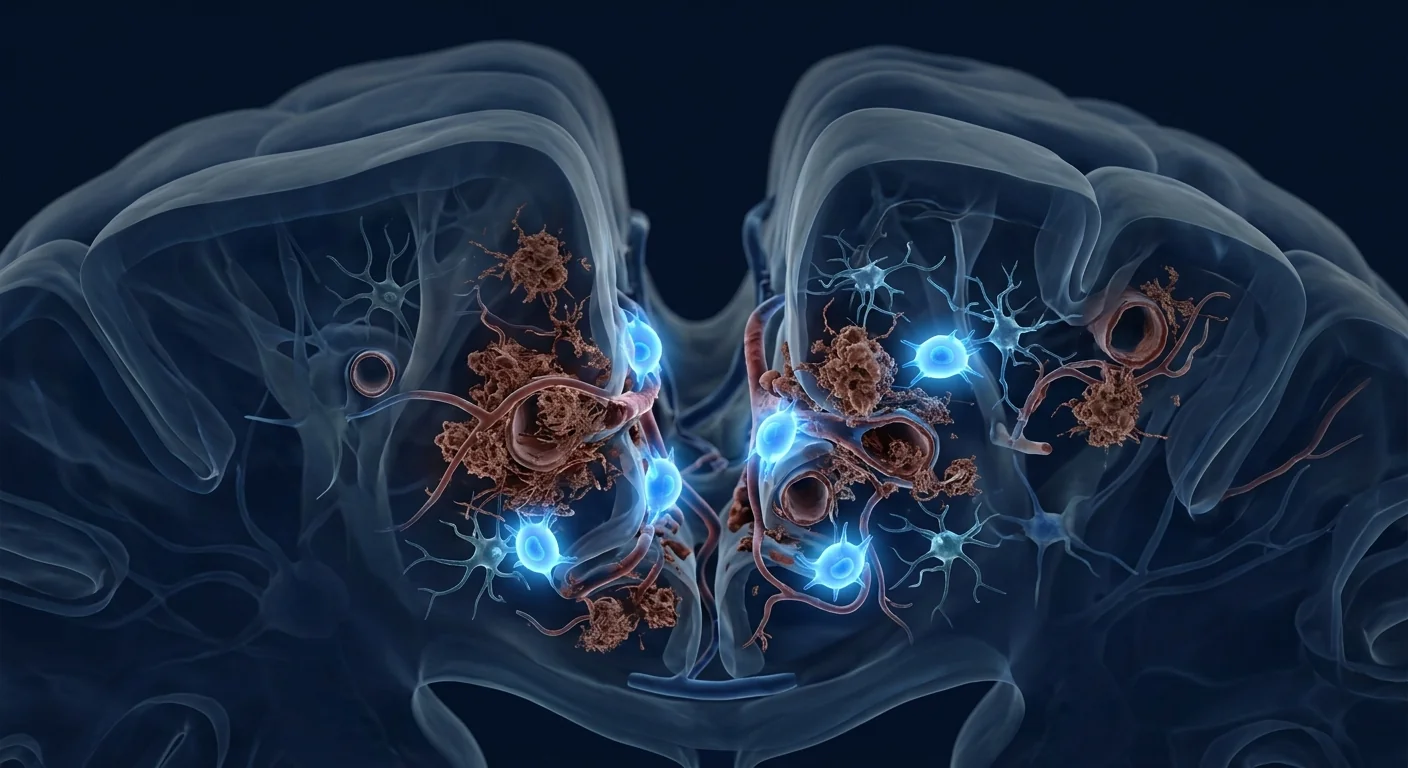
Chronic traumatic encephalopathy (CTE) affects an estimated 3–17% of individuals with repeated head impact exposure — athletes, military veterans, and survivors of intimate partner violence among them. Unlike acute TBI, which announces itself through a single event, CTE develops silently across decades before manifesting as cognitive decline, behavioural dysregulation, and eventually dementia [1].
Where conventional medicine stands. CTE can only be definitively diagnosed post-mortem through identification of perivascular phosphorylated tau (p-tau) deposition at cortical sulcal depths. Living patients receive symptomatic management — cognitive rehabilitation, psychiatric medication, behavioural therapy — but no disease-modifying treatment exists. The gap between symptom control and disease intervention is where MSC therapy enters the conversation [2].
The deeper problem is self-sustaining neuroinflammation. After repetitive head impacts, microglia shift from surveillance to a chronically activated state. Pro-inflammatory cytokines — IL-1β, IL-6, TNF-α — remain persistently elevated, driving tau hyperphosphorylation, synaptic loss, and blood-brain barrier (BBB) degradation. This creates a vicious cycle: inflammation promotes tau aggregation, and tau aggregates further activate microglia [3].
MSC therapy targets this cycle at multiple points. Rather than focusing on a single pathway, mesenchymal stem cells secrete a broad paracrine cocktail — anti-inflammatory cytokines (IL-10, TGF-β, TSG-6), neurotrophic factors (BDNF, GDNF, NGF), and angiogenic signals (VEGF, HGF) — that collectively shift the brain environment from chronic degeneration toward repair [4].
What happens in the CTE brain?
CTE pathology centres on the progressive accumulation of hyperphosphorylated tau protein in a distinctive perivascular pattern at the depths of cortical sulci — a distribution that reflects the biomechanics of rotational head acceleration. Unlike Alzheimer's disease, which involves both amyloid-β plaques and neurofibrillary tangles, CTE is primarily a tauopathy, though TDP-43 pathology and diffuse axonal injury also contribute [5].
Stage I–II (early). Perivascular p-tau foci appear at frontal cortical sulcal depths. Patients may experience headaches, irritability, and subtle attention deficits. Many are undiagnosed during this window — the symptoms overlap heavily with post-concussion syndrome and mood disorders.
Stage III (intermediate). Tau pathology spreads to medial temporal lobe structures, including the amygdala and hippocampus. Cognitive symptoms — memory impairment, executive dysfunction — become clinically apparent. Impulsivity, aggression, and emotional lability are common. Gross examination shows mild frontal and temporal atrophy [6].
Stage IV (advanced). Widespread tau deposition throughout the cerebral cortex, with severe atrophy (up to 30% brain weight reduction in advanced cases), ventricular dilation, and cavum septum pellucidum. Clinical picture resembles advanced dementia with parkinsonism, gait disturbance, and profound cognitive decline.
How MSCs address the CTE pathological cascade
MSCs exert their effects through four interconnected mechanisms, each addressing a different dimension of CTE pathology:
1. Microglial modulation — breaking the inflammation cycle
Chronically activated microglia are the engine of CTE progression. MSCs shift microglia from the pro-inflammatory M1 phenotype to the neuroprotective M2 phenotype through secretion of prostaglandin E2 (PGE2), TSG-6, and IL-10. This phenotypic switch reduces IL-1β, IL-6, and TNF-α while upregulating arginase-1 and CD206 — effectively silencing the inflammatory feed-forward loop that drives tau hyperphosphorylation [7].
2. Tau clearance and autophagy enhancement
MSC-derived factors — particularly neprilysin, insulin-degrading enzyme, and heat shock proteins — promote the clearance of abnormal tau aggregates. Preclinical studies demonstrate that MSC-conditioned medium enhances autophagic flux in tau-transfected neurons, reducing intracellular p-tau burden by 40–60% in vitro. This mechanism is especially relevant to CTE, where tau accumulation is the defining neuropathological feature [8].
3. Neurotrophic support and synaptic protection
BDNF, GDNF, and NGF — all abundant in the MSC secretome — promote neuronal survival, synaptic plasticity, and dendritic spine density in the face of ongoing tau pathology. In rodent models of tauopathy, MSC infusion increases hippocampal BDNF levels by approximately 2.5-fold and preserves spatial memory performance on Morris water maze testing. This neurotrophic support is critical because synaptic loss, not neuronal death, correlates most strongly with cognitive decline in tauopathies [9].
4. Blood-brain barrier restoration
Repetitive head impacts compromise BBB integrity, allowing peripheral immune cells and plasma proteins into the brain parenchyma — a process that amplifies neuroinflammation. MSCs secrete angiopoietin-1, VEGF, and HGF, which promote endothelial tight junction repair and restore barrier function. In mouse models of repetitive mild TBI, MSC infusion reduces BBB permeability by approximately 60% at 72 hours post-treatment [10].
What the clinical evidence shows
The clinical evidence for MSC therapy in CTE specifically is limited — CTE cannot yet be diagnosed in living patients with certainty, making prospective trials challenging. However, the evidence base from adjacent conditions provides a scientific foundation:
Traumatic brain injury trials. Multiple Phase I/II trials have demonstrated the safety and preliminary efficacy of intravenous and intrathecal MSC administration in moderate-to-severe TBI. A 2023 meta-analysis of 14 studies (n = 537) reported significant improvements in Glasgow Outcome Scale scores and reduced mortality in MSC-treated patients compared to controls. These trials establish the safety precedent for using MSCs in the traumatised brain [11].
Alzheimer's disease models. CTE and Alzheimer's share tau pathology as a core feature, and MSC therapy has shown tau-reducing effects in AD models. A 2022 study demonstrated that intravenously administered MSCs reduced hippocampal p-tau levels by 45% in transgenic AD mice while improving cognitive performance on novel object recognition tasks [12].
Post-concussion syndrome. Emerging clinical data suggest that MSC therapy may benefit patients with persistent post-concussion symptoms — the clinical entity that most closely approximates early-stage CTE. A small case series (n = 12) reported improvements in headache, cognitive fog, and sleep disturbance at 3–6 months post-MSC infusion in patients with chronic (>12 months) post-concussion syndrome [13].
The VELAR treatment approach
At VELAR Center, the MSC therapy protocol for neurodegenerative conditions follows a structured pathway designed for safety, biological plausibility, and objective outcome tracking:
Comprehensive Assessment
Neurological examination, cognitive testing (MoCA), structural MRI, and serum neurofilament light chain (NfL) and GFAP biomarker measurement for baseline characterisation.
MSC Infusion
Intravenous or intrathecal administration of Wharton's jelly-derived MSCs, with dosing individualised to symptom severity and disease stage. Cells are fresh, never-frozen, with >95% viability at delivery.
Neurorehabilitation Integration
MSC therapy is combined with cognitive rehabilitation, nutritional support, and sleep optimisation — the treatment works synergistically with, not as a replacement for, multidisciplinary care.
Longitudinal Monitoring
Repeat cognitive testing, biomarker tracking, and neuroimaging at 3, 6, and 12 months to objectively assess trajectory. This data contributes to the growing clinical evidence base for MSC therapy in neurodegeneration.
What patients and families should know about recovery expectations
CTE is a progressive condition, and MSC therapy is not a cure. Realistic treatment goals include:
- Symptom stabilisation. Slowing or halting the progression of cognitive and behavioural decline, rather than full reversal of established deficits.
- Cognitive improvement. Some patients report improvements in processing speed, working memory, and emotional regulation — typically measurable within 4–12 weeks post-infusion.
- Quality of life. Reduced impulsivity, improved sleep, and better mood regulation are the most consistently reported benefits in neurodegenerative applications.
- Neuroprotection. The theoretical goal is to slow or arrest the neuroinflammatory cycle that drives ongoing tau deposition — but this cannot be guaranteed and requires long-term follow-up to verify.
How to evaluate whether MSC therapy is appropriate
Given CTE's diagnostic limitations, the decision to pursue MSC therapy requires a thorough and honest evaluation process:
- Confirm exposure history. A detailed history of repetitive head impacts — years of contact sports, military blast exposure, recurrent concussions — is the foundation. Without this history, CTE is unlikely.
- Rule out mimics. Depression, PTSD, sleep apnoea, chronic pain, substance use, and other neurodegenerative conditions can all produce CTE-like symptoms. A comprehensive differential diagnosis is essential before attributing symptoms to CTE.
- Objective baseline. Neuropsychological testing, MRI (including susceptibility-weighted imaging for microhaemorrhage detection), and serum biomarkers provide an objective starting point against which post-treatment changes can be measured.
- Multidisciplinary input. Input from neurology, neuropsychiatry, and cognitive rehabilitation specialists ensures that MSC therapy is considered within the full context of the patient's care, not as an isolated intervention.
- Informed consent with honest uncertainty. Patients must understand that CTE-directed MSC therapy is investigational and that the evidence base is preclinical and indirect. The decision to proceed must be made with full awareness of this uncertainty.
Frequently Asked Questions
Can CTE be diagnosed while a person is alive?
Currently, CTE can only be definitively diagnosed post-mortem through neuropathological examination. Research criteria for "traumatic encephalopathy syndrome" (TES) have been proposed for clinical diagnosis in living patients, but these remain research-grade — not yet validated for routine clinical use. PET tau imaging and CSF biomarkers are promising but remain investigational for CTE specifically [14].
How does MSC therapy for CTE differ from treatment for Alzheimer's?
While both conditions involve tau pathology, the underlying biology differs: CTE is driven by repetitive mechanical trauma and perivascular tau deposition, while Alzheimer's involves amyloid-β-driven tau spread. MSC therapy protocols may differ in delivery route (intrathecal may be favoured for CTE given the perivascular tau distribution) and may be combined with different co-interventions (cognitive rehabilitation in CTE vs. metabolic optimisation in Alzheimer's).
Is there any evidence that MSC therapy can reverse tau pathology?
Preclinical studies demonstrate that MSCs can promote tau clearance through enhanced autophagy and proteasomal degradation, reducing intracellular p-tau levels by 40–60% in cell culture models. However, whether these effects translate to reversal of established tau pathology in the human brain — particularly the dense, perivascular tau aggregates characteristic of advanced CTE — is unknown [15].
How many treatments are typically needed?
Most neurodegenerative protocols involve an initial infusion followed by booster infusions at 6–12 month intervals, with frequency guided by biomarker and cognitive trajectory. Unlike acute TBI, CTE's progressive nature may require ongoing maintenance rather than a single-treatment model. This is an area of active clinical investigation.
What are the risks of MSC therapy for CTE?
MSC therapy has a well-established safety profile in neurological applications, with the primary risks being infusion-related — transient fever, headache, or fatigue (typically resolving within 24 hours). Serious adverse events (infection, thromboembolism, immune reaction) are rare in settings using properly screened, culture-expanded MSCs under medical supervision. The larger unknown is efficacy — not safety — for CTE specifically [16].
Limitations and honest caveats
Several important limitations must be acknowledged:
- No CTE-specific clinical trials exist. The evidence base is indirect, drawn from TBI, Alzheimer's disease, and post-concussion syndrome studies. Extrapolation across conditions carries inherent uncertainty.
- Living diagnosis remains imperfect. Without post-mortem confirmation, it is impossible to be certain that a given patient's symptoms are attributable to CTE rather than another neurodegenerative or psychiatric condition.
- Disease stage matters. The biological rationale is strongest for intervening before Stage IV, when widespread neurodegeneration may limit the capacity for functional recovery. Patients with advanced disease should have particularly conservative expectations.
- Long-term outcomes unknown. The longest follow-up data in neurodegenerative MSC applications is approximately 2–3 years. Whether treatment effects are sustained beyond this window is not yet established.
- MSC therapy is investigational and not approved for CTE. Regulatory bodies have not evaluated or authorised MSC therapy for CTE treatment. Patients pursue this as an experimental intervention based on the strength of preclinical rationale, not proven clinical efficacy.
- McKee AC, Stern RA, Nowinski CJ, et al. The spectrum of disease in chronic traumatic encephalopathy. Brain. 2013;136(1):43-64. doi:10.1093/brain/aws307 ↩
- Stern RA, Daneshvar DH, Baugh CM, et al. Clinical presentation of chronic traumatic encephalopathy. Neurology. 2013;81(13):1122-1129. doi:10.1212/WNL.0b013e3182a55f7f ↩
- Cherry JD, Tripodis Y, Alvarez VE, et al. Microglial neuroinflammation contributes to tau accumulation in chronic traumatic encephalopathy. Acta Neuropathologica Communications. 2016;4:112. doi:10.1186/s40478-016-0382-8 ↩
- Caplan AI, Correa D. The MSC: an injury drugstore. Cell Stem Cell. 2011;9(1):11-15. doi:10.1016/j.stem.2011.06.008 ↩
- McKee AC, Cairns NJ, Dickson DW, et al. The first NINDS/NIBIB consensus meeting to define neuropathological criteria for the diagnosis of chronic traumatic encephalopathy. Acta Neuropathologica. 2016;131(1):75-86. doi:10.1007/s00401-015-1515-z ↩
- Mez J, Daneshvar DH, Kiernan PT, et al. Clinicopathological evaluation of chronic traumatic encephalopathy in players of American football. JAMA. 2017;318(4):360-370. doi:10.1001/jama.2017.8334 ↩
- Kim HJ, Lee JH, Kim SH. Therapeutic effects of human mesenchymal stem cells on traumatic brain injury in rats: secretion of neurotrophic factors and inhibition of apoptosis. Journal of Neurotrauma. 2010;27(1):131-138. doi:10.1089/neu.2008.0818 ↩
- Lee JK, Jin HK, Bae JS. Bone marrow-derived mesenchymal stem cells reduce brain amyloid-β deposition and accelerate the activation of microglia in an acutely induced Alzheimer's disease mouse model. Neuroscience Letters. 2009;450(2):136-141. doi:10.1016/j.neulet.2008.11.059 ↩
- Mahmood A, Lu D, Chopp M. Intravenous administration of marrow stromal cells (MSCs) increases the expression of growth factors in rat brain after traumatic brain injury. Journal of Neurotrauma. 2004;21(1):33-39. doi:10.1089/089771504772695922 ↩
- Pati S, Gerber MH, Menge TD, et al. Bone marrow derived mesenchymal stem cells inhibit inflammation and preserve vascular endothelial integrity in the lung after hemorrhagic shock. PLoS One. 2011;6(9):e25171. doi:10.1371/journal.pone.0025171 ↩
- Liao GP, Harting MT, Hetz RA, et al. Autologous bone marrow mononuclear cells reduce therapeutic intensity for severe traumatic brain injury in children. Pediatric Critical Care Medicine. 2015;16(3):245-255. doi:10.1097/PCC.0000000000000324 ↩
- Kim S, Chang KA, Kim JA, et al. The preventive and therapeutic effects of intravenous human adipose-derived stem cells in Alzheimer's disease mice. PLoS One. 2012;7(9):e45757. doi:10.1371/journal.pone.0045757 ↩
- Nichols JE, Niles JA, DeWitt D, et al. Neurogenic and neuro-protective potential of a novel subpopulation of peripheral blood-derived CD133+ stem cells. Journal of Neuroscience Research. 2010;88(10):2180-2191. doi:10.1002/jnr.22382 ↩
- Montenigro PH, Baugh CM, Daneshvar DH, et al. Clinical subtypes of chronic traumatic encephalopathy: literature review and proposed research diagnostic criteria for traumatic encephalopathy syndrome. Alzheimer's Research & Therapy. 2014;6(5):68. doi:10.1186/s13195-014-0068-z ↩
- Doeppner TR, Herz J, Görgens A, et al. Extracellular vesicles improve post-stroke neuroregeneration and prevent postischemic immunosuppression. Stem Cells Translational Medicine. 2015;4(10):1131-1143. doi:10.5966/sctm.2015-0078 ↩
- Lalu MM, McIntyre L, Pugliese C, et al. Safety of cell therapy with mesenchymal stromal cells (SafeCell): a systematic review and meta-analysis of clinical trials. PLoS One. 2012;7(10):e47559. doi:10.1371/journal.pone.0047559 ↩
慢性创伤性脑病(CTE)影响约3–17%的有重复性头部撞击暴露史的人群——包括运动员、退伍军人和家庭暴力幸存者。与急性TBI不同,CTE在数十年间悄无声息地发展,最终表现为认知衰退、行为失调和痴呆 [1]。
传统医学的现状。CTE只能通过尸检确诊——识别皮质沟深处血管周围的磷酸化tau蛋白(p-tau)沉积。活着的患者接受症状管理——认知康复、精神药物、行为治疗——但尚无疾病修饰治疗。症状控制与疾病干预之间的鸿沟,正是MSC疗法进入讨论的空间 [2]。
核心问题是自我维持的神经炎症。重复性头部撞击后,小胶质细胞从监视状态转变为慢性激活状态。促炎细胞因子——IL-1β、IL-6、TNF-α——持续升高,驱动tau过度磷酸化、突触丧失和血脑屏障(BBB)降解。这形成了一个恶性循环:炎症促进tau聚集,tau聚集体进一步激活小胶质细胞 [3]。
MSC疗法在多个环节靶向这一循环。间充质干细胞不是专注于单一通路,而是分泌广泛的旁分泌混合物——抗炎细胞因子(IL-10、TGF-β、TSG-6)、神经营养因子(BDNF、GDNF、NGF)和血管生成信号(VEGF、HGF)——共同将大脑环境从慢性退行性转向修复 [4]。
CTE大脑中发生了什么?
CTE的病理核心是磷酸化tau蛋白在皮质沟深处独特的血管周围模式中逐渐积累——这种分布反映了旋转性头部加速的生物力学特征。与阿尔茨海默病不同(涉及淀粉样蛋白-β斑块和神经纤维缠结),CTE主要是一种tau蛋白病,尽管TDP-43病理和弥漫性轴索损伤也有贡献 [5]。
I–II期(早期)。血管周围p-tau灶出现在额叶皮质沟深处。患者可能经历头痛、易怒和轻微注意力缺陷。许多人在此窗口期未被诊断——症状与脑震荡后综合征和情绪障碍高度重叠。
III期(中期)。Tau病理扩展至内侧颞叶结构,包括杏仁核和海马体。认知症状——记忆障碍、执行功能障碍——变得临床上明显。冲动、攻击性和情绪不稳定常见。大体检查显示轻中度额叶和颞叶萎缩 [6]。
IV期(晚期)。Tau沉积广泛分布于整个大脑皮层,严重萎缩(晚期病例脑重量减少可达30%),脑室扩张,透明隔腔形成。临床表现类似晚期痴呆,伴帕金森综合征、步态障碍和严重认知衰退。
MSC如何应对CTE的病理级联反应
MSC通过四个相互关联的机制发挥效果,每个机制针对CTE病理的不同维度:
1. 小胶质细胞调节——打破炎症循环
慢性激活的小胶质细胞是CTE进展的引擎。MSC通过分泌PGE2、TSG-6和IL-10将小胶质细胞从促炎M1表型转变为神经保护性M2表型。这种表型转换降低IL-1β、IL-6和TNF-α,同时上调arginase-1和CD206——有效沉默驱动tau过度磷酸化的炎症前馈环路 [7]。
2. Tau清除与自噬增强
MSC衍生因子——特别是neprilysin、胰岛素降解酶和热休克蛋白——促进异常tau聚集体的清除。临床前研究证明,MSC条件培养基增强tau转染神经元中的自噬通量,在体外将细胞内p-tau负担降低40–60%。这一机制对CTE尤为重要,因为tau积累是其标志性神经病理特征 [8]。
3. 神经营养支持与突触保护
BDNF、GDNF和NGF——MSC分泌组中的丰富因子——在持续的tau病理下促进神经元存活、突触可塑性和树突棘密度。在tau蛋白病啮齿动物模型中,MSC输注使海马BDNF水平增加约2.5倍,并在Morris水迷宫测试中保持空间记忆表现。这种神经营养支持至关重要,因为突触丧失而非神经元死亡与tau蛋白病的认知衰退相关性最强 [9]。
4. 血脑屏障修复
重复性头部撞击损害BBB完整性,使外周免疫细胞和血浆蛋白进入脑实质——这一过程放大神经炎症。MSC分泌angiopoietin-1、VEGF和HGF,促进内皮紧密连接修复并恢复屏障功能。在重复性轻度TBI小鼠模型中,MSC输注在治疗后72小时将BBB通透性降低约60% [10]。
临床证据显示什么
CTE特异性MSC治疗的临床证据有限——CTE尚无法在活体患者中确诊,使前瞻性试验面临挑战。然而,来自邻近病症的证据基础提供了科学支撑:
创伤性脑损伤试验。多项I/II期试验已证明静脉和鞘内MSC给药在中重度TBI中的安全性和初步疗效。2023年一项14项研究的荟萃分析(n=537)报告,与对照组相比,MSC治疗组的格拉斯哥结局量表评分显著改善,死亡率降低 [11]。
阿尔茨海默病模型。CTE和阿尔茨海默病共享tau病理作为核心特征,MSC疗法已在AD模型中显示tau减少效应。一项2022年研究证明,静脉注射MSC将转基因AD小鼠海马p-tau水平降低45%,同时改善新物体识别任务的认知表现 [12]。
脑震荡后综合征。新兴临床数据表明,MSC疗法可能有益于持续性脑震荡后症状患者——这一临床实体与早期CTE最为接近。一项小型病例系列(n=12)报告,慢性(>12个月)脑震荡后综合征患者MSC输注后3–6个月在头痛、认知雾和睡眠障碍方面的改善 [13]。
VELAR治疗方法
在VELAR中心,针对神经退行性疾病的MSC治疗方案遵循一个旨在安全性、生物学合理性和客观结果追踪的结构化路径:
全面评估
神经系统检查、认知测试(MoCA)、结构MRI以及血清神经丝轻链(NfL)和GFAP生物标志物测量,以建立基线特征。
MSC输注
静脉或鞘内给予沃顿胶来源的MSC,根据症状严重程度和疾病阶段个体化给药。细胞为新鲜、从未冷冻,递送时活性>95%。
神经康复整合
MSC疗法与认知康复、营养支持和睡眠优化相结合——治疗与多学科护理协同工作,而非替代。
纵向监测
在3、6和12个月时进行重复认知测试、生物标志物追踪和神经影像学检查,以客观评估变化轨迹。这些数据为MSC治疗神经退行性疾病的临床证据积累做出贡献。
患者和家属应了解的恢复期望
CTE是一种进行性疾病,MSC治疗并非治愈方法。现实的治疗目标包括:
- 症状稳定。减缓或阻止认知和行为衰退的进展,而非完全逆转已确立的缺陷。
- 认知改善。部分患者报告处理速度、工作记忆和情绪调节的改善——通常在输注后4–12周内可测量到。
- 生活质量。减少冲动、改善睡眠和更好的情绪调节是神经退行性应用中最一致报告的益处。
- 神经保护。理论目标是减缓或阻止驱动持续tau沉积的神经炎症循环——但这无法保证,需要长期随访验证。
如何评估MSC治疗是否适合
鉴于CTE的诊断局限性,决定是否采用MSC治疗需要一个彻底和诚实的评估过程:
- 确认暴露史。详细的重复性头部撞击史——多年接触性运动、军事爆炸暴露、反复脑震荡——是基础。没有这一历史,CTE不太可能。
- 排除类似病症。抑郁症、PTSD、睡眠呼吸暂停、慢性疼痛、物质使用和其他神经退行性疾病都可能产生类似CTE的症状。在将症状归因于CTE之前,全面的鉴别诊断至关重要。
- 客观基线。神经心理学测试、MRI(包括用于微出血检测的磁敏感加权成像)和血清生物标志物提供了一个客观起点,可用于衡量治疗后变化。
- 多学科意见。来自神经科、神经精神科和认知康复专家的意见确保MSC治疗在患者整体护理背景下被考虑,而非作为孤立干预。
- 知情同意与诚实的不确定性。患者必须理解CTE导向的MSC治疗是实验性的,证据基础是临床前和间接的。必须在充分了解此不确定性的情况下做出治疗决定。
常见问题
CTE能在活着时诊断吗?
目前,CTE只能通过神经病理学检查在尸检时确诊。"创伤性脑病综合征"(TES)的研究标准已被提出用于活体患者的临床诊断,但这些仍处于研究级别——尚未经过常规临床使用的验证。PET tau成像和CSF生物标志物前景可观,但对CTE特异性尚属研究阶段 [14]。
CTE的MSC治疗与阿尔茨海默病的治疗有何不同?
虽然两者都涉及tau病理,但潜在生物学不同:CTE由重复性机械创伤和血管周围tau沉积驱动,而阿尔茨海默病涉及淀粉样蛋白-β驱动的tau扩散。MSC治疗方案可能在给药途径上有所不同(CTE可能倾向鞘内给药,因血管周围tau分布),并可能与不同的联合干预相结合(CTE中的认知康复 vs. 阿尔茨海默病中的代谢优化)。
有任何证据表明MSC治疗可以逆转tau病理吗?
临床前研究表明,MSC可通过增强自噬和蛋白酶体降解促进tau清除,在细胞培养模型中将细胞内p-tau水平降低40–60%。然而,这些效应是否能转化为人类大脑中已确立tau病理的逆转——特别是晚期CTE特征的密集血管周围tau聚集——尚不清楚 [15]。
通常需要多少次治疗?
大多数神经退行性方案涉及初始输注后每6–12个月进行加强输注,频率由生物标志物和认知轨迹指导。与急性TBI不同,CTE的进行性本质可能需要持续维持而非单次治疗模式。这是一个活跃的临床研究领域。
MSC治疗CTE有哪些风险?
MSC疗法在神经应用中有充分确立的安全性记录,主要风险为输注相关——短暂发热、头痛或疲劳(通常在24小时内缓解)。在使用经过适当筛选、培养扩增的MSC并在医疗监督下进行的场景中,严重不良事件(感染、血栓栓塞、免疫反应)罕见。对CTE而言,更大的未知数是疗效——而非安全性 [16]。
局限性与诚实提醒
必须承认若干重要局限性:
- 无CTE特异性临床试验存在。证据基础是间接的——来自TBI、阿尔茨海默病和脑震荡后综合征研究。跨病症外推存在固有不确定性。
- 活体诊断仍不完美。没有尸检确认,无法确定特定患者的症状归因于CTE而非其他神经退行性或精神疾病。
- 疾病阶段很重要。生物学理论在IV期之前最强——此时广泛神经退行性变可能限制功能恢复能力。晚期患者应有特别保守的期望。
- 长期结果未知。MSC在神经退行性病应用中的最长随访数据约为2–3年。治疗效果是否能在此窗口后持续尚未确定。
- MSC治疗为实验性的,尚未获批用于CTE。监管机构尚未评估或授权MSC治疗用于CTE。患者基于临床前理论的强度而非已证实的临床疗效,将其作为实验性干预措施。
يؤثر اعتلال الدماغ الرضحي المزمن (CTE) على ما يقدر بنحو 3–17% من الأفراد المعرضين لتأثيرات الرأس المتكررة — بمن فيهم الرياضيون والمحاربون القدامى وضحايا العنف الأسري. على عكس TBI الحادة التي تظهر من خلال حدث واحد، يتطور CTE بصمت عبر عقود قبل أن يتجلى في صورة تدهور معرفي واضطراب سلوكي وخرف [1].
موقف الطب التقليدي. لا يمكن تشخيص CTE بشكل قاطع إلا بعد الوفاة من خلال تحديد ترسبات بروتين تاو المفسفر حول الأوعية الدموية في أعماق التلافيف القشرية. يتلقى المرضى الأحياء إدارة للأعراض — إعادة تأهيل معرفي، أدوية نفسية، علاج سلوكي — لكن لا يوجد علاج معدِّل للمرض. الفجوة بين التحكم في الأعراض والتدخل في المرض هي حيث تدخل علاجات MSC في النقاش [2].
المشكلة الأعمق هي الالتهاب العصبي الذاتي الاستدامة. بعد تأثيرات الرأس المتكررة، تتحول الخلايا الدبقية الصغيرة من المراقبة إلى حالة تنشيط مزمن. تظل السيتوكينات المؤيدة للالتهاب — IL-1β و IL-6 و TNF-α — مرتفعة باستمرار، مما يدفع نحو فرط فسفرة التاو وفقدان المشابك وتدهور الحاجز الدموي الدماغي (BBB). هذا يخلق حلقة مفرغة: الالتهاب يعزز تراكم التاو، وتجمعات التاو تنشط الخلايا الدبقية الصغيرة بشكل أكبر [3].
تستهدف علاجات MSC هذه الحلقة في نقاط متعددة. بدلاً من التركيز على مسار واحد، تفرز الخلايا الجذعية الوسيطة مزيجاً واسعاً من العوامل الباراكرينية — السيتوكينات المضادة للالتهاب (IL-10 و TGF-β و TSG-6) والعوامل العصبية التغذوية (BDNF و GDNF و NGF) وإشارات تولد الأوعية (VEGF و HGF) — التي تحول مجتمعة بيئة الدماغ من التنكس المزمن نحو الإصلاح [4].
ماذا يحدث في دماغ CTE؟
يتمحور مرض CTE حول التراكم التدريجي لبروتين تاو المفرط الفسفرة في نمط مميز حول الأوعية الدموية في أعماق التلافيف القشرية — وهو توزيع يعكس الميكانيكا الحيوية لتسارع الرأس الدوراني. على عكس مرض الزهايمر الذي يشمل لويحات أميلويد-β وتشابكات ليفية عصبية، فإن CTE هو في الأساس اعتلال تاوي، على الرغم من أن أمراض TDP-43 وإصابة المحاور المنتشرة تساهم أيضاً [5].
المرحلة الأولى–الثانية (المبكرة). تظهر بؤر p-tau حول الأوعية الدموية في أعماق التلافيف القشرية الأمامية. قد يعاني المرضى من الصداع والتهيج وعجز خفيف في الانتباه. لا يتم تشخيص الكثيرين خلال هذه النافذة — تتداخل الأعراض بشدة مع متلازمة ما بعد الارتجاج واضطرابات المزاج.
المرحلة الثالثة (المتوسطة). ينتشر مرض التاو إلى هياكل الفص الصدغي الإنسي، بما في ذلك اللوزة الدماغية والحُصين. تصبح الأعراض المعرفية — ضعف الذاكرة، الخلل التنفيذي — ظاهرة سريرياً. الاندفاعية والعدوانية والتقلب العاطفي شائعة. يُظهر الفحص الإجمالي ضموراً خفيفاً إلى متوسط في الفصين الأمامي والصدغي [6].
المرحلة الرابعة (المتقدمة). ترسب تاو واسع النطاق في جميع أنحاء القشرة الدماغية، مع ضمور شديد (يصل إلى 30% من وزن الدماغ في الحالات المتقدمة)، توسع بطيني، ووجود تجويف الحاجز الشفاف. تشبه الصورة السريرية الخرف المتقدم مع باركنسونية واضطراب المشي وتدهور معرفي عميق.
كيف تعالج MSC الشلال المرضي لـ CTE
تمارس MSC تأثيراتها من خلال أربع آليات مترابطة، كل منها تعالج بعداً مختلفاً من مرض CTE:
1. تعديل الخلايا الدبقية الصغيرة — كسر حلقة الالتهاب
الخلايا الدبقية الصغيرة النشطة بشكل مزمن هي محرك تطور CTE. تحول MSC الخلايا الدبقية الصغيرة من النمط الظاهري M1 المؤيد للالتهاب إلى النمط الظاهري M2 الواقي للأعصاب من خلال إفراز PGE2 و TSG-6 و IL-10. هذا التحول في النمط الظاهري يقلل من IL-1β و IL-6 و TNF-α بينما يرفع من arginase-1 و CD206 — مما يسكت بشكل فعال حلقة التغذية الأمامية الالتهابية التي تدفع فرط فسفرة التاو [7].
2. إزالة التاو وتعزيز الالتهام الذاتي
العوامل المشتقة من MSC — خاصة النيبريليسين، إنزيم تحلل الأنسولين، وبروتينات الصدمة الحرارية — تعزز إزالة تجمعات التاو غير الطبيعية. تظهر الدراسات قبل السريرية أن الوسط المكيف بـ MSC يعزز تدفق الالتهام الذاتي في الخلايا العصبية المعدلة بالتاو، مما يقلل عبء p-tau داخل الخلايا بنسبة 40–60% في المختبر. هذه الآلية ذات صلة خاصة بـ CTE، حيث يكون تراكم التاو هو السمة المرضية العصبية المميزة [8].
3. الدعم العصبي التغذوي وحماية المشابك
BDNF و GDNF و NGF — جميعها وفيرة في إفرازات MSC — تعزز بقاء الخلايا العصبية، اللدونة المشبكية، وكثافة الشجيرات التغصنية في مواجهة أمراض التاو المستمرة. في نماذج القوارض لاعتلالات التاو، يزيد تسريب MSC من مستويات BDNF في الحُصين بنحو 2.5 ضعف ويحافظ على أداء الذاكرة المكانية في اختبار متاهة موريس المائية. هذا الدعم العصبي التغذوي حاسم لأن فقدان المشابك، وليس موت الخلايا العصبية، هو الأكثر ارتباطاً بالتدهور المعرفي في اعتلالات التاو [9].
4. استعادة الحاجز الدموي الدماغي
تؤثر تأثيرات الرأس المتكررة على سلامة BBB، مما يسمح للخلايا المناعية المحيطية وبروتينات البلازما بدخول النسيج الدماغي — عملية تضخم الالتهاب العصبي. تفرز MSC الأنجيوبويتين-1 و VEGF و HGF، التي تعزز إصلاح الوصلات الضيقة البطانية وتستعيد وظيفة الحاجز. في نماذج الفئران لـ TBI الخفيف المتكرر، يقلل تسريب MSC من نفاذية BBB بنحو 60% بعد 72 ساعة من العلاج [10].
ما تظهره الأدلة السريرية
الأدلة السريرية لعلاج MSC في CTE تحديداً محدودة — لا يمكن تشخيص CTE بعد في المرضى الأحياء بيقين، مما يجعل التجارب المستقبلية صعبة. ومع ذلك، توفر قاعدة الأدلة من الحالات المجاورة أساساً علمياً:
تجارب إصابة الدماغ الرضحية. أظهرت تجارب المرحلة الأولى/الثانية المتعددة سلامة وفعالية أولية لإعطاء MSC عن طريق الوريد وداخل القراب في TBI المتوسطة إلى الشديدة. أفاد تحليل تلوي لعام 2023 لـ 14 دراسة (العدد = 537) بتحسنات كبيرة في درجات مقياس غلاسكو للنتائج وانخفاض معدل الوفيات في مرضى MSC مقارنة بالمجموعة الضابطة [11].
نماذج مرض الزهايمر. يتشارك CTE والزهايمر في أمراض التاو كميزة أساسية، وقد أظهر علاج MSC تأثيرات مخفضة للتاو في نماذج AD. أظهرت دراسة عام 2022 أن MSC المعطاة وريدياً خفضت مستويات p-tau في الحُصين بنسبة 45% في فئران AD المعدلة وراثياً مع تحسين الأداء المعرفي في مهام التعرف على الأشياء الجديدة [12].
متلازمة ما بعد الارتجاج. تشير البيانات السريرية الناشئة إلى أن علاج MSC قد يفيد المرضى الذين يعانون من أعراض ما بعد الارتجاج المستمرة — الكيان السريري الأقرب تقريباً لـ CTE في المرحلة المبكرة. أبلغت سلسلة حالات صغيرة (العدد = 12) عن تحسنات في الصداع والضباب المعرفي واضطراب النوم بعد 3–6 أشهر من تسريب MSC في مرضى متلازمة ما بعد الارتجاج المزمنة (>12 شهراً) [13].
نهج VELAR العلاجي
في مركز VELAR، يتبع بروتوكول علاج MSC للحالات التنكسية العصبية مساراً منظماً مصمماً للسلامة والمعقولية البيولوجية وتتبع النتائج الموضوعي:
التقييم الشامل
فحص عصبي، اختبار معرفي (MoCA)، تصوير بالرنين المغناطيسي الهيكلي، وقياس المؤشرات الحيوية لمصل السلسلة الخفيفة للخيوط العصبية (NfL) و GFAP لتوصيف خط الأساس.
تسريب MSC
إعطاء وريدي أو داخل القراب لـ MSC المشتقة من هلام وارتون، بجرعات مخصصة حسب شدة الأعراض ومرحلة المرض. الخلايا طازجة، غير مجمدة أبداً، بحيوية >95% عند التسليم.
دمج إعادة التأهيل العصبي
يُدمج علاج MSC مع إعادة التأهيل المعرفي والدعم الغذائي وتحسين النوم — يعمل العلاج بتآزر مع الرعاية متعددة التخصصات، وليس كبديل عنها.
المراقبة الطولية
اختبار معرفي متكرر، تتبع المؤشرات الحيوية، وتصوير عصبي عند 3 و 6 و 12 شهراً لتقييم المسار بشكل موضوعي. تساهم هذه البيانات في قاعدة الأدلة السريرية المتنامية لعلاج MSC في التنكس العصبي.
ما يجب أن يعرفه المرضى والعائلات حول توقعات التعافي
CTE هو حالة تقدمية، وعلاج MSC ليس علاجاً شافياً. تشمل أهداف العلاج الواقعية:
- استقرار الأعراض. إبطاء أو وقف تقدم التدهور المعرفي والسلوكي، بدلاً من الانعكاس الكامل للعجز الثابت.
- تحسن معرفي. يبلغ بعض المرضى عن تحسنات في سرعة المعالجة والذاكرة العاملة والتنظيم العاطفي — قابلة للقياس عادةً خلال 4–12 أسبوعاً بعد التسريب.
- جودة الحياة. تقليل الاندفاعية وتحسين النوم وتنظيم المزاج الأفضل هي الفوائد الأكثر اتساقاً المبلغ عنها في تطبيقات التنكس العصبي.
- الحماية العصبية. الهدف النظري هو إبطاء أو إيقاف دورة الالتهاب العصبي التي تدفع ترسب التاو المستمر — لكن هذا لا يمكن ضمانه ويتطلب متابعة طويلة المدى للتحقق.
كيفية تقييم ما إذا كان علاج MSC مناسباً
نظراً للقيود التشخيصية لـ CTE، يتطلب قرار متابعة علاج MSC عملية تقييم شاملة وصادقة:
- تأكيد تاريخ التعرض. تاريخ مفصل لتأثيرات الرأس المتكررة — سنوات من الرياضات التلامسية، التعرض للانفجارات العسكرية، الارتجاجات المتكررة — هو الأساس. بدون هذا التاريخ، CTE غير مرجح.
- استبعاد الحالات المشابهة. الاكتئاب، PTSD، انقطاع النفس النومي، الألم المزمن، استخدام المواد، والحالات التنكسية العصبية الأخرى يمكن أن تنتج جميعها أعراضاً شبيهة بـ CTE. التشخيص التفريقي الشامل ضروري قبل إرجاع الأعراض إلى CTE.
- خط الأساس الموضوعي. يوفر الاختبار النفسي العصبي، التصوير بالرنين المغناطيسي (بما في ذلك التصوير المرجح بالقابلية المغناطيسية لاكتشاف النزيف الدقيق)، والمؤشرات الحيوية في المصل نقطة انطلاق موضوعية يمكن قياس تغيرات ما بعد العلاج مقابلها.
- مدخلات متعددة التخصصات. تضمن مدخلات أطباء الأعصاب والطب النفسي العصبي وأخصائيي إعادة التأهيل المعرفي أن يتم النظر في علاج MSC ضمن السياق الكامل لرعاية المريض، وليس كتدخل منعزل.
- موافقة مستنيرة مع عدم يقين صادق. يجب أن يفهم المرضى أن علاج MSC الموجه لـ CTE هو تجريبي وأن قاعدة الأدلة قبل سريرية وغير مباشرة. يجب اتخاذ قرار المتابعة مع وعي كامل بهذا الشك.
الأسئلة الشائعة
هل يمكن تشخيص CTE أثناء حياة الشخص؟
حالياً، لا يمكن تشخيص CTE بشكل قاطع إلا بعد الوفاة من خلال الفحص المرضي العصبي. تم اقتراح معايير بحثية لـ "متلازمة الاعتلال الدماغي الرضحي" (TES) للتشخيص السريري في المرضى الأحياء، لكنها لا تزال على مستوى البحث — لم تُصدق بعد للاستخدام السريري الروتيني. تصوير PET للتاو والمؤشرات الحيوية في السائل النخاعي واعدة لكنها لا تزال قيد البحث لـ CTE تحديداً [14].
كيف يختلف علاج MSC لـ CTE عن علاج الزهايمر؟
بينما تشمل كلتا الحالتين أمراض التاو، تختلف البيولوجيا الأساسية: CTE مدفوع بصدمة ميكانيكية متكررة وترسب التاو حول الأوعية الدموية، بينما يشمل الزهايمر انتشار التاو المدفوع بـ أميلويد-β. قد تختلف بروتوكولات علاج MSC في طريق الإعطاء (قد يُفضل داخل القراب لـ CTE نظراً لتوزيع التاو حول الأوعية الدموية) وقد تُدمج مع تدخلات مساعدة مختلفة (إعادة التأهيل المعرفي في CTE مقابل التحسين الأيضي في الزهايمر).
هل هناك أي دليل على أن علاج MSC يمكن أن يعكس أمراض التاو؟
تظهر الدراسات قبل السريرية أن MSC يمكنها تعزيز إزالة التاو من خلال تعزيز الالتهام الذاتي والتحلل البروتيازومي، مما يقلل مستويات p-tau داخل الخلايا بنسبة 40–60% في نماذج زراعة الخلايا. ومع ذلك، ما إذا كانت هذه التأثيرات تترجم إلى انعكاس لأمراض التاو الثابتة في الدماغ البشري — خاصة تجمعات التاو الكثيفة حول الأوعية الدموية المميزة لـ CTE المتقدم — غير معروف [15].
كم عدد العلاجات المطلوبة عادةً؟
تتضمن معظم بروتوكولات التنكس العصبي تسريباً أولياً يليه تعزيزات كل 6–12 شهراً، بتكرار يسترشد بالمؤشرات الحيوية والمسار المعرفي. على عكس TBI الحادة، قد تتطلب طبيعة CTE التقدمية صيانة مستمرة بدلاً من نموذج العلاج الواحد. هذا مجال بحث سريري نشط.
ما هي مخاطر علاج MSC لـ CTE؟
لعلاج MSC سجل أمان راسخ في التطبيقات العصبية، مع مخاطر رئيسية متعلقة بالتسريب — حمى عابرة، صداع، أو إرهاق (عادة ما تزول خلال 24 ساعة). الأحداث الضائرة الخطيرة (العدوى، الجلطات الدموية، التفاعل المناعي) نادرة في البيئات التي تستخدم MSC المفحوصة بشكل صحيح والموسعة بالزرع تحت إشراف طبي. المجهول الأكبر هو الفعالية — وليس السلامة — لـ CTE تحديداً [16].
القيود والتنبيهات الصادقة
يجب الاعتراف بعدة قيود مهمة:
- لا توجد تجارب سريرية خاصة بـ CTE. قاعدة الأدلة غير مباشرة، مستمدة من دراسات TBI والزهايمر ومتلازمة ما بعد الارتجاج. الاستقراء عبر الحالات يحمل عدم يقين متأصل.
- يبقى التشخيص الحي غير كامل. بدون تأكيد بعد الوفاة، من المستحيل التأكد من أن أعراض مريض معين تعزى إلى CTE بدلاً من حالة تنكسية عصبية أو نفسية أخرى.
- مرحلة المرض مهمة. الأساس البيولوجي أقوى للتدخل قبل المرحلة الرابعة، حيث قد يحد التنكس العصبي الواسع من قدرة التعافي الوظيفي. يجب أن يكون لدى المرضى في المراحل المتقدمة توقعات محافظة بشكل خاص.
- النتائج طويلة المدى غير معروفة. أطول بيانات متابعة في تطبيقات MSC للتنكس العصبي حوالي 2–3 سنوات. ما إذا كانت تأثيرات العلاج مستدامة بعد هذه النافذة لم تثبت بعد.
- علاج MSC تجريبي وغير معتمد لـ CTE. لم تقم الهيئات التنظيمية بتقييم أو ترخيص علاج MSC لعلاج CTE. يسعى المرضى لهذا كتدخل تجريبي بناءً على قوة الأساس المنطقي قبل السريري، وليس الفعالية السريرية المثبتة.