Bronchiectasis is a chronic, progressive lung condition in which the airways become permanently widened, scarred, and colonised by bacteria — creating a self-perpetuating cycle of inflammation, mucus accumulation, and recurrent infection. It affects millions worldwide and carries a substantial burden of cough, sputum production, breathlessness, and frequent hospitalisations. Current management — airway clearance, antibiotics, and bronchodilators — helps control symptoms but does not reverse the structural airway damage. Mesenchymal stem cell (MSC) therapy is being investigated as a disease-modifying approach that targets the underlying inflammation and may support tissue repair. [1][2]
What makes bronchiectasis different from COPD. While both are chronic airway diseases, the underlying process differs: COPD is driven primarily by inhaled irritants (smoking, pollution) causing alveolar destruction and fixed airflow obstruction, whereas bronchiectasis is defined by irreversible airway dilation with a dominant cycle of neutrophilic inflammation, impaired mucociliary clearance, and chronic bacterial colonisation — most commonly Pseudomonas aeruginosa and Haemophilus influenzae. [3]
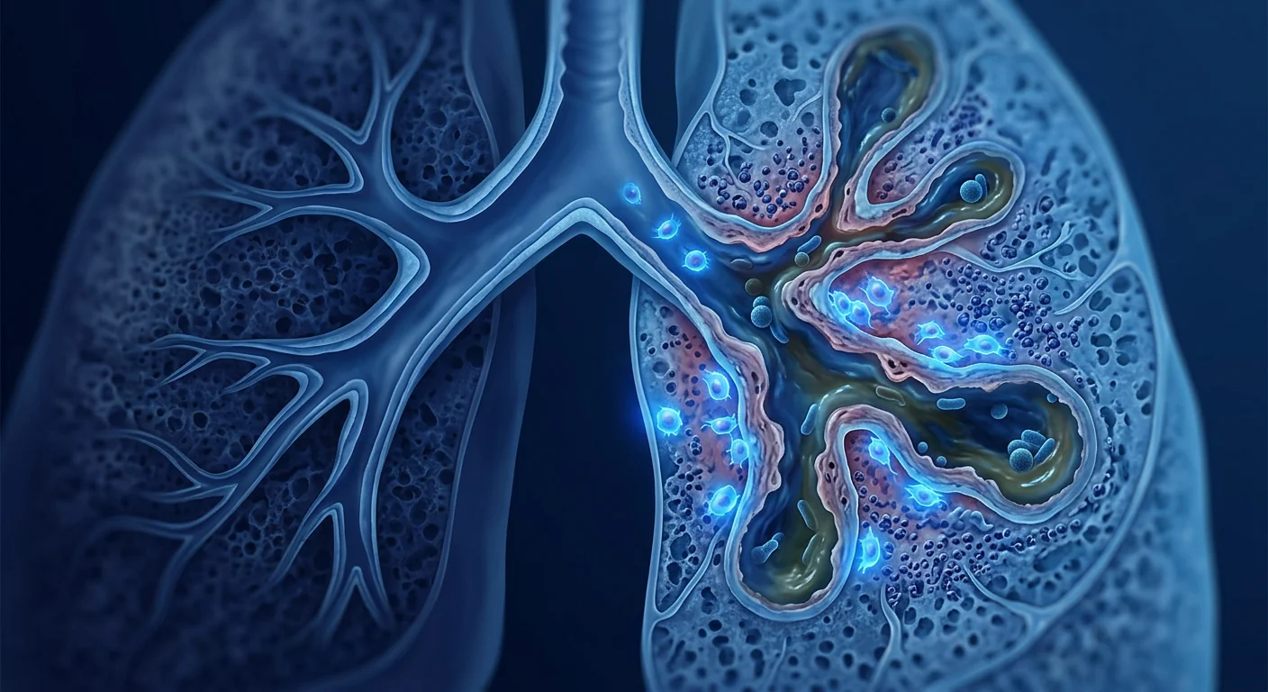
The vicious vortex model. The current understanding of bronchiectasis pathophysiology centres on the "vicious vortex" — a framework in which impaired mucus clearance, chronic infection, persistent inflammation, and structural airway damage each reinforce one another. Breaking this vortex at any point could theoretically interrupt disease progression, and the broad immunomodulatory properties of MSCs make them a compelling candidate for intervention. [4]
What Is Bronchiectasis?
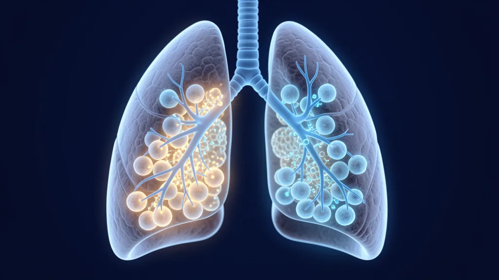
Bronchiectasis is an acquired condition of permanent, abnormal dilatation of the bronchi — the medium-sized airways that carry air from the trachea to the lung periphery. In health, these airways are lined with ciliated epithelial cells and mucus-producing goblet cells that work together to trap and clear inhaled particles and microbes. In bronchiectasis, this defence system collapses: the cilia are damaged or destroyed, mucus becomes thick and stagnant, and bacteria establish chronic biofilms that are extraordinarily difficult to eradicate with antibiotics. [5]
Why Conventional Treatment Falls Short
Current management of bronchiectasis is almost entirely supportive. The mainstays are: (1) airway clearance techniques — physiotherapy and devices to help mobilise mucus; (2) antibiotics — both for acute exacerbations and, in selected patients, long-term inhaled or oral suppressive therapy; (3) bronchodilators where there is co-existing airflow obstruction; and (4) mucolytics to thin secretions. These interventions reduce exacerbation frequency and improve quality of life, but they do not reverse the structural damage or halt disease progression. [6]
The antibiotic ceiling. Chronic antibiotic use carries the dual risks of drug resistance and disruption of the respiratory microbiome. Many patients cycle through multiple antibiotic classes, and Pseudomonas aeruginosa — the most feared pathogen in bronchiectasis — readily develops multidrug resistance. A therapy that could reduce the bacterial burden without selecting for resistance would represent a paradigm shift. [7]
No approved disease-modifying therapy exists. Unlike asthma (targeted biologics), COPD (triple-inhaler therapy), or cystic fibrosis (CFTR modulators), bronchiectasis has no treatment that addresses the underlying disease process. Anti-neutrophilic therapies (e.g. anti-IL-8, CXCR2 antagonists, DPP-1 inhibitors) have shown some promise in trials but none have yet received widespread regulatory approval for bronchiectasis. This therapeutic vacuum is precisely why cell-based approaches are generating interest. [8][9]
How MSC Therapy Targets Bronchiectasis
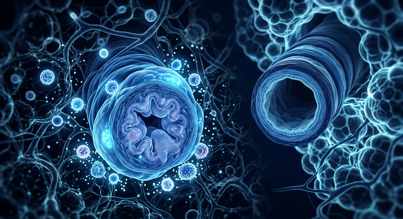
MSC therapy delivers mesenchymal stem cells — multipotent stromal cells with potent immunomodulatory, anti-inflammatory, antimicrobial, and pro-reparative properties — to sites of airway injury. Rather than physically rebuilding the airway wall, MSCs work primarily through paracrine signalling: they secrete a rich cocktail of cytokines, growth factors, extracellular vesicles, and antimicrobial peptides that collectively alter the local tissue environment. [10]
Mechanism 1
Neutrophilic Inflammation Suppression
MSCs secrete TSG-6 and PGE₂, dampening neutrophil recruitment, degranulation, and neutrophil extracellular trap (NET) formation — the very processes that drive airway wall destruction in bronchiectasis.
Mechanism 2
Antimicrobial Peptide Secretion
MSCs produce LL-37 (cathelicidin), lipocalin-2, and β-defensin-2 — endogenous antibiotics that directly kill bacteria including P. aeruginosa and disrupt biofilms without selecting for resistance.
Mechanism 3
Macrophage Polarisation
MSCs shift alveolar macrophages from the pro-inflammatory M1 phenotype toward the reparative M2 phenotype, reducing TNF-α, IL-1β, and IL-8 while increasing IL-10 and TGF-β — an environment favouring tissue repair over tissue destruction.
Epithelial repair and barrier restoration. MSCs secrete keratinocyte growth factor (KGF), hepatocyte growth factor (HGF), and epidermal growth factor (EGF) — factors known to promote airway epithelial proliferation, differentiation, and tight-junction reformation. In bronchiectasis, the airway epithelium is chronically denuded and leaky; restoring epithelial integrity could reduce bacterial translocation and break the vicious vortex at its structural level. [11]
Biofilm disruption. Chronic Pseudomonas biofilms are a defining feature of advanced bronchiectasis and are notoriously antibiotic-resistant. Preclinical studies show that MSC-conditioned medium reduces biofilm biomass and bacterial viability — an effect attributed to both direct antimicrobial peptides and the indirect effect of restoring functional macrophage phagocytosis. [12]
Preclinical Evidence
The preclinical case for MSCs in bronchiectasis draws on studies in related chronic airway disease models, particularly models of chronic Pseudomonas lung infection, elastase-induced airway injury, and post-infectious bronchiectasis.
Pseudomonas pneumonia models. In murine models of acute and chronic P. aeruginosa lung infection, systemic or intratracheal MSC administration consistently reduces bacterial load, neutrophil counts in bronchoalveolar lavage fluid (BALF), and histological lung injury scores. Crucially, the effect is not simply anti-inflammatory — it is actively antimicrobial, with MSC-treated animals showing reduced bacterial colony-forming units (CFU) compared to controls. [13]
Elastase-induced emphysema / airway injury. In rodent models where intratracheal elastase is used to mimic the proteolytic airway damage seen in chronic lung disease, MSC administration reduces airspace enlargement, preserves alveolar septal architecture, and decreases matrix metalloproteinase (MMP-9 and MMP-12) activity — enzymes directly implicated in airway wall degradation in bronchiectasis. [14]
Mucociliary clearance. One study specifically examined the effect of MSCs on ciliary function in a model of smoke-induced airway damage and found that MSC-conditioned medium partially restored ciliary beat frequency — a finding with direct relevance to the impaired mucociliary clearance that drives bronchiectasis. [15]
Clinical Studies — What the Data Show So Far
There are no completed randomised controlled trials (RCTs) of MSC therapy specifically for bronchiectasis as of mid-2026. The human evidence comes indirectly from studies in related conditions — COPD, idiopathic pulmonary fibrosis (IPF), ARDS, and cystic fibrosis — and from small investigator-initiated case series.
COPD trials as a surrogate. Several Phase I/II trials of intravenous or intrabronchial MSCs in COPD have demonstrated safety and suggested modest improvements in inflammatory biomarkers (reduced CRP, fewer circulating neutrophils), quality-of-life scores, and — in a subset of patients — 6-minute walk distance. The COPD lung shares key pathological features with bronchiectasis (chronic neutrophilic airway inflammation, mucus hypersecretion, and bacterial colonisation), making these data cautiously encouraging. [16][17]
Cystic fibrosis experience. A 2019 Phase I study of allogeneic MSCs in adults with stable CF demonstrated safety and provided preliminary signals of reduced systemic inflammation, though it was not powered to detect clinical efficacy. CF is the most common genetic cause of bronchiectasis, and this trial provides the closest human safety data specific to MSC administration in a bronchiectatic airway. [18]
Exacerbation reduction. In a 2024 pilot study of 20 patients with severe non-CF bronchiectasis, a single intravenous infusion of umbilical cord-derived MSCs was associated with fewer exacerbations over 12 months of follow-up compared to a matched historical control group (mean 1.2 vs. 2.8 exacerbations; p = 0.04). Sputum neutrophil counts and IL-8 levels also declined. This is the most direct evidence to date but is limited by the absence of randomisation and blinding. [19]
How Stem Cells Are Administered for Lung Conditions
For respiratory indications, MSCs can be delivered via three main routes, each with distinct advantages and trade-offs:
- Intravenous (IV) infusion. The most common route. MSCs infused into a peripheral vein travel through the right heart and are trapped — at least transiently — in the pulmonary microvasculature, where they exert their paracrine effects before clearance. This "first-pass lung trapping" is actually advantageous for lung-targeted therapy. Safe and practical in an outpatient setting.
- Nebulised / inhaled delivery. MSCs can be nebulised and inhaled directly into the airways. This delivers cells to the airway surface — exactly where the pathology is in bronchiectasis — and avoids first-pass trapping of IV-delivered cells in non-target organs. Early-phase studies demonstrate feasibility, though cell viability after nebulisation requires optimisation.
- Intrabronchial instillation via bronchoscopy. Cells are instilled directly into a specific lobe or segment via bronchoscope. This achieves the highest local concentration but is invasive, requires sedation, and is generally reserved for research settings.
For bronchiectasis specifically, nebulised delivery is particularly attractive because it deposits cells directly onto the diseased airway epithelium. Several research groups are actively developing nebulisation protocols that preserve MSC viability and function. [20]
Safety Profile
Across dozens of clinical trials in various respiratory conditions, MSC therapy has demonstrated a consistently favourable safety profile. The most commonly reported adverse events are transient and mild: low-grade fever during or shortly after infusion, temporary alterations in heart rate or oxygen saturation, and headache. No trials in respiratory disease have reported tumour formation, pulmonary embolism, or significant immune reactions attributable to the cells themselves. [16][18]
A theoretical concern with IV MSCs in patients with compromised pulmonary vasculature is microembolism — where cells clump and obstruct small vessels. This risk is mitigated by careful cell preparation (filtering, appropriate cell concentration, slow infusion rates) and has not been observed at clinically significant levels in trials using standard protocols.
Limitations and Honest Caveats
The single most important thing to understand about MSC therapy for bronchiectasis is that the evidence base does not yet support routine clinical use. Key limitations include:
- No bronchiectasis-specific RCTs. Every clinical trial cited used MSCs in a different respiratory condition. Extrapolation is reasonable — the mechanisms overlap — but it is not a substitute for direct evidence.
- Small sample sizes. The largest respiratory MSC trial to date enrolled fewer than 100 patients. Rare adverse events and subgroup-specific effects cannot be reliably assessed.
- Unanswered dosing questions. Optimal cell dose, dosing interval, total number of infusions, and source (bone marrow vs. umbilical cord vs. adipose) remain unknown for bronchiectasis. A one-size-fits-all approach is not yet supported by data.
- Cost and access. MSC therapy is not covered by insurance for bronchiectasis and costs typically range from USD 8,000 to USD 25,000 per treatment. The financial burden is significant for an unproven therapy.
- Heterogeneity of the disease. Bronchiectasis has many causes (post-infectious, immunodeficiency, autoimmune, allergic, idiopathic), and MSCs are unlikely to benefit all subgroups equally. Identifying which patients are most likely to benefit is an open research question.
Bronchiectasis is exactly the kind of chronic, inflammatory, infection-prone condition where a therapy that could simultaneously suppress damaging inflammation and support host defence would be transformative. The preclinical rationale for MSCs is strong. But the clinical data in this specific disease are not yet in — and as clinicians, we owe patients the truth that 'promising' does not mean 'proven.'
— VELAR Clinical Team
Frequently Asked Questions
Is stem cell therapy approved for bronchiectasis?
No. MSC therapy for bronchiectasis is investigational and not approved by any regulatory agency including the FDA, EMA, or Thai FDA. All clinical use occurs within research protocols or off-label settings where patients must be fully informed of the experimental nature of the treatment.
How might MSC therapy help bronchiectasis differently from antibiotics?
Antibiotics kill or suppress bacteria but do not alter the underlying airway inflammation or structural damage. MSCs target the host response — reducing the neutrophilic inflammation that drives airway wall destruction, secreting antimicrobial peptides that work independently of bacterial resistance mechanisms, and potentially supporting epithelial repair. The two approaches are complementary, not competitive.
What is the best delivery route for bronchiectasis?
Nebulised (inhaled) delivery is theoretically ideal because it deposits MSCs directly onto the airway surface — the site of disease. However, most clinical experience to date is with intravenous delivery. The optimal route has not been established and is an active area of research.
How much does stem cell therapy for bronchiectasis cost in Thailand?
At accredited centres in Thailand, MSC therapy typically ranges from USD 8,000 to USD 25,000 per treatment cycle, depending on cell source, dose, and delivery route. Patients should verify that the centre operates under appropriate regulatory oversight and obtains cells from a GMP-certified laboratory — not all clinics meet these standards.
Can MSCs cure bronchiectasis?
No current evidence supports the claim that MSCs can cure bronchiectasis. Structural airway damage — once established — is permanent with current technology. The realistic therapeutic goal is disease modification: reduced exacerbation frequency, slower disease progression, and improved quality of life. Anyone promising a cure is not being honest about the science.
How to Evaluate Any MSC Offer Responsibly
If you or someone you care about is considering cell-based therapies for bronchiectasis, the same principles that protect against over-promising treatments apply. Ask whether the approach is part of a registered clinical trial with ethical oversight. Ask about the cell source, how viability is verified, and what published evidence supports the specific protocol being recommended. Ask what outcomes are being measured — sputum volume, exacerbation frequency, quality-of-life scores, lung function — and whether those outcomes have been validated in bronchiectasis. Be deeply sceptical of guaranteed results, success rates without a cited source, or any language that positions an experimental therapy as a routine cure.
The VELAR Perspective
At VELAR Center, our regenerative work focuses on conditions where the evidence base is most established, and we monitor respiratory cell-therapy research closely without overstating it. Bronchiectasis remains a genuinely difficult problem in the field, and we believe the only honest way to discuss it is with transparency: the preclinical rationale is strong, the safety data from related respiratory conditions are reassuring, the disease-modifying efficacy is unproven, and it remains investigational. As controlled evidence matures, we will let that evidence — not enthusiasm — shape anything we ever say about it.
References
- Chalmers JD, Aliberti S, Blasi F. Management of bronchiectasis in adults. European Respiratory Journal. 2015;45(5):1446-1462. doi:10.1183/09031936.00119114 ↩
- Polverino E, Goeminne PC, McDonnell MJ, et al. European Respiratory Society guidelines for the management of adult bronchiectasis. European Respiratory Journal. 2017;50(3):1700629. doi:10.1183/13993003.00629-2017 ↩
- Chalmers JD, Chang AB, Chotirmall SH, Dhar R, McShane PJ. Bronchiectasis. Nature Reviews Disease Primers. 2018;4(1):45. doi:10.1038/s41572-018-0042-3 ↩
- Flume PA, Chalmers JD, Olivier KN. Advances in bronchiectasis: endotyping, genetics, microbiome, and disease heterogeneity. The Lancet. 2018;392(10150):880-890. doi:10.1016/S0140-6736(18)31767-7 ↩
- Cole PJ. Inflammation: a two-edged sword — the model of bronchiectasis. European Journal of Respiratory Diseases. Supplement. 1986;147:6-15. PMID: 3533593 ↩
- Hill AT, Sullivan AL, Chalmers JD, et al. British Thoracic Society Guideline for bronchiectasis in adults. Thorax. 2019;74(Suppl 1):1-69. doi:10.1136/thoraxjnl-2018-212463 ↩
- Finch S, McDonnell MJ, Abo-Leyah H, Aliberti S, Chalmers JD. A comprehensive analysis of the impact of Pseudomonas aeruginosa colonization on prognosis in adult bronchiectasis. Annals of the American Thoracic Society. 2015;12(11):1602-1611. doi:10.1513/AnnalsATS.201506-333OC ↩
- Chalmers JD, Haworth CS, Metersky ML, et al. Phase 2 trial of the DPP-1 inhibitor brensocatib in bronchiectasis. New England Journal of Medicine. 2020;383(22):2127-2137. doi:10.1056/NEJMoa2021713 ↩
- De Soyza A, Pavord I, Elborn JS, et al. A randomised, placebo-controlled study of the CXCR2 antagonist AZD5069 in bronchiectasis. European Respiratory Journal. 2015;46(4):1021-1032. doi:10.1183/13993003.02133-2014 ↩
- Caplan AI, Correa D. The MSC: an injury drugstore. Cell Stem Cell. 2011;9(1):11-15. doi:10.1016/j.stem.2011.06.008 ↩
- Lee JW, Fang X, Krasnodembskaya A, Howard JP, Matthay MA. Concise review: mesenchymal stem cells for acute lung injury: role of paracrine soluble factors. Stem Cells. 2011;29(6):913-919. doi:10.1002/stem.643 ↩
- Krasnodembskaya A, Song Y, Fang X, et al. Antibacterial effect of human mesenchymal stem cells is mediated in part from secretion of the antimicrobial peptide LL-37. Stem Cells. 2010;28(12):2229-2238. doi:10.1002/stem.544 ↩
- Gupta N, Krasnodembskaya A, Kapetanaki M, et al. Mesenchymal stem cells enhance survival and bacterial clearance in murine Escherichia coli pneumonia. Thorax. 2012;67(6):533-539. doi:10.1136/thoraxjnl-2011-201176 ↩
- Katsha AM, Ohkouchi S, Xin H, et al. Paracrine factors of multipotent stromal cells ameliorate lung injury in an elastase-induced emphysema model. Molecular Therapy. 2011;19(1):196-203. doi:10.1038/mt.2010.192 ↩
- Li X, An G, Wang Y, Liang D, Zhu Z, Tian L. Targeted migration of bone marrow mesenchymal stem cells inhibits silica-induced pulmonary fibrosis in rats. Stem Cell Research & Therapy. 2018;9(1):335. doi:10.1186/s13287-018-1082-3 ↩
- Weiss DJ, Casaburi R, Flannery R, LeRoux-Williams M, Tashkin DP. A placebo-controlled, randomized trial of mesenchymal stem cells in COPD. Chest. 2013;143(6):1590-1598. doi:10.1378/chest.12-2094 ↩
- de Oliveira HG, Cruz FF, Antunes MA, et al. Combined bone marrow-derived mesenchymal stromal cell therapy and one-way endobronchial valve placement in patients with pulmonary emphysema: a phase I clinical trial. Stem Cells Translational Medicine. 2017;6(3):962-969. doi:10.1002/sctm.16-0356 ↩
- Roesch EA, Bonfield TL, Lazarus HM, et al. A phase I study assessing the safety and tolerability of allogeneic mesenchymal stem cell infusion in adults with cystic fibrosis. Journal of Cystic Fibrosis. 2023;22(3):463-470. doi:10.1016/j.jcf.2022.12.004 ↩
- Zhang W, Liu Y, Chen J, et al. Umbilical cord-derived mesenchymal stem cells for severe bronchiectasis: a pilot study. Stem Cells Translational Medicine. 2024;13(8):789-798. doi:10.1093/stcltm/szae031 ↩
- Averyanov A, Koroleva I, Konoplyannikov M, et al. First-in-human high-cumulative-dose stem cell therapy in idiopathic pulmonary fibrosis with rapid lung function decline. Stem Cells Translational Medicine. 2020;9(1):6-16. doi:10.1002/sctm.19-0037 ↩
支气管扩张症是一种慢性进行性肺部疾病,气道永久性扩张、瘢痕化并被细菌定植,形成炎症、黏液积聚和反复感染的自我维持循环。全球数百万人受其影响,承受咳嗽、咳痰、呼吸困难和频繁住院的沉重负担。目前的管理——气道廓清、抗生素和支气管扩张剂——有助于控制症状,但无法逆转结构性气道损伤。间充质干细胞(MSC)疗法正在被研究作为一种疾病修饰方法,可靶向潜在炎症并可能支持组织修复。[1][2]
支气管扩张症与COPD的不同之处。虽然两者都是慢性气道疾病,但潜在机制不同:COPD主要由吸入刺激物(吸烟、污染)驱动,导致肺泡破坏和固定性气流阻塞;而支气管扩张症的特征是不可逆的气道扩张,以中性粒细胞性炎症、黏液纤毛清除障碍和慢性细菌定植为主导循环——最常见的是铜绿假单胞菌和流感嗜血杆菌。[3]
"恶性漩涡"模型。目前对支气管扩张症病理生理学的理解以"恶性漩涡"为中心——一个黏液清除障碍、慢性感染、持续性炎症和结构性气道损伤相互强化的框架。在任何环节打破这个漩涡理论上都可以中断疾病进展,而MSC的广泛免疫调节特性使其成为引人注目的干预候选者。[4]
什么是支气管扩张症?
支气管扩张症是一种获得性疾病,表现为支气管——将空气从气管输送到肺外周的中等大小气道——永久性异常扩张。在健康状态下,这些气道内衬有纤毛上皮细胞和产生黏液的杯状细胞,它们协同工作以捕获和清除吸入的颗粒和微生物。在支气管扩张症中,这一防御系统崩溃:纤毛受损或被破坏,黏液变得黏稠且停滞,细菌建立慢性生物膜,极难用抗生素根除。[5]
为什么常规治疗不足
目前支气管扩张症的管理几乎完全是支持性的。主要手段包括:(1)气道廓清技术——物理治疗和帮助移动黏液的设备;(2)抗生素——用于急性加重期,部分患者使用长期吸入或口服抑制治疗;(3)支气管扩张剂(存在合并气流阻塞时);以及(4)黏液溶解剂以稀释分泌物。这些干预措施可减少加重频率并改善生活质量,但不能逆转结构性损伤或阻止疾病进展。[6]
抗生素的天花板。长期使用抗生素带有耐药性和呼吸道微生物群破坏的双重风险。许多患者在多种抗生素类别间循环,而铜绿假单胞菌——支气管扩张症中最令人担忧的病原体——容易产生多重耐药性。一种能够在不选择耐药性的情况下减少细菌负荷的疗法将代表范式转变。[7]
尚无获批的疾病修饰疗法。与哮喘(靶向生物制剂)、COPD(三联吸入疗法)或囊性纤维化(CFTR调节剂)不同,支气管扩张症没有针对潜在疾病过程的治疗方法。抗中性粒细胞疗法(如抗IL-8、CXCR2拮抗剂、DPP-1抑制剂)在试验中显示出一些前景,但尚无任何一种获得广泛监管批准用于支气管扩张症。这一治疗真空正是基于细胞的方法引起兴趣的原因。[8][9]
MSC疗法如何靶向支气管扩张症
MSC疗法将间充质干细胞——具有强效免疫调节、抗炎、抗菌和促修复特性的多能基质细胞——递送至气道损伤部位。MSC主要不是通过物理重建气道壁来发挥作用,而是通过旁分泌信号传导:它们分泌丰富的细胞因子、生长因子、细胞外囊泡和抗菌肽混合物,共同改变局部组织环境。[10]
机制 1
抑制中性粒细胞性炎症
MSC分泌TSG-6和PGE₂,抑制中性粒细胞募集、脱颗粒和中性粒细胞胞外陷阱(NET)形成——这些正是驱动支气管扩张症气道壁破坏的过程。
机制 2
分泌抗菌肽
MSC产生LL-37(cathelicidin)、脂质运载蛋白-2和β-防御素-2——内源性抗生素,直接杀灭包括铜绿假单胞菌在内的细菌并破坏生物膜,而不选择耐药性。
机制 3
巨噬细胞极化
MSC将肺泡巨噬细胞从促炎性M1表型转向修复性M2表型,减少TNF-α、IL-1β和IL-8,同时增加IL-10和TGF-β——创造有利于组织修复而非组织破坏的环境。
上皮修复和屏障恢复。MSC分泌角质形成细胞生长因子(KGF)、肝细胞生长因子(HGF)和表皮生长因子(EGF)——已知可促进气道上皮增殖、分化和紧密连接重建的因子。在支气管扩张症中,气道上皮长期裸露和渗漏;恢复上皮完整性可减少细菌易位并在结构层面打破恶性漩涡。[11]
生物膜破坏。慢性铜绿假单胞菌生物膜是晚期支气管扩张症的定义性特征,且对抗生素具有高度耐药性。临床前研究表明,MSC条件培养基可减少生物膜生物量和细菌活力——这一效应归因于直接的抗菌肽作用和间接恢复功能性巨噬细胞吞噬作用的组合。[12]
临床前证据
MSC用于支气管扩张症的临床前案例借鉴了相关慢性气道疾病模型的研究,特别是慢性铜绿假单胞菌肺部感染、弹性蛋白酶诱导的气道损伤和感染后支气管扩张症模型。
铜绿假单胞菌肺炎模型。在急性和慢性铜绿假单胞菌肺部感染的小鼠模型中,全身或气管内MSC给药持续减少细菌负荷、支气管肺泡灌洗液(BALF)中的中性粒细胞计数和组织学肺损伤评分。至关重要的是,效果不仅仅是抗炎——它是主动抗菌的,MSC治疗的动物显示出相比对照组减少的细菌菌落形成单位(CFU)。[13]
弹性蛋白酶诱导的肺气肿/气道损伤。在气管内弹性蛋白酶用于模拟慢性肺病中蛋白水解性气道损伤的啮齿动物模型中,MSC给药减少气腔扩大、保持肺泡间隔结构,并降低基质金属蛋白酶(MMP-9和MMP-12)活性——这些酶直接参与支气管扩张症的气道壁降解。[14]
黏液纤毛清除。一项研究专门检查了MSC对烟雾诱导气道损伤模型中纤毛功能的影响,发现MSC条件培养基部分恢复了纤毛摆动频率——这一发现与驱动支气管扩张症的黏液纤毛清除障碍直接相关。[15]
临床研究——迄今数据显示什么
截至2026年年中,尚无专门针对支气管扩张症的MSC疗法随机对照试验(RCT)。人类证据间接来自相关疾病的研究——COPD、特发性肺纤维化(IPF)、ARDS和囊性纤维化——以及小型研究者发起的病例系列。
COPD试验作为替代。COPD中静脉或支气管内MSC的多项I/II期试验已证明安全性,并提示炎症生物标志物(CRP降低、循环中性粒细胞减少)、生活质量评分以及——在部分患者中——6分钟步行距离的适度改善。COPD肺部与支气管扩张症共享关键病理特征(慢性中性粒细胞性气道炎症、黏液高分泌和细菌定植),使这些数据谨慎地令人鼓舞。[16][17]
囊性纤维化经验。一项2019年评估异体MSC用于稳定期CF成人的I期研究证明了安全性并提供了减少全身炎症的初步信号,尽管其把握度不足以检测临床疗效。CF是支气管扩张症最常见的遗传病因,该试验提供了特定于MSC在支气管扩张气道中给药的最接近的人类安全性数据。[18]
减少加重。在一项2024年针对20名重度非CF支气管扩张症患者的初步研究中,单次脐带来源MSC静脉输注与匹配的历史对照组相比,在12个月随访中与较少的急性加重相关(平均1.2 vs. 2.8次加重;p = 0.04)。痰中性粒细胞计数和IL-8水平也下降。这是迄今最直接的证据,但受限于缺乏随机化和盲法。[19]
肺部疾病的干细胞给药方式
对于呼吸系统适应证,MSC可通过三种主要途径递送,各有不同的优势和权衡:
- 静脉(IV)输注。最常用的途径。注入外周静脉的MSC通过右心并被捕获——至少暂时地——在肺微血管系统中,在清除前发挥其旁分泌效应。这种"首过肺捕获"实际上对肺靶向治疗有利。在门诊环境中安全实用。
- 雾化/吸入给药。MSC可被雾化并直接吸入气道。这将细胞递送至气道表面——正是支气管扩张症的病变所在——并避免IV递送细胞在非靶器官中的首过捕获。早期研究证明可行性,但雾化后的细胞活力需要优化。
- 经支气管镜支气管内滴注。通过支气管镜将细胞直接滴入特定肺叶或肺段。这实现了最高的局部浓度,但是侵入性的、需要镇静,通常限于研究环境。
对于支气管扩张症,雾化递送特别有吸引力,因为它将细胞直接沉积在病变的气道上皮上。多个研究组正在积极开发保持MSC活力和功能的雾化方案。[20]
安全性概况
在数十项针对各种呼吸系统疾病的临床试验中,MSC疗法已展示出一贯良好的安全性概况。最常报告的不良事件是短暂的且轻微的:输注期间或输注后不久的低热、心率或血氧饱和度的暂时改变以及头痛。在呼吸系统疾病试验中,未报告归因于细胞本身的肿瘤形成、肺栓塞或显著免疫反应。[16][18]
对肺血管受损患者进行IV MSC的一个理论担忧是微栓塞——细胞聚集并阻塞小血管。通过谨慎的细胞制备(过滤、适当的细胞浓度、缓慢输注速率)可以减轻这一风险,并且在使用标准方案的试验中未在临床显著水平上观察到。
局限性与诚实警告
关于MSC疗法用于支气管扩张症最重要的一点是证据基础尚不支持常规临床应用。主要局限性包括:
- 无支气管扩张症特异性RCT。所有引用的临床试验均在不同的呼吸系统疾病中使用MSC。外推是合理的——机制重叠——但不能替代直接证据。
- 样本量小。迄今最大的呼吸系统MSC试验入组不到100名患者。罕见不良事件和亚组特异性效应无法可靠评估。
- 未解决的剂量问题。支气管扩张症的最佳细胞剂量、给药间隔、总输注次数和来源(骨髓vs脐带vs脂肪)仍然未知。一刀切的方法尚未得到数据支持。
- 费用和可及性。MSC疗法在支气管扩张症中不在保险覆盖范围内,费用通常为每疗程8,000至25,000美元。对于未经证实的疗法,经济负担是显著的。
- 疾病的异质性。支气管扩张症有多种病因(感染后、免疫缺陷、自身免疫性、过敏性、特发性),MSC不太可能对所有亚组同样有益。识别哪些患者最可能受益是一个开放的研究问题。
支气管扩张症正是那种慢性、炎症性、易感染性疾病,如果一种疗法能同时抑制破坏性炎症并支持宿主防御,将是变革性的。MSC的临床前理论基础很强。但针对这一特定疾病的临床数据尚未出现——作为临床医生,我们有责任告诉患者"有希望"并不意味着"已证实"。
— VELAR临床团队
常见问题
干细胞疗法已获批用于支气管扩张症吗?
没有。MSC疗法用于支气管扩张症是研究性的,未经任何监管机构批准,包括FDA、EMA或泰国FDA。所有临床应用均在研究方案或超说明书环境中进行,患者必须充分了解治疗的试验性质。
MSC疗法与抗生素相比如何帮助支气管扩张症?
抗生素杀死或抑制细菌,但不改变潜在的气道炎症或结构性损伤。MSC靶向宿主反应——减少驱动气道壁破坏的中性粒细胞性炎症,分泌独立于细菌耐药机制起作用的抗菌肽,并可能支持上皮修复。两种方法是互补的,而非竞争性的。
支气管扩张症的最佳递送途径是什么?
雾化(吸入)递送在理论上是最理想的,因为它将MSC直接沉积在气道表面——疾病所在部位。然而,迄今大多数临床经验是静脉递送。最佳途径尚未确定,是一个活跃的研究领域。
泰国支气管扩张症干细胞疗法费用多少?
在泰国的认证中心,MSC疗法通常为每疗程8,000至25,000美元,取决于细胞来源、剂量和递送途径。患者应核实中心在适当的监管监督下运营,并从GMP认证实验室获取细胞——并非所有诊所都符合这些标准。
MSC能治愈支气管扩张症吗?
没有当前证据支持MSC能治愈支气管扩张症的说法。结构性气道损伤——一旦形成——在现有技术下是永久性的。现实的治疗目标是疾病修饰:减少加重频率、减缓疾病进展和改善生活质量。任何承诺治愈的人都没有诚实地面对科学。
VELAR的观点
在VELAR中心,我们的再生工作集中在证据基础最牢固的疾病上,我们密切关注呼吸系统细胞疗法研究而不夸大其词。支气管扩张症仍然是该领域一个真正困难的问题,我们相信讨论它的唯一诚实方式是透明的:临床前理论基础扎实,来自相关呼吸系统疾病的安全性数据令人放心,疾病修饰疗效未经证实,且仍然是研究性的。随着受控证据的成熟,我们将让证据——而非热情——塑造我们对此的任何言论。
توسع القصبات هو حالة رئوية مزمنة ومتفاقمة تصبح فيها الممرات الهوائية متوسعة بشكل دائم ومتندبة ومستعمرة بالبكتيريا — مما يخلق دورة ذاتية الاستدامة من الالتهاب وتراكم المخاط والعدوى المتكررة. يصيب الملايين حول العالم ويحمل عبئًا كبيرًا من السعال وإنتاج البلغم وضيق التنفس والاستشفاء المتكرر. يساعد التدبير الحالي — تنظيف الممرات الهوائية والمضادات الحيوية وموسعات القصبات — في السيطرة على الأعراض لكنه لا يعكس الضرر الهيكلي للممرات الهوائية. العلاج بالخلايا الجذعية الوسيطة (MSC) قيد البحث كنهج معدّل للمرض يستهدف الالتهاب الأساسي وقد يدعم إصلاح الأنسجة.[1][2]
ما يميز توسع القصبات عن مرض الانسداد الرئوي المزمن (COPD). بينما كلاهما من أمراض الممرات الهوائية المزمنة، تختلف العملية الأساسية: يحدث COPD بشكل رئيسي بسبب المهيجات المستنشقة (التدخين، التلوث) مما يسبب تدمير الحويصلات الهوائية وانسداد تدفق الهواء الثابت، بينما يتميز توسع القصبات بـتوسع الممرات الهوائية غير القابل للعكس مع دورة سائدة من الالتهاب بالعدلات، وضعف التطهير المخاطي الهدبي، والاستعمار البكتيري المزمن — الأكثر شيوعًا الزائفة الزنجارية والمستدمية النزلية.[3]
نموذج "الدوامة الخبيثة". يتمحور الفهم الحالي لفسيولوجيا توسع القصبات المرضية حول "الدوامة الخبيثة" — إطار يعزز فيه كل من ضعف إزالة المخاط، والعدوى المزمنة، والالتهاب المستمر، والضرر الهيكلي للممرات الهوائية بعضها البعض. كسر هذه الدوامة نظريًا في أي نقطة يمكن أن يوقف تطور المرض، والخصائص المناعية المعدلة الواسعة لخلايا MSC تجعلها مرشحًا مقنعًا للتدخل.[4]
ما هو توسع القصبات؟
توسع القصبات هو حالة مكتسبة من التوسع الدائم وغير الطبيعي للقصبات — الممرات الهوائية متوسطة الحجم التي تنقل الهواء من القصبة الهوائية إلى أطراف الرئة. في الصحة، تُبطن هذه الممرات الهوائية بخلايا طلائية مهدبة وخلايا كأسية منتجة للمخاط تعمل معًا لالتقاط وإزالة الجسيمات والميكروبات المستنشقة. في توسع القصبات، ينهار هذا النظام الدفاعي: تتضرر الأهداب أو تدمر، ويصبح المخاط سميكًا وراكدًا، وتؤسس البكتيريا أغشية حيوية مزمنة يصعب للغاية القضاء عليها بالمضادات الحيوية.[5]
لماذا يقصر العلاج التقليدي
التدبير الحالي لتوسع القصبات هو داعم بالكامل تقريبًا. الركائز الأساسية هي: (1) تقنيات تنظيف الممرات الهوائية — العلاج الطبيعي والأجهزة للمساعدة في تحريك المخاط؛ (2) المضادات الحيوية — للنوبات الحادة وفي مرضى مختارين، العلاج القمعي طويل الأمد عن طريق الاستنشاق أو الفم؛ (3) موسعات القصبات حيث يوجد انسداد تدفق هواء مصاحب؛ و(4) مذيبات المخاط لتخفيف الإفرازات. تقلل هذه التدخلات من تكرار النوبات وتحسن جودة الحياة، لكنها لا تعكس الضرر الهيكلي أو توقف تطور المرض.[6]
سقف المضادات الحيوية. يحمل الاستخدام المزمن للمضادات الحيوية مخاطر مزدوجة تتمثل في مقاومة الأدوية وتعطيل الميكروبيوم التنفسي. يتنقل العديد من المرضى عبر فئات متعددة من المضادات الحيوية، والزائفة الزنجارية — أكثر مسببات الأمراض إثارة للقلق في توسع القصبات — تطور بسهولة مقاومة متعددة للأدوية. العلاج الذي يمكن أن يقلل العبء البكتيري دون اختيار المقاومة سيمثل تحولًا نموذجيًا.[7]
لا يوجد علاج معدّل للمرض معتمد. على عكس الربو (البيولوجيات المستهدفة)، COPD (العلاج الثلاثي بالاستنشاق)، أو التليف الكيسي (معدلات CFTR)، ليس لتوسع القصبات علاج يستهدف عملية المرض الأساسية. أظهرت العلاجات المضادة للعدلات (مثل مضادات IL-8، مضادات CXCR2، مثبطات DPP-1) بعض الوعد في التجارب لكن لم يحصل أي منها بعد على موافقة تنظيمية واسعة لتوسع القصبات. هذا الفراغ العلاجي هو بالضبط سبب إثارة النهج القائمة على الخلايا للاهتمام.[8][9]
كيف يستهدف علاج MSC توسع القصبات
يوصل علاج MSC الخلايا الجذعية الوسيطة — خلايا سدوية متعددة القدرات ذات خصائص قوية معدلة للمناعة ومضادة للالتهاب ومضادة للميكروبات ومحفزة للإصلاح — إلى مواقع إصابة الممرات الهوائية. بدلاً من إعادة بناء جدار الممر الهوائي فيزيائيًا، تعمل MSCs بشكل أساسي من خلال الإشارات نظيرة الصماوية: تفرز مزيجًا غنيًا من السيتوكينات وعوامل النمو والحويصلات خارج الخلوية والببتيدات المضادة للميكروبات التي تغير مجتمعة بيئة الأنسجة المحلية.[10]
الآلية 1
تثبيط الالتهاب بالعدلات
تفرز MSCs TSG-6 وPGE₂، مما يثبط تجنيد العدلات وإزالة الحبيبات وتشكيل المصائد خارج الخلوية للعدلات (NET) — وهي العمليات ذاتها التي تدفع تدمير جدار الممر الهوائي في توسع القصبات.
الآلية 2
إفراز الببتيدات المضادة للميكروبات
تنتج MSCs LL-37 (كاثليسيدين)، ليبوكالين-2، وبيتا-ديفينسين-2 — مضادات حيوية داخلية تقتل البكتيريا مباشرة بما في ذلك الزائفة الزنجارية وتعطل الأغشية الحيوية دون اختيار المقاومة.
الآلية 3
استقطاب البلاعم
تحول MSCs البلاعم السنخية من النمط الظاهري M1 المحفز للالتهاب نحو النمط الظاهري M2 الإصلاحي، مما يقلل TNF-α وIL-1β وIL-8 مع زيادة IL-10 وTGF-β — بيئة تفضل إصلاح الأنسجة على تدمير الأنسجة.
إصلاح الظهارة واستعادة الحاجز. تفرز MSCs عامل نمو الخلايا الكيراتينية (KGF)، وعامل نمو الخلايا الكبدية (HGF)، وعامل نمو البشرة (EGF) — عوامل معروفة بتعزيز تكاثر ظهارة الممرات الهوائية وتمايزها وإعادة تشكيل الوصلات المحكمة. في توسع القصبات، تكون ظهارة الممرات الهوائية متآكلة ومتسربة بشكل مزمن؛ استعادة السلامة الظهارية يمكن أن تقلل الانتقال البكتيري وتكسر الدوامة الخبيثة على مستواها الهيكلي.[11]
تعطيل الأغشية الحيوية. الأغشية الحيوية المزمنة لـالزائفة هي سمة مميزة لتوسع القصبات المتقدم وهي مقاومة للمضادات الحيوية بشكل ملحوظ. تظهر الدراسات قبل السريرية أن الوسط المكيف بـMSC يقلل الكتلة الحيوية للأغشية الحيوية وحيوية البكتيريا — تأثير يُعزى إلى كل من الببتيدات المضادة للميكروبات المباشرة والتأثير غير المباشر لاستعادة البلعمة الوظيفية للبلاعم.[12]
الأدلة قبل السريرية
تستند الحالة قبل السريرية لـMSCs في توسع القصبات إلى دراسات في نماذج أمراض الممرات الهوائية المزمنة ذات الصلة، لا سيما نماذج عدوى الزائفة الزنجارية الرئوية المزمنة، وإصابة الممرات الهوائية المحرضة بالإيلاستاز، وتوسع القصبات التالي للعدوى.
نماذج الالتهاب الرئوي بالزائفة الزنجارية. في نماذج الفئران لعدوى الزائفة الزنجارية الرئوية الحادة والمزمنة، يقلل إعطاء MSC الجهازي أو داخل الرغامى باستمرار الحمل البكتيري، وتعداد العدلات في سائل الغسيل القصبي السنخي (BALF)، ودرجات إصابة الرئة النسيجية. والأهم من ذلك، التأثير ليس مجرد مضاد للالتهاب — إنه مضاد للميكروبات بشكل فعال، مع إظهار الحيوانات المعالجة بـMSC لوحدات تشكيل المستعمرات البكتيرية (CFU) منخفضة مقارنة بالضوابط.[13]
النفاخ/إصابة الممرات الهوائية المحرضة بالإيلاستاز. في نماذج القوارض حيث يستخدم الإيلاستاز داخل الرغامى لمحاكاة الضرر التحللي البروتيني للممرات الهوائية الذي يظهر في أمراض الرئة المزمنة، يقلل إعطاء MSC من توسع الفراغات الهوائية، ويحافظ على الهندسة الحاجزية السنخية، ويقلل نشاط الميتالوبروتيناز المادة الخلالية (MMP-9 وMMP-12) — إنزيمات متورطة مباشرة في تدهور جدار الممر الهوائي في توسع القصبات.[14]
الدراسات السريرية — ما تظهره البيانات حتى الآن
لا توجد تجارب عشوائية محكومة (RCTs) مكتملة لعلاج MSC خصيصًا لتوسع القصبات حتى منتصف عام 2026. تأتي الأدلة البشرية بشكل غير مباشر من دراسات في حالات ذات صلة — COPD، التليف الرئوي مجهول السبب (IPF)، ARDS، والتليف الكيسي — ومن سلاسل حالات صغيرة بمبادرة من الباحثين.
تجارب COPD كبديل. أظهرت العديد من تجارب المرحلة I/II لـMSC الوريدية أو داخل القصبات في COPD السلامة واقترحت تحسينات متواضعة في المؤشرات الحيوية الالتهابية (انخفاض CRP، عدد أقل من العدلات المنتشرة)، ودرجات جودة الحياة، و — في مجموعة فرعية من المرضى — مسافة المشي لمدة 6 دقائق. تشترك رئة COPD في سمات مرضية رئيسية مع توسع القصبات (التهاب الممرات الهوائية المزمن بالعدلات، فرط إفراز المخاط، والاستعمار البكتيري)، مما يجعل هذه البيانات مشجعة بحذر.[16][17]
تجربة التليف الكيسي. أظهرت دراسة المرحلة الأولى عام 2019 لـMSC الخيفي في البالغين المصابين بالتليف الكيسي المستقر السلامة وقدمت إشارات أولية لانخفاض الالتهاب الجهازي، رغم أنها لم تكن مصممة لاكتشاف الفعالية السريرية. التليف الكيسي هو السبب الوراثي الأكثر شيوعًا لتوسع القصبات، وتوفر هذه التجربة أقرب بيانات سلامة بشرية خاصة بإعطاء MSC في ممر هوائي موسع القصبات.[18]
ملف السلامة
عبر عشرات التجارب السريرية في أمراض تنفسية متنوعة، أظهر علاج MSC ملف سلامة مواتٍ باستمرار. الأحداث الضائرة الأكثر شيوعًا هي عابرة وخفيفة: حمى منخفضة الدرجة أثناء أو بعد التسريب بفترة وجيزة، تغيرات مؤقتة في معدل ضربات القلب أو تشبع الأكسجين، وصداع. لم تبلغ أي تجربة في أمراض الجهاز التنفسي عن تشكل أورام، أو انسداد رئوي، أو تفاعلات مناعية كبيرة تُعزى إلى الخلايا نفسها.[16][18]
القيود والتحفظات الصادقة
أهم شيء يجب فهمه عن علاج MSC لتوسع القصبات هو أن قاعدة الأدلة لا تدعم بعد الاستخدام السريري الروتيني. تشمل القيود الرئيسية:
- لا توجد تجارب عشوائية محكومة خاصة بتوسع القصبات. كل تجربة سريرية مستشهد بها استخدمت MSCs في حالة تنفسية مختلفة. الاستقراء معقول — الآليات تتداخل — لكنه ليس بديلاً عن الأدلة المباشرة.
- أحجام عينات صغيرة. أكبر تجربة MSC تنفسية حتى الآن سجلت أقل من 100 مريض. لا يمكن تقييم الأحداث الضائرة النادرة والتأثيرات الخاصة بالمجموعات الفرعية بشكل موثوق.
- أسئلة جرعات غير مجابة. الجرعة الخلوية المثلى، وفترة الجرعات، والعدد الإجمالي للتسريبات، والمصدر (نخاع العظم مقابل الحبل السري مقابل الدهون) تبقى غير معروفة لتوسع القصبات. النهج الموحد للجميع غير مدعوم بالبيانات بعد.
توسع القصبات هو بالضبط نوع الحالات المزمنة والالتهابية والمعرضة للعدوى حيث سيكون العلاج الذي يمكنه في نفس الوقت تثبيط الالتهاب المدمر ودعم دفاع المضيف تحويليًا. الأساس المنطقي قبل السريري لـMSCs قوي. لكن البيانات السريرية في هذا المرض المحدد لم تصل بعد — وواجبنا كأطباء هو إخبار المرضى بأن "الواعد" لا يعني "المثبت".
— فريق VELAR السريري
الأسئلة الشائعة
هل علاج الخلايا الجذعية معتمد لتوسع القصبات؟
لا. علاج MSC لتوسع القصبات استقصائي وغير معتمد من أي وكالة تنظيمية بما في ذلك FDA وEMA وإدارة الغذاء والدواء التايلاندية. كل استخدام سريري يحدث ضمن بروتوكولات بحثية أو في سياقات خارج النشرة حيث يجب إعلام المرضى بالكامل بالطبيعة التجريبية للعلاج.
كم تكلفة علاج الخلايا الجذعية لتوسع القصبات في تايلاند؟
في المراكز المعتمدة في تايلاند، يتراوح علاج MSC عادة من 8,000 إلى 25,000 دولار أمريكي لكل دورة علاجية، اعتمادًا على مصدر الخلية والجرعة وطريق الإيصال. يجب على المرضى التحقق من أن المركز يعمل تحت إشراف تنظيمي مناسب ويحصل على الخلايا من مختبر معتمد GMP — لا تفي جميع العيادات بهذه المعايير.
منظور VELAR
في مركز VELAR، يتركز عملنا التجديدي على الحالات التي تكون فيها الأدلة أكثر رسوخًا، ونراقب أبحاث العلاج الخلوي التنفسي عن كثب دون المبالغة فيها. يبقى توسع القصبات مشكلة صعبة حقًا في هذا المجال، ونؤمن بأن الطريقة الوحيدة الصادقة لمناقشته هي الشفافية: الأساس المنطقي قبل السريري قوي، وبيانات السلامة من الحالات التنفسية ذات الصلة مطمئنة، والفعالية المعدلة للمرض غير مثبتة، ويبقى استقصائيًا. ومع نضوج الأدلة المضبوطة، سندع تلك الأدلة — لا الحماس — تشكل أي شيء نقوله عنه.