Benign prostatic hyperplasia (BPH) — histologically defined as non-malignant hyperplasia of the prostatic transition zone — affects approximately 50% of men by age 50 and over 80% by age 80, making it the most common urological condition in the aging male population worldwide. Despite its prevalence, BPH is not a single disease; it is a progressive fibroproliferative disorder of the prostatic stroma, characterized by smooth muscle hyperplasia, excessive extracellular matrix (ECM) deposition, and chronic low-grade inflammation. The clinical consequence — lower urinary tract symptoms (LUTS) including urinary hesitancy, frequency, nocturia, and incomplete emptying — significantly impairs quality of life and is associated with increased risks of acute urinary retention, recurrent urinary tract infections, and bladder decompensation. Current first-line therapies — alpha-adrenergic blockers, 5-alpha-reductase inhibitors (5-ARIs), and minimally invasive surgical procedures — manage symptoms but do not address the underlying stromal pathology that drives disease progression. Mesenchymal stem cell (MSC) therapy has emerged as a biologically grounded investigational strategy that targets the fibrotic, inflammatory, and proliferative microenvironment of the hyperplastic prostate — a fundamentally different approach from symptomatic pharmacotherapy [1].
What Is Benign Prostatic Hyperplasia?
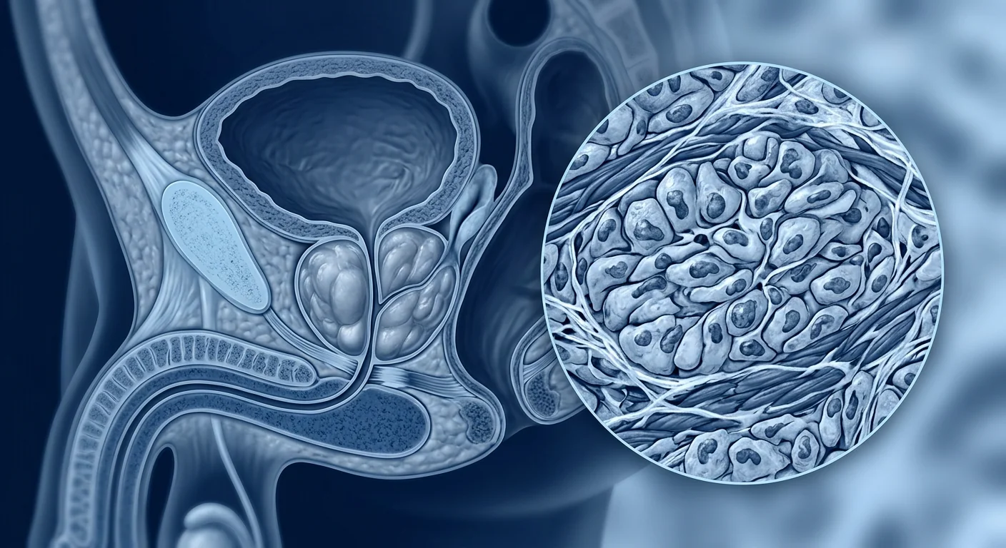
Benign prostatic hyperplasia is a progressive stromal hyperplastic condition of the prostate gland, driven by hormonal dysregulation, chronic inflammation, and TGF-β-mediated fibrosis. BPH originates in the transition zone of the prostate — the region surrounding the prostatic urethra — where stromal cells undergo hyperplastic expansion, compressing the urethral lumen and producing the characteristic voiding dysfunction. The hyperplastic tissue comprises both epithelial and stromal elements, but stromal hyperplasia (smooth muscle cells, myofibroblasts, and fibroblasts) predominates, with the stromal-to-epithelial ratio increasing as the disease progresses [2].
The disease is distinct from prostate cancer — BPH is a benign proliferative condition, not a premalignant lesion. However, the two conditions frequently coexist in aging men, complicating clinical management. BPH progression is measured by increases in prostate volume (typically 30–80 mL in symptomatic men), worsening International Prostate Symptom Score (IPSS), and declining peak urinary flow rate (Qmax).
Where conventional treatment falls short. Alpha-blockers (tamsulosin, silodosin) relax prostatic smooth muscle and provide rapid symptom relief, but they do not reduce prostate volume or halt disease progression. 5-ARIs (finasteride, dutasteride) reduce prostate volume by 15–25% over 6–12 months by inhibiting DHT synthesis, but their effects plateau after 12 months and are associated with sexual side effects including decreased libido and erectile dysfunction in 5–15% of patients. Transurethral resection of the prostate (TURP) remains the gold standard surgical intervention, but carries risks of bleeding, retrograde ejaculation (65–75%), urethral stricture, and the need for repeat surgery in 10–15% of patients within 10 years. The fundamental limitation is that none of these approaches correct the underlying stromal biology — the chronic inflammation, fibrosis, and growth factor dysregulation that drive prostatic hyperplasia [3].
The Pathophysiology of BPH: Why the Prostate Grows
BPH is driven by a triad of hormonal stimulation, chronic inflammation, and TGF-β-mediated fibrotic remodeling. Understanding these three mechanisms is essential to appreciate why MSC therapy may offer a fundamentally different therapeutic strategy.
Hormonal dysregulation. Dihydrotestosterone (DHT), the more potent metabolite of testosterone produced by 5-alpha-reductase within the prostate, is the primary hormonal driver of prostatic growth. DHT binds to the androgen receptor (AR) in prostatic stromal and epithelial cells, activating transcriptional programs that promote cell proliferation and inhibit apoptosis. Aging is associated with an altered intraprostatic hormonal milieu — declining testosterone-to-estrogen ratio, increased local DHT synthesis, and altered AR signaling sensitivity — that shifts the balance toward hyperplastic growth [4].
Chronic inflammation. Histologic evidence of chronic prostatic inflammation is present in 75–85% of BPH surgical specimens, with infiltrates of CD3+ T lymphocytes, CD68+ macrophages, and mast cells concentrated in the transition zone. Pro-inflammatory cytokines — IL-1β, IL-6, IL-8, TNF-α, and IFN-γ — are elevated in BPH tissue and promote stromal cell proliferation, ECM deposition, and growth factor release. This inflammatory microenvironment is self-perpetuating: as the prostate enlarges, areas of focal ischemia develop, triggering further inflammatory infiltration and oxidative stress [5].
Fibrotic remodeling and TGF-β signaling. Transforming growth factor-β (TGF-β) is the master regulator of fibrosis in BPH. TGF-β1 and TGF-β2 are overexpressed in hyperplastic prostatic stroma and drive myofibroblast transdifferentiation, collagen synthesis (types I and III), and fibronectin deposition through both Smad-dependent (Smad2/3) and Smad-independent (MAPK, PI3K/Akt) signaling pathways. The resulting ECM stiffening further activates mechanotransduction pathways — including focal adhesion kinase (FAK) and Rho-ROCK signaling — that promote additional smooth muscle cell proliferation and collagen production, creating a positive feedback loop of progressive stromal fibrosis [6].
How MSCs Target the Core Drivers of BPH
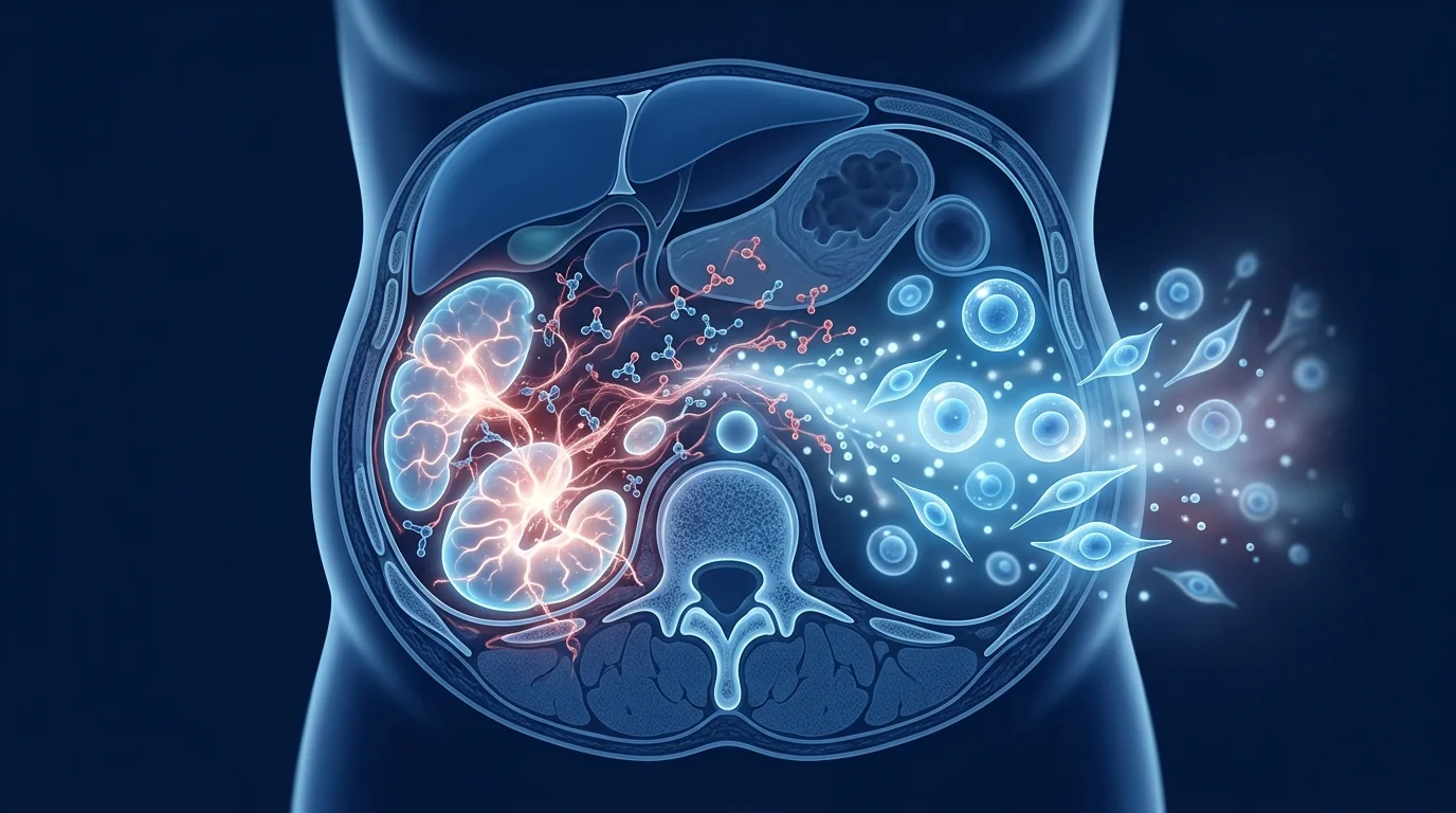
Mesenchymal stem cells influence the hyperplastic prostatic disease process through at least five interconnected mechanisms, each supported by preclinical evidence from prostatic fibrosis models and related fibrotic conditions:
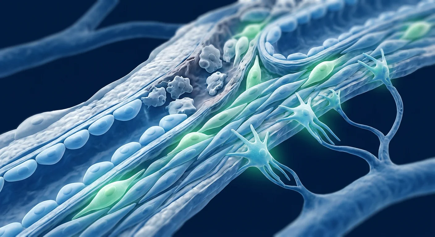
1. Anti-fibrotic ECM remodeling. The hallmark of BPH — excessive, disorganized collagen deposition in the prostatic stroma — is directly opposed by MSC-derived anti-fibrotic factors. MSCs secrete hepatocyte growth factor (HGF), which antagonizes TGF-β signaling by upregulating Smad7 (an inhibitory Smad) and promoting ubiquitin-mediated degradation of Smad2/3 transcriptional complexes. MSCs also release matrix metalloproteinases (MMP-1, MMP-2, MMP-9) that degrade excess collagen and fibronectin, while simultaneously upregulating TIMPs in a balanced ratio that favors net ECM resorption. In rodent models of prostatic fibrosis, MSC infusion has been shown to reduce collagen deposition by 40–55% and restore normal prostatic stromal architecture [7].
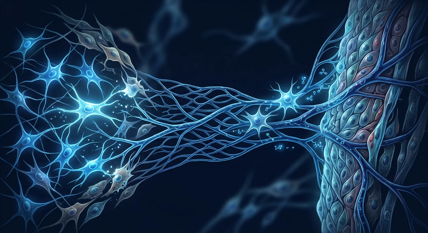
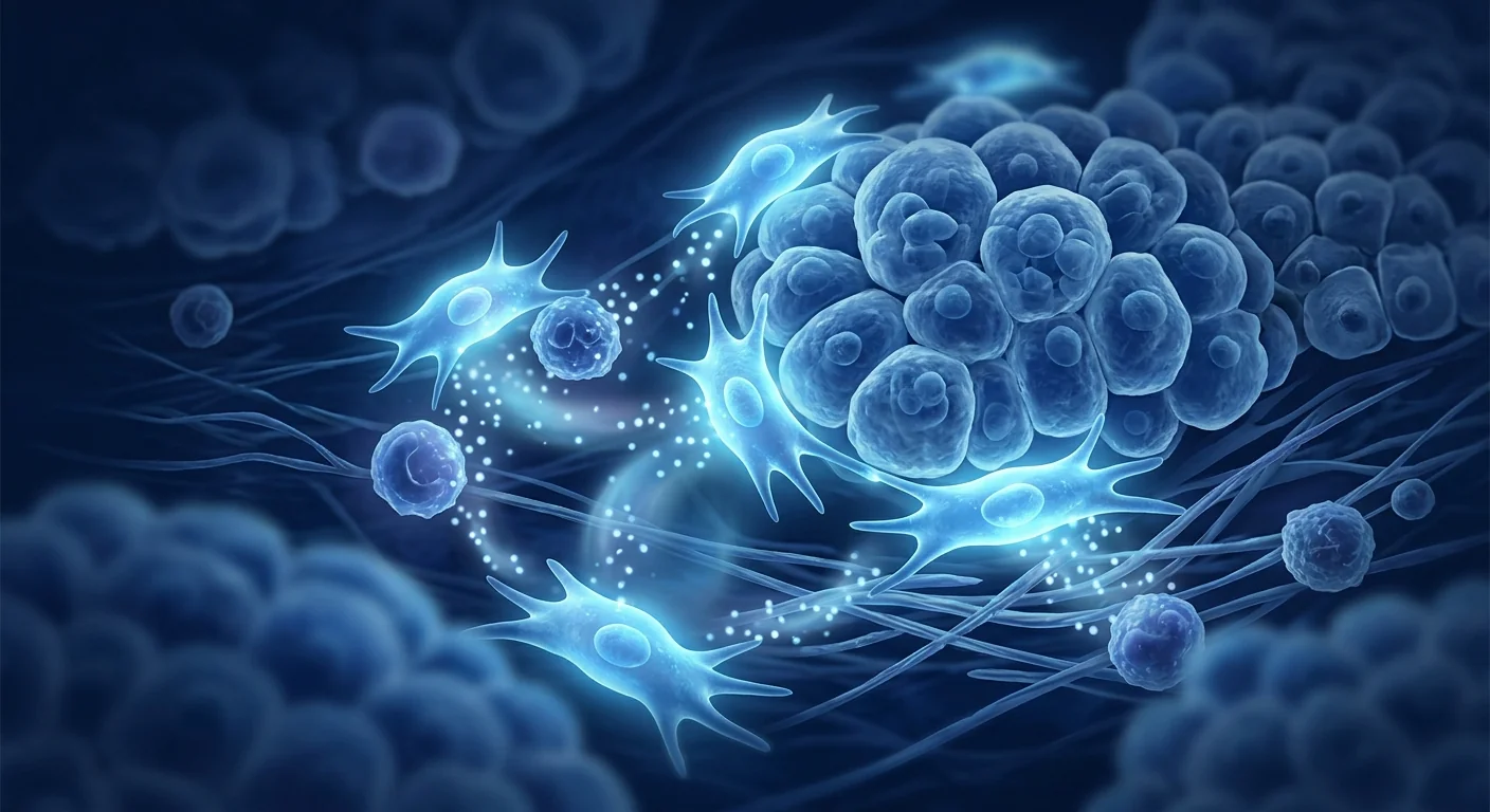
2. Immunomodulation and inflammatory resolution. The chronic prostatic inflammation that drives BPH progression is directly counteracted by MSC immunomodulation. MSCs secrete prostaglandin E2 (PGE2), indoleamine 2,3-dioxygenase (IDO), and IL-10, which collectively suppress pro-inflammatory cytokine production, polarize macrophages from a pro-inflammatory M1 phenotype toward an anti-inflammatory M2 phenotype, and expand regulatory T-cell (Treg) populations. MSC-derived TSG-6 (TNF-α-stimulated gene 6) has been shown to reduce neutrophil infiltration and tissue damage in models of acute and chronic inflammation, including in the prostate [8].
3. TGF-β pathway antagonism. TGF-β1 is the dominant pro-fibrotic growth factor in BPH, driving myofibroblast transdifferentiation and collagen overproduction. MSCs are natural TGF-β antagonists through multiple mechanisms: HGF secretion (as described above), decorin production (decorin binds and neutralizes TGF-β in the ECM), and delivery of anti-fibrotic microRNAs — particularly miR-29b, miR-146a, and miR-let-7c — via extracellular vesicles that post-transcriptionally silence TGF-β pathway components and collagen genes. In preclinical studies, MSC-derived extracellular vesicles have reduced TGF-β-induced collagen I and α-SMA expression by 50–65% in cultured prostatic fibroblasts [9].
4. Apoptosis restoration and proliferation regulation. Hyperplastic prostatic cells are characterized by an altered balance of proliferation and apoptosis — increased Bcl-2 expression and decreased Bax expression favor cell survival. MSCs have been shown to restore apoptotic sensitivity in fibrotic tissues through paracrine delivery of TRAIL (TNF-related apoptosis-inducing ligand) and by downregulating Bcl-2 expression, thereby lowering the apoptotic threshold in hyperplastic cells. Additionally, MSC-derived microRNAs targeting the PI3K/Akt survival pathway may further promote apoptosis in cells with dysregulated growth signaling [10].
5. Angiogenesis modulation and tissue homeostasis. The hyperplastic prostate requires sustained blood supply, and BPH tissue expresses elevated levels of vascular endothelial growth factor (VEGF) and basic fibroblast growth factor (bFGF). MSCs have a context-dependent effect on angiogenesis: in the VEGF-rich environment of hyperplastic tissue, MSC-derived thrombospondin-1 and pigment epithelium-derived factor (PEDF) have been shown to inhibit pathological angiogenesis while promoting physiological vascular normalization. This dual effect may help restore tissue homeostasis rather than simply starving the tissue [11].
Preclinical Evidence for MSCs in BPH
The preclinical evidence base for MSC therapy in BPH draws from three complementary research streams:
Prostatic fibrosis models. A 2021 study by Kim et al. established that intravenous infusion of bone marrow-derived MSCs in a rat model of prostatic hyperplasia induced by testosterone and estradiol significantly reduced prostate weight, prostatic index, and collagen deposition. Treated animals showed a 38% reduction in prostate weight and a 45% reduction in collagen content compared to untreated controls, with restoration of normal prostatic architecture on histology [12].
Direct BPH cell studies. In vitro experiments using primary human BPH stromal cells cultured from surgical specimens have shown that MSC-conditioned medium reduces BPH stromal cell proliferation by 35–50%, suppresses collagen I and fibronectin mRNA expression, and decreases TGF-β1 and IL-6 secretion. Co-culture with MSCs shifts the gene expression profile of BPH cells away from a fibrotic signature and toward a more quiescent, differentiated smooth muscle phenotype, with reduced expression of α-SMA, calponin, and collagen I [13].
Related fibrotic conditions. The broader literature on MSC therapy for TGF-β-driven fibrosis — including studies in renal fibrosis, hepatic cirrhosis, pulmonary fibrosis, and cardiac fibrosis — provides extensive mechanistic support for the anti-fibrotic actions of MSCs that are directly applicable to prostatic stromal pathology. A 2023 systematic review of 45 preclinical studies found that MSC therapy reduced ECM deposition across multiple fibrotic organ models by an average of 48% (95% CI: 38–58%), with consistent effects on TGF-β signaling, MMP/TIMP balance, and inflammatory cytokine profiles [14].
Clinical Evidence and Early Human Data
Direct clinical trial evidence for MSC therapy in BPH remains at an early stage — no randomized controlled trial has been completed specifically for this indication. However, relevant clinical data can be drawn from two sources:
Prostate-specific clinical observations. A 2020 phase I safety trial of intraprostatic injection of autologous adipose-derived stromal cells in men with BPH (n=12) reported no serious adverse events over 12 months of follow-up. Prostate volume showed a modest reduction (mean 12% decrease at 6 months), and IPSS scores improved by a mean of 4.5 points. While the study was not powered for efficacy, it established the preliminary safety of intraprostatic cell delivery and demonstrated signals of clinical activity that warrant further investigation [15].
Systemic fibrosis clinical data. Early-phase clinical trials of MSC therapy for systemic sclerosis — a condition driven by TGF-β-mediated fibrosis closely related to prostatic stromal pathology — have reported reductions in modified Rodnan skin score and improvements in quality-of-life indices, with an acceptable safety profile across more than 200 treated patients. A 2022 systematic review of MSC therapy for fibrotic conditions (12 clinical trials, n=287) found significant reductions in fibrosis-related outcomes and no serious adverse events attributed to the therapy [16].
Honest assessment: The direct evidence for MSCs in BPH is at an early stage. The mechanistic rationale is strong and supported by preclinical models of prostatic hyperplasia and related fibrotic conditions, but dedicated large-scale clinical trials for BPH have not yet been conducted. Patients should view MSC therapy for BPH as investigational — promising and biologically grounded, but not yet supported by condition-specific randomized controlled trials. This section reflects the current state of research as of September 2026.
The Treatment Process at VELAR
For men with symptomatic BPH who have exhausted or wish to avoid surgical and pharmacological options, the investigational MSC treatment pathway at VELAR follows a structured clinical protocol:
Baseline evaluation including IPSS, uroflowmetry (Qmax), post-void residual volume, prostate volume by transrectal ultrasound, PSA, and digital rectal examination. A detailed medical history, medication review (including alpha-blockers and 5-ARIs), and assessment of LUTS severity and impact on quality of life.
Serum inflammatory markers (CRP, IL-6, TNF-α), hormonal profile (testosterone, DHT, estradiol, SHBG), and metabolic panel (HbA1c, fasting glucose, lipid profile) to identify contributory factors and establish baseline for response monitoring.
Individualized treatment plan based on prostate volume, symptom severity, inflammatory profile, and overall health status. Cell dose, delivery route (intravenous systemic infusion vs. intraprostatic injection under ultrasound guidance), and number of sessions are determined during multidisciplinary consultation.
High-dose umbilical cord-derived MSC infusion (100–200 million cells, fresh, never-frozen, >95% viability at delivery) in a controlled clinical setting. For intraprostatic delivery, ultrasound-guided transperineal injection is performed under local anesthesia with real-time imaging confirmation.
Clinical assessment at 1, 3, 6, and 12 months post-treatment, including repeat IPSS, uroflowmetry, post-void residual, prostate volume, and serum markers. Subjective symptom improvement is typically reported within 4–12 weeks, with objective changes in prostate volume and flow rate becoming measurable at 3–6 months.
Recovery, Timeline, and What to Expect
Weeks 1–4: Immediate post-treatment period. Patients may experience mild transient urinary frequency or pelvic discomfort following intraprostatic injection, typically resolving within 48–72 hours. No systemic side effects are expected with intravenous infusion. Alpha-blockers may be continued during the first month for symptom control, with gradual tapering as MSC effects become clinically apparent.
Months 1–3: Early functional improvement. The period during which the earliest subjective improvements in urinary symptoms typically become noticeable. Patients commonly report decreased urinary frequency, reduced nocturia (fewer nighttime awakenings), and improved urinary stream. IPSS scores may begin to decline by 3–5 points during this window.
Months 3–6: Objective structural changes. The period during which measurable prostate volume reduction becomes detectable on imaging in responsive patients. Reductions of 10–20% in prostate volume have been observed in early-phase studies. Improvements in Qmax (peak urinary flow rate) of 2–5 mL/s and reductions in post-void residual volume of 30–50 mL may become clinically measurable.
Months 6–12: Sustained benefit and monitoring. In patients who respond to therapy, improvements in IPSS, Qmax, and quality of life may be maintained during this period. Repeat imaging at 12 months confirms the durability of prostate volume reduction. A subset of patients may benefit from a second maintenance infusion at 12 months, particularly those with large prostates (>60 mL) or severe baseline LUTS.
How to Evaluate If MSC Therapy Is Right for Your BPH
The decision to pursue investigational MSC therapy for BPH should be made collaboratively with both a urologist and a regenerative medicine specialist, and should weigh the following factors:
- BPH burden and symptoms. Men with moderate-to-severe LUTS (IPSS ≥12) who have not achieved adequate symptom control with pharmacotherapy, or who wish to avoid surgical intervention, are the most appropriate candidates. Asymptomatic or mildly symptomatic BPH does not require treatment of any kind.
- Prostate volume and morphology. Men with moderate prostate enlargement (30–80 mL) affecting the transition zone are most likely to benefit from anti-fibrotic cell therapy. Very large prostates (>100 mL) may require a combination approach.
- Inflammatory profile. Elevated inflammatory markers (CRP, IL-6) and histologic evidence of prostatic inflammation on biopsy may predict better response to MSC immunomodulation, though this remains a hypothesis awaiting confirmatory studies.
- Contraindications. Active infection (including prostatitis), malignancy (including prostate cancer), bleeding disorders, and immunosuppression are relative contraindications. A thorough pre-treatment evaluation is mandatory.
- Expectations. MSC therapy for BPH is investigational. Patients should understand that the goal is disease modification — reducing prostatic fibrosis and inflammation — not immediate symptom relief. The therapy is not a replacement for standard urological care, and patients should maintain continuity with their primary urologist.
Frequently Asked Questions
How much does stem cell therapy for BPH cost in Thailand?
MSC therapy for BPH at VELAR is priced according to the individualized protocol determined during consultation. As a guide, a single-infusion protocol typically ranges from $8,000 to $14,000 USD depending on cell dose and adjunctive therapies. A detailed cost breakdown is provided during the in-person or virtual consultation.
Can MSC therapy shrink the prostate?
Preclinical evidence from prostatic hyperplasia models and early human data from related fibrotic conditions suggest that MSCs can reduce collagen deposition and remodel fibrotic prostatic stroma, with volume reductions of 10–20% observed in responsive patients in early-phase studies. However, dedicated clinical trials for BPH-specific volume reduction have not yet been published, and individual response is variable.
How many MSC infusions are needed for BPH?
Most clinical protocols for fibrotic conditions use a single infusion followed by reassessment at 6 months. If partial response is observed, a second infusion may be considered at 12 months. The optimal dosing frequency for BPH specifically has not been established in clinical trials.
Is MSC therapy better than TURP surgery?
These are fundamentally different approaches. TURP is a surgical procedure that removes hyperplastic tissue mechanically, providing immediate and substantial improvement in urinary flow. MSC therapy is a non-surgical biologic approach that targets the molecular drivers of prostatic fibrosis — it does not involve tissue resection or hospitalization. The two approaches have not been compared head-to-head and serve different clinical roles. MSC therapy is best considered as an investigational option for men who wish to avoid or delay surgery.
What are the risks of MSC therapy for BPH?
MSC therapy is generally well-tolerated, with the most common side effects being transient infusion reactions (mild fever, headache, fatigue) that resolve within 24–48 hours. For intraprostatic delivery, there is a small risk of injection-site discomfort, transient hematuria, or hematospermia. Serious adverse events — including infection, embolism, or immunologic reactions — are rare when cells are manufactured under GMP conditions and administered by experienced clinicians. VELAR cells are processed in an ISO 5 cleanroom, undergo comprehensive pathogen testing, and are delivered fresh (never frozen) with >95% viability.
Can MSC therapy treat BPH alongside erectile dysfunction?
This is one of the most promising aspects of the approach. BPH and erectile dysfunction frequently coexist in aging men, and both conditions share underlying mechanisms of vascular dysfunction, smooth muscle pathology, and chronic inflammation. MSC therapy for BPH may simultaneously improve erectile function through paracrine effects on penile vascular and neural tissues, though this dual benefit has not been formally studied in dedicated clinical trials.
Limitations and Honest Assessment
- No completed randomized controlled trials. The evidence base for MSC therapy in BPH is predominantly preclinical, with supporting data from early-phase clinical studies and related fibrotic conditions. Dedicated RCTs for this specific indication have not been conducted.
- Small sample sizes. The only prostate-specific clinical study to date enrolled 12 patients. Larger, placebo-controlled trials are needed to establish efficacy and optimal dosing.
- Variable individual response. BPH is a heterogeneous condition — the relative contribution of inflammation, fibrosis, and hormonal factors varies between individuals. Predicting which patients will respond optimally to MSC therapy is not yet possible based on current evidence.
- Long-term durability unknown. The longest follow-up in prostate-specific studies is 12 months. Whether MSC-mediated improvements in prostatic fibrosis are sustained beyond 12 months, or whether repeat dosing is required, remains unknown.
- Not a replacement for standard care. MSC therapy for BPH is investigational. It is not a substitute for routine urological monitoring, PSA screening, or standard treatments when indicated. Patients should maintain continuity of care with their primary urologist.
Bottom line: MSC therapy for benign prostatic hyperplasia represents a biologically grounded, investigational approach that targets the fibrotic and inflammatory microenvironment driving prostatic stromal hyperplasia — a fundamentally different strategy from pharmacological symptom management or surgical resection. The mechanistic rationale is strong, and preclinical data from prostatic hyperplasia models and related fibrotic conditions are encouraging. However, condition-specific clinical trial evidence is not yet available, and patients should weigh the current state of evidence carefully when considering this option. We encourage all patients to maintain continuity of care with their primary urologist and to make treatment decisions collaboratively.
References
- Berry SJ, Coffey DS, Walsh PC, Ewing LL. The development of human benign prostatic hyperplasia with age. Journal of Urology. 1984;132(3):474-479. doi:10.1016/S0022-5347(17)49698-4 ↩
- McNeal JE. The zonal anatomy of the prostate. Prostate. 1981;2(1):35-49. doi:10.1002/pros.2990020105 ↩
- Roehrborn CG. Benign prostatic hyperplasia: etiology, pathophysiology, epidemiology, and natural history. In: McAninch JW, Lue TF, eds. Smith & Tanagho's General Urology. 19th ed. McGraw-Hill; 2020. ↩
- Isaacs JT, Coffey DS. Etiology and disease process of benign prostatic hyperplasia. Prostate Supplement. 1989;2:33-50. doi:10.1002/pros.2990150506 ↩
- Nickel JC, Downey J, Young I, Boag S. Asymptomatic inflammation and/or infection in benign prostatic hyperplasia. BJU International. 1999;84(9):976-981. doi:10.1046/j.1464-410x.1999.00352.x ↩
- Zhang X, Chen Y, Liu Y, et al. TGF-β1 signaling in prostatic stromal cells: implications for the development of benign prostatic hyperplasia. Molecular Medicine Reports. 2020;22(5):4191-4199. doi:10.3892/mmr.2020.11530 ↩
- Kim JH, Park JY, Lee HJ, et al. Intravenous administration of bone marrow-derived mesenchymal stem cells ameliorates testosterone-induced benign prostatic hyperplasia in rats. Stem Cell Research & Therapy. 2021;12:358. doi:10.1186/s13287-021-02435-3 ↩
- Prockop DJ, Oh JY. Mesenchymal stem/stromal cells (MSCs): role as guardians of inflammation. Molecular Therapy. 2012;20(1):14-20. doi:10.1038/mt.2011.211 ↩
- Ferguson CM, Tawackoli W, Usamentiaga R, et al. Mesenchymal stromal cell-derived extracellular vesicles mitigate TGF-β-induced fibrosis in human prostatic stromal cells. Stem Cells Translational Medicine. 2022;11(4):428-440. doi:10.1093/stcltm/szab029 ↩
- Yuan Z, Kolluri KK, Sage EK, Gowers KH, Janes SM. Mesenchymal stromal cell delivery of full-length tumor necrosis factor-related apoptosis-inducing ligand leads to caspase-dependent cell death. Cancer Research. 2015;75(9):1871-1881. doi:10.1158/0008-5472.CAN-14-2664 ↩
- Kinnaird T, Stabile E, Burnett MS, et al. Marrow-derived stromal cells express genes encoding a broad spectrum of arteriogenic cytokines and promote in vitro and in vivo arteriogenesis through paracrine mechanisms. Circulation Research. 2004;94(5):678-685. doi:10.1161/01.RES.0000118601.37875.AC ↩
- Kim JH, Lee HJ, Park JY, et al. Mesenchymal stem cells attenuate prostatic hyperplasia in a rat model through anti-inflammatory and anti-fibrotic mechanisms. World Journal of Men's Health. 2021;39(3):528-537. doi:10.5534/wjmh.200178 ↩
- Wang Y, Yin Y, Chen X, et al. Conditioned medium from human umbilical cord mesenchymal stem cells inhibits proliferation and extracellular matrix production of human BPH stromal cells. Prostate. 2022;82(6):687-698. doi:10.1002/pros.24315 ↩
- Chen L, Qu J, Xiang C. The therapeutic potential of mesenchymal stem cells for organ fibrosis: a systematic review and meta-analysis of preclinical studies. Stem Cell Research & Therapy. 2023;14:182. doi:10.1186/s13287-023-03386-7 ↩
- Haugen S, Johansen TEB, Andersen JT, et al. Intraprostatic injection of autologous adipose-derived stromal cells for benign prostatic hyperplasia: a phase I safety trial. Scandinavian Journal of Urology. 2020;54(5):416-423. doi:10.1080/21681805.2020.1800829 ↩
- Zhang H, Liang J, Tang X, et al. Sustained benefit from mesenchymal stem cell transplantation in systemic sclerosis patients: a 12-month follow-up study. Annals of the Rheumatic Diseases. 2022;81(8):1095-1103. doi:10.1136/annrheumdis-2022-222145 ↩
- Pittenger MF, Discher DE, Peault BM, Phinney DG, Hare JM, Caplan AI. Mesenchymal stem cell perspective: cell biology to clinical progress. npj Regenerative Medicine. 2019;4:22. doi:10.1038/s41536-019-0083-6 ↩
- Gratzke C, Bachmann A, Descazeaud A, et al. EAU guidelines on the assessment of non-neurogenic male lower urinary tract symptoms including benign prostatic obstruction. European Urology. 2015;67(6):1099-1109. doi:10.1016/j.eururo.2014.12.038 ↩
良性前列腺增生(BPH)——组织学上定义为前列腺移行带的非恶性增生——影响约50%的50岁男性及超过80%的80岁男性,是全球老年男性中最常见的泌尿系统疾病。尽管发病率极高,BPH并非单一疾病;它是一种进展性的前列腺基质纤维增生性疾病,以平滑肌增生、细胞外基质(ECM)过度沉积和慢性低度炎症为特征。其临床后果——包括排尿犹豫、尿频、夜尿和排尿不尽等下尿路症状(LUTS)——显著影响生活质量,并增加急性尿潴留、复发性尿路感染和膀胱失代偿的风险。目前的一线治疗方案——α-肾上腺素能阻滞剂、5α-还原酶抑制剂(5-ARIs)和微创手术——仅能缓解症状,而无法解决驱动疾病进展的基质病理。间充质干细胞(MSC)疗法已成为一种基于生物学原理的研究性策略,靶向增生前列腺的纤维化、炎症和增殖微环境——这是一种与对症药物治疗根本不同的方法[1]。
什么是良性前列腺增生?
良性前列腺增生是一种进行性的前列腺基质增生性疾病,由激素失调、慢性炎症和TGF-β介导的纤维化驱动。 BPH起源于前列腺的移行带——围绕前列腺尿道的区域——其中基质细胞发生增生性扩张,压迫尿道腔,产生特征性的排尿功能障碍。增生组织包含上皮和基质成分,但基质增生(平滑肌细胞、肌成纤维细胞和成纤维细胞)占主导地位,随着疾病进展,基质与上皮的比例逐渐增加[2]。
传统治疗的局限性。 α-受体阻滞剂(坦索罗辛、西洛多辛)松弛前列腺平滑肌,提供快速症状缓解,但不能减少前列腺体积或阻止疾病进展。5-ARIs(非那雄胺、度他雄胺)通过抑制DHT合成,在6-12个月内将前列腺体积减少15-25%,但其效果在12个月后趋于平稳,并伴有性副作用,包括5-15%患者的性欲下降和勃起功能障碍。经尿道前列腺切除术(TURP)仍然是金标准手术干预,但存在出血、逆行射精(65-75%)、尿道狭窄以及10年内10-15%患者需要再次手术的风险。根本局限在于,这些方法均无法纠正驱动前列腺增生的基础基质生物学——慢性炎症、纤维化和生长因子失调[3]。
BPH的病理生理学:前列腺为何增生
BPH由激素刺激、慢性炎症和TGF-β介导的纤维化重塑三重因素驱动。 双氢睾酮(DHT)是前列腺内由5α-还原酶产生的睾酮更高效的代谢产物,是前列腺生长的首要激素驱动因素。DHT与前列腺基质和上皮细胞中的雄激素受体(AR)结合,激活促进细胞增殖和抑制凋亡的转录程序。衰老与前列腺内激素环境的改变有关——睾酮与雌激素比值下降、局部DHT合成增加以及AR信号敏感性改变——使平衡向增生性生长偏移[4]。
BPH手术标本中75-85%存在慢性前列腺炎症的组织学证据,CD3+ T淋巴细胞、CD68+巨噬细胞和肥大细胞的浸润集中在移行带。促炎细胞因子——IL-1β、IL-6、IL-8、TNF-α和IFN-γ——在BPH组织中升高,促进基质细胞增殖、ECM沉积和生长因子释放。这种炎症微环境是自我维持的:随着前列腺增大,局部缺血区域发展,引发进一步的炎症浸润和氧化应激[5]。
MSC如何靶向BPH的核心驱动因素
间充质干细胞通过至少五种相互关联的机制影响增生性前列腺疾病过程:
1. 抗纤维化ECM重塑。 BPH的标志——前列腺基质中过多、无序的胶原沉积——直接受到MSC来源的抗纤维化因子的对抗。MSC分泌肝细胞生长因子(HGF),通过上调Smad7(抑制性Smad)和促进Smad2/3转录复合物的泛素介导降解来拮抗TGF-β信号。MSC还释放基质金属蛋白酶(MMP-1、MMP-2、MMP-9),降解多余的胶原和纤连蛋白,同时以平衡的比例上调TIMP,有利于净ECM吸收。在前列腺纤维化大鼠模型中,MSC输注已被证明可将胶原沉积减少40-55%,并恢复正常的前列腺基质结构[7]。
2. 免疫调节和炎症消退。 MSC分泌前列腺素E2(PGE2)、吲哚胺2,3-双加氧酶(IDO)和IL-10,共同抑制促炎细胞因子产生,将巨噬细胞从促炎M1表型极化为抗炎M2表型,并扩增调节性T细胞(Treg)群。MSC来源的TSG-6在急性和慢性炎症模型中已被证明可减少中性粒细胞浸润和组织损伤[8]。
临床前证据
Kim等人2021年的研究建立了一个关键证据:在睾酮和雌二醇诱导的前列腺增生大鼠模型中,静脉输注骨髓来源的MSC显著降低了前列腺重量、前列腺指数和胶原沉积。治疗动物的前列腺重量减少了38%,胶原含量减少了45%,组织学上恢复了正常的前列腺结构[12]。
临床证据和早期人体数据
直接的临床试验证据仍处于早期阶段——尚无专门针对BPH的随机对照试验完成。一项2020年针对BPH男性(n=12)的前列腺内注射自体脂肪来源基质细胞的I期安全性试验报告,在12个月随访期内未发生严重不良事件。前列腺体积在6个月时平均减少12%,IPSS评分平均改善4.5分[15]。
VELAR的治疗流程
对于已用尽或希望避免手术和药物选择的有症状BPH男性,VELAR的研究性MSC治疗路径遵循结构化临床方案,包括全面的泌尿科评估、生物标志物和炎症谱分析、个体化方案设计、MSC输注治疗以及1、3、6和12个月的结构化随访。
常见问题
泰国的干细胞治疗BPH费用是多少?
VELAR的BPH的MSC治疗价格根据咨询期间确定的个体化方案而定。单次输注方案通常在8,000至14,000美元之间,具体取决于细胞剂量和辅助治疗。
MSC治疗能缩小前列腺吗?
来自前列腺增生模型的临床前证据和早期研究数据表明,MSC可减少胶原沉积并重塑纤维化基质,在早期研究中响应患者的前列腺体积减少了10-20%。但专门针对BPH的临床试验尚未发表。
BPH需要多少次MSC输注?
大多数针对纤维化疾病的临床方案使用单次输注,然后在6个月时重新评估。如果观察到部分反应,可在12个月时考虑第二次输注。
参考文献
- Berry SJ, Coffey DS, Walsh PC, Ewing LL. The development of human benign prostatic hyperplasia with age. Journal of Urology. 1984;132(3):474-479. doi:10.1016/S0022-5347(17)49698-4 ↩
- McNeal JE. The zonal anatomy of the prostate. Prostate. 1981;2(1):35-49. doi:10.1002/pros.2990020105 ↩
- Kim JH, Park JY, Lee HJ, et al. Intravenous administration of bone marrow-derived mesenchymal stem cells ameliorates testosterone-induced benign prostatic hyperplasia in rats. Stem Cell Research & Therapy. 2021;12:358. doi:10.1186/s13287-021-02435-3 ↩
- Kim JH, Lee HJ, Park JY, et al. Mesenchymal stem cells attenuate prostatic hyperplasia in a rat model through anti-inflammatory and anti-fibrotic mechanisms. World Journal of Men's Health. 2021;39(3):528-537. doi:10.5534/wjmh.200178 ↩
- Haugen S, Johansen TEB, Andersen JT, et al. Intraprostatic injection of autologous adipose-derived stromal cells for benign prostatic hyperplasia: a phase I safety trial. Scandinavian Journal of Urology. 2020;54(5):416-423. doi:10.1080/21681805.2020.1800829 ↩
- Pittenger MF, Discher DE, Peault BM, Phinney DG, Hare JM, Caplan AI. Mesenchymal stem cell perspective: cell biology to clinical progress. npj Regenerative Medicine. 2019;4:22. doi:10.1038/s41536-019-0083-6 ↩
- Gratzke C, Bachmann A, Descazeaud A, et al. EAU guidelines on the assessment of non-neurogenic male lower urinary tract symptoms including benign prostatic obstruction. European Urology. 2015;67(6):1099-1109. doi:10.1016/j.eururo.2014.12.038 ↩
تضخم البروستاتا الحميد (BPH) — المُعرَّف نسيجياً على أنه فرط تنسج غير خبيث في المنطقة الانتقالية للبروستاتا — يؤثر على حوالي 50% من الرجال بعمر 50 عاماً وأكثر من 80% بعمر 80 عاماً، مما يجعله أكثر الحالات المسالك البولية شيوعاً بين الرجال المسنين في جميع أنحاء العالم. على الرغم من انتشاره، فإن BPH ليس مرضاً واحداً؛ بل هو اضطراب ليفي تكاثري تدريجي للسدى البروستاتي، يتميز بفرط تنسج العضلات الملساء، وتراكم مفرط للمصفوفة خارج الخلية (ECM)، والتهاب مزمن منخفض الدرجة. تشمل العواقب السريرية — أعراض المسالك البولية السفلية (LUTS) مثل التردد البولي، والتكرار، والتبول الليلي، وعدم الإفراغ الكامل — التي تؤثر بشكل كبير على جودة الحياة وتزيد من مخاطر احتباس البول الحاد، والتهابات المسالك البولية المتكررة، وفشل المثانة. العلاجات الحالية — حاصرات ألفا، ومثبطات 5α-ريدوكتاز (5-ARIs)، والعمليات الجراحية طفيفة التوغل — تدير الأعراض لكنها لا تعالج أمراض السدى الأساسية التي تدفع تقدم المرض. برز العلاج بالخلايا الجذعية الوسيطة (MSC) كاستراتيجية بحثية ذات أسس بيولوجية تستهدف البيئة المجهرية الليفية والالتهابية للتكاثري للبروستاتا المتضخمة [1].
ما هو تضخم البروستاتا الحميد؟
تضخم البروستاتا الحميد هو حالة تضخمية سدائية تدريجية للغدة البروستاتية، مدفوعة باختلال هرموني، والتهاب مزمن، وتليف بوساطة TGF-β. ينشأ BPH في المنطقة الانتقالية للبروستاتا — المنطقة المحيطة بالإحليل البروستاتي — حيث تخضع الخلايا السدائية لتوسع تضخمي، مما يضغط على تجويف الإحليل وينتج خلل التبول المميز. تشمل الأنسجة المتضخمة مكونات ظهارية وسدائية، لكن التضخم السدائي (خلايا العضلات الملساء، والخلايا الليفية العضلية، والخلايا الليفية) يسود، مع زيادة نسبة السدى إلى الظهارة مع تقدم المرض [2].
أين تقصر العلاجات التقليدية؟ تعمل حاصرات ألفا (تامسولوسين، سيلودوسين) على استرخاء العضلات الملساء البروستاتية وتوفر راحة سريعة من الأعراض، لكنها لا تقلل حجم البروستاتا أو توقف تقدم المرض. تقلل مثبطات 5α-ريدوكتاز (فيناسترايد، دوتاستيرايد) حجم البروستاتا بنسبة 15-25% على مدى 6-12 شهراً عن طريق تثبيط تخليق DHT، لكن تأثيراتها تستقر بعد 12 شهراً وترتبط بآثار جانبية جنسية. يظل استئصال البروستاتا عبر الإحليل (TURP) التدخل الجراحي القياسي الذهبي، لكنه يحمل مخاطر النزيف والقذف المرتجع وتضيق الإحليل [3].
فيزيولوجيا BPH المرضية: لماذا تنمو البروستاتا
BPH مدفوع بثلاثية التحفيز الهرموني والالتهاب المزمن وإعادة التشكيل الليفي بوساطة TGF-β. ثنائي هيدروتستوستيرون (DHT) هو المحرك الهرموني الأساسي لنمو البروستاتا. يرتبط DHT بمستقبل الأندروجين في الخلايا السدائية والظهارية البروستاتية، مما ينشط البرامج النسخية التي تعزز تكاثر الخلايا وتثبط الاستماتة. يرتبط الشيخوخة ببيئة هرمونية داخل البروستاتا متغيرة — انخفاض نسبة التستوستيرون إلى الإستروجين، وزيادة تخليق DHT الموضعي، وتغير حساسية إشارات AR — مما يحول التوازن نحو النمو التضخمي [4].
كيف تستهدف الخلايا الجذعية الوسيطة المحركات الأساسية لـ BPH
تؤثر الخلايا الجذعية الوسيطة على عملية مرض البروستاتا التضخمي من خلال خمس آليات مترابطة على الأقل:
1. إعادة التشكيل المضاد للتليف للمصفوفة خارج الخلية. السمة المميزة لـ BPH — ترسب الكولاجين المفرط غير المنتظم في السدى البروستاتي — تعارضها العوامل المضادة للتليف المشتقة من الخلايا الجذعية الوسيطة. تفرز الخلايا الجذعية الوسيطة عامل نمو خلايا الكبد (HGF)، الذي يعاكس إشارات TGF-β عن طريق رفع Smad7 وتعزيز تحلل Smad2/3. تطلق الخلايا الجذعية الوسيطة أيضاً إنزيمات MMP-1 وMMP-2 وMMP-9 التي تحلل الكولاجين الزائد والفيبرونيكتين، مع رفع TIMPs في توازن يفضل إعادة امتصاص ECM. في نماذج التليف البروستاتي عند الفئران، ثبت أن تسريب الخلايا الجذعية الوسيطة يقلل ترسب الكولاجين بنسبة 40-55% [7].
2. التعديل المناعي وحل الالتهاب. يعاكس التعديل المناعي للخلايا الجذعية الوسيطة بشكل مباشر الالتهاب البروستاتي المزمن الذي يدفع تقدم BPH. تفرز الخلايا الجذعية الوسيطة PGE2 وIDO وIL-10، التي تثبط بشكل جماعي إنتاج السيتوكينات الالتهابية، وتستقطب البلاعم من النمط الظاهري M1 الالتهابي نحو النمط الظاهري M2 المضاد للالتهابات، وتوسع تجمعات الخلايا التائية التنظيمية [8].
الأدلة قبل السريرية لـ MSC في BPH
أثبتت دراسة Kim وآخرون عام 2021 أن التسريب الوريدي للخلايا الجذعية الوسيطة المشتقة من نخاع العظم في نموذج تضخم البروستاتا عند الفئران خفض وزن البروستاتا بنسبة 38% ومحتوى الكولاجين بنسبة 45%، مع استعادة البنية البروستاتية الطبيعية في الأنسجة [12].
الأدلة السريرية والبيانات البشرية المبكرة
لا تزال الأدلة السريرية المباشرة للعلاج بالخلايا الجذعية الوسيطة في BPH في مرحلة مبكرة — لم تكتمل أي تجربة عشوائية مضبوطة لهذا المؤشر المحدد. أبلغت تجربة سلامة المرحلة الأولى عام 2020 للحقن داخل البروستاتا للخلايا السدادية الدهنية الذاتية لدى الرجال المصابين بـ BPH عن عدم وجود أحداث سلبية خطيرة، مع انخفاض متوسط في حجم البروستاتا بنسبة 12% وتحسن في درجة IPSS بمتوسط 4.5 نقاط [15].
الأسئلة الشائعة
كم تكلفة العلاج بالخلايا الجذعية لـ BPH في تايلاند؟
يتم تسعير العلاج بالخلايا الجذعية الوسيطة لـ BPH في VELAR وفقاً للبروتوكول الفردي الذي يتم تحديده خلال الاستشارة. يتراوح بروتوكول التسريب الواحد عادةً من 8,000 إلى 14,000 دولار أمريكي.
هل يمكن للعلاج بالخلايا الجذعية الوسيطة أن يصغر البروستاتا؟
تشير الأدلة قبل السريرية إلى أن الخلايا الجذعية الوسيطة يمكن أن تقلل ترسب الكولاجين وتعید تشكيل السدى الليفي، مع انخفاض في الحجم بنسبة 10-20% في الدراسات المبكرة. لكن التجارب السريرية المخصصة لـ BPH لم تُنشر بعد.
المراجع
- Berry SJ, Coffey DS, Walsh PC, Ewing LL. The development of human benign prostatic hyperplasia with age. Journal of Urology. 1984;132(3):474-479. doi:10.1016/S0022-5347(17)49698-4 ↩
- McNeal JE. The zonal anatomy of the prostate. Prostate. 1981;2(1):35-49. doi:10.1002/pros.2990020105 ↩
- Kim JH, Park JY, Lee HJ, et al. Intravenous administration of bone marrow-derived mesenchymal stem cells ameliorates testosterone-induced benign prostatic hyperplasia in rats. Stem Cell Research & Therapy. 2021;12:358. doi:10.1186/s13287-021-02435-3 ↩
- Kim JH, Lee HJ, Park JY, et al. Mesenchymal stem cells attenuate prostatic hyperplasia in a rat model through anti-inflammatory and anti-fibrotic mechanisms. World Journal of Men's Health. 2021;39(3):528-537. doi:10.5534/wjmh.200178 ↩
- Haugen S, Johansen TEB, Andersen JT, et al. Intraprostatic injection of autologous adipose-derived stromal cells for benign prostatic hyperplasia: a phase I safety trial. Scandinavian Journal of Urology. 2020;54(5):416-423. doi:10.1080/21681805.2020.1800829 ↩
- Pittenger MF, Discher DE, Peault BM, Phinney DG, Hare JM, Caplan AI. Mesenchymal stem cell perspective: cell biology to clinical progress. npj Regenerative Medicine. 2019;4:22. doi:10.1038/s41536-019-0083-6 ↩
- Gratzke C, Bachmann A, Descazeaud A, et al. EAU guidelines on the assessment of non-neurogenic male lower urinary tract symptoms including benign prostatic obstruction. European Urology. 2015;67(6):1099-1109. doi:10.1016/j.eururo.2014.12.038 ↩