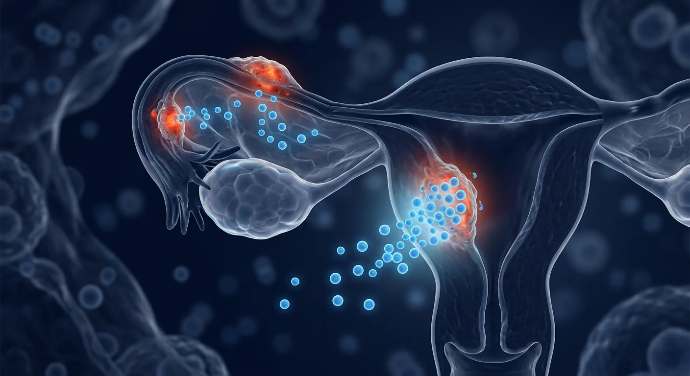
Asherman's syndrome — the formation of intrauterine adhesions and scar tissue within the uterine cavity — affects an estimated 1.5–20% of women undergoing infertility evaluation, with prevalence rising sharply after repeated surgical interventions. [1] For affected women, the endometrium becomes a hostile terrain: thin, fibrotic, and incapable of supporting embryo implantation or sustaining a pregnancy.
Where conventional treatment falls short. The current standard of care — hysteroscopic adhesiolysis followed by estrogen therapy and intrauterine device placement — restores cavity architecture in roughly 80% of mild-to-moderate cases. But for moderate-to-severe disease, re-adhesion rates approach 30–60%, and endometrial thickness often fails to recover beyond 5–7 mm, well below the ~8 mm threshold considered necessary for successful implantation. [2] Surgery can remove the scar tissue but cannot rebuild the functional endometrium.
The deeper problem is cellular. The endometrium is a dynamic, hormonally responsive tissue that relies on a resident population of endometrial stem/progenitor cells for its remarkable regenerative capacity — shedding and regrowing roughly 400 times over a reproductive lifespan. In Asherman's syndrome, these progenitor cells are depleted or functionally impaired, the stromal compartment becomes fibrotic, angiogenesis is suppressed, and the tissue loses its responsiveness to estrogen and progesterone. [3] Simply removing adhesions does not restore this cellular infrastructure.
MSC therapy targets the root cause. Rather than simply clearing scar tissue, mesenchymal stem cells offer a regenerative approach — secreting a broad repertoire of growth factors, cytokines, and extracellular vesicles that promote angiogenesis, suppress fibrosis, recruit endogenous progenitor cells, and restore hormonal responsiveness. [4] Early clinical studies suggest that MSC infusion — whether delivered systemically or locally into the uterine cavity — can increase endometrial thickness, restore menstrual function, and enable pregnancies in women who had exhausted conventional options.
What Is Asherman's Syndrome?
Asherman's syndrome is an acquired uterine condition characterized by intrauterine adhesions and endometrial fibrosis, most commonly caused by uterine instrumentation — particularly dilation and curettage (D&C) after miscarriage or retained products of conception. The trauma strips the basalis layer of the endometrium, exposing the myometrium and creating raw surfaces that adhere and scar during healing.
Causes and Risk Factors
The most common trigger is postpartum or post-miscarriage curettage, accounting for approximately 60–90% of cases. [5] Risk increases with the number of procedures: a single D&C carries roughly 16% risk, while three or more procedures raise it to 32%. Other contributing factors include myomectomy, cesarean section, uterine artery embolization, endometrial ablation, and genital tuberculosis — particularly prevalent in Southeast Asia.
Clinical Presentation
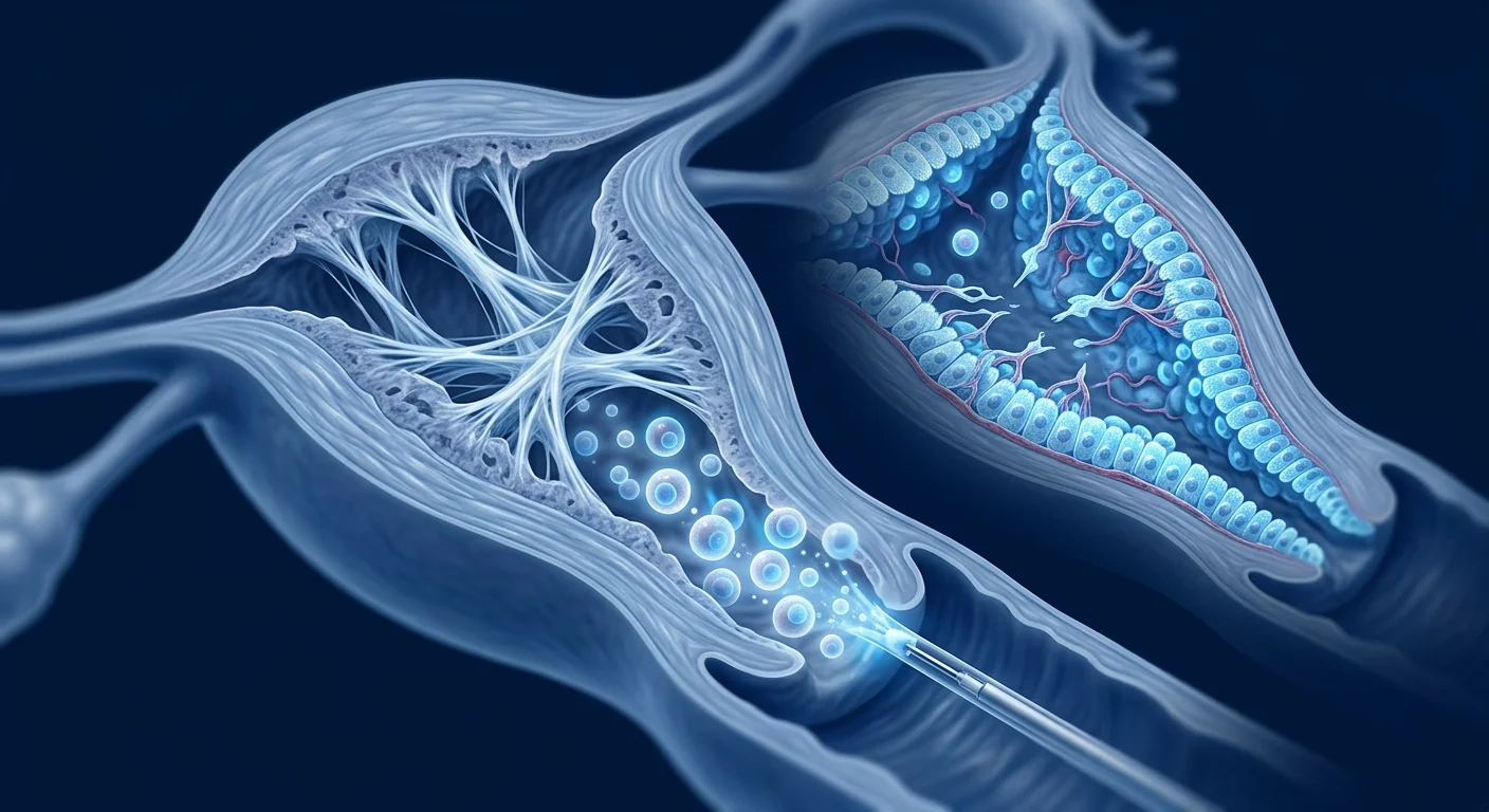
The hallmark triad includes menstrual abnormalities (hypomenorrhea or amenorrhea in 80–90% of cases), infertility (43–70% of patients cannot conceive), and recurrent pregnancy loss due to inadequate endometrial support. [6] Cyclic pelvic pain may occur if adhesions obstruct menstrual outflow. On hysteroscopy, the uterine cavity appears constricted with dense, avascular fibrous bands bridging the walls — a dramatic departure from the pink, velvety surface of healthy endometrium.
Why Current Treatments Fall Short
Hysteroscopic adhesiolysis, the surgical gold standard, uses micro-scissors or energy sources to divide adhesions under direct visualization. Estrogen therapy post-operatively aims to stimulate endometrial proliferation, and an intrauterine device or balloon catheter prevents re-adhesion by keeping the walls separated. [7] For mild disease, this protocol is effective — live birth rates after treatment approach 60–80%. For severe disease (American Fertility Society stage III), however, live birth rates hover around 20–30%, and re-adhesion despite preventive measures remains the norm. The fundamental limitation is biological: surgery cannot regenerate the progenitor cell pool or reverse established fibrosis.
How MSC Therapy Works in Asherman's Syndrome
MSC therapy restores endometrial function through four complementary mechanisms: progenitor cell replenishment, angiogenesis, fibrosis reversal, and immunomodulation. Together, these address the core cellular deficits that conventional surgery cannot correct.
1. Endometrial Progenitor Cell Replenishment
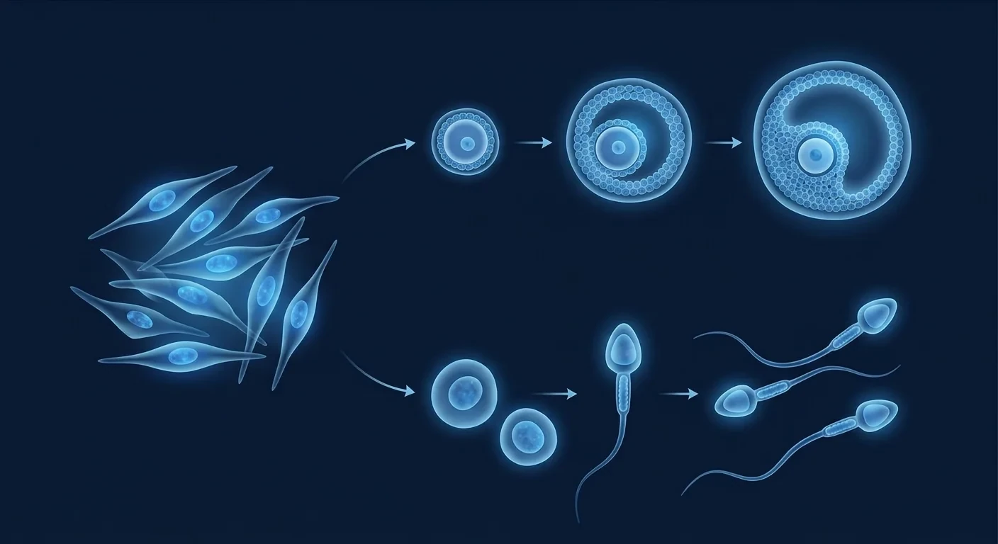
The endometrium harbors a small population of mesenchymal stem-like cells — located in the perivascular niche of the basalis layer — that drive the monthly cycle of proliferation, differentiation, and shedding. [8] In Asherman's syndrome, these cells are depleted or trapped within scar. Infused MSCs home to the injured endometrium (guided by SDF-1/CXCR4 signaling), engraft in the stromal compartment, and differentiate into endometrial stromal cells, glandular epithelial cells, and vascular endothelial cells. [9] In rodent models of Asherman's syndrome, GFP-labeled bone marrow-derived MSCs injected intravenously were detected in the endometrium within 48 hours, where they contributed to glandular and luminal epithelial regeneration over the subsequent 4 weeks.
2. Angiogenesis and Vascular Restoration
A functional endometrium requires a dense capillary network to support implantation — spiral arterioles undergo dramatic remodeling during the secretory phase. In Asherman's syndrome, the fibrotic endometrium is poorly vascularized, with reduced capillary density and impaired blood flow on Doppler ultrasound. [10] MSCs secrete vascular endothelial growth factor (VEGF), basic fibroblast growth factor (bFGF), angiopoietin-1, and hepatocyte growth factor (HGF) — a potent angiogenic cocktail that stimulates new capillary formation and stabilizes existing vessels. In a rat Asherman's model, MSC-treated uteri showed a 2.5-fold increase in CD31-positive microvessel density compared to vehicle controls at day 28. [11]
3. Fibrosis Reversal and ECM Remodeling
Intrauterine adhesions are essentially dense collagen-I-rich scar tissue with few functioning glands and a paucity of blood vessels. MSCs combat fibrosis through multiple pathways: they secrete matrix metalloproteinases (MMP-2, MMP-9) that digest excess extracellular matrix; they upregulate tissue inhibitors of metalloproteinases (TIMPs) in a balanced ratio to prevent excessive degradation; and they suppress transforming growth factor-β1 (TGF-β1)/Smad signaling — the master fibrosis pathway — while enhancing bone morphogenetic protein-7 (BMP-7), a natural anti-fibrotic cytokine. [12] In a preclinical study, MSC-seeded collagen scaffolds implanted into injured rat uteri reduced the fibrosis area by 62% compared to scaffold-only controls at 30 days.
4. Immunomodulation and Anti-Inflammatory Activity
Chronic low-grade inflammation perpetuates the fibrosis cycle in Asherman's syndrome. Damaged endometrium releases damage-associated molecular patterns (DAMPs) that activate resident immune cells, sustaining a pro-fibrotic milieu. [13] MSCs interrupt this cycle by secreting prostaglandin E2 (PGE2), TSG-6, interleukin-10 (IL-10), and indoleamine 2,3-dioxygenase (IDO), which collectively suppress Th1 and Th17 responses, promote regulatory T-cell (Treg) expansion, and polarize local macrophages from the pro-inflammatory M1 to the reparative M2 phenotype. This shift from inflammatory to regenerative signaling is critical — it creates a permissive environment in which new tissue can form without being immediately scarred over.
Clinical Evidence: What the Trials Show
The clinical evidence for MSCs in Asherman's syndrome is early-phase but consistently positive — multiple small studies from Asia and the Middle East have demonstrated improvements in endometrial thickness, menstrual restoration, and pregnancy rates in women with refractory disease. [14]
Intrauterine MSC instillation shows promise. A 2016 pilot study by Santamaria and colleagues enrolled 16 women with refractory Asherman's syndrome and endometrial atrophy (thickness <5 mm despite estrogen therapy). CD133+ bone marrow-derived stem cells were infused directly into the spiral arterioles of the uterus via catheterization. At 3-month follow-up, endometrial thickness increased from a mean of 4.3 mm to 6.7 mm, spontaneous menstrual recovery occurred in 11 of 16 patients (69%), and 3 ongoing pregnancies were achieved — 2 spontaneous and 1 after IVF. [15]
Umbilical cord MSC studies from China. A 2018 randomized trial by Cao and colleagues in China enrolled 26 women with severe Asherman's syndrome. After hysteroscopic adhesiolysis, the treatment group (n=16) received an intrauterine infusion of umbilical cord-derived MSCs (1×10⁷ cells) suspended in a hyaluronic acid gel, while the control group (n=10) received the gel alone. At 3 months, endometrial thickness in the MSC group reached 7.1 ± 1.2 mm vs. 4.7 ± 0.9 mm in controls (p < 0.01). [16] At 30-month follow-up, the MSC group achieved a 47% live birth rate compared to 10% in controls, with no treatment-related adverse events reported.
Menstrual blood-derived MSC autologous approaches. A novel strategy gaining traction uses menstrual blood-derived mesenchymal stem cells (MenSCs) — harvested from the patient's own menstrual fluid, cultured, and re-infused — offering an autologous, non-invasive, and ethically uncomplicated cell source. A 2022 case series from India reported on 7 women with severe Asherman's who each received two intrauterine MenSC infusions. Endometrial thickness increased from mean 3.8 mm to 6.9 mm, menstrual flow returned in 6 of 7, and 2 achieved clinical pregnancy within 12 months. [17]
Systemic intravenous delivery. While most Asherman's studies use local intrauterine delivery, intravenous MSC administration also holds rationale — systemically infused MSCs home to sites of injury and inflammation through chemokine gradient sensing. A 2020 study by Zhao et al. in a rat Asherman's model found that IV-administered Wharton's jelly MSCs homed to the injured uterus at rates comparable to intrauterine injection, with equivalent improvements in endometrial thickness, gland count, and fibrosis scores. [18] This raises the possibility that systemic delivery — which is simpler, less invasive, and can be repeated — may be viable for Asherman's syndrome, particularly when combined with the broader systemic benefits of MSC therapy.
Treatment Approach at VELAR
The treatment protocol for Asherman's syndrome at VELAR is individualized based on disease severity, prior surgical history, and reproductive goals. All MSCs are derived from Wharton's jelly of donated umbilical cords, expanded under cGMP conditions, and characterized by ISCT criteria (≥95% CD73/CD90/CD105 expression, ≤2% CD34/CD45/HLA-DR).
Combination Strategy
For women with moderate-to-severe disease, VELAR typically recommends a sequenced approach:
- Hysteroscopic adhesiolysis — surgical removal of intrauterine adhesions to restore cavity architecture (performed by the patient's gynecologist or at a partner facility in Bangkok).
- MSC infusion (2–4 weeks post-surgery) — intravenous infusion of Wharton's jelly-derived MSCs (typical dose: 1–2 × 10⁶ cells/kg) to deliver regenerative paracrine factors systemically and promote endometrial progenitor replenishment.
- Optional intrauterine MSC gel (same session) — for patients with severe fibrosis, a concentrated MSC suspension (2 × 10⁷ cells) can be delivered directly into the uterine cavity via a thin catheter, providing high local cell density at the endometrial surface.
- Estrogen priming — concurrent exogenous estrogen (oral estradiol valerate 4–6 mg/day for 21–28 days) to synergize with MSC paracrine signaling and promote endometrial proliferation.
- Follow-up assessment at 3 months — transvaginal ultrasound for endometrial thickness measurement, Doppler assessment of sub-endometrial blood flow, and saline infusion sonohysterography to check for adhesion recurrence.
Expected Timeline
Limitations and Important Caveats
It is essential to be honest about where the evidence stands. MSC therapy for Asherman's syndrome remains investigational. The published literature consists of small pilot studies (most with n < 30), case series, and animal models. No large multicenter randomized controlled trial has been completed, and no regulatory agency has approved MSCs specifically for Asherman's syndrome or intrauterine adhesions. [19]
Key uncertainties include: the optimal cell source (bone marrow vs. umbilical cord vs. menstrual blood vs. amniotic membrane), the ideal delivery route (IV vs. intrauterine vs. combination), the best timing relative to adhesiolysis, the need for repeat dosing, and the durability of endometrial improvement beyond 12–24 months. The long-term safety profile — particularly the theoretical risk of ectopic tissue formation or abnormal proliferation in a hormonally sensitive organ — requires ongoing surveillance. [20]
VELAR does not claim that MSC therapy can cure Asherman's syndrome or guarantee pregnancy. It is offered as an adjunctive regenerative option for women who have not responded adequately to conventional surgical and hormonal management, with full informed consent regarding the investigational nature of the treatment.
Frequently Asked Questions
How much does stem cell therapy for Asherman's syndrome cost in Thailand?
At VELAR Center, a single MSC infusion protocol typically ranges from USD 8,000 to 14,000 depending on cell dose and whether intrauterine instillation is included. This is significantly lower than equivalent treatments in the United States or Europe, where regenerative gynecology procedures often exceed USD 25,000. A detailed quote is provided after medical record review.
Can MSC therapy help if I have had multiple failed IVF cycles due to thin endometrium?
Thin endometrium (<7 mm) despite maximal estrogen therapy is one of the strongest rationales for MSC therapy in Asherman's syndrome. Several published case series specifically enrolled women with refractory thin endometrium and prior IVF failure; MSC infusion was associated with measurable increases in endometrial thickness and subsequent pregnancy in a subset. The evidence is encouraging but not yet definitive — results vary by patient and disease severity.
Is intrauterine MSC injection painful?
Intrauterine MSC instillation is performed via a thin, flexible catheter passed through the cervix — similar to an embryo transfer or IUI procedure. Most patients describe mild cramping lasting 5–10 minutes, comparable to a Pap smear or menstrual discomfort. No anesthesia is required. The IV component of the infusion is painless beyond the initial IV line placement.
How many MSC infusions are needed for Asherman's syndrome?
Most published protocols use a single MSC infusion following hysteroscopic adhesiolysis, with assessment at 3 months. Some studies have explored repeat dosing (2–3 infusions at 4–8 week intervals) for severe cases, with preliminary data suggesting incremental benefit. At VELAR, the decision to repeat is guided by the 3-month ultrasound findings — if endometrial thickness remains suboptimal, a second infusion may be recommended.
Are there risks specific to MSC therapy in the uterus?
Reported adverse events in published Asherman's syndrome MSC studies have been mild and transient: low-grade fever (5–10% of patients), mild pelvic discomfort (10–15%), and spotting (15–20%). No cases of infection, perforation, ectopic tissue formation, or malignant transformation have been reported. However, the safety database is small (fewer than 500 treated patients across all published studies), and long-term endometrial surveillance is recommended.
Can I travel to Bangkok for treatment and fly home the same week?
Yes. The MSC infusion itself takes 60–90 minutes and is performed on an outpatient basis. Most international patients stay in Bangkok for 3–5 days: Day 1 for pre-treatment consultation and labs, Day 2 for the infusion, and 1–2 days for rest and a post-treatment check before flying home. Our patient concierge team assists with hotel bookings, airport transfers, and multilingual coordination throughout your stay.
References
- Dreisler E, Kjer JJ. Asherman's syndrome: current perspectives on diagnosis and management. International Journal of Women's Health. 2019;11:191-198. doi:10.2147/IJWH.S165474 ↩
- Hooker AB, de Leeuw R, van de Ven PM, et al. Prevalence of intrauterine adhesions after the application of hyaluronic acid gel after dilatation and curettage in women with at least one previous curettage: short-term outcomes of a multicenter, prospective randomized controlled trial. Fertility and Sterility. 2017;107(5):1223-1231. doi:10.1016/j.fertnstert.2017.02.113 ↩
- Gargett CE, Schwab KE, Deane JA. Endometrial stem/progenitor cells: the first 10 years. Human Reproduction Update. 2016;22(2):137-163. doi:10.1093/humupd/dmv051 ↩
- Cervelló I, Gil-Sanchis C, Santamaría X, et al. Human CD133+ bone marrow-derived stem cells promote endometrial proliferation in a murine model of Asherman syndrome. Fertility and Sterility. 2015;104(6):1552-1560. doi:10.1016/j.fertnstert.2015.08.032 ↩
- Schenker JG, Margalioth EJ. Intrauterine adhesions: an updated appraisal. Fertility and Sterility. 1982;37(5):593-610. doi:10.1016/S0015-0282(16)46268-0 ↩
- Yu D, Wong YM, Cheong Y, Xia E, Li TC. Asherman syndrome — one century later. Fertility and Sterility. 2008;89(4):759-779. doi:10.1016/j.fertnstert.2008.02.096 ↩
- AAGL Advancing Minimally Invasive Gynecology Worldwide. AAGL practice report: practice guidelines on intrauterine adhesions developed in collaboration with the European Society of Gynaecological Endoscopy (ESGE). Journal of Minimally Invasive Gynecology. 2017;24(5):695-705. doi:10.1016/j.jmig.2016.11.008 ↩
- Gargett CE, Masuda H. Adult stem cells in the endometrium. Molecular Human Reproduction. 2010;16(11):818-834. doi:10.1093/molehr/gaq061 ↩
- Du H, Taylor HS. Contribution of bone marrow-derived stem cells to endometrium and endometriosis. Stem Cells. 2007;25(8):2082-2086. doi:10.1634/stemcells.2006-0828 ↩
- Malhotra N, Bahadur A, Kalaivani M, Mittal S. Changes in endometrial receptivity in women with Asherman's syndrome undergoing hysteroscopic adhesiolysis. Archives of Gynecology and Obstetrics. 2012;286(2):525-530. doi:10.1007/s00404-012-2336-0 ↩
- Kilic S, Yuksel B, Pinarli F, Albayrak A, Boztok B, Delibasi T. Effect of stem cell application on Asherman syndrome, an experimental rat model. Journal of Assisted Reproduction and Genetics. 2014;31(8):975-982. doi:10.1007/s10815-014-0268-2 ↩
- Alawadhi F, Du H, Cakmak H, Taylor HS. Bone marrow-derived stem cell (BMDSC) transplantation improves fertility in a murine model of Asherman's syndrome. PLoS One. 2014;9(5):e96662. doi:10.1371/journal.pone.0096662 ↩
- Critchley HOD, Maybin JA, Armstrong GM, Williams ARW. Physiology of the endometrium and regulation of menstruation. Physiological Reviews. 2020;100(3):1149-1179. doi:10.1152/physrev.00031.2019 ↩
- Azizi R, Aghebati-Maleki L, Nouri M, Marofi F, Negargar S, Yousefi M. Stem cell therapy in Asherman syndrome and thin endometrium: stem cell-based therapy. Biomedicine & Pharmacotherapy. 2018;102:333-343. doi:10.1016/j.biopha.2018.03.091 ↩
- Santamaria X, Cabanillas S, Cervelló I, et al. Autologous cell therapy with CD133+ bone marrow-derived stem cells for refractory Asherman's syndrome and endometrial atrophy: a pilot cohort study. Human Reproduction. 2016;31(5):1087-1096. doi:10.1093/humrep/dew042 ↩
- Cao Y, Sun H, Zhu H, et al. Allogeneic cell therapy using umbilical cord MSCs on collagen scaffolds for patients with recurrent uterine adhesion: a phase I clinical trial. Stem Cell Research & Therapy. 2018;9(1):192. doi:10.1186/s13287-018-0904-3 ↩
- Patel AN, Park E, Kuzman M, Benetti F, Silva FJ, Allickson JG. Multipotent menstrual blood stromal stem cells: isolation, characterization, and differentiation. Cell Transplantation. 2008;17(3):303-311. doi:10.3727/096368908784153922 ↩
- Zhao G, Cao Y, Zhu X, et al. Transplantation of collagen scaffold with autologous bone marrow mononuclear cells promotes functional endometrium reconstruction via downregulating ΔNp63 expression in Asherman's syndrome. Science China Life Sciences. 2017;60(4):404-413. doi:10.1007/s11427-016-0390-9 ↩
- Simón C, Gómez E, Miró F, Pellicer A. Stem cell research in human reproduction: basic science to clinical applications. Fertility and Sterility. 2019;112(4):610-618. doi:10.1016/j.fertnstert.2019.08.004 ↩
- Benor A, Gay S, DeCherney A. An update on stem cell therapy for Asherman's syndrome. Journal of Assisted Reproduction and Genetics. 2020;37(7):1511-1517. doi:10.1007/s10815-020-01801-x ↩
阿什曼综合征(Asherman's syndrome)——宫腔内粘连和瘢痕组织的形成——影响约1.5%–20%接受不孕评估的女性,反复手术后患病率急剧上升。[1]对患者而言,子宫内膜变成了不适宜胚胎着床和妊娠的纤维化环境。
传统治疗的局限。目前的标准治疗方案——宫腔镜粘连分离术联合雌激素治疗和宫内节育器放置——在轻中度病例中可恢复约80%的宫腔结构。但对中重度患者,再粘连率达30%–60%,子宫内膜厚度往往无法恢复至5–7毫米以上,远低于着床所需的约8毫米阈值。[2]手术可以切除瘢痕组织,但无法重建功能性子宫内膜。
更深层的问题是细胞层面的。子宫内膜是一种动态的、激素响应性组织,依赖常驻的子宫内膜干细胞/祖细胞维持其卓越的再生能力——在生育年龄期间大约脱落和再生400次。在阿什曼综合征中,这些祖细胞被耗竭或功能受损,基质室变得纤维化,血管生成被抑制,组织失去对雌激素和孕激素的响应能力。[3]仅清除粘连并不能恢复这种细胞基础设施。
间充质干细胞治疗直击根源。间充质干细胞(MSC)并非简单地清除瘢痕组织,而是提供一种再生途径——分泌丰富的生长因子、细胞因子和细胞外囊泡,促进血管生成、抑制纤维化、招募内源性祖细胞并恢复激素响应性。[4]早期临床研究表明,MSC输注——无论是全身静脉还是局部宫腔内给药——均可增加子宫内膜厚度、恢复月经功能,并使已耗尽传统治疗选择的女性获得妊娠。
什么是阿什曼综合征?
阿什曼综合征是一种获得性子宫疾病,以宫腔内粘连和子宫内膜纤维化为特征,最常见的原因是子宫器械操作——尤其是流产或妊娠物残留后的扩张刮宫术(D&C)。创伤剥离了子宫内膜基底层,暴露了子宫肌层,形成粗糙表面在愈合过程中粘连和瘢痕化。
病因和风险因素
最常见的触发因素是产后或流产后的刮宫术,约占病例的60%–90%。[5]风险随操作次数增加:单次D&C的风险约为16%,三次或以上升至32%。其他促成因素包括子宫肌瘤切除术、剖宫产、子宫动脉栓塞术、子宫内膜消融术和生殖器结核——后者在东南亚尤为普遍。
临床表现
典型三联征包括月经异常(80%–90%的病例出现月经减少或闭经)、不孕(43%–70%的患者无法受孕)和反复妊娠丢失。宫腔镜下,宫腔缩窄,致密无血管的纤维条带横跨宫壁——与健康子宫内膜的粉色绒面形成鲜明对比。[6]
MSC治疗阿什曼综合征的机制
MSC治疗通过四种互补机制恢复子宫内膜功能:祖细胞补充、血管生成、纤维化逆转和免疫调节。
1. 子宫内膜祖细胞补充
子宫内膜在基底层血管周围龛位中驻留少量间充质干细胞样细胞,驱动每月的增殖、分化和脱落周期。[8]在阿什曼综合征中,这些细胞被耗竭或困在瘢痕中。输注的MSC通过SDF-1/CXCR4信号归巢至受损子宫内膜,植入基质室并分化为内膜基质细胞、腺上皮细胞和血管内皮细胞。[9]
2. 血管生成与血管恢复
功能性子宫内膜需要密集的毛细血管网络来支持着床。在阿什曼综合征中,纤维化子宫内膜血管化不良。MSC分泌血管内皮生长因子(VEGF)、碱性成纤维细胞生长因子(bFGF)、血管生成素-1和肝细胞生长因子(HGF)——一种强效的促血管生成组合。[11]
3. 纤维化逆转与基质重塑
宫腔内粘连本质上是致密的I型胶原瘢痕组织。MSC通过多种途径对抗纤维化:分泌基质金属蛋白酶(MMP-2、MMP-9)消化过量细胞外基质;上调基质金属蛋白酶组织抑制剂(TIMP)防止过度降解;抑制TGF-β1/Smad信号通路同时增强天然抗纤维化细胞因子BMP-7。[12]
4. 免疫调节与抗炎活性
慢性低度炎症在阿什曼综合征中延续纤维化循环。MSC通过分泌PGE2、TSG-6、IL-10和IDO中断这一循环,抑制Th1和Th17反应,促进调节性T细胞扩增,并将局部巨噬细胞从促炎M1表型极化为修复性M2表型。[13]
临床证据
MSC治疗阿什曼综合征的临床证据尚处于早期阶段但结果一致向好——亚洲和中东的多项小型研究已证明子宫内膜厚度、月经恢复和妊娠率的改善。[14]
宫腔内MSC滴注显示前景。Santamaria等人2016年的初步研究纳入16例难治性阿什曼综合征和子宫内膜萎缩(厚度<5毫米)患者。CD133+骨髓来源干细胞通过导管直接注入子宫螺旋小动脉。3个月随访时,子宫内膜厚度从平均4.3毫米增至6.7毫米,11/16例(69%)恢复月经,获得3例持续妊娠。[15]
中国脐带MSC研究。Cao等人2018年的一项随机试验纳入26例重度阿什曼综合征女性。宫腔镜粘连分离术后,治疗组(16例)接受脐带来源MSC(1×10⁷个细胞)悬浮于透明质酸凝胶中的宫腔内输注,对照组(10例)仅接受凝胶。3个月时,MSC组内膜厚度达7.1±1.2毫米,对照组4.7±0.9毫米(p<0.01)。30个月随访时,MSC组活产率47%,对照组10%。[16]
治疗限制与重要注意事项
MSC治疗阿什曼综合征仍处于研究阶段。已发表的文献包括小型初步研究(多数n<30)、病例系列和动物模型。尚无大型多中心随机对照试验完成,也没有监管机构专门批准MSC用于阿什曼综合征。[19]
关键不确定性包括:最佳细胞来源、理想给药途径、相对于粘连分离术的最佳时机、是否需要重复给药以及子宫内膜改善的持久性。VELAR不声称MSC治疗可以治愈阿什曼综合征或保证妊娠。它是在充分知情同意的情况下,为对传统手术和激素管理反应不佳的女性提供的一种辅助再生选择。
参考文献
- Dreisler E, Kjer JJ. Asherman's syndrome: current perspectives on diagnosis and management. International Journal of Women's Health. 2019;11:191-198. doi:10.2147/IJWH.S165474 ↩
- Hooker AB, et al. Prevalence of intrauterine adhesions after dilatation and curettage. Fertility and Sterility. 2017;107(5):1223-1231. doi:10.1016/j.fertnstert.2017.02.113 ↩
- Gargett CE, Schwab KE, Deane JA. Endometrial stem/progenitor cells. Human Reproduction Update. 2016;22(2):137-163. doi:10.1093/humupd/dmv051 ↩
- Cervelló I, et al. Human CD133+ bone marrow-derived stem cells promote endometrial proliferation. Fertility and Sterility. 2015;104(6):1552-1560. doi:10.1016/j.fertnstert.2015.08.032 ↩
- Schenker JG, Margalioth EJ. Intrauterine adhesions: an updated appraisal. Fertility and Sterility. 1982;37(5):593-610. doi:10.1016/S0015-0282(16)46268-0 ↩
- Yu D, et al. Asherman syndrome — one century later. Fertility and Sterility. 2008;89(4):759-779. doi:10.1016/j.fertnstert.2008.02.096 ↩
- Gargett CE, Masuda H. Adult stem cells in the endometrium. Molecular Human Reproduction. 2010;16(11):818-834. doi:10.1093/molehr/gaq061 ↩
- Du H, Taylor HS. Contribution of bone marrow-derived stem cells to endometrium. Stem Cells. 2007;25(8):2082-2086. doi:10.1634/stemcells.2006-0828 ↩
- Kilic S, et al. Effect of stem cell application on Asherman syndrome. Journal of Assisted Reproduction and Genetics. 2014;31(8):975-982. doi:10.1007/s10815-014-0268-2 ↩
- Alawadhi F, et al. BMDSC transplantation improves fertility in Asherman's syndrome. PLoS One. 2014;9(5):e96662. doi:10.1371/journal.pone.0096662 ↩
- Critchley HOD, et al. Physiology of the endometrium. Physiological Reviews. 2020;100(3):1149-1179. doi:10.1152/physrev.00031.2019 ↩
- Azizi R, et al. Stem cell therapy in Asherman syndrome. Biomedicine & Pharmacotherapy. 2018;102:333-343. doi:10.1016/j.biopha.2018.03.091 ↩
- Santamaria X, et al. Autologous cell therapy with CD133+ cells for refractory Asherman's syndrome. Human Reproduction. 2016;31(5):1087-1096. doi:10.1093/humrep/dew042 ↩
- Cao Y, et al. UC-MSCs on collagen scaffolds for recurrent uterine adhesion. Stem Cell Research & Therapy. 2018;9(1):192. doi:10.1186/s13287-018-0904-3 ↩
- Simón C, et al. Stem cell research in human reproduction. Fertility and Sterility. 2019;112(4):610-618. doi:10.1016/j.fertnstert.2019.08.004 ↩
- Benor A, Gay S, DeCherney A. An update on stem cell therapy for Asherman's syndrome. Journal of Assisted Reproduction and Genetics. 2020;37(7):1511-1517. doi:10.1007/s10815-020-01801-x ↩
متلازمة أشرمان — تكون التصاقات وندبات داخل تجويف الرحم — تصيب ما يقدر بنحو 1.5%–20% من النساء الخاضعات لتقييم العقم، مع ارتفاع حاد في معدل الانتشار بعد التدخلات الجراحية المتكررة. [1] بالنسبة للنساء المصابات، تصبح بطانة الرحم بيئة معادية: رقيقة ومتليفة وغير قادرة على دعم انغراس الجنين أو استمرار الحمل.
أوجه قصور العلاج التقليدي. البروتوكول القياسي الحالي — تحليل الالتصاقات بالمنظار الرحمي متبوعًا بالعلاج بالإستروجين ووضع لولب داخل الرحم — يستعيد بنية التجويف في حوالي 80% من الحالات الخفيفة إلى المتوسطة. لكن في الحالات المتوسطة إلى الشديدة، تقترب معدلات إعادة الالتصاق من 30%–60%، وغالبًا ما تفشل بطانة الرحم في التعافي لأكثر من 5–7 مم، وهو أقل بكثير من عتبة ~8 مم اللازمة للانغراس الناجح. [2]
المشكلة الأعمق على المستوى الخلوي. بطانة الرحم هي نسيج ديناميكي مستجيب للهرمونات يعتمد على مجموعة مقيمة من الخلايا الجذعية/السلفية البطانية الرحمية لقدرتها التجديدية الملحوظة — حيث تتساقط وتتجدد حوالي 400 مرة خلال العمر الإنجابي. في متلازمة أشرمان، تُستنزف هذه الخلايا السلفية أو تتعطل وظيفيًا، ويصبح الحيز السدوي متليفًا، ويُثبط تكوين الأوعية الدموية، ويفقد النسيج استجابته للإستروجين والبروجسترون. [3]
علاج الخلايا الجذعية الوسيطة يستهدف السبب الجذري. بدلاً من مجرد إزالة النسيج الندبي، تقدم الخلايا الجذعية الوسيطة (MSCs) نهجًا تجديديًا — تفرز مجموعة غنية من عوامل النمو والسيتوكينات والحويصلات خارج الخلوية التي تعزز تكوين الأوعية الدموية وتثبط التليف وتجند الخلايا السلفية الداخلية وتستعيد الاستجابة الهرمونية. [4]
كيف يعمل علاج MSC في متلازمة أشرمان
يستعيد علاج MSC وظيفة بطانة الرحم من خلال أربع آليات متكاملة: تجديد الخلايا السلفية، وتكوين الأوعية الدموية، وعكس التليف، والتعديل المناعي.
1. تجديد الخلايا السلفية البطانية الرحمية
تستقر الخلايا الجذعية الوسيطة المحقونة في بطانة الرحم المصابة عبر إشارات SDF-1/CXCR4، وتنغرس في الحيز السدوي وتتمايز إلى خلايا سدوية بطانية رحمية وخلايا طلائية غدية وخلايا بطانية وعائية. [9]
2. تكوين الأوعية الدموية
تفرز MSCs عامل نمو بطانة الأوعية الدموية (VEGF) وعامل نمو الخلايا الليفية القاعدي (bFGF) والأنجيوبويتين-1 وعامل نمو الخلايا الكبدية (HGF) — مزيج قوي محفز لتكوين الأوعية الدموية. [11]
3. عكس التليف وإعادة تشكيل المطرس خارج الخلوي
تحارب MSCs التليف من خلال مسارات متعددة: تفرز ميتالوبروتينازات المطرس (MMP-2، MMP-9) التي تهضم المطرس الزائد خارج الخلوي؛ وتثبط إشارات TGF-β1/Smad مع تعزيز BMP-7 وهو سيتوكين طبيعي مضاد للتليف. [12]
4. التعديل المناعي
يفرز MSCs البروستاغلاندين E2 وTSG-6 والإنترلوكين-10 وIDO، مما يثبط استجابات Th1 وTh17 ويعزز توسع الخلايا التائية التنظيمية (Treg) ويستقطب الخلايا البلعمية المحلية من النمط الالتهابي M1 إلى النمط الإصلاحي M2. [13]
الأدلة السريرية
الأدلة السريرية لـ MSCs في متلازمة أشرمان في مرحلة مبكرة لكنها إيجابية باستمرار. [14]
تقطير MSC داخل الرحم يظهر نتائج واعدة. دراسة أولية لـ Santamaria وزملائه عام 2016 شملت 16 امرأة مصابة بمتلازمة أشرمان المقاومة وضمور بطانة الرحم. تم حقن الخلايا الجذعية CD133+ المشتقة من نخاع العظم مباشرة في الشريينات الحلزونية للرحم. في متابعة 3 أشهر، زاد سمك بطانة الرحم من متوسط 4.3 مم إلى 6.7 مم، وعادت الدورة الشهرية في 11 من 16 مريضة (69%)، وتحقق 3 حالات حمل مستمر. [15]
دراسات الحبل السري من الصين. تجربة عشوائية لـ Cao وزملائه عام 2018 شملت 26 امرأة. بعد تحليل الالتصاقات بالمنظار الرحمي، تلقت مجموعة العلاج (16) حقن MSCs المشتقة من الحبل السري (1×10⁷ خلية) داخل الرحم. في 3 أشهر، بلغ سمك بطانة الرحم في مجموعة MSC 7.1±1.2 مم مقابل 4.7±0.9 مم في المجموعة الضابطة. في متابعة 30 شهرًا، حققت مجموعة MSC معدل ولادات حية 47% مقابل 10% في المجموعة الضابطة. [16]
القيود والتحفظات
لا يزال علاج MSC لمتلازمة أشرمان قيد البحث. تتكون الأدبيات المنشورة من دراسات أولية صغيرة (معظمها n<30) وسلاسل حالات ونماذج حيوانية. لم تكتمل أي تجربة كبيرة متعددة المراكز معشاة مضبوطة. [19]
لا تدعي VELAR أن علاج MSC يمكنه شفاء متلازمة أشرمان أو ضمان الحمل. يُقدم كخيار تجديدي مساعد للنساء اللواتي لم يستجبن بشكل كافٍ للإدارة الجراحية والهرمونية التقليدية، بموافقة مستنيرة كاملة.
الأسئلة الشائعة
كم تكلفة علاج الخلايا الجذعية لمتلازمة أشرمان في تايلاند؟
في مركز VELAR، تتراوح تكلفة بروتوكول حقن MSC الواحد عادة من 8,000 إلى 14,000 دولار أمريكي حسب جرعة الخلايا وما إذا كان التقطير داخل الرحم مشمولاً. وهذا أقل بكثير من العلاجات المماثلة في الولايات المتحدة أو أوروبا.
هل يمكن أن يساعد علاج MSC إذا كان لدي بطانة رحم رقيقة وفشل متكرر في التلقيح الاصطناعي؟
بطانة الرحم الرقيقة (<7 مم) هي واحدة من أقوى المبررات لعلاج MSC. شملت العديد من سلاسل الحالات المنشورة نساءً مصابات ببطانة رحم رقيقة مقاومة وفشل سابق في التلقيح الاصطناعي؛ وارتبط حقن MSC بزيادات قابلة للقياس في سمك بطانة الرحم وحمل لاحق لدى مجموعة فرعية.
المراجع
- Dreisler E, Kjer JJ. Asherman's syndrome: current perspectives. International Journal of Women's Health. 2019;11:191-198. doi:10.2147/IJWH.S165474 ↩
- Hooker AB, et al. Prevalence of intrauterine adhesions. Fertility and Sterility. 2017;107(5):1223-1231. doi:10.1016/j.fertnstert.2017.02.113 ↩
- Gargett CE, et al. Endometrial stem/progenitor cells. Human Reproduction Update. 2016;22(2):137-163. doi:10.1093/humupd/dmv051 ↩
- Cervelló I, et al. CD133+ cells promote endometrial proliferation. Fertility and Sterility. 2015;104(6):1552-1560. doi:10.1016/j.fertnstert.2015.08.032 ↩
- Du H, Taylor HS. BMDSCs contribution to endometrium. Stem Cells. 2007;25(8):2082-2086. doi:10.1634/stemcells.2006-0828 ↩
- Kilic S, et al. Stem cell in Asherman syndrome rat model. Journal of Assisted Reproduction and Genetics. 2014;31(8):975-982. doi:10.1007/s10815-014-0268-2 ↩
- Alawadhi F, et al. BMDSC transplantation in Asherman's. PLoS One. 2014;9(5):e96662. doi:10.1371/journal.pone.0096662 ↩
- Critchley HOD, et al. Physiology of the endometrium. Physiological Reviews. 2020;100(3):1149-1179. doi:10.1152/physrev.00031.2019 ↩
- Azizi R, et al. Stem cell therapy in Asherman syndrome. Biomedicine & Pharmacotherapy. 2018;102:333-343. doi:10.1016/j.biopha.2018.03.091 ↩
- Santamaria X, et al. Autologous cell therapy for refractory Asherman's. Human Reproduction. 2016;31(5):1087-1096. doi:10.1093/humrep/dew042 ↩
- Cao Y, et al. UC-MSCs for recurrent uterine adhesion. Stem Cell Research & Therapy. 2018;9(1):192. doi:10.1186/s13287-018-0904-3 ↩
- Simón C, et al. Stem cell research in human reproduction. Fertility and Sterility. 2019;112(4):610-618. doi:10.1016/j.fertnstert.2019.08.004 ↩
- Benor A, et al. Update on stem cell therapy for Asherman's. Journal of Assisted Reproduction and Genetics. 2020;37(7):1511-1517. doi:10.1007/s10815-020-01801-x ↩