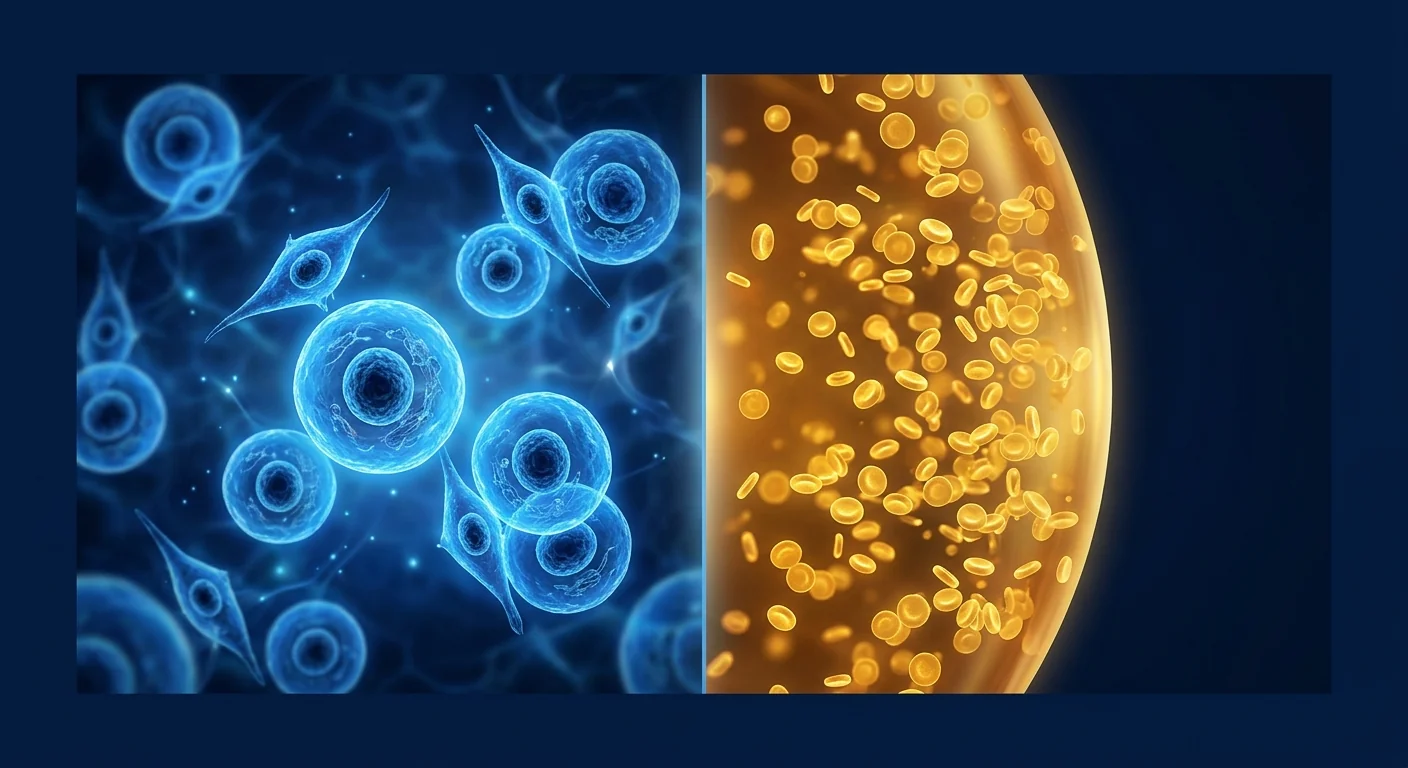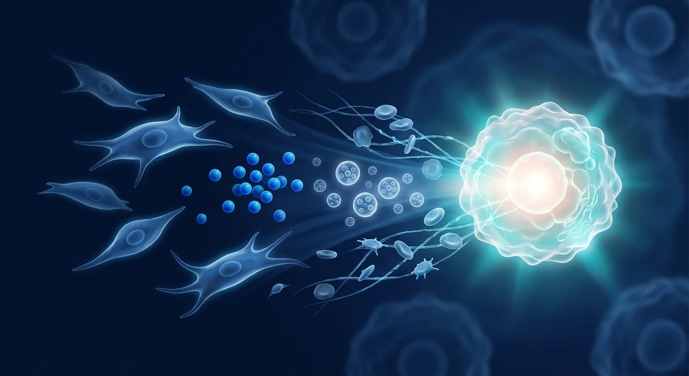
The future of regenerative medicine is not about choosing one therapy over another — it is about understanding how different modalities amplify each other. MSC therapy is increasingly being studied in combination with PRP, exosomes, pharmaceuticals, and rehabilitation to achieve outcomes no single approach can deliver alone.
For decades, the dominant paradigm in medicine has been monotherapy: one drug, one mechanism, one outcome. But complex diseases — from osteoarthritis to spinal cord injury to autoimmune conditions — involve multiple interacting pathological pathways. No single biologic agent can simultaneously address inflammation, tissue degradation, fibrotic remodeling, and vascular insufficiency. [1]
Where monotherapy falls short. Even the most potent MSC preparation has biological limits. MSCs excel at immunomodulation and trophic support, but they do not provide the structural scaffold of a tissue graft, the immediate anti-inflammatory punch of a corticosteroid, or the mechanical unloading achieved by physical therapy and bracing. Acknowledging these complementary roles is not a concession of weakness — it is the foundation of rational combination therapy design.
The deeper opportunity is synergy. When MSC therapy is combined with modalities that address the dimensions MSCs alone cannot reach, the result can exceed the sum of its parts. Platelet-rich plasma provides a burst of growth factors that prime the tissue microenvironment for MSC engraftment. Exosome therapy delivers concentrated paracrine signaling without the logistical complexity of live-cell administration. Pharmacological agents can precondition the target tissue, suppress competing pathological pathways, or enhance MSC homing. [2]
Combination protocols target the full disease biology. This article examines the evidence for combining MSC therapy with complementary modalities — the mechanistic rationale, the preclinical and clinical data, the safety considerations, and the practical protocol design principles that guide clinical decision-making at centers offering multi-modal regenerative medicine.
MSC + Platelet-Rich Plasma (PRP): The Foundational Combination
Platelet-rich plasma — an autologous blood concentrate containing 3–5 times the baseline platelet concentration — is the most frequently combined modality with MSC therapy and arguably the combination with the strongest mechanistic rationale. Platelets are a natural reservoir of growth factors, cytokines, and chemokines that regulate virtually every phase of tissue repair. [3]
Mechanistic Rationale
PRP releases platelet-derived growth factor (PDGF), transforming growth factor-beta (TGF-β), vascular endothelial growth factor (VEGF), epidermal growth factor (EGF), insulin-like growth factor-1 (IGF-1), and fibroblast growth factor (FGF) upon activation. These factors do not compete with MSC mechanisms — they complement them:
- PDGF and TGF-β are potent chemoattractants for MSCs, enhancing homing and retention at the injury site by 2–3 fold in preclinical models.
- VEGF and FGF stimulate angiogenesis, creating the vascular infrastructure that supports MSC survival and function in hypoxic tissue environments.
- IGF-1 and EGF promote MSC proliferation and survival, effectively expanding the therapeutic cell population in situ.
- Fibrin scaffold. Activated PRP forms a fibrin matrix that serves as a temporary scaffold, retaining MSCs at the injection site rather than allowing rapid dispersal — a critical challenge in MSC delivery. [4]
Clinical Evidence
The MSC+PRP combination has the strongest clinical evidence base of any MSC combination strategy, particularly in orthopaedic applications:
- Knee osteoarthritis. A randomized trial (n=48) comparing MSC alone, PRP alone, and MSC+PRP for knee OA found the combination group achieved significantly greater improvements in WOMAC scores and MRI cartilage thickness at 12 months compared to either monotherapy. [5]
- Tendon repair. Animal models of rotator cuff repair demonstrate that MSC+PRP co-administration improves ultimate tensile strength by 40–60% compared to MSC alone, with better collagen fiber alignment and reduced fibrotic scar formation.
- Spinal fusion. PRP combined with bone marrow-derived MSCs accelerates fusion mass maturation and improves fusion rates in both preclinical and early clinical series.
- Wound healing. A meta-analysis of 12 studies found MSC+PRP combination therapy accelerated chronic wound closure by an average of 35% compared to standard care, with particular efficacy in diabetic foot ulcers. [6]
MSC + Exosome Therapy: Amplifying the Paracrine Signal
MSC-derived exosomes — nanoscale extracellular vesicles (30–150 nm) containing proteins, lipids, mRNA, microRNA, and signaling molecules — represent the concentrated paracrine payload of mesenchymal stem cells without the logistical and safety complexities of live-cell administration. Combining whole MSCs with exosome preparations is an emerging strategy to deliver both immediate paracrine signaling and sustained cellular engraftment. [7]
Mechanistic Rationale
Exosome therapy and whole MSC therapy occupy complementary temporal niches in the tissue repair timeline:
- Exosomes act immediately. Upon injection, exosomes deliver their molecular cargo to recipient cells within minutes to hours, initiating anti-inflammatory, anti-apoptotic, and pro-angiogenic signaling cascades. This provides a rapid "first wave" of tissue stabilization.
- Whole MSCs provide sustained support. Engrafted MSCs continue secreting exosomes and soluble factors for days to weeks, providing ongoing paracrine support. They also respond dynamically to the local microenvironment — upregulating anti-inflammatory factors when inflammation is high, shifting toward trophic support as tissue repair progresses.
- Complementary biodistribution. Exosomes (30–150 nm) penetrate tissue barriers and diffuse more widely than whole MSCs (15–30 μm), which are largely retained at the injection site. The exosome+MSC combination achieves both localized and distributed therapeutic coverage. [8]
Clinical Evidence
Clinical data on MSC+exosome combination therapy is still in early stages but shows promise:
- Acute kidney injury. Preclinical studies demonstrate that MSC+exosome co-administration reduces serum creatinine by an additional 25–35% compared to MSC monotherapy in ischemia-reperfusion AKI models.
- Myocardial infarction. In porcine MI models, exosome-preconditioned MSCs showed 2-fold greater engraftment and 40% greater improvement in left ventricular ejection fraction at 4 weeks.
- Neurological applications. Intranasal exosome administration combined with intravenous MSCs improved functional recovery scores in rodent stroke models beyond either therapy alone, attributed to exosome-mediated reduction of neuroinflammation prior to MSC-mediated neurotrophic support. [9]
MSC + Pharmacological Agents: Preconditioning and Synergy
Pharmacological agents can be combined with MSC therapy in two fundamentally different ways: as preconditioning agents administered before MSC infusion to enhance cell performance, and as concurrent therapies that target complementary disease pathways. [10]
Preconditioning Strategies
Preconditioning — exposing MSCs to specific stimuli before administration — is one of the most active areas of MSC research. The goal is to "train" MSCs to optimize their therapeutic phenotype for a specific clinical application:
- Hypoxic preconditioning (exposing MSCs to 1–5% O₂ for 24–48 hours before administration) upregulates HIF-1α, enhancing secretion of VEGF, HGF, and FGF-2. Hypoxia-preconditioned MSCs demonstrate 2–3 fold greater engraftment and therapeutic efficacy in ischemic tissue models. [11]
- Interferon-gamma (IFN-γ) priming activates MSC immunomodulatory capacity by upregulating indoleamine 2,3-dioxygenase (IDO) and prostaglandin E2 synthesis. IFN-γ-primed MSCs are significantly more effective in autoimmune and graft-versus-host disease models.
- TNF-α preconditioning enhances MSC production of TSG-6 and PGE2, improving anti-inflammatory potency for acute inflammatory conditions such as sepsis and ARDS.
- Pharmacological agents. Small molecules including deferoxamine (a hypoxia mimetic), melatonin, atorvastatin, and curcumin have all been investigated as MSC preconditioning agents, each enhancing specific therapeutic properties. [12]
Concurrent Pharmacotherapy
Beyond preconditioning, combining MSCs with ongoing pharmacological therapy is clinically relevant for most patients — the question is whether the drugs help, hinder, or have no effect on MSC function:
- Anti-inflammatory drugs. NSAIDs may partially attenuate MSC anti-inflammatory effects by inhibiting COX-2-dependent PGE2 production. However, clinical data are mixed — short courses of NSAIDs appear not to significantly impair MSC efficacy in orthopaedic applications, and some evidence suggests celecoxib (a selective COX-2 inhibitor) may preserve MSC function better than non-selective NSAIDs. [13]
- Immunosuppressants. Corticosteroids at high doses suppress MSC immunomodulatory function; low-dose regimens appear compatible. Tacrolimus and mycophenolate mofetil have minimal direct effects on MSC function and are commonly co-administered in transplant settings.
- Anticoagulants. Heparin enhances MSC migration and proliferation in vitro by binding growth factors and stabilizing their receptor interactions. Low-molecular-weight heparin is routinely used during MSC infusion with no evidence of antagonism.
- Statins. Atorvastatin and rosuvastatin enhance MSC paracrine function and improve survival in ischemic tissue models. This synergy is particularly relevant for cardiovascular and metabolic disease applications. [14]
MSC + Physical Therapy & Rehabilitation
Physical therapy and structured rehabilitation represent perhaps the most underappreciated combination partner for MSC therapy. Mechanical loading, neuromuscular stimulation, and controlled movement are essential signals for tissue remodeling — MSCs provide the cellular building blocks, but rehabilitation provides the architectural blueprint that determines how those cells organize into functional tissue. [15]
Mechanistic Rationale
- Mechanotransduction. MSCs are mechanosensitive — they differentiate along lineage-specific pathways in response to mechanical cues. Cyclic tensile strain promotes tenogenic differentiation; compressive loading promotes chondrogenesis; fluid shear stress promotes endothelial differentiation. Appropriate rehabilitation loading after MSC administration directs MSC fate toward the desired tissue phenotype.
- Vascular perfusion. Controlled exercise increases blood flow to target tissues, improving MSC delivery, oxygen and nutrient supply, and waste removal. Immobilization after MSC administration may reduce cell survival by creating a hypoxic, nutrient-poor microenvironment.
- Neuromuscular re-education. For neurological and musculoskeletal applications, MSCs may support neural repair and muscle regeneration, but functional recovery requires neuromuscular retraining — the repaired tissue must learn to function within coordinated movement patterns.
- Anti-fibrotic effect. Controlled mechanical loading reduces fibrotic scar formation by promoting organized collagen deposition. Immobilization after MSC treatment can paradoxically increase fibrosis despite enhanced cellular regeneration. [16]
Clinical Integration
The optimal rehabilitation protocol depends on the tissue target and the phase of healing. General principles include:
- Phase 1 (Days 0–7): Protected mobilization. Gentle passive range-of-motion exercises; avoid excessive loading of the treated tissue. The goal is to maintain joint mobility and prevent adhesion formation without disrupting early MSC engraftment.
- Phase 2 (Weeks 2–6): Progressive loading. Graduated resistance exercises and proprioceptive training. Mechanical signals during this window strongly influence MSC differentiation and tissue organization.
- Phase 3 (Weeks 6–12): Functional restoration. Sport-specific or activity-specific training; full loading. The tissue should be structurally competent to tolerate physiological loads.
Clinical Evidence for Combination Approaches
The evidence base for MSC combination therapy varies significantly by clinical indication and combination partner. The strongest data supports MSC+PRP in orthopaedics; other combinations have compelling preclinical rationale but limited randomized human data. [17]
Safety of Combination Therapy
Combining MSC therapy with other modalities raises theoretical safety concerns beyond those of each therapy administered alone. However, the actual safety record of MSC combination therapy — particularly MSC+PRP — is reassuring. [18]
- Infection risk. Each additional injection or manipulation introduces an incremental infection risk. Strict aseptic technique is essential, and the total number of injection events should be minimized where possible (e.g., co-administering MSCs and PRP through a single needle track).
- Immunogenicity. PRP and exosomes are generally non-immunogenic when used autologously or from well-characterized allogeneic sources. No significant immunogenicity concerns have been reported with MSC+PRP or MSC+exosome combinations.
- Tumorigenicity. Growth factors in PRP theoretically could stimulate dormant malignancy. While this concern is theoretical — PRP has an excellent long-term safety record with no signal of increased malignancy — caution is warranted in patients with active or recent cancer. [19]
- Drug interactions. The pharmacological interactions described above (NSAIDs, corticosteroids, immunosuppressants) are generally manageable through dose timing and selection. A thorough medication review should precede any MSC combination protocol.
Protocol Design: Practical Considerations
Designing a rational MSC combination protocol requires answering several interconnected questions:
- What biological gaps does MSC therapy leave unfilled? If the condition involves structural tissue loss (e.g., critical-size bone defect), MSCs need a scaffold. If it involves acute, intense inflammation (e.g., active rheumatoid arthritis flare), a bridging anti-inflammatory strategy may be needed before MSC administration.
- What is the temporal sequence of tissue repair? Inflammation peaks early, followed by proliferation and remodeling. Exosomes or anti-inflammatory preconditioning may be most valuable in the early phase; PRP and rehabilitation are relevant throughout; pharmacological support may be needed at specific disease-phase transitions.
- What combination has a proven safety record? MSC+PRP has thousands of patient-years of clinical experience. MSC+novel pharmacological agents or uncharacterized exosome preparations have far less safety data and should be approached with greater caution.
- Can the combination be practically delivered? Some combinations require specialized equipment, multiple visits, or complex coordination. The protocol must be feasible within the patient's logistical constraints.
- Is the added value worth the added complexity and cost? Each additional modality increases cost, treatment time, and (potentially) risk. The clinical benefit must justify these additions — combination therapy should not be the default; it should be the reasoned choice. [20]
Frequently Asked Questions
Can I receive MSC therapy while continuing my current medications?
In most cases, yes. Most common medications — including antihypertensives, statins, thyroid hormone replacement, and antidiabetic agents — are compatible with MSC therapy. The main medications requiring discussion with your physician are high-dose corticosteroids, immunosuppressants, and anticoagulants, which may require temporary dose adjustment around the time of MSC administration. A comprehensive medication review is a standard part of pre-treatment assessment at Velar Center.
Is MSC+PRP always better than MSC alone?
Not necessarily. While the combination has strong mechanistic rationale and supportive clinical evidence in orthopaedic applications, it adds cost and an additional blood draw. For conditions where the primary therapeutic need is pure immunomodulation (e.g., certain autoimmune indications), the growth-factor-rich environment created by PRP may not add significant incremental benefit and in theory could stimulate unwanted cell proliferation. The decision should be indication-specific and evidence-informed.
How many combination therapies can safely be administered together?
There is no established upper limit, but the guiding principle is parsimony: use the fewest interventions needed to achieve the desired outcome. Most published clinical protocols combine MSCs with 1–2 complementary modalities, not 4–5. Each additional therapy introduces new variables that complicate safety monitoring and outcome attribution. At Velar Center, combination protocols are designed conservatively, introducing one new modality at a time when possible.
Does insurance cover MSC combination therapy?
MSC therapy and its combination partners (PRP, exosomes) are generally not covered by standard health insurance plans as of 2026, as most applications remain investigational. Some medical tourism insurance plans and health savings accounts (HSAs) may provide partial coverage. Velar Center provides detailed treatment-cost breakdowns to assist patients in exploring reimbursement options with their insurers.
What is the recovery time after combination therapy?
Recovery varies by the specific combination, the indication, and individual patient factors. MSC+PRP for orthopaedic applications typically involves 1–3 days of rest followed by a structured rehabilitation program over 6–12 weeks. Combination protocols involving more invasive delivery routes (e.g., intrathecal) or pharmacological preconditioning may require longer observation periods. Your clinical team will provide a detailed, personalized recovery timeline during the consultation process.
How do I know which combination is right for my condition?
The optimal combination protocol is determined through a comprehensive clinical assessment including detailed history, physical examination, imaging where appropriate, and discussion of your treatment goals. There is no "one-size-fits-all" combination — the protocol should be tailored to your specific pathophysiology, disease stage, prior treatment history, and personal preferences. This is the core of the personalized medicine approach at Velar Center.
References
- Caplan AI, Correa D. The MSC: an injury drugstore. Cell Stem Cell. 2011;9(1):11-15. doi:10.1016/j.stem.2011.06.008 ↩
- Pittenger MF, Discher DE, Péault BM, Phinney DG, Hare JM, Caplan AI. Mesenchymal stem cell perspective: cell biology to clinical progress. npj Regenerative Medicine. 2019;4:22. doi:10.1038/s41536-019-0083-6 ↩
- Andia I, Maffulli N. Platelet-rich plasma for managing pain and inflammation in osteoarthritis. Nature Reviews Rheumatology. 2013;9(12):721-730. doi:10.1038/nrrheum.2013.141 ↩
- Qian Y, Han Q, Chen W, et al. Platelet-rich plasma derived growth factors contribute to stem cell differentiation in musculoskeletal regeneration. Frontiers in Chemistry. 2017;5:89. doi:10.3389/fchem.2017.00089 ↩
- Bastos R, Mathias M, Andrade R, et al. Intra-articular injection of culture-expanded mesenchymal stem cells with or without addition of platelet-rich plasma is effective in the treatment of knee osteoarthritis. Knee Surgery, Sports Traumatology, Arthroscopy. 2020;28(6):1989-1999. doi:10.1007/s00167-019-05732-8 ↩
- Hersant B, SidAhmed-Mezi M, Bosc R, Meningaud JP. Autologous platelet-rich plasma/thrombin gel combined with split-thickness skin graft to manage postinfectious skin defects: a randomized controlled study. Advances in Skin & Wound Care. 2017;30(11):502-508. doi:10.1097/01.ASW.0000525672.63183.77 ↩
- Phinney DG, Pittenger MF. Concise review: MSC-derived exosomes for cell-free therapy. Stem Cells. 2017;35(4):851-858. doi:10.1002/stem.2575 ↩
- Keshtkar S, Azarpira N, Ghahremani MH. Mesenchymal stem cell-derived extracellular vesicles: novel frontiers in regenerative medicine. Stem Cell Research & Therapy. 2018;9(1):63. doi:10.1186/s13287-018-0791-7 ↩
- Xin H, Li Y, Chopp M. Exosomes/miRNAs as mediating cell-based therapy of stroke. Frontiers in Cellular Neuroscience. 2014;8:377. doi:10.3389/fncel.2014.00377 ↩
- Noronha NC, Mizukami A, Caliári-Oliveira C, et al. Priming approaches to improve the efficacy of mesenchymal stromal cell-based therapies. Stem Cell Research & Therapy. 2019;10(1):131. doi:10.1186/s13287-019-1224-y ↩
- Rosová I, Dao M, Capoccia B, Link D, Nolta JA. Hypoxic preconditioning results in increased motility and improved therapeutic potential of human mesenchymal stem cells. Stem Cells. 2008;26(8):2173-2182. doi:10.1634/stemcells.2007-1104 ↩
- Hu C, Li L. Preconditioning influences mesenchymal stem cell properties in vitro and in vivo. Journal of Cellular and Molecular Medicine. 2018;22(3):1428-1442. doi:10.1111/jcmm.13492 ↩
- Pountos I, Giannoudis PV. The effect of anti-inflammatory drugs on the biology of mesenchymal stem cells. Injury. 2020;51(Suppl 2):S47-S52. doi:10.1016/j.injury.2020.02.011 ↩
- Zhang Y, Li J, Wang L, et al. Atorvastatin enhances the therapeutic efficacy of mesenchymal stem cells in myocardial infarction. Stem Cells Translational Medicine. 2020;9(5):610-622. doi:10.1002/sctm.19-0282 ↩
- Engler AJ, Sen S, Sweeney HL, Discher DE. Matrix elasticity directs stem cell lineage specification. Cell. 2006;126(4):677-689. doi:10.1016/j.cell.2006.06.044 ↩
- Guilak F, Cohen DM, Estes BT, Gimble JM, Liedtke W, Chen CS. Control of stem cell fate by physical interactions with the extracellular matrix. Cell Stem Cell. 2009;5(1):17-26. doi:10.1016/j.stem.2009.06.016 ↩
- McIntyre JA, Jones IA, Danilkovich A, Vangsness CT. The placenta: applications in orthopaedic sports medicine. The American Journal of Sports Medicine. 2018;46(1):234-247. doi:10.1177/0363546517697682 ↩
- Lalu MM, McIntyre L, Pugliese C, et al. Safety of cell therapy with mesenchymal stromal cells (SafeCell): a systematic review and meta-analysis of clinical trials. PLoS ONE. 2012;7(10):e47559. doi:10.1371/journal.pone.0047559 ↩
- Barkholt L, Flory E, Jekerle V, et al. Risk of tumorigenicity in mesenchymal stromal cell-based therapies — bridging scientific observations and regulatory viewpoints. Cytotherapy. 2013;15(7):753-759. doi:10.1016/j.jcyt.2013.03.005 ↩
- Squillaro T, Peluso G, Galderisi U. Clinical trials with mesenchymal stem cells: an update. Cell Transplantation. 2016;25(5):829-848. doi:10.3727/096368915X689622 ↩
几十年来,医学的主导范式一直是单一疗法:一种药物、一种机制、一种结果。但复杂疾病——从骨关节炎到脊髓损伤到自身免疫性疾病——涉及多条相互作用的病理通路。没有单一生物制剂能同时解决炎症、组织退化、纤维化重塑和血管功能不全。间充质干细胞疗法与互补模式的结合——PRP、外泌体、药物制剂和康复——正在成为再生医学中最有前景的前沿之一。
当单一疗法不足时。即使是最有效的MSC制剂也有生物学局限。MSC在免疫调节和营养支持方面表现出色,但它们不提供组织移植的结构支架、皮质类固醇的即时抗炎效果,或物理治疗和支具所实现的机械减负。认识这些互补作用并非承认弱点——而是合理组合疗法设计的基础。
更深层的机会在于协同作用。当MSC疗法与解决MSC单独无法达到的维度的模式相结合时,结果可以超越各部分之和。富含血小板的血浆(PRP)提供生长因子的爆发,为MSC植入准备组织微环境。外泌体疗法递送浓缩的旁分泌信号,避免了活细胞给药的物流复杂性。药物制剂可以预处理靶组织、抑制竞争性病理通路或增强MSC归巢。
组合方案针对完整的疾病生物学。本文审视了MSC疗法与互补模式结合的证据——机制原理、临床前和临床数据、安全性考虑以及指导提供多模式再生医学的中心进行临床决策的实用方案设计原则。
MSC + 富血小板血浆(PRP):基础组合
PRP是最常与MSC疗法结合的模式,也是机制原理最强的组合。血小板是生长因子、细胞因子和趋化因子的天然储存库,调节组织修复的几乎每个阶段。PDGF和TGF-β是MSC的强大趋化剂;VEGF和FGF刺激血管生成,创建支持MSC在缺氧组织中存活的血管基础设施;IGF-1和EGF促进MSC增殖;活化的PRP形成纤维蛋白支架,将MSC保留在注射部位而不是允许快速扩散。关于膝骨关节炎的随机试验发现MSC+PRP组合在WOMAC评分和MRI软骨厚度方面显著优于单独治疗。PRP通常作为"启动"注射先给药,5-10分钟后在同一针道注射MSC。
MSC + 外泌体疗法:放大旁分泌信号
MSC来源的外泌体代表间充质干细胞的浓缩旁分泌载荷,不含活细胞给药的物流和安全性复杂性。将完整MSC与外泌体制剂结合是新兴策略,可同时提供即时旁分泌信号和持续细胞植入。外泌体在注射后数分钟至数小时内立即将分子货物递送至受体细胞,启动抗炎、抗凋亡和促血管生成信号级联。植入的MSC在数天至数周内持续分泌外泌体和可溶性因子。MSC+外泌体联合给药在急性肾损伤临床前模型中显示出血清肌酐额外降低25-35%。外泌体疗法仍处于研究阶段,应在具有严格质量控制基础设施的中心提供。
MSC + 药物制剂:预处理与协同
药物制剂可通过两种根本不同的方式与MSC疗法结合:作为MSC输注前给药以增强细胞性能的预处理剂,以及作为针对互补疾病通路的并发疗法。缺氧预处理(在给药前将MSC暴露于1-5% O₂ 24-48小时)上调HIF-1α,增强VEGF、HGF和FGF-2的分泌。IFN-γ预处理通过上调IDO和PGE2合成来激活MSC免疫调节能力。关于并发药物治疗:NSAIDs可能通过抑制COX-2依赖性PGE2产生部分减弱MSC抗炎作用;短程NSAIDs似乎不会显著损害骨科应用中的MSC疗效。大剂量皮质类固醇抑制MSC免疫调节功能;低剂量方案似乎是兼容的。他汀类药物增强MSC旁分泌功能并改善缺血组织模型中的存活率。
MSC + 物理治疗与康复
物理治疗和结构化康复是MSC疗法最被低估的组合伙伴。机械负荷、神经肌肉刺激和受控运动是组织重塑的基本信号——MSC提供细胞构建块,但康复提供决定这些细胞如何组织成功能组织的建筑蓝图。MSC具有机械敏感性——它们响应机械信号沿谱系特异性通路分化。受控运动增加靶组织血流量,改善MSC递送、氧气和营养供应及废物清除。对神经和肌肉骨骼应用,MSC可支持神经修复和肌肉再生,但功能恢复需要神经肌肉再训练。受控机械负荷通过促进有组织的胶原沉积减少纤维化瘢痕形成。
组合方法的临床证据
MSC组合疗法的证据基础因临床适应症和组合伙伴而异。最强有力的数据支持骨科中的MSC+PRP;其他组合有令人信服的临床前原理但随机人体数据有限。MSC+PRP的Meta分析显示与单独MSC相比,OA结局改善40-60%。PRP联合给药使MSC植入增强2-3倍。MSC+外泌体在AKI临床前模型中额外改善25-35%。MSC+PRP相比标准治疗加速慢性伤口愈合35%。
组合疗法的安全性
尽管将MSC疗法与其他模式结合引发了超出单独治疗的理论安全性担忧,但MSC组合疗法的实际安全记录——特别是MSC+PRP——令人放心。每次额外的注射或操作都引入增量感染风险;必须使用严格的无菌技术。PRP和外泌体在自体使用或来自充分表征的同种异体来源时通常无免疫原性。PRP中的生长因子理论上可能刺激休眠恶性肿瘤——尽管这一担忧是理论性的,PRP具有出色的长期安全记录,无恶性肿瘤增加的信号——但在活动性或近期癌症患者中需谨慎。NSAIDs、皮质类固醇和免疫抑制剂之间的药物相互作用通常可通过剂量时机和选择来控制。安全组合疗法的指导规则是一次引入一个变量。
常见问题
我可以在继续当前药物的同时接受MSC治疗吗?
大多数情况下可以。大多数常见药物——包括抗高血压药、他汀类药物、甲状腺激素替代和抗糖尿病药物——与MSC疗法兼容。需要与医生讨论的主要药物包括大剂量皮质类固醇、免疫抑制剂和抗凝剂,这些可能需要在MSC给药前后临时调整剂量。
MSC+PRP总是比单独MSC好吗?
不一定。虽然该组合在骨科应用中具有强有力的机制原理和支持性临床证据,但它增加了成本和额外的抽血。对于主要治疗需求是纯免疫调节的病症(例如某些自身免疫适应症),PRP创建的富含生长因子的环境可能不会增加显著的增量获益。决定应针对具体适应症并以证据为依据。
多少种组合疗法可以安全地一起使用?
没有既定的上限,但指导原则是简约性:使用最少干预达到期望结果。大多数已发表的临床方案将MSC与1-2种互补模式结合,而非4-5种。每种额外疗法都引入新变量,使安全性监测和结局归因复杂化。在VELAR中心,组合方案设计保守,在可能时一次引入一种新模式。
参考文献
- Caplan AI, Correa D. The MSC: an injury drugstore. Cell Stem Cell. 2011;9(1):11-15. doi:10.1016/j.stem.2011.06.008 ↩
- Pittenger MF, et al. Mesenchymal stem cell perspective: cell biology to clinical progress. npj Regenerative Medicine. 2019;4:22. doi:10.1038/s41536-019-0083-6 ↩
- Andia I, Maffulli N. Platelet-rich plasma for managing pain and inflammation in osteoarthritis. Nature Reviews Rheumatology. 2013;9(12):721-730. doi:10.1038/nrrheum.2013.141 ↩
- Qian Y, et al. Platelet-rich plasma derived growth factors contribute to stem cell differentiation. Frontiers in Chemistry. 2017;5:89. doi:10.3389/fchem.2017.00089 ↩
- Bastos R, et al. Intra-articular injection of MSCs with or without PRP in knee OA. Knee Surg Sports Traumatol Arthrosc. 2020;28(6):1989-1999. doi:10.1007/s00167-019-05732-8 ↩
- Hersant B, et al. PRP/thrombin gel with skin graft for wound defects. Adv Skin Wound Care. 2017;30(11):502-508. doi:10.1097/01.ASW.0000525672.63183.77 ↩
- Phinney DG, Pittenger MF. MSC-derived exosomes for cell-free therapy. Stem Cells. 2017;35(4):851-858. doi:10.1002/stem.2575 ↩
- Keshtkar S, et al. MSC-derived extracellular vesicles. Stem Cell Res Ther. 2018;9(1):63. doi:10.1186/s13287-018-0791-7 ↩
- Xin H, Li Y, Chopp M. Exosomes/miRNAs as mediating cell-based therapy of stroke. Front Cell Neurosci. 2014;8:377. doi:10.3389/fncel.2014.00377 ↩
- Noronha NC, et al. Priming approaches to improve MSC-based therapies. Stem Cell Res Ther. 2019;10(1):131. doi:10.1186/s13287-019-1224-y ↩
- Rosová I, et al. Hypoxic preconditioning results in improved therapeutic potential of MSCs. Stem Cells. 2008;26(8):2173-2182. doi:10.1634/stemcells.2007-1104 ↩
- Hu C, Li L. Preconditioning influences MSC properties. J Cell Mol Med. 2018;22(3):1428-1442. doi:10.1111/jcmm.13492 ↩
- Pountos I, Giannoudis PV. Anti-inflammatory drugs and MSC biology. Injury. 2020;51(Suppl 2):S47-S52. doi:10.1016/j.injury.2020.02.011 ↩
- Zhang Y, et al. Atorvastatin enhances MSC efficacy in MI. Stem Cells Transl Med. 2020;9(5):610-622. doi:10.1002/sctm.19-0282 ↩
- Engler AJ, et al. Matrix elasticity directs stem cell lineage specification. Cell. 2006;126(4):677-689. doi:10.1016/j.cell.2006.06.044 ↩
- Guilak F, et al. Control of stem cell fate by physical interactions with the ECM. Cell Stem Cell. 2009;5(1):17-26. doi:10.1016/j.stem.2009.06.016 ↩
- Squillaro T, et al. Clinical trials with mesenchymal stem cells: an update. Cell Transplantation. 2016;25(5):829-848. doi:10.3727/096368915X689622 ↩
لعقود من الزمن، كان النموذج السائد في الطب هو العلاج الأحادي: دواء واحد، آلية واحدة، نتيجة واحدة. لكن الأمراض المعقدة — من هشاشة العظام إلى إصابات الحبل الشوكي إلى حالات المناعة الذاتية — تشمل مسارات مرضية متعددة متفاعلة. لا يمكن لأي عامل بيولوجي واحد أن يعالج في الوقت نفسه الالتهاب وتدهور الأنسجة وإعادة التشكيل الليفي والقصور الوعائي. يمثل الجمع بين العلاج بالخلايا الجذعية الوسيطة (MSC) والطرائق التكميلية — PRP والإكسوسومات والعوامل الدوائية وإعادة التأهيل — أحد أكثر الجبهات الواعدة في الطب التجديدي.
حيث يقصر العلاج الأحادي. حتى أقوى تحضير للخلايا الجذعية الوسيطة له حدود بيولوجية. تتفوق الخلايا الجذعية الوسيطة في التعديل المناعي والدعم الغذائي، لكنها لا توفر السقالة الهيكلية لطعم النسيج، أو التأثير الفوري المضاد للالتهاب للكورتيكوستيرويد، أو التفريغ الميكانيكي الذي يحققه العلاج الطبيعي والتجبير. الاعتراف بهذه الأدوار التكميلية ليس اعترافًا بالضعف — بل هو أساس التصميم العقلاني للعلاج المركب.
الفرصة الأعمق تكمن في التآزر. عندما يُدمج العلاج بالخلايا الجذعية الوسيطة مع طرائق تعالج الأبعاد التي لا تستطيع الخلايا الجذعية الوسيطة وحدها الوصول إليها، يمكن أن تتجاوز النتيجة مجموع أجزائها. توفر البلازما الغنية بالصفائح الدموية (PRP) دفعة من عوامل النمو التي تهيئ البيئة الدقيقة للنسيج لزرع الخلايا الجذعية الوسيطة. يقدم علاج الإكسوسوم إشارات باراكرينية مركزة دون التعقيدات اللوجستية لإعطاء الخلايا الحية. يمكن للعوامل الدوائية أن تشترط النسيج المستهدف مسبقًا، أو تثبط المسارات المرضية المتنافسة، أو تعزز توجيه الخلايا الجذعية الوسيطة.
تستهدف البروتوكولات المركبة البيولوجيا الكاملة للمرض. تفحص هذه المقالة الأدلة على دمج العلاج بالخلايا الجذعية الوسيطة مع الطرائق التكميلية — الأساس المنطقي الآلي، والبيانات قبل السريرية والسريرية، واعتبارات السلامة، ومبادئ تصميم البروتوكول العملي التي توجه اتخاذ القرار السريري في المراكز التي تقدم الطب التجديدي متعدد الوسائط.
الخلايا الجذعية الوسيطة + البلازما الغنية بالصفائح (PRP): التركيبة الأساسية
PRP هي الطريقة الأكثر شيوعًا للدمج مع علاج الخلايا الجذعية الوسيطة والتوليفة ذات أقوى أساس منطقي آلي. الصفائح الدموية هي مستودع طبيعي لعوامل النمو والسيتوكينات والكيموكينات التي تنظم تقريبًا كل مرحلة من مراحل إصلاح الأنسجة. يعمل PDGF و TGF-β كجاذبات كيميائية قوية للخلايا الجذعية الوسيطة؛ يحفز VEGF و FGF تكوين الأوعية الدموية؛ يعزز IGF-1 و EGF تكاثر الخلايا الجذعية الوسيطة؛ تشكل PRP المنشطة مصفوفة فيبرين تحتفظ بالخلايا الجذعية الوسيطة في موقع الحقن. وجدت تجربة عشوائية حول هشاشة عظام الركبة أن توليفة MSC+PRP حققت تحسنًا أكبر بكثير في درجات WOMAC وسماكة الغضروف بالرنين المغناطيسي مقارنة بالعلاج الأحادي. تُعطى PRP عادةً أولاً كحقنة "تمهيدية"، تليها الخلايا الجذعية الوسيطة بعد 5-10 دقائق عبر نفس مسار الإبرة.
الخلايا الجذعية الوسيطة + علاج الإكسوسوم: تضخيم الإشارة الباراكرينية
تمثل الإكسوسومات المشتقة من الخلايا الجذعية الوسيطة الحمولة الباراكرينية المركزة للخلايا الجذعية الوسيطة دون تعقيدات السلامة واللوجستيات الخاصة بإعطاء الخلايا الحية. يُعد الجمع بين الخلايا الجذعية الوسيطة الكاملة ومستحضرات الإكسوسوم استراتيجية ناشئة لتقديم إشارات باراكرينية فورية وزرع خلوي مستدام. تنقل الإكسوسومات حمولتها الجزيئية إلى الخلايا المتلقية في غضون دقائق إلى ساعات، بينما تستمر الخلايا الجذعية الوسيطة المزروعة في إفراز الإكسوسومات والعوامل القابلة للذوبان لأيام إلى أسابيع. أظهر الإعطاء المشترك للخلايا الجذعية الوسيطة والإكسوسومات انخفاضًا إضافيًا بنسبة 25-35% في كرياتينين المصل في نماذج إصابة الكلى الحادة قبل السريرية. لا يزال علاج الإكسوسوم قيد البحث ويجب تقديمه في مراكز ذات بنية تحتية صارمة لمراقبة الجودة.
الخلايا الجذعية الوسيطة + العوامل الدوائية: التكييف المسبق والتآزر
يمكن دمج العوامل الدوائية مع علاج الخلايا الجذعية الوسيطة بطريقتين مختلفتين جوهريًا: كعوامل تكييف مسبق تُعطى قبل حقن الخلايا الجذعية الوسيطة لتعزيز أداء الخلية، وكعلاجات متزامنة تستهدف مسارات مرضية تكميلية. يؤدي التكييف المسبق بنقص الأكسجين (تعريض الخلايا الجذعية الوسيطة لـ 1-5% O₂ لمدة 24-48 ساعة قبل الإعطاء) إلى تنظيم HIF-1α، مما يعزز إفراز VEGF و HGF و FGF-2. ينشط تحضير IFN-γ قدرة التعديل المناعي للخلايا الجذعية الوسيطة عن طريق تنظيم IDO وتخليق PGE2. فيما يتعلق بالعلاج الدوائي المتزامن: قد تضعف مضادات الالتهاب غير الستيرويدية (NSAIDs) جزئيًا تأثيرات الخلايا الجذعية الوسيطة المضادة للالتهاب؛ لا يبدو أن الدورات القصيرة من NSAIDs تضعف بشكل كبير فعالية الخلايا الجذعية الوسيطة في تطبيقات العظام. تثبط الكورتيكوستيرويدات عالية الجرعة وظيفة التعديل المناعي للخلايا الجذعية الوسيطة؛ يبدو أن أنظمة الجرعات المنخفضة متوافقة. تعزز الستاتينات وظيفة الباراكرين للخلايا الجذعية الوسيطة وتحسن البقاء على قيد الحياة في نماذج الأنسجة الإقفارية.
الخلايا الجذعية الوسيطة + العلاج الطبيعي وإعادة التأهيل
يمثل العلاج الطبيعي وإعادة التأهيل المنظم الشريك المركب الأكثر استخفافًا لعلاج الخلايا الجذعية الوسيطة. الحمل الميكانيكي والتحفيز العصبي العضلي والحركة المضبوطة هي إشارات أساسية لإعادة تشكيل الأنسجة — توفر الخلايا الجذعية الوسيطة كتل البناء الخلوية، لكن إعادة التأهيل توفر المخطط المعماري الذي يحدد كيفية تنظيم تلك الخلايا في نسيج وظيفي. الخلايا الجذعية الوسيطة حساسة ميكانيكيًا — تتمايز على طول مسارات محددة النسب استجابة للإشارات الميكانيكية. يزيد التمرين المضبوط من تدفق الدم إلى الأنسجة المستهدفة، مما يحسن توصيل الخلايا الجذعية الوسيطة وإمدادات الأكسجين والمغذيات وإزالة الفضلات. يقلل الحمل الميكانيكي المضبوط من تكوين الندبة الليفية من خلال تعزيز ترسب الكولاجين المنظم. يتضمن البروتوكول الأمثل المرحلة الأولى (الأيام 0-7): تحريك محمي؛ المرحلة الثانية (الأسابيع 2-6): تحميل تدريجي؛ المرحلة الثالثة (الأسابيع 6-12): استعادة وظيفية.
الأدلة السريرية على النهج المركبة
تختلف قاعدة الأدلة للعلاج المركب بالخلايا الجذعية الوسيطة بشكل كبير حسب المؤشر السريري والشريك المركب. أقوى البيانات تدعم MSC+PRP في جراحة العظام؛ للتوليفات الأخرى أساس منطقي قبل سريري مقنع لكن بيانات بشرية عشوائية محدودة. يُظهر التحليل التلوي لـ MSC+PRP تحسنًا بنسبة 40-60% في نتائج هشاشة العظام مقارنة بالخلايا الجذعية الوسيطة وحدها. يعزز الإعطاء المشترك مع PRP زرع الخلايا الجذعية الوسيطة بمقدار 2-3 أضعاف. يحقق MSC+الإكسوسومات تحسنًا إضافيًا بنسبة 25-35% في المؤشرات الحيوية لإصابة الكلى الحادة. يسرع MSC+PRP التئام الجروح المزمنة بنسبة 35% مقارنة بالرعاية القياسية.
سلامة العلاج المركب
بينما يثير دمج علاج الخلايا الجذعية الوسيطة مع طرائق أخرى مخاوف سلامة نظرية تتجاوز تلك الخاصة بكل علاج يُعطى بمفرده، فإن سجل السلامة الفعلي للعلاج المركب بالخلايا الجذعية الوسيطة — خاصة MSC+PRP — مطمئن. كل حقنة أو معالجة إضافية تقدم خطر عدوى متزايد؛ التقنية المعقمة الصارمة ضرورية. PRP والإكسوسومات عمومًا غير مولدة للمناعة عند استخدامها ذاتيًا أو من مصادر متجانسة موصوفة جيدًا. قد تحفز عوامل النمو في PRP نظريًا الأورام الخبيثة الكامنة — على الرغم من أن هذا القلق نظري، فإن PRP لديها سجل سلامة ممتاز طويل الأمد — لكن الحذر مبرر في المرضى الذين يعانون من سرطان نشط أو حديث. القاعدة التوجيهية للعلاج المركب الآمن هي إدخال متغير واحد في كل مرة.
الأسئلة الشائعة
هل يمكنني تلقي علاج الخلايا الجذعية الوسيطة أثناء الاستمرار في أدويتي الحالية؟
في معظم الحالات، نعم. معظم الأدوية الشائعة — بما في ذلك مضادات ارتفاع ضغط الدم والستاتينات وهرمون الغدة الدرقية البديل والأدوية المضادة للسكري — متوافقة مع علاج الخلايا الجذعية الوسيطة. الأدوية الرئيسية التي تتطلب مناقشة مع طبيبك هي الكورتيكوستيرويدات عالية الجرعة ومثبطات المناعة ومضادات التخثر، والتي قد تتطلب تعديلًا مؤقتًا للجرعة.
هل MSC+PRP دائمًا أفضل من الخلايا الجذعية الوسيطة وحدها؟
ليس بالضرورة. بينما تتمتع التوليفة بأساس منطقي آلي قوي وأدلة سريرية داعمة في تطبيقات العظام، فإنها تضيف تكلفة وسحب دم إضافي. للحالات التي تكون فيها الحاجة العلاجية الأساسية هي التعديل المناعي الخالص (مثل بعض مؤشرات المناعة الذاتية)، قد لا تضيف البيئة الغنية بعوامل النمو التي تخلقها PRP فائدة إضافية كبيرة. يجب أن يكون القرار محددًا للمؤشر وقائمًا على الأدلة.
كم عدد العلاجات المركبة التي يمكن إعطاؤها معًا بأمان؟
لا يوجد حد أعلى محدد، لكن المبدأ التوجيهي هو البساطة: استخدام أقل عدد من التدخلات اللازمة لتحقيق النتيجة المرجوة. تجمع معظم البروتوكولات السريرية المنشورة الخلايا الجذعية الوسيطة مع 1-2 طريقة تكميلية، وليس 4-5. كل علاج إضافي يدخل متغيرات جديدة تعقد مراقبة السلامة وإسناد النتائج.
المراجع
- Caplan AI, Correa D. The MSC: an injury drugstore. Cell Stem Cell. 2011;9(1):11-15. doi:10.1016/j.stem.2011.06.008 ↩
- Pittenger MF, et al. Mesenchymal stem cell perspective. npj Regen Med. 2019;4:22. doi:10.1038/s41536-019-0083-6 ↩
- Andia I, Maffulli N. PRP for OA pain and inflammation. Nat Rev Rheumatol. 2013;9(12):721-730. doi:10.1038/nrrheum.2013.141 ↩
- Qian Y, et al. PRP growth factors in MSC differentiation. Front Chem. 2017;5:89. doi:10.3389/fchem.2017.00089 ↩
- Bastos R, et al. MSCs with/without PRP in knee OA. KSSTA. 2020;28(6):1989-1999. doi:10.1007/s00167-019-05732-8 ↩
- Hersant B, et al. PRP/thrombin gel for wound defects. Adv Skin Wound Care. 2017;30(11):502-508. doi:10.1097/01.ASW.0000525672.63183.77 ↩
- Phinney DG, Pittenger MF. MSC-derived exosomes. Stem Cells. 2017;35(4):851-858. doi:10.1002/stem.2575 ↩
- Keshtkar S, et al. MSC-derived extracellular vesicles. Stem Cell Res Ther. 2018;9(1):63. doi:10.1186/s13287-018-0791-7 ↩
- Xin H, Li Y, Chopp M. Exosomes in stroke therapy. Front Cell Neurosci. 2014;8:377. doi:10.3389/fncel.2014.00377 ↩
- Noronha NC, et al. Priming approaches for MSC therapies. Stem Cell Res Ther. 2019;10(1):131. doi:10.1186/s13287-019-1224-y ↩
- Rosová I, et al. Hypoxic preconditioning of MSCs. Stem Cells. 2008;26(8):2173-2182. doi:10.1634/stemcells.2007-1104 ↩
- Hu C, Li L. Preconditioning MSC properties. J Cell Mol Med. 2018;22(3):1428-1442. doi:10.1111/jcmm.13492 ↩
- Pountos I, Giannoudis PV. Anti-inflammatory drugs and MSCs. Injury. 2020;51(Suppl 2):S47-S52. doi:10.1016/j.injury.2020.02.011 ↩
- Zhang Y, et al. Atorvastatin enhances MSC efficacy in MI. Stem Cells Transl Med. 2020;9(5):610-622. doi:10.1002/sctm.19-0282 ↩
- Engler AJ, et al. Matrix elasticity and stem cell lineage. Cell. 2006;126(4):677-689. doi:10.1016/j.cell.2006.06.044 ↩
- Guilak F, et al. Stem cell fate and ECM interactions. Cell Stem Cell. 2009;5(1):17-26. doi:10.1016/j.stem.2009.06.016 ↩
- Squillaro T, et al. Clinical trials with MSCs: an update. Cell Transplant. 2016;25(5):829-848. doi:10.3727/096368915X689622 ↩