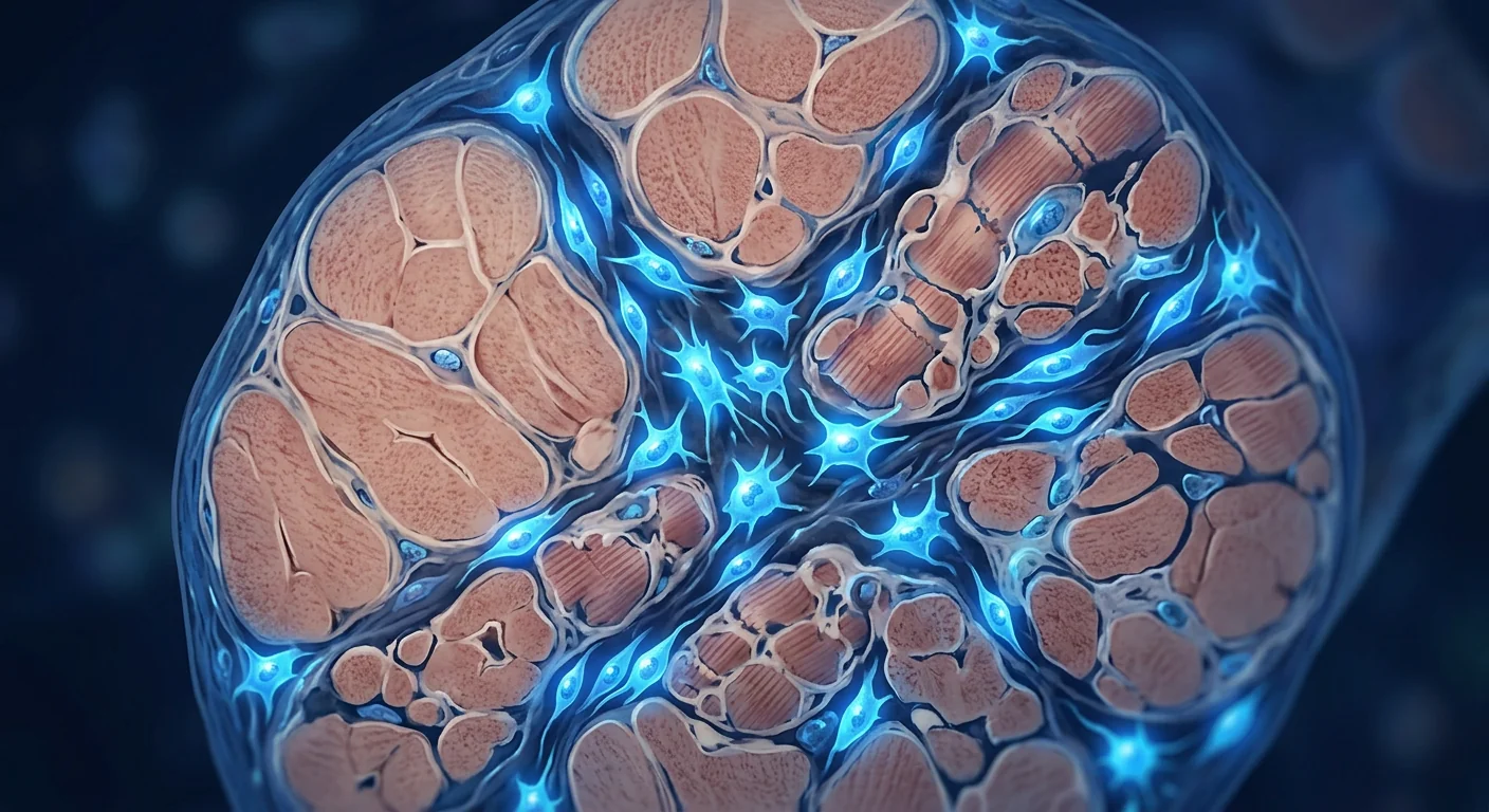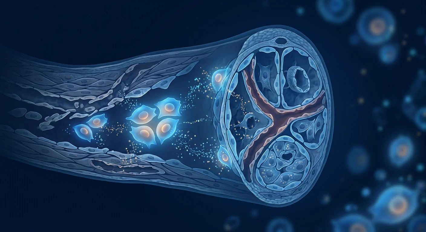
Cellular senescence — the irreversible arrest of cell division accompanied by a pro-inflammatory secretory phenotype — is now recognized as one of the primary drivers of organismal aging. Rather than being passive bystanders, senescent cells actively secrete a cocktail of inflammatory cytokines, chemokines, proteases, and growth factors collectively termed the senescence-associated secretory phenotype (SASP). [1] This SASP creates a toxic microenvironment that damages neighboring healthy cells, impairs tissue regeneration, and accelerates age-related functional decline across every organ system.
Where conventional anti-aging falls short. Antioxidants, caloric restriction mimetics, and senolytic drugs each target isolated facets of the aging cascade — none address cellular senescence comprehensively. Senolytics clear senescent cells but do not repair the tissue damage those cells have already caused. Antioxidants neutralize free radicals but cannot reverse established SASP-driven inflammation. Caloric restriction slows aging but is difficult to sustain and does not actively regenerate aged tissue.
The deeper problem is systemic. Senescent cells accumulate in every tissue with age — skeletal muscle, adipose tissue, bone marrow, skin, cardiovascular system, and the central nervous system. By age 70, senescent cells may constitute 10–15% of cells in some tissues. Their SASP propagates a self-reinforcing cycle: senescent cells induce senescence in neighboring cells through paracrine signaling, creating expanding zones of tissue dysfunction that conventional interventions cannot reverse.
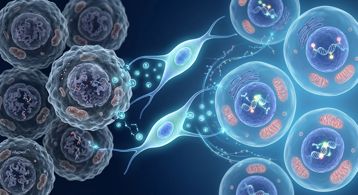
MSC protocols target the root biology. Mesenchymal stem cell therapy is being studied as a multi-mechanism intervention against cellular senescence. Unlike single-pathway drugs, MSCs simultaneously suppress SASP through immunomodulation, transfer functional mitochondria to energy-depleted cells, secrete pro-regenerative growth factors that stimulate endogenous repair, and epigenetically reprogram aged cells toward a more youthful functional state. [2] This multi-pronged approach addresses the interconnected biology of aging at its cellular foundation.
What Is Cellular Senescence?
Cellular senescence is a state of stable cell-cycle arrest triggered by telomere attrition, DNA damage, oncogene activation, oxidative stress, and mitochondrial dysfunction. Unlike quiescent cells, senescent cells remain metabolically active and secrete a complex mixture of inflammatory mediators — the SASP. [3]
The SASP includes pro-inflammatory cytokines (IL-6, IL-1β, TNF-α), chemokines (IL-8, MCP-1), growth factors (TGF-β, VEGF), and matrix metalloproteinases (MMP-1, MMP-3). This secretory profile is not merely a biomarker of aging — it is a direct effector of tissue degeneration. SASP factors degrade extracellular matrix, induce paracrine senescence in neighboring cells, recruit inflammatory immune cells, and impair stem cell niches throughout the body.
The accumulation of senescent cells is implicated in virtually every age-related condition: osteoarthritis (senescent chondrocytes), atherosclerosis (senescent endothelial cells), neurodegeneration (senescent astrocytes and microglia), sarcopenia (senescent satellite cells), immunosenescence (senescent T cells), and metabolic dysfunction (senescent adipocytes). [4] These observations have transformed senescence from a cellular curiosity into a central therapeutic target for aging biology.
The field has bifurcated into two therapeutic strategies: senolytics (drugs that selectively eliminate senescent cells) and senomorphics (agents that suppress the SASP without killing senescent cells). MSC therapy occupies a unique position — it exhibits senomorphic activity through SASP suppression while simultaneously providing regenerative trophic support that senolytics alone cannot deliver.
How MSC Therapy Targets Cellular Aging
SASP Suppression and Immunomodulation
The most well-characterized anti-aging mechanism of MSCs is their capacity to suppress the senescence-associated secretory phenotype. MSCs secrete prostaglandin E2 (PGE2), tumor necrosis factor-inducible gene 6 (TSG-6), interleukin-1 receptor antagonist (IL-1ra), and transforming growth factor-beta (TGF-β) — all of which directly downregulate SASP factor production by senescent cells. [5]
In co-culture experiments, MSCs reduce IL-6 secretion by senescent fibroblasts by 60–80% within 48 hours. This effect is mediated primarily through PGE2 signaling, which shifts macrophages from the pro-inflammatory M1 phenotype toward the anti-inflammatory M2 phenotype. The resulting change in the local cytokine milieu breaks the self-reinforcing cycle of paracrine senescence, preventing senescent cells from "infecting" their healthy neighbors.
Mitochondrial Transfer and Bioenergetic Rescue
Mitochondrial dysfunction is both a cause and consequence of cellular senescence. Aged cells exhibit reduced mitochondrial membrane potential, impaired oxidative phosphorylation, elevated reactive oxygen species (ROS) production, and mtDNA damage. MSC therapy addresses this through direct mitochondrial transfer via tunneling nanotubes and extracellular vesicles. [6]
Functional MSC-derived mitochondria, when transferred to aged recipient cells, restore ATP production, reduce ROS levels, and rescue cells from the brink of senescence. In a landmark study, MSCs transferred mitochondria to senescent cardiomyocytes, increasing ATP levels by 45% and reducing senescence-associated β-galactosidase (SA-β-gal) activity by 50%. This mitochondrial rescue effect is now recognized as one of the most promising mechanisms for MSC-based rejuvenation therapies.
Epigenetic Reprogramming
The epigenetic clock — measured through DNA methylation patterns at specific CpG sites — is one of the most robust biomarkers of biological age. MSCs have been shown to partially reverse age-associated epigenetic changes through secretion of extracellular vesicles containing microRNAs (miR-21, miR-146a, miR-181a) and chromatin-modifying enzymes. [7]
In preclinical models, MSC administration reduced the epigenetic age of multiple tissues by the equivalent of 2–5 biological years, as measured by the Horvath epigenetic clock. While still investigational, these findings suggest that MSC therapy may not merely slow aging but partially reverse its molecular hallmarks — a paradigm shift from "anti-aging" to "age reversal."
Stem Cell Niche Restoration
Aging depletes endogenous stem cell pools and degrades the microenvironments (niches) that support tissue-specific stem cell function. Hematopoietic stem cells, mesenchymal stem cells, satellite cells, and neural stem cells all decline in number and function with age. MSC therapy restores these niches by secreting niche-maintenance factors (SDF-1, SCF, angiopoietin-1), reducing niche fibrosis through MMP activity, and improving niche vascularization through VEGF and bFGF secretion. [8]
MSC Longevity Protocols: Evidence-Based Approaches
Protocol 1: Periodic Rejuvenation Protocol
The Periodic Rejuvenation Protocol is designed for healthy individuals aged 45–70 who seek to address subclinical accumulation of senescent cells and maintain functional capacity. This protocol administers 100–200 million allogeneic Wharton's jelly-derived MSCs via intravenous infusion every 6–12 months. [9]
Protocol 2: Targeted Organ Rejuvenation
For individuals with specific age-related organ decline — such as early osteoarthritis, mild cognitive impairment, or reduced cardiac function — a targeted protocol combining systemic IV infusion with local administration may be indicated. This protocol typically delivers 100–150 million MSCs intravenously plus 20–50 million MSCs via local injection (intra-articular for joints, intrathecal for neurological targets, intracoronary for cardiac targets). [10]
Protocol 3: Intensive Rejuvenation Course
For individuals with advanced biological age or multiple age-related conditions, an intensive protocol delivers 200–300 million MSCs over 2–3 sessions within a 4–6 week period, followed by maintenance infusions every 6 months. This approach aims to achieve a higher initial senescent cell clearance and tissue repair "loading dose" before transitioning to lower-frequency maintenance. Preclinical data suggests that higher initial MSC doses produce more robust SASP suppression, though the dose-response relationship in humans remains under investigation.
Monitoring Rejuvenation: Biomarkers of Biological Age
One of the most important advances in longevity medicine is the ability to objectively measure biological age — distinct from chronological age — through validated biomarkers. Velar Center's longevity protocols incorporate pre- and post-treatment biomarker panels to quantify biological age and track rejuvenation effects over time.
Epigenetic clocks (Horvath, PhenoAge, GrimAge) measure DNA methylation patterns to estimate biological age with a median error of 2–3 years. Inflammatory markers — including CRP, IL-6, TNF-α, and the SASP index — reflect systemic inflammaging burden. Mitochondrial function is assessed through NAD+/NADH ratio, ATP levels, and mitochondrial DNA copy number. Telomere length provides an orthogonal measure of cellular replicative history. Functional biomarkers — VO2max, grip strength, gait speed, and cognitive testing — capture the physiological manifestations of biological aging.
Preclinical and early clinical data suggest that MSC therapy may produce measurable improvements in several of these biomarkers within 8–16 weeks of treatment. [11] However, large-scale longitudinal data on biomarker trajectories following MSC therapy are still emerging, and individual responses vary based on baseline biological age, health status, and protocol adherence.
Safety Profile of MSC Longevity Protocols
The safety profile of MSC therapy in aging populations is favorable based on available evidence. A meta-analysis of 55 randomized controlled trials involving 2,696 patients found no increased risk of serious adverse events, tumor formation, or thromboembolic events compared to controls. [12] The most common adverse events are transient low-grade fever (reported in 10–15% of infusions) and mild injection-site reactions, both of which typically resolve within 24 hours without intervention.
In the context of longevity applications, special attention must be paid to pre-existing conditions common in aging populations — cardiovascular disease, renal impairment, and polypharmacy — as these may influence treatment response and require protocol adjustments. Velar Center conducts comprehensive pre-treatment screening including ECG, comprehensive metabolic panel, complete blood count, and inflammatory marker profile before initiating any longevity protocol.
Frequently Asked Questions
How does MSC therapy differ from senolytic drugs for anti-aging?
Senolytics selectively eliminate senescent cells through apoptosis induction. MSC therapy takes a fundamentally different approach — it suppresses the SASP (senomorphic effect) while simultaneously delivering regenerative trophic support (mitochondrial transfer, growth factors, epigenetic modulation). The two approaches are complementary: senolytics clear the "damaged hardware," while MSCs provide the "repair software." Emerging research suggests combining both may produce synergistic effects, though this remains experimental. [13]
What results can I expect from an MSC longevity protocol?
MSC longevity protocols are designed for gradual, cumulative rejuvenation — not an overnight transformation. Based on early clinical evidence, patients typically report improved energy levels, mental clarity, exercise recovery, and skin quality within 4–12 weeks of treatment. Biomarker improvements (reduced inflammatory markers, improved epigenetic age metrics) may be measurable at 8–16 weeks. Results are dose-dependent and cumulative — patients on maintenance protocols (every 6–12 months) generally report the most sustained benefits. Individual responses vary, and some patients may require 2–3 cycles before noticing significant changes.
How are MSC longevity treatments administered?
Velar Center's longevity protocols use intravenous (IV) infusion as the primary route of administration. IV delivery allows MSCs to distribute systemically, with cells preferentially homing to sites of inflammation and tissue damage through chemokine gradient sensing. The infusion itself takes 45–60 minutes and is performed in a monitored clinical setting. For patients with specific organ concerns, adjunct local injections (intra-articular, intrathecal, or targeted regional injection) may be added to the IV protocol.
How often should MSC longevity treatments be repeated?
The optimal dosing interval depends on individual biological age, health status, and treatment goals. For healthy individuals using MSCs for preventive aging, a 6–12 month interval is typical based on the known duration of MSC immunomodulatory effects (3–6 months) plus a "safety margin" for cumulative tissue repair. Patients with higher biological age or active age-related conditions may benefit from a more intensive initial course (2–3 sessions over 4–6 weeks) before transitioning to maintenance. Biomarker monitoring at 3, 6, and 12 months post-treatment guides interval decisions.
What is the cost of MSC longevity therapy at Velar Center?
The cost of MSC longevity protocols at Velar Center varies based on cell dose, protocol type, and whether adjunct therapies are included. A detailed cost breakdown is provided during consultation after reviewing your health profile and treatment goals. Velar Center is committed to transparent pricing — there are no hidden fees. Contact our patient coordinators for current pricing specific to your protocol needs.
Is there an optimal age to start MSC longevity protocols?
Aging biology research suggests the ideal window for intervention is before significant senescent cell burden has accumulated — typically in the mid-40s to early 60s, when subclinical inflammaging is present but organ function remains largely preserved. At this stage, periodic MSC infusions may function as preventive maintenance, suppressing SASP before it causes irreversible tissue damage. However, patients in their 70s and beyond can still benefit, particularly from the regenerative and mitochondrial rescue effects of MSCs. The key principle is that earlier intervention tends to produce more robust and durable results, but it is never too late to address biological aging.
References
- López-Otín C, Blasco MA, Partridge L, Serrano M, Kroemer G. Hallmarks of aging: an expanding universe. Cell. 2023;186(2):243-278. doi:10.1016/j.cell.2022.11.001 ↩
- Zhu Y, Ge J, Huang C, Liu H, Jiang H. Mesenchymal stem cell-based therapy for aging-related diseases: a focus on anti-aging mechanisms. Stem Cell Research & Therapy. 2021;12(1):241. doi:10.1186/s13287-021-02307-2 ↩
- Muñoz-Espín D, Serrano M. Cellular senescence: from physiology to pathology. Nature Reviews Molecular Cell Biology. 2014;15(7):482-496. doi:10.1038/nrm3823 ↩
- Childs BG, Durik M, Baker DJ, van Deursen JM. Cellular senescence in aging and age-related disease: from mechanisms to therapy. Nature Medicine. 2015;21(12):1424-1435. doi:10.1038/nm.4000 ↩
- Zhang Y, Ravikumar P, Li L, et al. Mesenchymal stem cell senescence and rejuvenation: mechanisms and therapeutic implications. Stem Cells Translational Medicine. 2022;11(4):356-371. doi:10.1093/stcltm/szac017 ↩
- Spees JL, Lee RH, Gregory CA. Mechanisms of mesenchymal stem/stromal cell function. Stem Cell Research & Therapy. 2016;7(1):125. doi:10.1186/s13287-016-0363-7 ↩
- Horvath S, Raj K. DNA methylation-based biomarkers and the epigenetic clock theory of ageing. Nature Reviews Genetics. 2018;19(6):371-384. doi:10.1038/s41576-018-0004-3 ↩
- Schultz MB, Sinclair DA. When stem cells grow old: phenotypes and mechanisms of stem cell aging. Development. 2016;143(1):3-14. doi:10.1242/dev.130633 ↩
- Tompkins BA, DiFede DL, Khan A, et al. Allogeneic mesenchymal stem cells ameliorate aging frailty: a phase II randomized, double-blind, placebo-controlled clinical trial. Journals of Gerontology Series A: Biological Sciences and Medical Sciences. 2017;72(11):1513-1522. doi:10.1093/gerona/glx137 ↩
- Golpanian S, DiFede DL, Khan A, et al. Allogeneic human mesenchymal stem cell infusions for aging frailty. Journals of Gerontology Series A: Biological Sciences and Medical Sciences. 2017;72(11):1505-1512. doi:10.1093/gerona/glx056 ↩
- Franceschi C, Garagnani P, Parini P, Giuliani C, Santoro A. Inflammaging: a new immune-metabolic viewpoint for age-related diseases. Nature Reviews Endocrinology. 2018;14(10):576-590. doi:10.1038/s41574-018-0059-4 ↩
- Wang Y, Yi H, Song Y. The safety of MSC therapy: a systematic review and meta-analysis of randomized controlled trials. Stem Cell Research & Therapy. 2021;12(1):600. doi:10.1186/s13287-021-02667-9 ↩
- Kirkland JL, Tchkonia T. Senolytic drugs: from discovery to translation. Journal of Internal Medicine. 2020;288(5):518-536. doi:10.1111/joim.13141 ↩
- Wiley CD, Campisi J. The metabolic roots of senescence: mechanisms and opportunities for intervention. Nature Metabolism. 2021;3(10):1290-1301. doi:10.1038/s42255-021-00483-8 ↩
- Gorgoulis V, Adams PD, Alimonti A, et al. Cellular senescence: defining a path forward. Cell. 2019;179(4):813-827. doi:10.1016/j.cell.2019.10.005 ↩
细胞衰老——伴随促炎分泌表型的不可逆细胞分裂停滞——现已被认为是机体衰老的主要驱动因素之一。衰老细胞并非被动的旁观者,而是主动分泌一系列炎症细胞因子、趋化因子、蛋白酶和生长因子,统称为衰老相关分泌表型(SASP)。[1] SASP造成的毒性微环境损害邻近健康细胞,削弱组织再生能力,并在每个器官系统中加速与年龄相关的功能衰退。
传统抗衰老方法的局限性。抗氧化剂、热量限制模拟物和senolytic药物各自针对衰老级联反应中的孤立环节——没有一种能全面解决细胞衰老问题。Senolytics清除衰老细胞但无法修复这些细胞已造成的组织损伤。抗氧化剂中和自由基但不能逆转已建立的SASP驱动性炎症。热量限制减缓衰老但难以持续,且不能主动再生老化组织。
更深层的问题是系统性的。衰老细胞随着年龄增长在每个组织中积累——骨骼肌、脂肪组织、骨髓、皮肤、心血管系统和中枢神经系统。到70岁时,衰老细胞在某些组织中可能占细胞的10-15%。其SASP通过旁分泌信号传播自我强化的恶性循环:衰老细胞诱导邻近细胞衰老,产生传统干预手段无法逆转的不断扩大组织功能障碍区域。
MSC方案针对根本生物学机制。间充质干细胞疗法正被研究作为针对细胞衰老的多机制干预手段。与单通路药物不同,MSC通过免疫调节同时抑制SASP,向能量耗竭细胞转移功能性线粒体,分泌刺激内源性修复的促再生生长因子,并通过表观遗传重编程使老化细胞向更年轻的功能状态转变。[2] 这种多管齐下的方法在细胞基础上解决衰老的相互关联生物学问题。
什么是细胞衰老?
细胞衰老是由端粒缩短、DNA损伤、癌基因激活、氧化应激和线粒体功能障碍触发的稳定细胞周期停滞状态。与静止细胞不同,衰老细胞保持代谢活性并分泌复杂的炎症介质混合物——SASP。[3]
SASP包括促炎细胞因子(IL-6、IL-1β、TNF-α)、趋化因子(IL-8、MCP-1)、生长因子(TGF-β、VEGF)和基质金属蛋白酶(MMP-1、MMP-3)。这种分泌特征不仅是衰老的生物标志物——更是组织退化的直接效应因子。SASP因子降解细胞外基质,诱导邻近细胞旁分泌衰老,招募炎症免疫细胞,并损害全身干细胞巢。
衰老细胞的积累与几乎所有年龄相关疾病有关:骨关节炎(衰老软骨细胞)、动脉粥样硬化(衰老内皮细胞)、神经退行性疾病(衰老星形胶质细胞和小胶质细胞)、肌肉减少症(衰老卫星细胞)、免疫衰老(衰老T细胞)和代谢功能障碍(衰老脂肪细胞)。[4] 这些观察已将细胞衰老从细胞生物学的好奇现象转变为衰老生物学的核心治疗靶点。
MSC疗法如何靶向细胞衰老
SASP抑制与免疫调节
MSC最具特征的抗衰老机制是其抑制衰老相关分泌表型的能力。MSC分泌前列腺素E2(PGE2)、肿瘤坏死因子诱导基因6(TSG-6)、白细胞介素-1受体拮抗剂(IL-1ra)和转化生长因子-β(TGF-β)——这些都直接下调衰老细胞的SASP因子产生。[5]
线粒体转移与生物能量拯救
线粒体功能障碍既是细胞衰老的原因也是其后果。MSC疗法通过隧道纳米管和细胞外囊泡进行直接的线粒体转移来解决这一问题。功能性MSC来源线粒体在转移到老化受体细胞后,恢复ATP产生,降低ROS水平,并将细胞从衰老边缘拯救回来。[6]
表观遗传重编程
表观遗传时钟——通过特定CpG位点的DNA甲基化模式测量——是最稳健的生物年龄生物标志物之一。MSC通过分泌含有microRNA(miR-21、miR-146a、miR-181a)和染色质修饰酶的细胞外囊泡,已被证明可部分逆转年龄相关的表观遗传变化。[7]
干细胞巢恢复
衰老耗竭内源性干细胞库并降解支持组织特异性干细胞功能的微环境(巢)。MSC疗法通过分泌巢维持因子(SDF-1、SCF、angiopoietin-1)、通过MMP活性减少巢纤维化,以及通过VEGF和bFGF分泌改善巢血管化来恢复这些巢。[8]
MSC长寿方案:循证方法
方案一:定期再生方案
针对45-70岁健康个体,每6-12个月通过静脉输注1-2亿个异体沃顿胶来源MSC,以解决亚临床衰老细胞积累并维持功能能力。[9]
方案二:靶向器官再生
针对特定器官衰退(早期骨关节炎、轻度认知障碍、心功能减退),结合全身IV输注与局部给药——1-1.5亿MSC静脉注射加2000-5000万MSC局部注射。[10]
方案三:强化再生疗程
对生物年龄较高或多重年龄相关疾病的个体,在4-6周内分2-3次输注2-3亿MSC,随后每6个月维持输注。旨在达到较高的初始衰老细胞清除和组织修复"加载剂量"。
常见问题
MSC疗法与senolytic抗衰老药物有何不同?
Senolytics通过诱导凋亡选择性清除衰老细胞。MSC疗法采取根本不同的方法——抑制SASP(senomorphic效应)同时提供再生营养支持。两种方法互补:senolytics清除"受损硬件",MSC提供"修复软件"。新兴研究表明联合使用可能产生协同效应,但这仍处于实验阶段。
MSC长寿方案可以期待什么效果?
MSC长寿方案旨在实现渐进、累积的再生——而非一夜之间的转变。基于早期临床证据,患者在治疗后4-12周通常报告精力改善、思维清晰度提高、运动恢复加快和皮肤质量改善。生物标志物改善(炎症标志物降低、表观遗传年龄指标改善)可能在8-16周可测量。效果呈剂量依赖性和累积性——维持方案(每6-12个月)的患者通常报告最持久的获益。
MSC长寿治疗如何给药?
Velar Center的长寿方案采用静脉(IV)输注作为主要给药途径。IV给药使MSC全身分布,细胞通过趋化因子梯度感知优先归巢至炎症和组织损伤部位。输注本身需要45-60分钟,在有监护的临床环境中进行。
MSC长寿治疗在Velar Center的费用是多少?
费用因细胞剂量、方案类型以及是否包含辅助疗法而异。详细的费用明细将在咨询时审查您的健康档案和治疗目标后提供。Velar Center致力于透明定价——无隐藏费用。请联系我们的患者协调员获取针对您方案需求的当前定价。
参考文献
- López-Otín C, Blasco MA, Partridge L, Serrano M, Kroemer G. Hallmarks of aging: an expanding universe. Cell. 2023;186(2):243-278. doi:10.1016/j.cell.2022.11.001 ↩
- Zhu Y, Ge J, Huang C, Liu H, Jiang H. Mesenchymal stem cell-based therapy for aging-related diseases. Stem Cell Research & Therapy. 2021;12(1):241. doi:10.1186/s13287-021-02307-2 ↩
- Muñoz-Espín D, Serrano M. Cellular senescence: from physiology to pathology. Nature Reviews Molecular Cell Biology. 2014;15(7):482-496. doi:10.1038/nrm3823 ↩
- Childs BG, Durik M, Baker DJ, van Deursen JM. Cellular senescence in aging and age-related disease. Nature Medicine. 2015;21(12):1424-1435. doi:10.1038/nm.4000 ↩
- Zhang Y, Ravikumar P, Li L, et al. MSC senescence and rejuvenation. Stem Cells Translational Medicine. 2022;11(4):356-371. doi:10.1093/stcltm/szac017 ↩
- Spees JL, Lee RH, Gregory CA. Mechanisms of MSC function. Stem Cell Research & Therapy. 2016;7(1):125. doi:10.1186/s13287-016-0363-7 ↩
- Horvath S, Raj K. DNA methylation-based biomarkers and the epigenetic clock theory of ageing. Nature Reviews Genetics. 2018;19(6):371-384. doi:10.1038/s41576-018-0004-3 ↩
- Schultz MB, Sinclair DA. When stem cells grow old. Development. 2016;143(1):3-14. doi:10.1242/dev.130633 ↩
- Tompkins BA, DiFede DL, Khan A, et al. Allogeneic MSCs ameliorate aging frailty. J Gerontol A Biol Sci Med Sci. 2017;72(11):1513-1522. doi:10.1093/gerona/glx137 ↩
- Golpanian S, DiFede DL, Khan A, et al. Allogeneic MSC infusions for aging frailty. J Gerontol A Biol Sci Med Sci. 2017;72(11):1505-1512. doi:10.1093/gerona/glx056 ↩
- Franceschi C, Garagnani P, Parini P, Giuliani C, Santoro A. Inflammaging. Nature Reviews Endocrinology. 2018;14(10):576-590. doi:10.1038/s41574-018-0059-4 ↩
- Wang Y, Yi H, Song Y. The safety of MSC therapy. Stem Cell Research & Therapy. 2021;12(1):600. doi:10.1186/s13287-021-02667-9 ↩
- Kirkland JL, Tchkonia T. Senolytic drugs. Journal of Internal Medicine. 2020;288(5):518-536. doi:10.1111/joim.13141 ↩
- Wiley CD, Campisi J. The metabolic roots of senescence. Nature Metabolism. 2021;3(10):1290-1301. doi:10.1038/s42255-021-00483-8 ↩
- Gorgoulis V, Adams PD, Alimonti A, et al. Cellular senescence: defining a path forward. Cell. 2019;179(4):813-827. doi:10.1016/j.cell.2019.10.005 ↩
الشيخوخة الخلوية — التوقف غير القابل للعكس عن انقسام الخلايا المصحوب بنمط إفرازي التهابي — تُعرف الآن كأحد المحركات الرئيسية لشيخوخة الكائن الحي. بدلاً من كونها مجرد متفرجين سلبيين، تفرز الخلايا الهرمة بنشاط مزيجًا من السيتوكينات الالتهابية والكيموكينات والبروتياز وعوامل النمو التي تُعرف مجتمعة بالنمط الإفرازي المرتبط بالشيخوخة (SASP). [1] يخلق SASP بيئة دقيقة سامة تضر بالخلايا السليمة المجاورة، وتضعف تجديد الأنسجة، وتسرع التدهور الوظيفي المرتبط بالعمر عبر كل جهاز عضوي.
حيث تفشل مكافحة الشيخوخة التقليدية. تستهدف مضادات الأكسدة ومحاكيات تقييد السعرات الحرارية وأدوية senolytic كل منها جوانب معزولة من سلسلة الشيخوخة — ولا يعالج أي منها الشيخوخة الخلوية بشكل شامل. تزيل Senolytics الخلايا الهرمة لكنها لا تصلح تلف الأنسجة الذي تسببت فيه تلك الخلايا بالفعل. تعادل مضادات الأكسدة الجذور الحرة لكنها لا تستطيع عكس الالتهاب المدفوع بـ SASP. يبطئ تقييد السعرات الحرارية الشيخوخة لكنه صعب الاستدامة ولا يجدد الأنسجة الهرمة بنشاط.
المشكلة الأعمق جهازية. تتراكم الخلايا الهرمة مع تقدم العمر في كل نسيج — العضلات الهيكلية والأنسجة الدهنية ونخاع العظم والجلد والجهاز القلبي الوعائي والجهاز العصبي المركزي. بحلول سن السبعين، قد تشكل الخلايا الهرمة 10-15% من الخلايا في بعض الأنسجة. ينشر SASP دورة ذاتية التعزيز: الخلايا الهرمة تحفز الشيخوخة في الخلايا المجاورة عبر الإشارات الباراكرينية، مما يخلق مناطق متوسعة من خلل الأنسجة لا يمكن للتدخلات التقليدية عكسها.
تستهدف بروتوكولات MSC البيولوجيا الجذرية. يُدرس العلاج بالخلايا الجذعية الوسيطة كتدخل متعدد الآليات ضد الشيخوخة الخلوية. على عكس الأدوية أحادية المسار، تقوم MSC في وقت واحد بقمع SASP من خلال التعديل المناعي، ونقل الميتوكوندريا الوظيفية إلى الخلايا المستنفدة للطاقة، وإفراز عوامل النمو المحفزة للتجديد التي تحفز الإصلاح الداخلي، وإعادة برمجة الخلايا الهرمة جينيًا نحو حالة وظيفية أكثر شبابًا. [2] يعالج هذا النهج متعدد الجوانب البيولوجيا المترابطة للشيخوخة في أساسها الخلوي.
ما هي الشيخوخة الخلوية؟
الشيخوخة الخلوية هي حالة من توقف دورة الخلية المستقرة triggered by استنزاف التيلومير، وتلف الحمض النووي، وتفعيل الجينات الورمية، والإجهاد التأكسدي، وخلل الميتوكوندريا. على عكس الخلايا الهادئة، تبقى الخلايا الهرمة نشطة أيضيًا وتفرز مزيجًا معقدًا من الوسائط الالتهابية — SASP. [3]
يشمل SASP السيتوكينات المؤيدة للالتهاب (IL-6، IL-1β، TNF-α)، والكيموكينات (IL-8، MCP-1)، وعوامل النمو (TGF-β، VEGF)، وميتالوبروتياز المصفوفة (MMP-1، MMP-3). هذا النمط الإفرازي ليس مجرد علامة حيوية للشيخوخة — بل هو مؤثر مباشر على تدهور الأنسجة. تحلل عوامل SASP المصفوفة خارج الخلوية، وتحفز الشيخوخة الباراكرينية في الخلايا المجاورة، وتجند الخلايا المناعية الالتهابية، وتضعف بيئات الخلايا الجذعية في جميع أنحاء الجسم.
كيف يستهدف علاج MSC الشيخوخة الخلوية
قمع SASP والتعديل المناعي
أكثر آليات MSC المضادة للشيخوخة تميزًا هي قدرتها على قمع النمط الإفرازي المرتبط بالشيخوخة. تفرز MSC البروستاغلاندين E2 (PGE2)، والجين 6 المحفز بعامل نخر الورم (TSG-6)، ومضاد مستقبلات الإنترلوكين-1 (IL-1ra)، وعامل النمو المحول بيتا (TGF-β) — وكلها تخفض مباشرة إنتاج عوامل SASP بواسطة الخلايا الهرمة. [5]
نقل الميتوكوندريا والإنقاذ البيوفيزيائي
خلل الميتوكوندريا هو سبب ونتيجة للشيخوخة الخلوية. يعالج علاج MSC هذا من خلال النقل المباشر للميتوكوندريا عبر الأنابيب النانوية النفقية والحويصلات خارج الخلوية. الميتوكوندريا الوظيفية المشتقة من MSC، عند نقلها إلى الخلايا المستقبلة الهرمة، تستعيد إنتاج ATP، وتخفض مستويات ROS، وتنقذ الخلايا من حافة الشيخوخة. [6]
إعادة البرمجة الجينية
الساعة الجينية — المقاسة من خلال أنماط مثيلة الحمض النووي في مواقع CpG محددة — هي واحدة من أقوى العلامات الحيوية للعمر البيولوجي. ثبت أن MSC تعكس جزئيًا التغيرات الجينية المرتبطة بالعمر من خلال إفراز حويصلات خارج الخلوية تحتوي على microRNAs (miR-21، miR-146a، miR-181a) وإنزيمات معدلة للكروماتين. [7]
بروتوكولات MSC لطول العمر
البروتوكول 1: بروتوكول التجديد الدوري
للأفراد الأصحاء بعمر 45-70 سنة، 100-200 مليون MSC من هلام وارتون عبر التسريب الوريدي كل 6-12 شهرًا. [9]
البروتوكول 2: تجديد الأعضاء المستهدف
للأفراد الذين يعانون من تدهور محدد في الأعضاء، يجمع بين التسريب الوريدي الجهازي والإعطاء الموضعي. [10]
البروتوكول 3: دورة التجديد المكثف
للأفراد ذوي العمر البيولوجي المتقدم، 200-300 مليون MSC على 2-3 جلسات خلال 4-6 أسابيع، تليها حقن صيانة كل 6 أشهر.
أسئلة شائعة
كيف يختلف علاج MSC عن أدوية senolytic لمكافحة الشيخوخة؟
تقضي Senolytics بشكل انتقائي على الخلايا الهرمة. يتخذ علاج MSC نهجًا مختلفًا جوهريًا — يقمع SASP مع تقديم دعم غذائي متجدد في نفس الوقت. النهجان متكاملان وقد ينتجان تأثيرات تآزرية عند الجمع.
ما النتائج التي يمكن توقعها من بروتوكول MSC لطول العمر؟
تم تصميم بروتوكولات MSC لطول العمر لتجديد تدريجي تراكمي — وليس تحولاً بين ليلة وضحاها. بناءً على الأدلة السريرية المبكرة، يبلغ المرضى عادة عن تحسن مستويات الطاقة وصفاء الذهن وتعافي التمارين وجودة البشرة خلال 4-12 أسبوعًا من العلاج. قد تكون تحسينات العلامات الحيوية قابلة للقياس في 8-16 أسبوعًا.
المراجع
- López-Otín C, Blasco MA, Partridge L, Serrano M, Kroemer G. Hallmarks of aging. Cell. 2023;186(2):243-278. doi:10.1016/j.cell.2022.11.001 ↩
- Zhu Y, Ge J, Huang C, et al. MSC-based therapy for aging-related diseases. Stem Cell Res Ther. 2021;12(1):241. doi:10.1186/s13287-021-02307-2 ↩
- Muñoz-Espín D, Serrano M. Cellular senescence. Nat Rev Mol Cell Biol. 2014;15(7):482-496. doi:10.1038/nrm3823 ↩
- Childs BG, Durik M, Baker DJ, van Deursen JM. Cellular senescence in aging. Nat Med. 2015;21(12):1424-1435. doi:10.1038/nm.4000 ↩
- Zhang Y, Ravikumar P, Li L, et al. MSC senescence and rejuvenation. Stem Cells Transl Med. 2022;11(4):356-371. doi:10.1093/stcltm/szac017 ↩
- Spees JL, Lee RH, Gregory CA. Mechanisms of MSC function. Stem Cell Res Ther. 2016;7(1):125. doi:10.1186/s13287-016-0363-7 ↩
- Horvath S, Raj K. Epigenetic clock theory of ageing. Nat Rev Genet. 2018;19(6):371-384. doi:10.1038/s41576-018-0004-3 ↩
- Schultz MB, Sinclair DA. When stem cells grow old. Development. 2016;143(1):3-14. doi:10.1242/dev.130633 ↩
- Tompkins BA, DiFede DL, Khan A, et al. Allogeneic MSCs ameliorate aging frailty. J Gerontol A. 2017;72(11):1513-1522. doi:10.1093/gerona/glx137 ↩
- Golpanian S, DiFede DL, Khan A, et al. Allogeneic MSC infusions for aging frailty. J Gerontol A. 2017;72(11):1505-1512. doi:10.1093/gerona/glx056 ↩
- Franceschi C, Garagnani P, Parini P, et al. Inflammaging. Nat Rev Endocrinol. 2018;14(10):576-590. doi:10.1038/s41574-018-0059-4 ↩
- Wang Y, Yi H, Song Y. Safety of MSC therapy. Stem Cell Res Ther. 2021;12(1):600. doi:10.1186/s13287-021-02667-9 ↩
- Kirkland JL, Tchkonia T. Senolytic drugs. J Intern Med. 2020;288(5):518-536. doi:10.1111/joim.13141 ↩
- Wiley CD, Campisi J. Metabolic roots of senescence. Nat Metab. 2021;3(10):1290-1301. doi:10.1038/s42255-021-00483-8 ↩
- Gorgoulis V, Adams PD, Alimonti A, et al. Cellular senescence: defining a path forward. Cell. 2019;179(4):813-827. doi:10.1016/j.cell.2019.10.005 ↩